Embed Size (px)
Citation preview
1
Supported by the EU-Commission Public Health Directorate Programme of Community Action on Rare
Diseases WHO Collaborating Centre for the Epidemiology Surveillance of Congenital Anomalies
A Review of Environmental Risk Factors for Congenital Anomalies
Edition 1
(uploaded to website 29 April 2004)
EUROCAT Central Registry Room 15E12
University of Ulster Newtownabbey
Co Antrim Northern Ireland
BT37 0QB Tel: +44 (0)28 9036 6639 Fax: +44 (0)28 9036 8341
Email: [email protected] Website: www.eurocat.ulster.ac.uk
ISBN 1 85923 187 X

2
ACKNOWLEDGEMENTS This review of the literature has been supported to date by the DH/DETR/Environment Agency Joint Research Programme on the Possible Health Effects of Landfill Sites Project LSHTM/00/1 and by the European Commission Rare Diseases Programme.
NOTES TO THE USE OF THIS REVIEW This literature review will be expanded and updated over time. When quoting the review, please quote as follows e.g. Little J (2002) Smoking. In: EUROCAT Special Report. The environmental causes of congenital anomalies: a review of the literature. [online] www.eurocat.ulster.ac.uk/pubdata [accessed date]. It is not the aim of this review to be systematic in all parts, but to be an informative starting point for access to the literature, and for understanding what factors may underlie variations in prevalence of congenital anomalies in time or space. We set the evidence regarding congenital anomalies in the context of evidence related to other pregnancy outcomes, mainly spontaneous abortion (miscarriage), low birthweight and prematurity. Our review in relation to those outcomes is not exhaustive, and relates only to those risk factors for which there is evidence regarding the risk of congenital anomalies. We mention research on neurodevelopmental outcomes in relation to specific prenatal chemical exposures, but do not touch on wider areas such as neurodevelopmental outcomes in relation to nutrition. This review is mainly concerned with epidemiologic studies. The assessment of environmental causes of congenital anomalies also draws on toxicological data, data from animal studies, and detailed exposure data. A (future) chapter will discuss these sources of evidence, but we do not review them systematically, nor do we attempt risk assessments for any of the environmental exposures reviewed.

3
FURTHER RESOURCES Sullivan F, Barlow S, McElhatton P. A review of the potential teratogenicity of substances emanating from landfill sites. Available from [online] www.advisorybodies.doh.gov.uk/landup.htm EUROCAT Special Report (2003) Prevention of Neural Tube Defects by Periconceptional Folic Acid Supplementation in Europe. Available from [online] www.eurocat.ulster.ac.uk/pubdata/ Schardein JL (2000) Chemically induced birth defects, 3rd Ed, Marcel Dekker, New York Ferencz C, Rubin JD, Loffredo CA, Magee CA (1993) Epidemiology of Congenital Heart Disease: the Baltimore Washington Infant Study 1981-89. Perspectives in Pediatric Cardiology, Vol 4, Futura, New York Elwood JM, Little J, Elwood JH (1992) Epidemiology and control of neural tube defects. Oxford Medical Publications Brown N, Lumley J, Tickle C, Keene J. Congenital limb reduction defects: clues from developmental biology, teratology and epidemiology. The Stationery Office, London WHO Human Genetics Programme (2002) Global strategies to reduce the health care burden of craniofacial anomalies. Report of WHO meetings on International Collaborative Research on Craniofacial Anomalies. For information on the genetics of congenital anomalies, refer to www.ncbi.nlm.nih.gov/entrez/query.fcgi?db=OMIM

4
LIST OF CONTRIBUTORS Professor Helen Dolk EUROCAT Central Registry University of Ulster, Room 15E03 Newtownabbey, Co Antrim, Northern Ireland BT37 0QB Dr Pat Doyle Department of Epidemiology & Population Sciences London School of Hygiene & Tropical Medicine Keppel Street London WC1E 7HT Dr Ester Garne Epidemiology University of Southern Denmark Sdr Boulevard 23a DK – 5000, Odense C Denmark Professor Julian Little University of Aberdeen Department of Medicine & Therapeutics Polwarth Building, Foresterhill Aberdeen, Scotland AB25 2ZD Maria Loane EUROCAT Central Registry University of Ulster, Room 15E12 Newtownabbey Co Antrim, Northern Ireland BT37 0QB Dr Elisabeth Robert France Central-East Registry of Congenital Malformations Institut Europeen des Genomutations Rue Edmond Locard 86 F-69005 Lyon France Professor Mary Seller Division of Medical & Molecular Genetics 7th Floor Guy’s Tower Guy’s Hospital LONDON SE1 9RT Dr Martine Vrijheid Scientist Unit of Radiation & Cancer International Agency for Research on Cancer 150 Cours Albert Thomas 69372 Lyon Cedex France

5
TABLE OF CONTENTS
Page PART I. Overview of principles and methods in identifying
the causes of congenital anomalies
Chapter I.1 Genetic causes of congenital anomalies and their interaction with environmental factors
Mary Seller January 2004 7 Chapter I.2 Epidemiological evidence regarding environmental
causes of congenital anomalies: interpretational issues Helen Dolk January 2004 30
Coming soon: The interpretation of evidence based on animal studies Nigel Brown PART II. Selected environmental risk factors for congenital anomalies Chapter II.1 Nutrition Julian Little May 2002 51 Chapter II.2 Smoking Julian Little May 2002 83 Chapter II.3 Ionising Radiation Pat Doyle February 2003 89 Chapter II.4 Maternal diabetes Ester Garne January 2004 94 Chapter II.5 Maternal epilepsy Elizabeth Robert January 2004 98 Coming soon: Therapeutic and recreational drugs Pat McElhatton Ethnicity Maria Loane Socio-economic status and maternal age Helen Dolk Maternal infections Araceli Busby Assisted Reproduction Alcohol Part III. Environmental pollution and congenital anomalies Chapter III.1 Chemical environmental Martine Vrijheid, Maria Loane
and occupational exposures and Helen Dolk 107 Coming soon: Assessing evidence from animal studies – dioxin and benomyl as case studies Nigel Brown

6
PART I
Overview of Principles & Methods in Identifying the Causes of Congenital
Anomalies

7
CHAPTER I.1 GENETIC CAUSES OF CONGENITAL
ANOMALIES AND THEIR INTERACTION WITH ENVIRONMENTAL FACTORS
Mary Seller, January 2004
1. Congenital anomalies The term ‘congenital anomaly’ is used for all types of structural defects with which a baby can be
born. Overall, these abnormalities can be classified broadly into malformations, malformation
syndromes, disruptions and deformations, which provides some insight into possible underlying
aetiology.
A malformation is a localised error of normal development, a primary structural defect occurring
during the morphogenesis of an organ or tissue. Examples are isolated cleft lip, cleft palate, spina
bifida or a ventricular septal defect in the heart. Such an isolated malformation occurs in an otherwise
normal child. Single malformations are relatively common and usually have a multifactorial
aetiology: that is, both genes and environment are important (see later).
A malformation syndrome is the occurrence together in one individual of several different
malformations as primary events arising from the same underlying cause. There is a specific
recognisable pattern of abnormalities according to each causative factor. The cause is usually a single
gene mutation or a chromosome abnormality, or occasionally an environmental agent.
A disruption is where there is a major destruction or alteration of a body part that had previously
formed, or which had the intrinsic potential to form, quite normally. An example is a missing limb, or
part thereof. Usually the causes of disruptions are not genetic, but environmental, such as drugs,
maternal illness or infections, or strands of amnion becoming detached and encircling a limb or digit.
A deformation is an alteration of shape or structure imposed on a body part after, or during, its normal
formation, usually by mechanical forces. The cause may be external or internal to the fetal body. For
example talipes (club foot) can be produced by an external factor, oligohydramnios (lack of amniotic
fluid), or be secondary to open spina bifida, an intrinsic cause, where damage to the spinal cord causes
paralysis of leg muscles and then positional abnormalities of the foot. Thus, deformations may have a
genetic or part genetic cause, or an environmental one. But as often happens, the situation may not be
8
clearly categorised, such as when the oligohydramnios producing the talipes is secondary to renal
agenesis in the fetus, a multifactorial condition.
It is generally estimated that around 14% of babies are born with a single minor malformation, for
example, a skin tag that can easily be removed, and is of little consequence. However, around 2-3% of
neonates have a single major malformation like spina bifida that will require more extensive medical
treatment, or perhaps even be lethal. Just under 1% of neonates have multiple malformations. These
are the prevalence figures, the number observed at birth. However, the occurrence of malformations is
much higher, but many affected pregnancies are aborted spontaneously, especially during the first
trimester of pregnancy.
The precise cause of congenital malformations is not known for as many as 50-60% of the total. It is
believed that overall, multifactorial aetiology accounts for 20-25% of all abnormalities; 6-8% are
monogenic, that is, caused by mutations in a single gene; 6-8% by chromosome abnormalities; and 6-
8% by environmental factors such as maternal illness, infections, drugs, radiation and alcohol.
2. Monogenic disorders In monogenic disorders, a mutation in a single gene results in malformations, usually a spectrum of
abnormalities: eg Meckel syndrome, in which there is encephalocoele, polydactyly and polycystic
kidneys. All known diseases resulting from single gene mutations are listed in OMIM (Online
Mendelian Inheritance in Man at http://www3.ncbi.nlm.nih.gov/Omim/searchomim.html). Such
disorders are inherited, and the mode of inheritance may be autosomal dominant, autosomal recessive
or sex-linked. Within families, each type of inheritance has a characteristic pedigree (family tree).
In autosomal dominant inheritance, manifestation of the disease occurs in heterozygotes when only
one copy of the disease gene is present. An affected individual usually has an affected parent, and has
a 50% chance of passing the gene on to each of his/her offspring. The pedigree thus shows affected
individuals in each generation, there is a ‘family history’, and both males and females are equally
likely to be affected.
Autosomal recessive conditions manifest only when an individual has two copies of the mutant gene.
One is received from each parent who are both carriers, but completely unaffected, for one copy of the
abnormal gene in this case is not sufficient to cause the disease. Usually there is no family history, an
affected individual occurs solely in one generation, although there may be more than one case within
a single sibship. An exception occurs when there is consanguinity in the family.
9
In sex-linked inheritance, the disease causing mutation is carried on one of the sex chromosomes. In
the most common form, X-linked recessive inheritance, males only are affected because they have
only one X chromosome. Their daughters have a 50% chance of receiving the mutant gene from
them, but if they do so, they are unaffected as they have two X chromosomes. However, they are
carriers and have a 50% chance of passing the defective gene on to their own sons who, if they
receive it, will be affected. Once more, there is a family history, but the pattern is different from that
of autosomal dominant inheritance. Males alone are affected, comprising affected grandfathers and
grandsons with the intermediate generation unaffected, and there is no male-to-male transmission of
the disease.
In dominant conditions, the genetic background might influence the phenotype (the clinical features),
giving variable expressivity between different affected individuals or causing incomplete penetrance
of the gene. Variable expressivity is precisely what its name suggests. An example occurs in
neurofibromatosis Type I, in which the clinical manifestation may involve hundreds of cutaneous
neurofibromata, that can be extremely disfiguring, or just a few, which may be hardly noticeable. In
incomplete penetrance, an individual carries the mutant gene but is completely devoid of any clinical
symptoms. Both these phenomena may be explained by specific ‘modifier’ genes in the genome, the
products of which influence either the expression of, or the product of, the mutant gene, or more non-
specifically by the products of many other genes in the genome.
In a few dominant conditions, there are instances where an affected individual does not have an
affected parent. One way this can occur is when new mutations arise in the gene relatively frequently.
This is found in achondroplasia, so that often the affected individual has arisen as a new mutation of
the gene in one of the parental germ cells. Another way is when there is germ-line mosaicism in one
parent, in which a proportion of the cells of the gonad bear the mutation while the rest do not.
Occasionally, a specific environmental factors is required for the mutant gene to be manifest. An
example is the gene that causes malignant hyperthermia. The mutation is manifest only if the
individual undergoes halothene anaesthesia with succinylcholine as the muscle relaxant, when muscle
rigidity and hyperthermia occur that can be lethal. But under normal circumstances, and with other
types of anaesthesia, the individual is unaffected and blissfully unaware that they carry that particular
mutation.

10
3. Molecular pathogenesis in single gene malformation syndromes As the genes that cause specific malformation syndromes are identified, and the molecular structure of
their protein product is determined, so we are beginning to understand the molecular pathogenesis of
congenital malformations which underlies the developmental pathogenesis. A few examples only can
be given. An interesting point emerging, which was totally unexpected, is that sometimes, different
mutations in the same gene cause different syndromes. This is allelic heterogeneity. Three distinct
skeletal dysplasias arise from a mutation in the fibroblast growth factor receptor-3 (FGFR-3) gene.
They are achondroplasia (marked limb shortening and relatively large head), thanatophoric dysplasia
(severe limb shortening, small chest, lethal at birth) and hypochondroplasia (less severe limb
shortening). A different domain of FGFR-3 is affected in each case. In achondroplasia, the mutation
affects the transmembrane domain (Rousseau et al, 1994); in thanatophoric dysplasia, there is an
amino acid substitution in the link between the second and third immunoglobulin-like extracellular
domains (Tavotmina et al, 1995), and in hypochondroplasia, the mutation affects the intracellular
proximal tyrosine kinase domain (Bellus et al, 1995).
Mutations in another of the fibroblast growth factor receptor genes, the FGFR-2 gene, does not
produce a skeletal dysplasia but either, Crouzon syndrome (Reardon and Winter, 1996), a
craniosynostosis with normal stature, or, Apert syndrome (Wilkie et al, 1996), a craniosynostosis with
severe syndactyly of the hands and feet. There are a number of fibroblast growth factor receptors
known and they are ligands for fibroblast growth factors, of which again, there are several. They
function at a very early stage of embryogenesis and are involved in cell growth, differentiation, spatial
patterning and programmed cell death.
Another example of allelic heterogeneity involves the GLI3 gene, a zinc finger transcription factor.
Greig cephalopolysyndactyly, where there is a high forehead with frontal bossing, macrocephaly,
broad thumbs and postaxial polydactyly and syndactyly of the hands as well as preaxial polydactyly
of the feet, is produced by large deletions in the GLI3 gene (Vortkamp et al, 1991), while smaller
frameshift mutations in this gene produce Pallister Hall syndrome (Kang et al, 1997), which has
central polydactyly and hamartoma, ear abnormalities, macrognathia and anal defects.
Holoprosencephaly (failure of division of the cerebral hemispheres) is a malformation that can be
produced by mutations in a number of different genes. This is locus heterogeneity. The first gene to
be discovered in humans was the sonic hedgehog gene (Roessler et al, 1996). The secreted product of
this gene is a signalling molecule that affects ventral patterning of the early embryo (Echelard et al,
1993), being expressed in the notochord, the floorplate of the neural tube, and ventral midline pre-
somitic mesoderm (Chiang et al, 1996). The sonic hedgehog protein promotes the expression of other
11
developmental genes. The membrane receptors named ‘patched’ and ‘smoothened’ exist as a complex
whereby normally, patched prevents smoothened from being expressed. But when the sonic hedgehog
protein binds to patched, this repression is released, allowing smoothened to signal, resulting in Gli
protein function (Ingham, 1998). Other genes which, when mutated, cause holoprosencephaly,
include SIX3, a homeobox containing transcription factor (Wallis et al, 1999), and TGIF, a
transcription factor that competitively inhibits the binding of the retinoic acid receptor to a retinoid-
responsive promoter (Bertolino et al, 1995).
In many instances, the fact that multiple abnormalities affecting several different body parts occur
when there is only one mutation in a single gene, a phenomenon referred to as ‘pleiotropy’ and
hitherto enigmatic, can now be explained by our contemporary molecular knowledge. The genes
concerned are those involved in early developmental processes and their control. The expression of
these genes is seldom restricted either temporally or spatially and these genes are usually expressed
several times over at different times and in different organs.
4. Mutations A mutation is a change in the DNA. Throughout life, mutations are constantly occurring in the DNA
of both somatic and germ-line cells as a result of factors arising both internally or externally. Every
time a cell divides, DNA replication has to precede it and this process is prone to error. Also, we are
often exposed, usually unknowingly, to environmental agents, such as radiation and chemical agents
that can damage the DNA. Most of the errors and some of the damage are detected and corrected by
an intrinsic cellular DNA repair mechanism. But those faults that escape can have far-reaching
effects. If they are in germ-line cells, then their consequences can extend even further, for they may
be inherited.
One far-reaching effect of mutations in somatic cells, arising spontaneously or as a result of
environmental insults, is that they may accumulate during life, and eventually cause cancer. The
mutations often affect the tumour suppressor genes and oncogenes that are involved in the normal
control and regulation of cell division and cell proliferation. Chromosome breakage has long been
known to be associated with tumour cells and early studies of some of these led to the identification of
some of the cancer causing genes; genes involved in normal cell function were disrupted by the
chromosome breakage.

12
Mutations can occur anywhere in the entire genome, but since only 3% of our DNA comprises
protein-coding regions, that is, genes, a mutation is usually only of consequence to the individual
when it occurs in these stretches of DNA or in the associated gene regulatory and controlling
elements. The malformation syndromes mentioned above have arisen in this way and are said to be
the result of ‘spontaneous mutations’ because the cause of the mutation is unknown.
Mutations may involve a single purine or pyrimidine base change in the DNA, or the deletion or
addition of a few or many bases. While occurring throughout the genome, mutations do not happen
uniformly, certain areas, such as those rich in CG dinucleotide sequences and known as ‘hotspots’, are
more prone than others. Large genes are more likely to have mutations than smaller genes. Also,
mutation rates vary with age, and there is a paternal age effect in some dominant disorders. The risk
of a father producing a child with achondroplasia or Marfan’s syndrome or Apert’s syndrome is
markedly increased after the age of 40 years. This is explained by the fact that the progenitor cells of
the sperm divide continuously during life, and DNA replication errors accumulate with advancing
age. This contrasts with the situation in females where cell division in the egg is suspended during
fetal life and is not resumed until ovulation. In females, increase in maternal age is associated with
the other major class of mutation where there are observable numerical changes to the chromosomes,
as distinct from minute changes in the DNA at the molecular level (see below). The addition or the
loss of a whole chromosome, aneuploidy, is the result of the lack of separation of the two chromatids
at mitosis. Such non-disjunction seems to happen more commonly the older a woman is, implying
that the longer cell division is suspended, the more likely the disjunction process is to be faulty.
Radiation and an uncertain number of chemicals can cause mutations. Ionising radiation from either
nuclear fallout or X-rays can break the bonds between the double stranded DNA and also alter the
bases within the strands. In addition, they can cause chromosome breakage. The degree of genetic
damage is directly related to the amount of exposure and the effects are cumulative. This type of
radiation penetrates deep into the body tissues and can reach the gonads, so potentially, both somatic
and germ-line mutations can occur. There is evidence on both counts. Survivors of the Japanese
atomic bombings and also of the explosion at the Chernobyl nuclear power plant, developed cancers,
showing that the expected somatic mutations occur. Dubrova et al (2002) used minisatellites for
mutation detection and studied Ukrainian people exposed to the Chernobyl fallout. They found a near
twofold increase in the rate of mutations being passed on by exposed males to their offspring,
providing evidence that the germ-line is also affected.
Non-ionising UV radiation does not penetrate the body further than the skin. It causes the formation
of covalent bonds between neighbouring pyrimidine bases, so that when the DNA replicates these
pyrimidine dimers are unable to pair properly with purines. The ultimate result is skin cancer.

13
As previously mentioned, errors in DNA replication constantly occur naturally, and the body has an
innate and complex DNA repair system that rectifies these mistakes and minor environmental
damage. Mutations in the genes encoding the enzymes involved in this system can have severe
effects and render individuals hugely sensitive to radiation, and even to normal exposure to daylight.
Xeroderma pigmentosum is one of several disorders arising in this way, where skin damage and
cancers occur in childhood, and neurological impairment.
An enormous number of chemicals, including even commonly used substances like caffeine, are
mutagenic in experimental animals. It is tempting to extrapolate this information to humans, but there
is good evidence for some chemicals. Many of these substances alter the DNA helical structure or
component bases, or interfere with DNA replication. Somatic cells are primarily affected. If a
chemically induced mutation on a germ cell does have a deleterious effect on the embryo, it usually
leads to death at an early cleavage stage. But exogenous chemicals and drugs may not necessarily
cause mutations. Instead, they may directly or indirectly affect the embryo during development and
produce congenital malformations (see below). In this case they are called ‘teratogens’. They
influence crucial developmental processes in a variety of ways.
In addition to innate DNA repair systems, the body also has some complex systems of enzymes that
metabolise and detoxify certain environmental chemicals and drugs when they enter the body. A
mutation in any of the genes encoding these enzymes, for instance in the family of P450 genes, can
lead to increased susceptibility to diseases and cancers. In addition, many of these genes are
polymorphic, the various alleles producing different isozymes that have different properties. These
allelic variants are responsible for the variation within the population in the response and reaction to
individual drugs and chemicals.
5. Chromosome disorders Chromosome abnormalities are another form of mutation, and there are many different types:
duplication of an entire chromosome, or of the entire set, or of only part of a chromosome, or similar
deficiencies of chromosomal material. If the chromosome involved is an autosome (all the
chromosomes except the two sex chromosomes), then the consequences of such duplications or
deficiencies are usually severe. Many affected individuals are lost shortly after conception. Around
60% of all miscarriages in the first trimester have a chromosome abnormality. Those that survive to
14
be born usually have mental retardation, intrauterine and postnatal growth retardation, dysmorphic
features, and there may often be major congenital malformations too.
The most common type of chromosome anomaly is trisomy, where there is an extra version of a
single chromosome. Trisomy can arise for any of the chromosomes, but it affects some more
frequently than others. Trisomy for chromosome 16 is the most common, but no real embryo forms in
the conceptus and none survive more than a few weeks post-conception. Trisomy 1 is extremely rare.
Although most of the trisomies 13, 18 and 21 that are conceived are aborted prenatally, these
particular trisomies are the only instances where a few cases do survive to be born at term. Trisomy
21 alone has the ability to achieve adulthood. Each of these three trisomies has its own distinct
pattern of abnormalities. Trisomy 13 (Patau syndrome) is characterised by microcephaly, scalp
defects, microphthalmia, ocular hypotelorism, cleft lip and palate, flexed fingers, polydactyly,
prominent heels, heart defects and severe developmental delay, with death usually within the first few
months of life. Trisomy 18 (Edwards syndrome) has an elongate skull, small, lowset ears,
micrognathia, flexed and overlapping fingers, prominent heels, heart defects, horseshoe kidney and
severe developmental delay, few surviving longer than the first year. Trisomy 21 (Down syndrome)
has brachycephaly, ocular hypertelorism, upslanting and short palpebral fissures, epicanthic folds,
small nose and flat face, small, lowset ears, single transverse palmar crease, stubby fingers with short
incurved fifth finger, broad feet, heart defects and learning difficulties.
Sex chromosome aberrations have less severe effects than autosomal abnormalities and in some types,
affected individuals can go through life unaware of their condition. Some prenatal loss occurs, but
postnatal survival is usually not compromised. The two that may have overt clinical features are
Klinefelter syndrome where there is an extra X chromosome in a male (47, XXY), and Turner
syndrome, where an X chromosome is missing in a female (45,X). The rarer 47, XXX and 47, XYY
conditions usually do not have any phenotypic effects except occasional educational or behavioural
problems in childhood and possible infertility in adulthood. In Klinefelter syndrome there is
hypogonadism with sterility, tall stature, and gynaecomastia. In Turner syndrome exceptionally, 99%
of cases die in utero. Survivors exhibit short stature, webbing of the neck, broad chest, increased
carrying angle of the arms, co-arctation of the aorta and sterility.
As mentioned earlier, extra, or missing chromosomes largely arise because of faulty separation or
disjunction of the two chromatids during cell division. The other cause is anaphase lag, where one
chromosome fails to be incorporated within the newly forming nuclear membrane at the end of
mitosis, and is ‘lost’. Non-disjunction in the female is the most common cause of the common
autosomal trisomies, and as already described, is often related to raised maternal age, but, young
15
mothers do have Down’s babies and a proportion of cases arise because of non-disjunction in the
male, and this is not necessarily related to age.
Less commonly, malformation syndromes may be associated with chromosome translocations. These
are structural rearrangements of the chromosomes. The most common form is a reciprocal
translocation in which there is a break in each of two unrelated chromosomes of the set. The pieces
do not rejoin in the same way as before, but differently. There is exchange of genetic material and
two new chromosomes result. The translocations can exist as ‘balanced’, when there is no overall
loss or gain of DNA, and ‘unbalanced’, where there is either a loss or gain of genetic material that
usually has severe clinical consequences. Individuals who bear a balanced translocation are usually
normal and unaware of their situation, but they have a high risk of producing offspring with the
translocation in the unbalanced form who may be abnormal. This is because segregation of
translocated chromosomes at meiosis during formation of the gametes often results in unequal
products and consequent chromosome imbalance. Such imbalance can be highly detrimental resulting
in congenital malformations. Translocations can occur between any chromosomes, although some are
more commonly involved than others.
A few microscopically visible partial chromosome deletions have long been known to be associated
with malformation syndromes. Examples are: Wolf-Hirschhorn syndrome where the deletion is on
the short arm of chromosome 4 (4p-), and Cri du chat syndrome where the deletion is on 5p. But new
cytogenetic techniques, such as high resolution banding on extended chromosomes, and FISH
analysis (in situ hybridisation using specific fluorescently labelled DNA probes), have revealed that
some rare syndromes with multiple abnormalities that have apparently normal chromosomes actually
have a sub-microscopic deletion of a chromosome hitherto too small to be detected. Examples are:
DiGeorge, Williams and Rubenstein-Taybi syndromes. The deletion involves the loss of several, or
possibly many, genes, and the disorders are called the micro-deletion syndromes.
If only a few genes at adjacent loci are deleted, the indivdual may exhibit the features of several
genetic diseases. These are the contiguous gene syndromes. For instance, Duchenne muscular
dystrophy is often caused by a deletion of a large part of the dystrophin gene. Occasionally however,
an affected boy also has adrenal hypoplasia and glycerol kinase deficiency because the deletion
extends to the adjacent DNA where the loci for those genes occur.
16
6. Genomic imprinting As a general rule, a specific allele of a particular gene produces the same phenotype regardless of
whether that allele is derived from the father or the mother. But there are instances where an allele
from one parent is not expressed because it is transcriptionally inactive. This is called ‘genomic
imprinting’. Only a few genes in only a few areas of some chromosomes appear to be subject to this
phenomenon. It involves inactivation of the genes during gametogenesis. At one stage in the
maturation of both the ova and sperm, the existing imprinting is erased, and new gamete-specific
imprinting is established. The precise mechanism is not known but methylation of the DNA around
the gene seems to occur. This is an epigenetic effect, no mutations of the DNA are involved, rather,
DNA modification.
In the group of diseases referred to as imprinting disorders, the clinical picture that arises depends on
which parent the specific allele comes from. An example of such a disorder is Beckwith-Wiedemann
syndrome. Normally, only the paternal allele of the insulin growth factor gene 2 (IGF2) on
chromosome 11 is expressed, that on the chromosome 11 derived from the mother being imprinted.
In the rare instance when both copies of chromosome 11 are derived from the father (a phenomenon
known as uniparental disomy) both IGF2 alleles are expressed, there is no imprinted allele. Double
the normal amount of gene product occurs, and Beckwith-Wiedemann syndrome arises, the features
of which are obesity, omphalocoele and a tendency to develop Wilms’ tumour (a kidney cancer).
The converse situation, absent gene product, occurs when micro-deletions occur in the region of
imprinted genes. The consequences differ according to parental origin. A region of the long arm of
chromosome 15 is susceptible to macroscopic and micro-deletions involving the removal of many
genes. Normally within that region, a cluster of genes is paternally imprinted while another set of
genes is maternally imprinted. If the maternally imprinted genes are present, but since they are
imprinted are inactive, and the paternally derived, but normally active genes, are and are deleted,
Prader-Willi syndrome results. The absence of the paternal gene products has detrimental phenotypic
effects: short stature, hypogonadism, obesity and learning difficulties. Where the deletion instead
involves the maternally active genes, Angelman syndrome arises, where there are profound learning
difficulties, a happy disposition, ataxic gait and epilepsy. These two rare conditions can also occur
when the relevant parental genes are missing because there is uniparental disomy.
Recently, an unexpectedly high incidence of Beckwith-Wiedemann syndrome has been noted
amongst children born as a result of assisted conception techniques (ACT) in three different countries,
the UK (Maher et al, 2003), France (Gicquel et al, 2003) and the United States (Debaun et al, 2003).
Earlier, an association between Angelman syndrome and one particular type of ACT, intra-

17
cytoplasmic sperm injection (ICSI) was found (Cox et al, 2002). No direct causal link between ACT
and these imprinting disorders has yet been established, but concern has been expressed that some
elements of the procedures, perhaps the female hormone priming, or sperm manipulation, or early
embryo culture, interferes with the establishment of the genomic imprints. There is some evidence
from animal studies to support this idea, but proof in human subjects is awaited.
7. Multifactorial disorders Many of the common congenital abnormalities that occur as isolated abnormalities rather than as part
of a syndrome, such as spina bifida and cleft lip and palate, have a multifactorial aetiology. Each
disorder occurs more frequently within families than in the general population, but there is no
recognisable pattern of Mendelian inheritance discernable. Many of the common acquired diseases of
adult life too, like diabetes mellitus, coronary artery disease and hypertension show familial
clustering, but also do not follow regular inheritance patterns. It is believed that many factors
contribute to the cause of all these disorders, both multiple genetic and environmental factors. Neither
group of factors alone is sufficient to cause the disorder, all components interact and contribute to its
occurrence. This is multifactorial inheritance, about which much still remains to be elucidated.
The basis for ideas about the nature of the genetic components comes from earlier genetic theory on
the inheritance of normal variation. Many normal human characteristics such as height, blood
pressure and intelligence that are measured on a numerical scale are regarded as quantitative traits.
Within the general population, a range of height for example, is found, from a few very small
individuals to a few very tall people, with most people being of average height. The population
distribution of height is continuous and follows a normal, bell-shaped, curve. Many years ago, it was
suggested that such quantitative characters are determined by many genes, probably hundreds, all at
different loci, each of which exerted a small and additive effect, cumulatively producing the
phenotype. Each gene of small effect was termed a ‘polygene’, and quantitative traits were said to
arise by polygenic inheritance. However, it is well known that height is also influenced by
environmental factors such as nutrition and exercise, so is really multifactorial. In fact, the only
human trait that is nearest to being truly polygenic is the finger ridge count. Finger ridges
(fingerprints) are genetically determined and are laid down in the first trimester of pregnancy when
environmental influences are minimal. The total number on all ten fingers shows a near 100%
correlation between monozygotic twins and a near 50% correlation between first degree relatives, as
they should do if they are polygenic. But the fact that the correlation is not absolute shows that even
in this trait, there are minor environmental influences as well.
18
This old idea of hundreds of polygenes being involved in quantitative characters was transferred to the
aetiolology of multifactorial diseases. However, this concept is now having to be modified in the
light of contemporary genetic research. Particularly in some of the common adult onset diseases, a
start is being made in the identification of some of the genetic components by the use of high density
genetic maps and highly polymorphic markers. Now, rather than hundreds of genes, fewer are
implicated. Further, it appears that they do not necessarily all have an equal, small effect. Although
still perhaps quite a number of genes are involved, a few loci seem to contribute a significant
proportion of the variance. For example, in insulin dependent diabetes mellitus (type I), it is possible
that around 40% of the genetic contribution may come from only two genes. The specific human
leucocyte antigen (HLA) class II alleles, DR3 and/or DR4 possibly account for around 30% of the
genetic susceptibility (Mein et al, 1998), while allelic variation in a specific polymorphism located 5’
to the insulin gene (Bell et al, 1984) may contribute a further, say, 10%. However, there are at least
another 20 regions of the genome being investigated as they are considered to contain additional
susceptibility genes.
Congenital malformations that have a multifactorial aetiology are explained on the basis of the
liability-threshold model (Falconer, 1965). It is accepted that each disorder is caused by both multiple
genetic and environmental factors. Together, these factors are considered to create a liablity to the
condition. For a particular disorder, the liability of the whole population is normally distributed.
Most people have an average number of liability factors, a few people have a small number, and a few
people have many. For the disease to be expressed, a certain number of liability factors are needed
and this is called the threshold. Below the threshold, individuals do not have the disorder, even those
close to the threshold also do not have it, but once the threshold has been crossed, the disease
phenotype is manifest. The genetic component alone cannot cause the disease, but it creates a
predisposition that, if enough of the environmental factors too are present, together, they push the
individual over the threshold of liability and an abnormal phenotype results.
There are many multifactorial congenital abnormalities, but very little is yet known in any of them
about the identity of either the genetic components or the environmental factors. Congenital
dislocation of the hip is a possible exception, and a folic acid deficiency, as yet unspecified, is
implicated in the aetiology of neural tube defects (see below). In congenital dislocation of the hip
(Carter, 1969), three of the genetic factors involved are a) having the autosomal dominant gene for
familial generalised joint laxity, b) being over the threshold for the multifactorial condition acetabular
dysplasia resulting in a shallow acetabulum, and c) being female. Two of the contributory
environmental factors are position of the legs in utero, and position of the legs after birth. Other, as
yet unknown, factors may also contribute. Further, in multifactorial conditions, there is generally no
19
knowledge of the relative contribution that individual genetic and environmental components make to
the cause. However, the relative contribution that the composite genetic and environmental factors
make overall to the causation can be calculated from a comparison of the occurrence of the disorder in
monozygotic and dizygotic twins. This is the heritability. It is expressed as a percentage that
represents the proportion of the genetic contribution. For instance in cleft lip and palate, the
heritability is 76%, while in congenital heart disease, it is 35%. Thus genes are more important than
environmental factors in the causation of cleft lip and palate, but the reverse is the case in congenital
heart disease.
8. Folates and the prevention of neural tube defects Long ago it was noted that women who had given birth to infants with neural tube defects (NTD)
(referred to as NTD women) had low serum folate levels (Hibbard and Smithells, 1965). Another
study showed that NTD women generally had a poor diet compared with their sisters who had normal
children (Laurence et al, 1980). An intervention study which administered periconceptional
multivitamins including folate to NTD women resulted in a significant reduction in recurrence of the
NTD, and for the first time showed that primary prevention of a major congenital malformation was
possible (Smithells et al, 1980). A four arm, double blind, placebo trial, again in “at risk” women,
proved conclusively that folate was the preventive agent (MRC Vitamin Study Research Group,
1990); and a Hungarian population study showed that first occurrences of an NTD in a family as well
as recurrences, were prevented by such therapy (Czeizel and Dudas, 1992).
There is some evidence that other congenital malformations may be prevented to some extent by
folate treatment too, notably cleft lip with or without cleft palate (Tolarova, 1982) and also, less
convincingly, obstructive urogenital defects and limb reduction defects (Czeizel, 1993).
Encouragingly, data continue to come forth suggesting that congenital malformations other than NTD
are prevented by maternal periconceptional therapy (for example, de Walle et al, 2003).
The mechanism for the preventive effect of folate on NTD is not yet known. Is it correcting a defect
in the embryo or in the mother? Simplistically, it is assumed that the administered folate overcomes a
genetically determined defect in folate metabolism that is important in neural tube closure.
Folate metabolism is complicated (see for example, Kelly et al, 1997). Folate occurs in plasma and
other body fluids as 5-methyl-tetrahydrofolate (5-methyl-THF) and diffuses into cells in this form, but
if it is to be retained intracellularly it must be metabolised by methionine synthase, a vitamin B12

20
dependent enzyme, to tetrahydrofolate (THF). Folate has a number of functions. Intracellular folate
has one-carbon units attached. These are used in the biosynthesis of purines and pyrimidines. Folate is
also essential for the action of the enzyme thymidylate synthase. Thus folate is crucial for the de novo
synthesis of DNA, a process which is extremely important to a rapidly dividing embryo undergoing
embryogenesis. In addition, folate participates in the methylation cycle. Through the action of
methylene tetrahydrofolate reductase (MTHFR), 5-methyl-THF is formed from which a methyl group
is transferred to methionine. The end point of this methyl chain reaction is homocysteine, which can
be broken down by cystathionine β-synthase to cysteine and pyruvate, which can be used as a source
of energy.
A genetic variant of MTHFR inherited in an autosomal recessive manner produces a thermolabile
form of the enzyme that is only partially active. Homozygotes have low folate levels, increased
homocysteine levels and an increased risk for NTD and ischaemic heart disease (van de Put et al,
1995). The allele frequency varies in different populations (Schneider et al 1998), in Europeans it is
24-40% (van der Put et al, 1997). In one study, homozygosity was found to be 3 times higher in a
group of NTD individuals than in a normal study group (Whitehead et al, 1995). In Ireland, it is
regarded as a significant risk factor and is estimated to account for 12% of the genetic risk (Kirke et
al, 1996). However, as not every affected individual is of this genotype, and in some countries where
NTD are found the allele is uncommon, this can only be one risk factor among many others, and in
some individuals it is not a factor.
The possibility that other variants in MTHFR and/or inherited deficiencies in other enzymes involved
in folate pathways could be contributory to the cause of NTD are being investigated. Methionine
synthase is of especial interest because a low maternal serum vitamin B12 level is an independent risk
factor for NTD (Kirke et al, 1993). There are no significantly positive results and we are still far from
knowing how a folate deficiency causes NTD.
9. Other environmental effects in congenital malformations More subtle genetic effects that more obviously involve the environment as well, occur through some
maternal illnesses. Insulin-dependent diabetes mellitus is, as previously mentioned, a multifactorial
condition. If a pregnant mother has this disease and it is not well controlled, an unfavourable
environment for the developing embryo arises, and malformations may occur, typically, caudal
regression syndrome, heart defects and neural tube defects. The severity of the defects is related to
the level of diabetic control.

21
Many forms of epilepsy have a genetic or part-genetic cause. Maternal epilepsy too, is associated
with an increased risk of malformations, although here, the medication can also be teratogenic and it
can be difficult to separate the two effects. Several anti-convulsants, for example, phenytoin,
demonstrate another important principle of teratogenesis. The genotype of the embryo may be crucial
in determining the susceptibility of the embryo to a particular teratogen. It is known that only around
half of all fetuses exposed to phenytoin show any adverse effects, 5-10% having severe
malformations, 40% having only growth retardation and mental delay (Hanson et al, 1976). Which
response occurs, or whether there is no response at all, depends on the fetal genotype. The deleterious
effects of phenytoin arise, not directly from the drug itself, but indirectly from the toxic oxidative
metabolites that occur during its biotransformation in the body (Marlz et al, 1977). The fetal response
is determined by which combinations of two alleles the fetus has of the gene for the enzyme epoxide
hydrolase, that detoxifies the phenytoin metabolites (Buehler et al, 1990). The various isozymes differ
in the efficiency with which they carry out this reaction.
Another anti-convulsant, valproic acid, known to cause spina bifida, provides an example of how
contemporary research is revealing the molecular pathways underlying the teratogenic action. In the
mouse, Wlodarczyk et al, (1996) have shown that during neural tube closure in the embryo, valproic
acid causes the over expression of certain transcription factor genes and cell cycle checkpoint genes.
Organogenesis is brought about by hundreds of genes acting in an elaborately controlled and
regulated cascade and transcription factors regulate the expression of downstream developmental
genes. The fact that valproic acid alters the expression of transcription factors means that its effects
are far-reaching. Indirectly, many other genes are altered too.
Vitamin A, in the form of retinoic acid, is an effective treatment of teenage acne. However, retinoic
acid is a potent teratogen causing microcephaly, microtia, micrognathia, hydrocephaly, cardiac, aortic
arch and other abnormalities. Retinoic acid is itself a morphogen, and there also exists a complex
system of retinoic acid receptors. In particular, retinoic acid regulates the Hox genes, an important
family of transcription factors arranged in four gene clusters. Amongst other things they are involved
in patterning and conferring axial identity. And so are crucial in development.
Phenylketonuria is a purely genetic condition, being inherited in an autosomal recessive manner. The
enzyme phenylalanine hydroxylase is absent, and unless controlled by a special diet, excess
phenylalanine accumulates in the blood. This is toxic, especially to the brain, and irreversible mental
retardation results. If an affected woman is untreated in pregnancy, the excess phenylalanine reaches
the embryonic environment. As a result, and even if, as is usual, the baby is genotypically normal, the
phenylalanine-rich environment will produce microcephaly, heart defects and mental retardation in
the baby.

22
10. Associations
There are a number of spectra of malformations that occur together more frequently than can be
accounted for by chance, but their aetiology is unknown. This group of disorders is called
‘associations’. Each one is usually referred to by an acronym embracing the main abnormalities
involved, for example, OEIS: omphalocoele, exstrophy of the bladder, imperforate anus and spinal
defect, and VATER: vertebral defects, anal atresia, tracheo-oesophageal fistula and radial and renal
defects. There is no clustering of these disorders in families and no environmental agents appear to be
responsible. The causes of these non-random associations remain a mystery.
11. Evidence for genetic differences leading to geographical heterogeneity in the prevalence of congenital malformations in the UK today
Data on the geographical heterogeneity in the prevalence of congenital malformations with respect to
NTD in the past, are well known, and summarised by Elwood, Little and Elwood (1992). There are
geographical variations in NTD prevalence within the British Isles. Basically, there is a gradient of
increasing prevalence from the south-east to the north-west. This suggests a high risk of NTD for
people of Irish, Scottish or Welsh origins, which could support the idea that genetic factors are
important. However, the gross geographic pattern also correlates with the socioeconomic status, the
prosperous south-east and the deprived north-west, which lends support to the theory that it is
environmental factors that are important, rather than genetic ones.
Worldwide, there are marked geographic differences in NTD prevalence between and within
countries. Within China, for instance, the north has a high NTD prevalence while the south has a low
prevalence (Berry et al, 1999). In Africa, the fact that poverty is gross and NTD are generally
uncommon, could suggest that genetic factors are important.
It is particularly complex to unravel the effects of genetic and environmental factors in geographical
variation. A study of migrant populations may help, but ethnic groups usually transport their dietary
patterns and lifestyle with them wherever they go. Elwood, Little and Elwood (1992) conclude that
NTD prevalence depends on both geographic and ethnic factors, but the precise contribution of
genetic factors cannot be determined. Within the UK, certain migrant populations do have an
increased prevalence of malformations overall for whatever reason, for instance, the Sikhs in the
Birmingham region, and particular immigrant groups in East London. This must be borne in mind in
any study that is undertaken.

23
12. What can clinical studies tell us concerning the specificity of action of chemicals for certain types of congenital abnormality, or certain pregnancy outcomes such as birth weight, prematurity, and fetal death?
Most environmental factors that have a deleterious action on the developing embryo produce both
non-specific and specific effects. The non-specific effects, common to most agents, are an increase in
intrauterine death, intrauterine growth retardation, decreased birth weight, and general dysmorphic
features. The specific effects are a spectrum of congenital abnormalities that appears consistently with
a particular agent, which characterises that particular agent, and which is different from that produced
by another environmental agent. It is important to remember that a specific malformation may result
from the action of a number of different environmental agents. For example, spina bifida can be
produced by the drug valproic acid and by maternal insulin-dependent diabetes. There is no
difference between the two in the clinical appearance of the spina bifidas, but it is the other
abnormalities found in addition to the NTD that together comprise the specific spectrum of
abnormalities that characterises the particular environmental agent.
Clinical studies of affected individuals, together with pregnancy and family history analyses, have
allowed the delineation of specific patterns of defects associated with particular environmental agents,
for example, the Rubella virus and the sedative drug, thalidomide. The fact that different
environmental agents produce different spectra of abnormalities shows that the mechanisms of action
of the various agents must differ. However, only rarely is the pathogenesis known.
Clinical studies have also shown that, as in animals, a specific teratogen may produce different
malformations, or no malformations at all, according to the time of exposure of the embryo during its
development. This reflects the critical periods of development of each organ or body part: namely that
there is only a short time, just before and during its genesis, that it is vulnerable to malformation by an
environmental agent. Before and after that time it will not be affected. For example, exposure to the
Rubella virus before 8-10 weeks of gestation causes cataracts and heart defects; from 10-16 weeks,
hearing loss and retinopathy; and after 16 weeks has no effect. Thalidomide only produced
devastating phocomelic effects if embryonic exposure was in a very specific and narrow time band.
Different defects arise at other times of gestation.
Overall, the environmental factors known to produce malformations can be classified into:- a)
infectious agents, b) maternal illnesses and deficiency states, c) physical agents including radiation,
hyperthermia, and d) drugs and alcohol. Clinical studies highlight the following malformation spectra
for each, in addition to generalised low birth weight and dysmorphism, and increased risk of fetal
loss:-

24
A. Infectious Agents
1. Rubella: before 10 weeks gestation, cataracts and heart defects; 10-16 weeks, hearing loss and
retinopathy. 2. Varicella: limb hypoplasia, scarring, microcephaly, chorioretinitis. 3. Cytomegalovirus: hydrocephalus, periventricular calcification, neurological problems. 4. Toxoplasmosis: hydrocephalus, microcephaly, cerebral calcification, neurological problems. B. Maternal Illnesses
1. Insulin-dependent diabetes: macrosomia, caudal regression syndrome, neural tube defects,
congenital heart disease especially VSD and transposition of the great vessels. These defects do not occur in children of mothers who develop gestational diabetes.
2. Phenylketonuria (if not controlled by diet): microcephaly, micrognathia, heart defects, mental
retardation. 3. Unspecified folate deficiency (see separate section): neural tube defects and possibly cleft lip
and palate. 4. Epilepsy: effects cannot be separated from the teratogenic effect of medication (see above). C. Physical Agents 1. Radiation: very high doses to the fetus in mid to late gestation can produce microcephaly. 2. Hyperthermia: neural tube defects especially anencephaly, microcephaly, microphthalmia,
cleft lip and palate; but it is difficult to dissociate the effects of viral or other agents producing pyrexia from the effects of hyperthermia. The data from hot baths and saunas are not convincing. However, evidence for these abnormalities arising with pure hyperthermia exists in animals.
D. Drugs 1. Thalidomide: phocomelia and other limb defects, cardiac defects, gut atresia, renal agenesis. 2. Diethylstilboestrol: females – vaginal adenosis. Males – micropenis, hypospadias,
cryptorchidism. 3. Warfarin: hypoplastic nose and bone dysplasia like Conradi disease, choanal atresia,
microcephaly, hydrocephaly. 4. Valproic acid: spina bifida, midface hypoplasia, long philtrum, small mouth, cardiac defects.

25
5. Phenytoin: typical facies of brachycephaly, high forehead, marked eyebrows, long philtrum, depressed nasal bridge, metopic ridging, bowed upper lip; cleft lip and palate; hypoplastic nails and distal phalanges; short neck; hirsutism.
6. Aminopterin/methotrexate: microcephaly, broad nasal bridge, micrognathia, short limbs,
talipes, hypodactyly, syndactyly. 7. Retinoic acid (vitamin A congeners): hydrocephalus, microcephaly, cardiac defects especially
conotruncal malformations, aortic arch hypoplasia; microtia/anotia, micrognathia, urogenital anomalies, thymic hypoplasia.
8. Alcohol: microcephaly, long face, smooth philtrum, thin vermilion border to upper lip,
abnormal palmar creases, short distal phalanges, heart defects, mental retardation.

26
REFERENCES
General
Much of the content of this review represents basic general knowledge of Medical Genetics. For further information see the following textbooks:-
O’Connor JM & Ferguson-Smith MA (1997) Essential Medical Genetics, 5th Ed, Blackwell Science, Oxford
Mueller RF & Young ID (2001) Emery’s Elements of Medical Genetics, 11th Ed, Churchill
Livingstone, Harcourt Publishers Ltd, Edinburgh Specific
Bell GI, Horita S & Karam JH (1984) A polymorphic locus near the human insulin gene is associated with insulin dependent diabetes mellitus. Diabetes 33: 176-183
Bellus GA, McIntosh I, Smith EA, Aylsworth EA, Kaitila I, Horton WA, Greenhaw GA, Hecht JI &
Francomano CA (1995) A recurrent mutation in the tyrosine kinase domain of fibroblast growth factor receptor 3 causes hypochondroplasia. Nature Genet 10: 357-359
Berry RJ, Li Z, Erickson JD, Moore CA, Wang H et al, for the China-US Collaborative Project for
Neural Tube Defect Prevention. (1999) Prevention of neural tube defects with folic acid in China. New England Journal of Medicine 343: 1485-1490
Bertolino E, Reimund B, Wildt-Perinic D & Clerc RG (1995) A novel homeobox protein which
recognises a TGT core and functionally interferes with a retinoid-responsive motif. J Biol Chem 270: 31178-31188
Buehler BA, Delimomt D, Waes M van & Finnell R (1990) Prenatal prediction of risk of the fetal
hydantoin syndrome. New England Journal of Medicine 322: 1567-1572 Carter CO (1969) Genetics of common disorders. British Medical Bulletin 25: 52-57 Chiang C, Litingtung Y, Lee E, Young KE, Corden JL, Westphal H & Beachy PA (1996) Cyclopia
and defective xial patterning in mice lacking Sonic Hedgehog gene function. Nature 383: 407-413
Cox GF, Burger J, Lip V, Mau UA, Sperling K, Wu BL & Horsthemke B (2002) Intracytoplasmic
sperm injection may increase the risk of imprinting defects. American Journal of Human Genetics 71: 162-164
Czeizel AE (1993) Prevention of congenital abnormalities by periconceptional multivitamin
supplementation. British Medical Journal 306: 1645-1648 Czeizel AE & Dudas I (1992) Prevention of first occurrence of neural tube defects by
periconceptional vitamin supplementation. New England Journal of Medicine 327: 1832-1835
Debaun MR, Niemitz EL & Feinberg AP (2003) Association of in vitro fertilisation with Beckwith-
Wiedemann syndrome and epigenetic alterations of LIT1 and H19. American Journal of Human Genetics 72: 156-160

27
De Walle HEK, Reefhuis J & Cornel MC (2003) Folic acid prevents more than neural tube defects: a registry based study in the northern Netherlands. European Journal of Epidemiology 18: 279-280
Dubrova YE, Grant G, Chumak AA, Stezha VA & Karakasian AN (2002) Elevated minisatellite
mutation rate in the post-Chernobyl families from Ukraine. American Journal of Human Genetics 71: 801-809
Echelard Y, Epstein DJ, St-Jacques BJ, Shen L, Mohler J, McMahon JA & McMahon AP (1993)
Sonic hedgehog, a member of a family of putative signalling molecules is implicated in the regulation of CNS polarity. Cell 75: 1417-1430
Elwood JM, Little J & Elwood JH (1992) Epidemiology and Control of Neural Tube Defects, Oxford
University Press, Oxford Falconer DS (1965) The inheritance of liability to certain diseases, estimated from the incidence
among relatives. Ann Hum Genet 29: 51-76 Gicquel C, Gaston V, Mandelbaum J, Siffroi JP, Flahault A & BoucY le (2003) In vitro fertilisation
may increase the risk of Beckwith-Wiedemann syndrome related to abnormal imprinting of the KCNQ10T gene. Am J Hum Genet 72: 1338-1341
Hanson JW, Myrioanthopoulos NC, Harvey MA & Smith DW (1976) Risk to the offspring of
women treated with hydantoin anticonvulsants, with emphasis on the fetal hydantoin syndrome. J Pediatr 89: 662-668
Hibbard ED & Smithells RW (1965) Folic acid metabolism and human embryopathy. Lancet I: 1254 Ingham PW (1998) Transducing Hedgehog: the story so far. EMBO J 17: 3505-3511 Kang S, Graham JM, Haskin S, Olney A & Biesecker LG (1997) Gli3 frameshift mutations cause
autosomal dominant Pallister-Hall syndrome. Nature Genet 15: 266-268 Kelly P, McPartlin J, Goggins M, Weir DG & Scott JM (1997) Unmetabolised folic acid in serum:
acute studies in subjects consuming fortified food and supplements. Am J Clin Nutr 65: 1790-1795
Kirke PN, Mills JL, Whitehead AS, Molloy A & Scott JM (1996) Methylenetetrahydrofolate
reductase mutation and neural tube defects. Lancet 348: 1037-1038 Kirke PN, Molloy AM, Daly LE, Burke H, Weir DG & Scott JM (1993) Maternal plasma folate and
vitamin B12 are independent risk factors for neural tube defects. Q J Med 86: 703-706 Laurence KM, James N, Miller M, Tennant G & Campbell H (1980) Increased risk of recurrence of
neural tube defects to mothers on poor diets and possible benefits of dietary counselling. British Medical Journal 281:1542-1544
Martz F, Failinger C & Blake DA (1977) Phenytoin teratogenesis: Correlation between
embryopathic effect and covalent binding of putative arene oxide metabolite in gestational tissue. J Pharmacol Exp Ther 203: 231-239
Maher ER. Brueton LA, Bowdin SC, Luharia A, Cooper W, Cole T, MacDonald F, Sampson JR,
Barratt CL, Reik W & Hawkins MM (2003) Beckwith-Wiedemann syndrome and assisted reproduction technology (ART). J Med Genet 40: 62-64

28
Mein CA, Esposito L, Dunn MG, Johnson GCL, Timms AE, Goy JV, Smith AN, Sebag-Montefiore L, Merriman ME, Wilson AJ, Pritchard LE, Cucca F, Barnett AH, Bain SC & Todd JA (1998) A search for type 1 diabetes susceptibility genes in families from the United Kingdom. Nature Genet 19: 297-300
MRC Vitamin Study Research group (1991) Prevention of neural tube defects: results of the MRC
vitamin study. Lancet 338: 131-137 Put NMJ van der, Steegers-Theunissen RPM, Frosst P, Trijbels FJM, Eskes TKAB, Heuvel LP van
den, Mariman ECM, Heyer M den, Rozen R & Blom HJ (1995) Mutated methylenetetrahydrofolate reductase as a risk factor for spina bifida. Lancet 346: 1071-1072
Put NMJ van der, Eskes TKAB & Blom HJ (1997) Is the common 677 C->T mutation in the
methylenetetrahydrofolate reductase gene a risk factor for neural tube defects? Q J Med 90: 111-115
Reardon W & Winter RM (1996) The molecular pathology of syndromic craniosynostosis. Med Mol
Today 1: 432-437 Roessler E, Belloni E, Gaudenz K, Jay P, Berta P, Scherer SW, Tsui L-C & Muenke M (1996)
Mutations in the human sonic hedgehog gene cause holoprosencephaly. Nature Genet 14: 357-360
Rousseau F, Bonaventure J, Legeai-Mallet L, Pelet A, Rozet JM, Maroteau P, Merrer M le &
Munnich A (1994) Mutations in the gene encoding fibroblast growth factor receptor 3 in achondroplasia. Nature 371: 252-254
Schneider JA, Rees DC, Liu Y-T & Clegg J (1998) Worldwide distribution of a common methylenetetrahydrofolate reductase mutation. Am J Hum Genet 62: 1258-1260 Smithells RW, Sheppard S, Schorah CJ, Seller MJ, Nevin NC, Harris R, Read AP & Fielding DW
(1980) Possible prevention of neural tube defects by periconceptional vitamin supplementation. Lancet I: 339-340
Tavormina PL, Thompson LM, Zhu YZ, Wilkin DJ, Lachmans RS, Wilcox WR, Rimoin DL, Cohn
DH & Wasmuth JJ (1995) Thanatophoric dysplasia (types I and II) caused by distinct mutations in fibroblast growth factor receptor 3. Nature Genet 9: 321-328
Tolarova, M (1982) Periconceptional supplementation with vitamins and folic acid to prevent
recurrence of cleft lip. Lancet ii: 217 Vortkamp A, Gessler M & Grzeschik KH (1991) GLI3 zinc-finger gene interrupted by translocations
in Greig syndrome families. Nature 352: 539-540 Wallis DE, Roessler E, Hehr U, Nanni L, Wiltshire T, Richieri-Costa A, Gillesses-Kaesbach G,
Zachai EH, Rommens J & Muenke M (1999) Mutations in the homeodomain of the human SIX3 gene cause holoprosencephaly. Nature Genet 22: 196-360
Whitehead AS, Gallagher P, Mills JL, Kirke PN, Burke H, Molloy AM, Weir DG, Shields DC &
Scott JM (1995) A genetic defect in 5,10 methylenetetrahydrofolate reductase in neural tube defects. Q J Med 88: 763-766

29
Wilkie AOM, Slaney SF, Oldridge M, Poole MD, Ashworth GJ, Hockley AD, Hayward RD, David DJ, Pulleyn LJ, Rutland P, Malcolm S, Winter RM & Reardon W (1995) Apert syndrome results from localised mutations of FGFR2 and is allelic with Crouzon syndrome. Nature Genet 9: 165-172
Wlodarczyk BC, Craig JC, Bennett GD, Calvin JA & Finnell RH (1996) Valproic acid-induced
changes in gene expression during neurulation in the mouse. Teratology 54: 284-297

30
CHAPTER I.2 EPIDEMIOLOGICAL EVIDENCE REGARDING ENVIRONMENTAL CAUSES OF CONGENITAL ANOMALIES: INTERPRETATIONAL ISSUES
Helen Dolk, January 2004
Published versions of most of this chapter are available in: Dolk H (2004) Epidemiologic Approaches to Identifying Environmental Causes of Birth Defects. Am J Med Gen Part C (Semin Med Gen), 125C, 4-11 Dolk H, Vrijheid M (2003) The impact of environmental pollution on congenital anomalies. British Medical Bulletin, 68,1-21
Contents
1. What is an “environmental cause”? 2. The prevalence of congenital anomalies 3. The design of an epidemiological study 4. Sources of bias and confounding in epidemiologic studies 5. Exposure assessment and exposure misclassification bias 6. Statistical power 7. Association or causation? 8. “Clusters” and the environmental contamination of air, food and water 9. Genetic epidemiology 10. The state of the epidemiologic evidence
1. What is an “environmental cause”? In its widest sense, an “environmental cause” is any non-genetic factor which increases the risk of a
congenital anomaly for the exposed individual. Such factors include nutritional excesses and
deficiencies (e.g. folic acid) (MRC 1991, Rothman 1995), maternal illness or infection (e.g. diabetes,
rubella) (Gregg 1941, McLeod & Ray 2002), drugs taken during pregnancy (e.g. thalidomide,
valproic acid) (Lenz 1961, Schardein 2000), chemical exposures in the workplace or home (e.g. to
solvents or pesticides) (Cordier et al 1997, Garcia 1998), and radiation (e.g. medical Xrays, and
atomic bomb irradiation) (Lione 1987, Otake & Schull 1984). Reviews of many of these
environmental risk factors are given in Part II. There is considerable interest in the possible role of
chemical contaminants in air, food and water and some authors restrict the term “environmental” to
such factors. Contaminants which have been the focus of particular recent interest include byproducts
of drinking water chlorination (Niewenhuijsen et al 2000), endocrine disrupting chemicals,
31
particularly in relation to hypospadias and cryptorchidism, (Toppari et al 1996) and unspecified
releases from landfill sites (Vrijheid 2000). A review of environmental pollution as a cause of
congenital anomalies is given in Part III.
An environmental cause can broadly have preconceptional mutagenic action (maternal or paternal) or
postconceptional teratogenic action (maternal). Preconceptional effects may include chromosomal
anomalies and syndromes as a result of new mutations. Postconceptional action (the main focus of
this review) can generally be assumed to be during the first trimester of pregnancy when most
organogenesis occurs, although relevant exposures may have occurred earlier if their effects are
indirect (e.g. effects on endocrine function) or if a chemical has a long biological half life in the body
(e.g. PCBs). The precise timing of exposure to the fetus is important – each normal developmental
process occurs during a specific period of a few days or weeks, and it is during this “sensitive period”
that exposure to a teratogenic agent my lead to a specific anomaly. Where a child has more than one
anomaly (“multiply malformed”) this may be because exposure has covered a number of sensitive
periods for different congenital anomalies, or because exposure at one developmental stage has a
number of different effects on organogenesis. The development of the brain remains subject to
adverse influences well into the second trimester and beyond (Evrard et al 1989, Otake & Schull
1984).
All congenital anomalies can be presumed to be caused by a combination or interaction of genetic and
environmental factors. The epidemiologist is interested in whether it is genetic or environmental
factors or both which distinguish individuals with and without a congenital anomaly (epidemiology
cannot investigate factors within the causal mechanism that are uniform within the population). At
one extreme of the spectrum are the single gene or chromosomal syndromes where individuals with
and without the syndrome are distinguished by the genetic mutation alone. Nevertheless, the example
of phenylketonuria reminds us that even with a purely “genetic” condition, environmental factors may
be involved in the causal mechanism and indeed may provide the basis for therapeutic intervention.
At the other end of the spectrum, one can place in the “environmental” category cases with
environmental exposures known to carry a high relative and absolute risk of congenital anomaly, such
as maternal rubella. Many environmental exposures significantly raise the risk of congenital anomaly,
but only the minority of exposed individuals are affected. For example, valproic acid is now a well
established risk factor for spina bifida, but most fetuses exposed to maternal valproic acid intake are
not born with spina bifida (Robert & Rosa 1982). It is a logical sequence in aetiological research first
to identify a factor which raises the risk, and then subsequently identify the other factors that
distinguish why only some of those exposed are affected. These other factors may be genetic
susceptibility factors (in mother or fetus), and elucidation of these factors would for example help
target therapies such as anticonvulsant therapies more safely for pregnant women. These factors may

32
also be co-existing environmental exposures. A low absolute risk associated with exposure may also
indicate exposure misclassification i.e relevant aspects of exposure such as timing or dose or the
presence of a specific component of a complex exposure have not been properly identified and
measured so that those classified as exposed include fetuses without relevant exposure.
When dealing with “cause”, we do not simply have a horizontal array of different biological, chemical
or physical agents, but causal pathways and networks which determine exposure to these proximate
agents. For example, maternal rubella infection and rubella vaccination policy are at different levels in
this causal network, as are folic acid intake and social class or economic prosperity. Preventive
strategies use knowledge at more than one level in the causal network. “Risk factor”, as used in this
chapter, is a looser term than “cause”, referring to any factor associated with increased risk of
congenital anomaly, whether or not it is an established or agreed “cause”, including indicators of
causal agents. For example recent immigration from countries without rubella vaccination may be a
risk factor for congenital rubella syndrome, and such a risk factor can be used to target vaccination
information. The age and reproductive history of the mother may be an indicator of endocrine or other
biological factors, or an indicator of lifestyle or exogenous exposures.
Knowledge of socioeconomic inequalities can give clues to the proximate causal agents. For example,
early case-control studies of neural tube defects finding strong social class gradients in risk were part
of the evidence that finally implicated nutritional factors and then more specifically folic acid in
neural tube defect aetiology (Elwood et al 1992). If the proximate causal agents are known,
knowledge of social class gradients can help target preventive efforts. Whether the proximate causal
agents are known or not, studies of socioeconomic inequalities can suggest ways in which economic
and structural changes can be effective as a preventive strategy in addition to focusing directly on the
proximate agents, and moreover can achieve a wider range of health benefits. There is surprisingly
little evidence regarding socioeconomic inequalities in congenital anomaly prevalence, particularly in
relation to congenital anomaly subgroups. Existing evidence suggests that most non-chromosomal
anomalies increase in prevalence with increasing socioeconomic disadvantage (Vrijheid et al 2000).
Exceptions to this may prove particularly interesting as aetiologic clues (Dolk et al 1998).
The factors determining who gets a congenital anomaly within a population may not be the same as
those determining why some subgroups or populations have higher congenital anomaly rates than
others. For example, the demonstration that insufficient folic acid intake is a strong risk factor for
neural tube defects within populations does not mean that differences in folic acid intake necessarily
explain the huge differences in neural tube defect prevalence that exist between populations,
geographically or over time (Elwood et al 1992).
33
While further research continues into the environmental causes of congenital anomalies, there is also
evidence that existing knowledge is not being effectively incorporated into health care in all
communities, and thus poor health care becomes in this sense a “cause” of the congenital anomaly.
Even ten years after randomised controlled trials demonstrated that folic acid prevents neural tube
defects, a minority of women were taking periconceptional folic acid supplements, and only a few
countries have recently responded to this by introducing folic acid fortification of staple foods, albeit
at low levels (EUROCAT Working Group 2003, Honein et al 2001). Studies of epileptic women have
also shown that they are not always receiving optimal care for the prevention of congenital anomalies
(Fairgreave et al 2000). Thalidomide itself has continued to cause congenital anomalies in some
countries where its use was continued to treat leprosy (Orioli & Freire 2000). The epidemiologic
approach is not used just to elucidate environmental causes but to track progress towards prevention
by the reduction of known environmental causes.
2. The prevalence of congenital anomalies The first epidemiologic question to be asked is generally how frequent congenital anomalies are in the
population, and how this compares to other populations. “Incidence” strictly refers to the number of
new cases arising in a population during a defined time period, while prevalence refers to the number
of existing cases in a population over a time period. “Prevalence” is a function of both incidence and
survival. Embryos and fetuses with congenital anomalies are selectively lost as spontaneous abortions
(miscarriages) during pregnancy. Among recognized pregnancies, 33-55% of spontaneous abortions
have chromosomal anomalies (Roman 1983), many of these incompatible with continuing in utero life
which are therefore never seen at birth. Use of the term “prevalence” is seen as appropriate for the
number of cases diagnosed (surviving) in a population of births (Schulman et al 1988). It should be
remembered that differences in prevalence may reflect differences in survival of affected fetuses
during pregnancy rather than differences in incidence.
Registries of congenital anomaly report 2-4% of births with congenital anomaly, depending on
inclusion criteria and ascertainment methods. Cardiac defects account for over one quarter of all
cases, limb anomalies one fifth, chromosomal anomalies and urinary system anomalies each around
15%, central nervous system anomalies including neural tube defects 10% and oral clefts 7%
(EUROCAT Working Group 2002).
The problem of measuring frequency has been exacerbated in the last few decades by the practice of
prenatal screening and termination of pregnancy. For example, in 32 European regions 1995-99, 53%
of spina bifida cases and 33% of Down Syndrome cases were prenatally diagnosed leading to
34
termination of pregnancy (EUROCAT Working Group 2002). These are averages in a range from 0%
(in regions where termination is illegal) to over 75% for both of these conditions in four of the 32
regions. In order to compare prevalence rates between populations in relation to possible underlying
environmental causes, it is necessary to calculate a “total” or “adjusted” prevalence rate including
terminations of pregnancy. However, the inclusion of terminations, especially if they occur relatively
early in pregnancy and relate to congenital anomalies with a high spontaneous fetal death rate (like
Down Syndrome) can artificially inflate prevalence rates compared to those based on populations
without terminations, since they include affected fetuses which would otherwise have been lost as
unrecorded spontaneous abortions.
Registries of congenital anomalies are a principal source of prevalence data. An important principle
underlying most registries is a well defined geographical population-base of resident mothers. Basing
a registry on a single hospital or selected hospitals can create selection bias, where high risk mothers
are referred to or from the hospital for specialist services, thus resulting in prevalence rates which are
biased upwards or downwards compared to the general population. Prenatal screening has increased
the potential for such selective flow between hospitals, and emphasised the need for population-based
studies.
The interpretation of differences in prevalence between populations based on registry data needs to
take a number of factors into account, some related to diagnostic practice within the region, some
related to how the registry gathers and codes its information (EUROCAT Working Group 2002).
Factors related to diagnostic practice include variations in autopsy rates on terminations, stillbirths
and neonatal deaths, particularly for the detection of malformations not externally visible; whether
autopies are performed by specialized fetopathologists; variations in rates of karyotyping and DNA
typing, and indications for karyotyping, prenatally or postnatally. Factors related to registration
practice include variation in the age limit for inclusion of newly diagnosed cases (some registries use
sources of information which cover only the neonatal period, thus missing diagnoses of cardiac and
other anomalies made later in infancy); variation in the reporting and classification of component
malformations of syndromes and sequences (Jones 1997) - for example, whether Meckel Syndrome is
included in the reported prevalence of encephalocele, or whether hydrocephaly secondary to spina
bifida is included in the prevalence of hydrocephaly.
Two of the most important sources of differences in reported prevalence of congenital anomalies
between populations are a combination of diagnostic and registration practice. The first is variation in
diagnosis and reporting of more minor anomalies. Most registries employ exclusion lists of “minor
anomalies” which although they may be of relevance to teratogenic exposures are too inconsistently
diagnosed and reported to be useful as routinely collected population data (EUROCAT Working
35
Group 2002, Rasmussen et al 2003). Remaining difficulties lie where malformations range from
minor to major forms (such as microphthalmia, microcephaly, polydactyly or syndactyly), since
thresholds for diagnosis and reporting may vary, severity is often not reported and definition and
coding schemes for severity are lacking. In general, less severe forms are more common, and thus
thresholds of severity for inclusion can have a considerable impact on prevalence rates. Recently,
assessment of increasing trends in the prevalence of hypospadias has been of particular interest in
relation to hypotheses regarding population exposure to endocrine disrupting chemicals, but the
potential for variable recording of the more minor distal forms of hypospadias have made this
assessment very difficult, particularly as surgery practice for distal forms seems to differ between
regions and over time (Dolk 2003).
The second is variation in screening practice. With the increased use of ultrasound prenatally and in
early postnatal life, the detection of many non-externally visible anomalies (such as cystic kidneys
and some cardiac anomalies) can be brought forward to a much earlier age. Thus, for registries with
an early age limit for reporting, cases are being reported that would otherwise have been diagnosed
too late for inclusion among registrations. This has led to increases in reported prevalence of
anomalies in many areas over the last two decades (EUROCAT Working Group 2003).
Thus, it is not a simple matter to interpret differences in congenital anomaly prevalence between
populations, and to progress beyond possible artefacts related to diagnostic and registration practice to
environmental hypotheses.
3. The design of an epidemiological study The epidemiological approach can be contrasted to case reports or case series where one or more
cases are described where the mother took a certain drug (for example) and had a child with a
congenital anomaly. The more common the drug exposure in the population and the more common
the malformation, the more likely that this may be a chance association. Reporting the case may elicit
case reports from other clinicians, improving the assessment of whether this is a chance association.
However, there is then the danger of what Smithells described as the “me-too phenomenon……as
capable of confirming a myth as a truth” (Smithells 1974). This is not to suggest that case reports do
not have a very important role. The “alert clinician” reporting case series has been at the origin of
many hypotheses subsequently further investigated epidemiologically. These include rubella (Gregg
1941) and thalidomide (Lenz 1961), the most instrumental epidemics in focusing attention on the
potential for maternal exposures during pregnancy to affect the fetus. The study of the teratogenic
effects of alcohol also had its origin in the reporting of case series (Jones et al 1974).
36
The ideal epidemiologic design to investigate whether factor F leads to congenital anomaly D is to
organise an experiment whereby all factors other than F are held constant between the groups i.e. a
randomised trial. For example, randomised trials were carried out to determine whether
periconceptional folic acid supplementation could prevent neural tube defects (MRC 1991). An
experimental study is rarely practical or ethical when considering most environmental exposures.
There are a number of main designs for an observational epidemiological study investigating
environmental aetiology. Case-control designs select a group of “cases” with congenital anomaly D,
and a group of “controls” without congenital anomaly D, and then set about determining the presence
or strength of a set of hypothesised risk factors in each group. The question is whether a greater
proportion of cases than controls have a certain risk factor present. Cohort designs identify an
“exposed cohort” where a risk factor F is present (e.g an IVF cohort, an occupational cohort, or a
cohort of pregnant women exposed to anticonvulsants), and a control cohort where risk factor F is
absent, and then follow-up these pregnancies to ascertain congenital anomalies in each cohort. The
question is whether a greater proportion of pregnancies with a certain risk factor/exposure have a
diagnosed congenital anomaly than without that risk factor/exposure. Such a design is used when the
exposure is rare, or needs to be prospectively recorded. More needs to be done to link exposure
cohorts with congenital anomaly registries, but confidentiality problems have limited this approach.
An Ecological study considers population subgroups (for example defined by geographical region of
residence) rather than individuals as its units of observation. In each population subgroup, the
frequency of one or more risk factors is measured, as well as the frequency of one or more congenital
anomalies among births. The question then is whether population subgroups which have higher levels
of a particular risk factor also have higher proportions of affected births. This design is more common
for community exposures, for example a study correlating anencephalus mortality with drinking water
composition in 36 Canadian cities (Elwood 1977).
The two measures most frequently reported are odds ratios (mainly from case-control studies) and
relative risks (mainly from cohort and ecological studies). The odds ratio is the ratio of the odds of
congenital anomaly among the exposed to the odds of congenital anomaly among the unexposed. For
rare outcomes like congenital anomalies, it is equivalent to (and can be interpreted as) the much more
readily understandable measure relative risk. Relative risk in this context is equivalent to the
prevalence of congenital anomalies among the exposed divided by the prevalence among the
unexposed ie. how many times higher the prevalence is among the exposed.

37
4. Sources of bias and confounding in epidemiologic studies The design and interpretation of observational studies is centrally concerned with assessing the
potential for bias i.e. unrecognised differences between the groups being compared and how these
may be influenced by the method of selection of those groups. These differences are of three types:
(i) in the way that congenital anomalies have been diagnosed or ascertained
Bias can occur in a cohort study if congenital anomaly status is ascertained differently for exposed
and unexposed cohorts, for example if congenital anomalies rates from a population register are
compared with the results of sending a questionnaire to an occupational cohort of pregnant women, or
with the results of a follow-up of pregnant epileptic women with special paediatric examinations for
their children. If maternal report is used for ascertainment of congenital anomalies, mothers of
differing exposure status may be more or less likely to report more minor congenital anomalies,
especially if they are aware of the possible relation between exposure and outcome.
It should be clear from the previous section on Prevalence, that there are numerous artefactual reasons
why ecological studies may find congenital anomaly prevalence varying geographically and
temporally.
Bias can result in a case-control study from selection of cases and controls according to clinic
attendance, rather than with reference to a population base. For example, if the study is looking at
pesticide exposure in relation to cleft palate risk, it may be that the clinic serves a wide surrounding
urban and rural area for cleft palate, but only a relatively small urban surrounding population for other
(control) conditions, and thus pesticide exposure of the mother in agricultural occupation may be
spuriously associated with risk of cleft palate.
Bias can occur in studies limited to liveborn cases and controls, where difference in exposure may
relate to the probability of prenatal diagnosis and termination (such as social status or maternal age)
rather than causal risk factors for the congenital anomaly. Similarly, if cases are limited to survivors
of the neonatal period, then one might discover risk factors for severity, multiple congenital anomalies
or survival, rather than causal factors for the congenital anomaly itself. Of course, this also applies to
survival during pregnancy, as previously mentioned.
(ii) in the way that exposure has been measured
Maternal recall bias may occur in a case-control study if mothers of children with congenital
anomalies recall exposures during their early pregnancy differently from mothers of unaffected
children, either because they are more motivated to remember or because they feel guilty about early
38
exposures. However, circumstances leading to severe bias are probably uncommon (Khoury et al
1994, Drews & Greenland 1990).
Most case-control studies do not achieve a complete response rate, and bias may result if the
characteristics of non-respondents differ between cases and controls, if those characteristics are also
related to the probability of exposure.
(iii) in the presence of confounding factors A “confounder” is a factor associated with both the health outcome (congenital anomalies) and the
exposure). Social class is related to many exposures, and also to the risk of congenital anomalies,
leading to potential socioeconomic confounding. While social class differences in neural tube defect
prevalence were a clue to the possibility of nutritional aetiologic factors, social class also acted as a
“confounder” when differences in neural tube defect between groups with high and low folic acid
intake were being compared. Was it folic acid or any of the numerous difference in exposure between
the social classes that was causally associated with neural tube defects?
Typically, people of lower social status are more highly exposed to pollution, either because they
move where housing prices are lowest, have less power or advocacy skills to prevent exposure, have
less access to environmental health information, or because aspects of lifestyle associated with greater
deprivation (such as ability to buy bottled water) lead to higher exposure. Although information on
the extent to which congenital anomaly prevalence is linked to social status is rather limited, current
evidence does suggest that more socio-economically deprived groups have higher non-chromosomal
congenital anomaly rates (Vrijheid et al 2000), and part of this may be explained by nutritional status.
Thus we have to take this into account when interpreting an association between, for example,
residence near an incinerator and a raised prevalence of congenital anomaly, as a causal effect of
incinerator releases. Since, when assessing environmental pollution, we are usually interested in fairly
small increases in risk affecting large numbers of exposed people, socio-economic confounding is a
particular problem.
Chromosomal anomalies such as Down Syndrome are strongly related to maternal age, and since at
present average maternal age increases with social status, higher social status is associated with a
greater risk of a Down Syndrome affected pregnancy, leading to inverse socio-economic confounding.
Many potential risk factors are correlated and it can be difficult to disentangle confounding effects
e.g. distinguishing the effects of drugs from the disease or indication, or distinguishing the effects of
different nutrients in the diet.
39
5. Exposure assessment and exposure misclassification bias The sources of bias illustrated above can lead to either a lower or higher estimate of relative risk
compared to the “true” relative risk. “Exposure misclassification” is a form of bias that usually results
in a lower estimate of relative risk than the true relative risk i.e. obscures a true exposure related risk.
For example, this obscuring effect can occur in studies of drug exposure when timing or compliance
are not precisely known, in occupational studies when only occupational titles are available, or in
environmental studies of proximity to pollution sources when the dispersion pattern of relevant
exposures is not known and the migration of residents between organogenesis and birth is not taken
into account, or in studies of drinking water exposures which allocate exposure according to water
source of residence without information on other sources of drinking water or fluctuation of water
contamination over time in relation to the organogenetic period.
Biological markers of individual exposure (such as cotinine or arsenic in urine, or PCB levels in blood
serum) can improve exposure assessment, but since cost usually dictates that they must be taken
within the framework of a case-control study, there can be problems with relating measurements after
birth to early pregnancy. For example, cotinine levels in urine relate to recent exposure, but PCB
levels in serum indicate long term buildup of exposure in the body. Where the exposure is complex,
such as a hazardous waste landfill site, it may be difficult to identify the key chemicals to measure.
Where teratogenic (malformation causing) effects are concerned, it is usually assumed that there is a
threshold of exposure below which the exposure is insufficient to overcome the natural regulatory and
repair mechanisms during fetal development and therefore will not lead to a major malformation
(Wilson 1977). This contrasts with the stochastic model for cancer initiation, where ever-smaller
doses are simply associated with ever-smaller probabilities of effect and a linear dose-response curve
is often adequate. Especially where there is evidence from occupational or animal studies that high
dose exposures can be teratogenic, the relevant question is whether chronic low-level pollution
reaches the threshold for effect. Biologically, it is reasonable to assume that each individual has their
own threshold which an exposure would need to exceed to lead to major disturbance of development.
Epidemiologically, it is reasonable to assume that individuals have different thresholds, depending on
co-existing or previous environmental exposures as well as genetic susceptibility. Epidemiologic
studies determine whether the environmental exposure is sufficient to exceed the thresholds of a
significant proportion of the population. Under this model it is important not to engage in the
“averaging” of exposure used for cancer epidemiology, i.e. the distribution of exposure dose in the
population matters, and duration may have importance independently of total dose. Thus, the effects
in a population with uniform medium exposure may differ from the effects in a population with the
same average exposure but where some are unexposed and others highly exposed. This is relevant to

40
the type of exposure modelling one might undertake. For example, wind direction may be important
in determining which areas near a factory have the highest average exposure (it may be that further
residents downwind experience higher average exposure than nearer residents upwind), but maximum
exposure may occur on windless days near the factory and affect all those living closest to the source
regardless of wind direction.
6. Statistical power Generally, the rarer the congenital anomaly, the rarer the exposure, and the smaller the risk among the
exposed relative to that among the unexposed, the greater will be the population sample size needed to
have a study of adequate statistical power to detect a risk of clinical or public health significance.
Choice of appropriate epidemiological study designs are on the one hand about minimising bias, and
on the other hand about maximising statistical power for a given number of study subjects. For a rare
congenital anomaly , one might choose a case-control study, and for a rare exposure such as
anticonvulsants or occupation as a dry cleaner one might choose a cohort study. For example, every
10,000 pregnanies in the general population will yield only three or four cases of major congenital
malformation born to epileptic women, and a combination of cohort and case-control approaches have
been used. Some of the apparent “inconsistency” between studies is simply because some are too
small to precisely determine a relative risk, as revealed by wide confidence intervals around estimates
of risk. It is therefore important to either proceed with large multicentric studies, or to report data
from smaller studies in such a way that eventually meta-analysis of all published studies combined
will be possible. A problem with the latter approach can be publishing bias, where positive
associations are more likely to be published than negative studies. A problem of the former approach
is that the larger the study in terms of number of study subjects, the more expensive it is liable to be,
and the more likely that one will forego detail or consistency in congenital anomaly or exposure
classification.
A device commonly used to increase numbers and statistical power is to group different types of
congenital anomaly or exposure together. Major congenital anomalies as a whole, diagnosed
prenatally or neonatally, affect approximately 2% of all births. Specific anomalies may affect one in a
thousand births (e.g. neural tube defects) or one in ten thousand (e.g. gastroschisis). Since little is
known about the aetiology of the majority of congenital anomalies, it is not always clear whether and
how to group different anomalies together (Rasmussen et al 2003). By grouping, one might miss risks
confined to specific types of congenital anomaly. Some efforts have therefore been made to define
more pathogenetically homogeneous groups, such as defects related to vascular disruption, or to
cranial neural crest cells. There is also discussion as to whether isolated defects and multiple
41
malformations are aetiologically distinct. For example, there is conflicting evidence as to whether
isolated neural tube defects and neural tube defects with multiple malformations are aetiologically
distinct subgroups as revealed by their epidemiological characteristics (Dolk et al 1991, Khoury et al
1982).
It is important to decide which subgroups of congenital anomalies are to be combined before looking
at the data, since spurious statistical significance can be created by grouping together defects on the
basis of similar relative risks or odds ratios.
7. Association or causation? Most studies report results which are statistically “significant” at the conventional 5% level i.e. the
probability of a difference arising as great or greater than the one observed when there is no true
underlying association is less than 5 in 100. If one hundred environmental exposures are investigated,
one would expect five statistically “significant” results purely by chance. This problem of multiple
testing has led to a distinction being made between hypothesis generating studies (where there is no
prior evidence about the exposures), often called “fishing expeditions”, and hypothesis testing studies
(where previous studies provide some evidence, and the new study is providing independent
confirmation). Systematic reviews of all studies and meta-analyses are important to protect against
overinterpretation of single study results.
We have many reported “associations” between a risk factor and a congenital anomaly in the literature
which may be due to chance, bias or confounding. Bradford Hill in his influential 1965 paper asked
“What aspects of that association should we especially consider before deciding that the most likely
interpretation of it is causation?”. (Bradford Hill 1965). His list included the strength of the
association (a high relative risk is less likely to be explained by bias), consistency (in different
populations under different circumstances), specificity (a cause leads to a single effect), a biologic
gradient (presence of a dose-response effect) and coherence (between different types of evidence).
These aspects of an association help us to assess strength of evidence for causation, and also reveal
why it is sometimes difficult to infer causation. For example, environmental pollution is usually
present at levels predicted to lead to a small excess risk, if any, though widespread and therefore of
potential public health significance. Since the strength of the association observed is low, it is more
difficult to put together convincing evidence of causation.

42
8. “Clusters” and the environmental contamination of air, food and water Reports in the media of a “cluster” of congenital anomalies, often associated with suspected local
contamination of air or water, are relatively frequent. A random distribution of cases in space and
time is not a regular distribution, and there will be patches of denser concentration of cases. A
community may become aware of an aggregation of cases in their area, and seek the nearest reason
such as a waste site or power line. The problem has been likened to the “Texan sharpshooter” who
draws his gun and fires at the barn door, and only afterwards goes and draws the target in the middle
of the densest cluster of bullet holes. Since random “clusters” are expected to occur and there are
usually relatively few cases for investigation, some argue that the likelihood of finding a common
causal factor is so low that it may often be better not to investigate but instead to “clean up the mess”
of the suspected contaminant without demanding causal proof (Rothman 1990). Others have tried to
derive guidelines for deciding which clusters are worth investigating, often containing some reference
to the size in number of cases and the nominal “significance” of the difference between the observed
and expected number of cases, but increasingly also containing some appraisal of the type of concerns
expressed locally. Multisite studies can be a useful line of investigation in response to local clusters,
for example investigating all communities with municipal incinerators, rather than just the one where
a cluster of congenital anomalies was observed (Dolk 1999).
Most of the well documented instances in the literature where a cluster was observed which was
subsequently established as due to environmental contaminants have been related to food exposures,
involving both high numbers of cases and high relative risk, including the Minnamata incident in
Japan where fish and shellfish were contaminated with methylmercury (Harada 1986), incidents of
PCB contamination of cooking oil in Taiwan and Japan (Rogan 1986), and pesticide over-use at a fish
farm in Hungary (Czeizel et al 1993). These are discussed in Part III.
9. Genetic epidemiology Genetic epidemiology is a term used now to refer to “the study of the role of genetic factors and their
interaction with environmental factors in the occurrence of disease in human populations” (Khoury et
al 1993). Genes currently the focus of research are genes involved in folate metabolism (Botto and
Yang 2000) and genes involved in detoxification of xenobiotics (Shaw et al 2003, Van Rooij et al
2002). This approach holds considerable potential for the further elucidation of environmental factors
for several reasons. Firstly, if genetic susceptibility to an environmental exposure is relatively
uncommon in the population, then by being able to identify the genetic susceptibles we may be able to
study environmental factors more effectively in the relevant subpopulation. Secondly, when we are
uncertain about whether a statistical association between an environmental exposure and a congenital

43
anomaly represents a causal association, the finding of a specific relationship with a genetic factor
may allay concerns about confounding or other forms of bias if it is possible to suppose that those
with and without a specific genetic variant allele are unlikely to remember environmental exposures
differently, or differ in the relevant lifestyle factors. Thirdly, knowledge of genetic factors can help
elucidate the biological mechanism and thus the potential effect of different environmental agents.
However, the joint assessment of genetic and environmental factors also carries with it problems. The
number of statistical tests being made are multiplied, so increasing the number of chance false positive
associations found which need to be confirmed or refuted with follow-up studies (Shaw et al 2003,
Khoury et al 1993). Also, sample sizes needed for study are greater if combinations of environmental and
genetic factors are being assessed (Shaw et al 2003), although this depends on the balance between
relative risk and frequency of exposure.
Another approach which has been used in genetic epidemiology with potential application to finding
the environmental causes of congenital anomalies is “Mendelian randomisation” (Little & Khoury
2003, Davey Smith and Ebrahim 2003). Recognizing the problems of bias and confounding in
observational epidemiologic studies of environmental exposures, proponents of mendelian
randomisation recommend studying the association between gene variants of known function and
disease, where those gene variants can be considered markers of altered exposure to an environmental
factor.
10. The state of the epidemiologic evidence Pregnant women may with reason expect that every attempt has been made to establish the safety of
drugs or new reproductive assistive technology techniques or byproducts of agricultural or industrial
processes. In fact, after approval or licensing, epidemiologic studies of the effects of exposure of
pregnant women in the population are largely ad-hoc rather than part of a concerted strategy of
research or surveillance. Thus the evidence base for policy making and clinical practice continues to
be poor in many areas.
It is important to remember that “absence of evidence” is not “evidence of absence”. When reviewing
large amounts of literature as in this Report, one tends to concentrate on the evidence rather than its
absence. Given the observational nature of most epidemiologic studies, it is easy to find concerns
relating to potential bias or confounding for any one study, or to find apparent inconsistencies
between studies relating to sources of bias, multiple statistical testing, or qualitative or quantitative
differences in the exposure analysed. We have taken the approach in chapters to come of presenting

44
the evidence. Synthesising the evidence in terms of determining the weight of evidence for a causal
association, or its magnitude, is beyond the scope of this Report.

45
REFERENCES
Botto LD, Yang Q (2000) 5,10-Methylenetetrahydrofolate Reductase Gene Variants and Congenital Anomalies: a HuGE review. Am J Epid; 151: 862-77
Bradford Hill A (1965) The Environment and Disease: Association or Causation? Proceedings of the Royal
Society of Medicine; 58: 295-300. Cordier S, Bergeret A, Goujard J et al (1997) Congenital malformations and maternal occupational exposure
to glycol ethers. Epidemiology; 8: 355-63 Czeizel A, Elek C, Gundy S et al (1993) Envionmental trichloroform and cluster of congenital abnormalities.
Lancet; 341: 539-542 Davey Smith G, Ebrahim S (2003) “Mendelian randomisation”: can genetic epidemiology contribute to
understanding environmental determinants of disease? Int J Epidemiol; 32: 1-22 Dolk H (1999) The role of the assessment of spatial variation and clustering in the environmental
surveillance of congenital anomalies. Eur J Epid; 15: 839-845 Dolk H (2003) The Epidemiology of Hypospadias. In Hadidi A and Azmy AF “Hypospadias Surgery: Art and
Science” (in press) Dolk H, Busby A, Armstrong B, Walls P (1998) Geographical variation in anophthalmia and microphthalmia
in England, 1988-94. BMJ; 317:905-910 Dolk H et al. (1991) Heterogeneity of neural tube defects in Europe: the significance of site of defect and
presence of other major anomalies in relation to geographic differences in prevalence. Teratology; 44: 547-559
Elwood JM (1977) Anencephalus and drinking water composition. Am J Epidemiol; 105: 460-8 Elwood JM, Little J, Elwood JH (1992) Epidemiology and control of neural tube defects. Monographs in Epidemiology and Biostatistics no. 20. Oxford University Press EUROCAT Working Group (2002) EUROCAT Report 8: Surveillance of congenital anomalies in Europe 1980-1999. University of Ulster available at [online] www.eurocat.ulster.ac.uk/pubdata/ EUROCAT Working Group. (2003) EUROCAT Special Report : Neural Tube Defect Prevalence and
Periconceptional Folic Acid Supplementation in Europe. University of Ulster available at [online] www.eurocat.ulster.ac.uk/pubdata/
Evrard P, Kadhim HJ, Sasint-Goerges P, Gadisseux JF (1989) Abnormal development and destructive
processes of the human brain during the second half of gestation. IN; Developmental neurobiology. P Evrard and A Minkowski (Eds). Raven Press, NY
Fairgreave SD, Jackson M, Jonas P et al (2000) Population based prospective study of the care of women with
epilepsy in pregnancy. BMJ; 321: 674-5 Garcia AM & Fletcher T (1998) Maternal occupation in the leather industry and selected congenital
malformations. Occup Environ Med; 55: 284-6 Gregg NM (1941) Congenital cataract following German measles in the mother. Trans Ophthalmol Soc Aust;
3: 35-46 Hansen M, Kurinczuk JJ, Bower C, Webb S (2002) The risk of major congenital anomalies after
intracytoplasmic sperm injection and in vitro fertilisation. N Engl J Med; 346:725-30 Harada M (1986) Congenital Minamata Disease: intrauterine methylmercury poisoning. In: Teratogen
Update: Environmentally Induced Birth Defect Risks. Alan R Liss, pp 123-126
46
Honein M, Paulozzi LJ, Mathews TJ, Erickson JD and Wong LY (2001) Impact of folic acid fortification of the US food supply on the occurrence of neural tube defects. JAMA; 285:2981-2986
Jones KL (1997) Smith’s recognizable patterns of human malformation. Philadephia: WB Saunders & co.
Jones KL, Smith DW, Streissguth AP et al (1974) Outcome in offspring of chronic alcoholic women. Lancet; 1:
1076-8. Khoury JM, Erickson JD, James LM (1982) Etiologic heterogeneity of NTD: clues from epidemiology.
Am J Epidemiol;115: 538-548 Lenz W (1961) Kindliche Missbildungen nach medicament-einnahme wahrend der graviditat? Dtsch Med
Wochenschr; 86: 2555 Khoury MJ, James LM, Erickson JD (1994) On the use of affected controls to address recall bias in case-
control studies of congenital anomalies. Teratology; 49: 273-281 Kline J, Kinney A, Levin B, Warburton D (2000) Trisomic pregnancy and earlier age at menopause. Am J
Hum Genet; 67: 395-404 Lione A. (1987) Ionizing radiation and human reproduction. Reprod Toxicol; 1: 3-16 Little J, Khoury MJ (2003) Mendelian randomisation: a new spin or real progress? The Lancet;362:
930-931 McLeod L, Ray JG (2002) Prevention and detection of diabetic embyropathy. Community Genetics; 5:
33-39 MRC Vitamin Research Group (1991) Prevention of neural tube defects: results of the Medical Research
Council Vitamin Study. Lancet; 338: 131-137 Murphy M, Whiteman D, Stone D et al (2000) Dietary folate and the prevalence of neural tube defects in the
British Isles: the past two decades. Br J Obs Gyn; 107: 885-889 Niewenhuijsen MJ, Tolednano MB, Eaton NE et al (2000) Chlorination disinfection byproducts in water and
their association with adverse reproductive outcomes: a review. Occup Environ Med; 57: 73-85 Orioli I, Freire BM (2000) Availability of teratogens in Brazil:Misoprostol and Thalidomide. Frontiers in
Fetal Health 2:8-9 (abstract). www.sickkids.on.ca/FrontiersInFetalHealth Otake M, Schull WJ (1984) In utero exposure to A bomb radiation and mental retardation; a reassessment. Br
J Radiol; 57:409-414 Rasmussen SA, Olney RS, Holmes LB et al (2003) Guidelines for case classification for the National Birth
Defects Prevention Study. Birth Defects Research (Part A); 67: 193-201 Robert E, Rosa FW (1982) Maternal valproic acid and congenital neural tube defects. Lancet; ii: 937 Rogan WJ. PCBs and Cola Coloured Babies: Japan, 1968 and Taiwan 1979(1986) In: Teratogen Update:
Environmentally Induced Birth Defect Risks. Alan R Liss, pp 127-130 Roman E. Spontaneous Abortion. In: Obstetrical Epidemiology. Eds SL Barron, AM Thomson,
Academic Press, London Rothman KJ (1990) A sobering start for the cluster busters’ conference. Amer J Epid; 132, suppl 1: S6-
S13

47
Rothman KJ, Moore LL, Singer MR et al (1995) Teratogenicity of high vitamin A intake. New Engl J Med; 333: 1369-73
Schardein JL (2000) Chemically induced birth defects. Marcel Dekker, New York 3rd Ed, pp 179-236 Schulman J, Shaw G, Selvin S (1988) On “rates” of birth defects. Teratology; 38: 427-429 Shaw GM, Nelson V, Iovannisci DM et al (2003) Maternal occupational chemical exposures and bio-
transformation genotypes as risk factors for selected congenital anomalies. Am J Epid; 157: 475-484 Smithells RW (1974) Environmental teratogens of man. British Medical Bulletin; 32: 27-33. Toppari J, Larsen JC, Christiansen P et al (1996) Male Reproductive Health and Environmental
Xenoestrogens. Environmental Health Perspectives; 104: 741-803 Torfs CP, Velie EM, Oeschsli FW, Bateson TF, Curry CJ (1994) A population-based study of gastroschisis:
demographic, pregnancy and lifestyle factors. Teratology; 50:44-53 Van Rooij IA, Groenen PM, van Drongelen M, Te Morsche RH, Peters WH, Steegers-Theunissen RP (2002)
Orofacial clefts and spina bifida:N-actyltransferase phenotype, maternal smoking, and medication use. Teratology; 66: 260-6
Vrijheid M (2000) Health effects of residence near hazardous waste landfill sites: a review of epidemiologic
literature. Environmental Health Perspectives 108 (suppl 1): 101-112 Vrijheid M, Dolk H, Stone D, Abramsky L, Alberman E, Scott JES (2000) Socioeconomic inequalities in risk
of congenital anomaly. Arch Dis Childh; 82: 349-352
Wilson JG (1977) Current status of teratology. In: Handbook of teratology. I. General principles and etiology. JG Wilson, FC Fraser (Eds), 47-74, Plenum Press, NY and London

48

49
PART II
Selected Environmental Risk Factors for Congenital Anomalies

50

51
CHAPTER II.1 NUTRITION Julian Little, May 2002
1. Issues in the analysis and interpretation of dietary intake data There are a number of issues in the analysis of nutritional epidemiology studies. First, in the context
of congenital anomalies, the most commonly used design is the case-control study. Therefore, control
selection is an important issue (part 1.1). Second, between the late 1970s and the early 1990s, there
appears to have been considerable international variation in maternal use of vitamin supplements in
the periconceptional period and during pregnancy. For example, this varied between 3% and 92% in
one international study (Preston-Martin et al., 1998). Following publication of the randomised trial
showing that folate reduces the recurrence risk of neural tube defects (NTDs) by about two thirds
(MRC Vitamin Study Research Group, 1991), various national recommendations about
periconceptional folate acid intake to prevent NTDs were instituted in the early 1990s. These varied
in terms of target group, recommended intake and method of achieving this intake (Cornel &
Erickson, 1997). There is some evidence from the UK and The Netherlands that the proportion of
women taking supplements incorporating folic acid has been increasing, but still less than half start to
take these before conception, partly because so many pregnancies are unplanned (Wild et al., 1997;
De Walle et al., 1999). From the point of view of investigating possible associations between nutrient
intake and congenital anomalies, variation in intake is not solely between those who take supplements
and those that do not. Therefore, the question arises as to whether the variation in diet among those
who do not take supplements is important in terms of risk of adverse reproductive outcome.
However, there have been relatively few studies of diet and congenital anomalies. Third, in regard to
diet, validation of the method of assessment is required. It is important that the assessment method
takes account of food fortification, e.g. breakfast cereal with folic acid. Fourth, the need for
adjustment for total energy intake has been discussed extensively in the cancer literature. Such
adjustment enables systematic differences in the completeness of reporting of dietary intake between
cases and controls to be taken into account. The most commonly used method of adjusting for energy
intake in the reproductive epidemiology field appears to be the standard multivariate model (e.g.
Shaw et al., 1999a) (Shaw et al., 1999a). However, when many nutrients which are inter-correlated
are included, the model may become unstable. In addition, inclusion of a specific nutrient together
with energy changes the biological meaning for energy. For example, inclusion of fat would result in
the term for energy indicating the relationship with intake of carbohydrate plus protein (Willett,
1990). Among other methods of adjustment for energy intake that have been proposed, the nutrient
residuals technique, which allows assessment of the coefficient effects of total energy intake and
intake of a specific nutrient (Willett, 1990), appears to be the easiest to interpret. Fifth, the inter-
correlations between intakes of specific nutrients makes it difficult to identify the independent effects
52
of any one nutrient. This problem arises because people eat foods rather than nutrients, and because
supplements tend to contain several vitamins and minerals. Randomised controlled trials are the
definitive method of evaluating the effect of a single nutrient. Insight may also be gained from
investigations of genetic polymorphisms affecting nutrient metabolism (Little, 1999; Clayton &
McKeigue, 2001). So far, the most intensively investigated such polymorphism in relation to
congenital anomalies is the C677T mutation in the MTHFR gene (Botto & Yang, 2000). In regard to
possible gene-nutrient interaction, this has as yet been little studied in relation to congenital
anomalies. A further issue is statistical power to detect effects of specific nutrients, especially when
there is a need to adjust for other nutrients. Finally, most dietary hypotheses are formulated in terms
of nutrients which are included in nutrient databases or are present in supplements. However, other
biologically active constituents may be relevant.
2. Nutrients and congenital anomalies
As congenital anomalies are aetiologically heterogeneous, the association with nutrients will be
discussed for specific classes of congenital anomalies. It is recognised that in some studies, all
malformations or major malformations have been considered as a group. These studies will not be
covered in this review, except for those relating to vitamin A, because of the concern about a possible
teratogenic effect of this vitamin in humans (Rosa, 1983; Lammer et al., 1985). In the UK in 1985,
concern was expressed about a possible increase in self medication with vitamin A (Lancet, 1985)
and, more recently, it was pointed out that a possible consequence of recommendations to women of
reproductive age to consume a minimum daily amount of folic acid to prevent NTDs might be an
increased intake of supplemental vitamin A (Oakley & Erickson, 1995). Vitamin A is an established
teratogen in experimental animals (Cohlan, 1953; Shenefelt, 1972; Geelen, 1979; Nau et al., 1994).
Observations of specific types of anomalies among the offspring of mothers who took isotretinoin
during pregnancy, and in studies in experimental animals, suggested that vitamin A may have a
specific effect on cranial neural crest cells (Teratology Society, 1987). Orofacial clefts are the most
frequent anomalies of structures thought to be derived from cranial neural crests cells. The available
studies of reported intake of dietary and supplemental vitamin A during pregnancy and both (a)
congenial anomalies involving structures thought to be derived from cranial neural crest cells and (b)
major malformation (Table 1) are inconsistent (Miller et al., 1998; Dolk et al., 1999).
53
2.1 Neural tube defects In regard to the epidemiology of NTDs, secular and seasonal variation, and variation by social class
are the features most readily attributed to a dietary cause (Elwood et al., 1992a). Most dietary
hypotheses have not attempted to explain other features of the epidemiology of these defects, such as
the variations by ethnic group, maternal parity, or the sex of the infant. A supplement of 4mg of folic
acid per day in the periconceptional period was shown to reduce by two thirds the recurrence risk of
NTDs by about two thirds in a randomised trial published in 1991 (MRC Vitamin Study Research
Group, 1991). A year later, a multivitamin and mineral supplement containing 0.8 mg of folic acid
was found to reduce the risk of first occurrence of NTDs in a Hungarian randomised trial (Czeizel &
Dudás, 1992). These findings were consistent with the results of an earlier non-randomised trial of a
multivitamin supplement containing 0.4 mg of folic acid (Smithells et al., 1980; Smithells et al.,
1983). These results were supported by cohort studies (Smithells et al., 1976; Milunsky et al., 1989),
most case-control studies (Schorah et al., 1983; Winship et al., 1984; Molloy et al., 1985; Yates et al.,
1987; Mulinare et al., 1988; Bower & Stanley, 1989; Mills et al., 1989; Werler et al., 1993; Friel et
al., 1995; Shaw et al., 1995a; Suarez et al., 2000) and two further small randomised trials (Laurence et
al., 1981; Kirke et al., 1992). These studies have been reviewed by Little (1995) (Little, 1995) and by
Botto et al. (2000) (Botto et al., 2000a) and there have been several reviews of the trials (Lumley et
al., 2000; Moore, 2001; Turner et al., 2001). The observational studies suggest a halving of risk
associated with periconceptional use of multivitamins or supplements containing folic acid. Blood
folic acid levels are lower in mothers of NTD cases than controls (Daly et al., 1995; Wald et al.,
1996).
In a community based intervention study in China in areas of low risk (southern) and high risk
(northern) in China, a daily 0.4 mg folic acid supplement reduced the risk of NTDs (Berry et al.,
1999). A 70% reduction in risk occurred among the fetuses or infants of women resident in northern
China who took folic acid, compared with a 16% reduction in the south. The reduction in risk in the
subgroup of women who had the highest level of compliance with the instruction to take the daily
supplement from the time of their premarital examination until the end of the first trimester of
pregnancy, i.e. who took folic acid more than 80% of the time, was 85% in the northern region and
40% in the southern region.
In March 1996, the US Food and Drug Administration (FDA) authorised the addition of folic acid to
enriched grain products, and made compliance mandatory by January 1998 (Honein et al., 2001).
Substantial increases in serum and red blood cell folate levels of among women of reproductive age in
the US population have been reported following fortification (Centers for Disease Control, 2000).
Using national birth certificate data on live births in the United States for the period 1990 to 1999, the

54
prevalence at birth of NTDs declined from 3.7 per 10,000 live births in the period October 1995 to
December 1996, i.e. before fortification, to 3.05 per 10,000 live births in the period October 1998 to
December 1999, i.e. after mandatory fortification (Honein et al., 2001). This represents a 19%
decline. The decline for spina bifida was 23%, and for anencephalus 11%. There are two important
limitations of using birth certificate data. First, fetal deaths and still births are not included. Both are
common in pregnancies complicated by NTD (Little & Elwood, 1992). Second, birth certificate data
excludes pregnancies terminated following prenatal diagnosis of NTD‚ and this complicates the
interpretation of time trends (Elwood & Little, 1992; Mills & England, 2001). In an attempt to
address this problem‚ Honein et al. (Honein et al., 2001) examined the changes in NTD prevalence in
the sub-set of women who received prenatal care only in the third trimester of pregnancy or who did
not receive any prenatal care this suggested a 17% decline. Overall, these data suggest that the effect
of folic acid fortification is less than the reduction suggested on the basis of case-control studies
(Mills & England, 2001).
In a multi-centre case-control study in the United States and Canada in which data on 1242 infants or
fetuses with NTDs were compared with 6660 infants or fetuses with malformations not related to
vitamin supplementation‚ the odds ratio (OR) of NTD associated with use folic acid antagonists
during the first or second months after the last menstrual period was 2.8 (95% CI‚ 1.4-4.6)
(Hernández-Diaz et al., 2001). The prevalence of exposure to folic acid antagonists was low – 2.2%
in cases and 1% in controls. The major types of anomaly included in the control group were
hypertrophic pyloric stenosis‚ indeterminate sex for pseudo-hermaphroditism, musculo-skeletal
anomalies of the skull or face‚ foot deformities‚ anomalies of the diaphragm‚
gastroschisis/omphalocoele‚ oesophageal stenosis‚ stenosis of the large intestine or anus‚ congenital
dislocation of the hip and hypospadias. A comparison was also made with 1626 non-malformed
infants and 2138 infants with chromosomal or Mendelian anomalies. The use of the alternative
control groups had little effect on the results observed.
In view of the importance of folate in the aetiology of NTDs, and because some studies suggest that
some women who had a pregnancy complicated by NTD also had abnormal homocysteine
metabolism (Steegers-Theunissen et al., 1991; Steegers-Theunissen et al., 1994; Mills et al., 1995),
there has been considerable interest in the possible role of genetic variation in the metabolism of
folate and homocysteine. The most studied genetic variant has been the C677T allele of the gene
coding for 5, 10-metheylenetetrahydrofolate reductase (MTHFR). Homozygotes for this allele have
reduced enzyme activity and higher plasma homocysteine levels in persons whose folate status is not
optimal (Botto & Yang, 2000). Compared to infants without the allele, infants homozygous for the
C677T allele are at a moderately increased risk of NTDs, and mothers homozygous for the allele also
appear to have an increased risk of having a pregnancy complicated by NTD.

55
In general‚ homocysteine levels in mothers of infants/fetuses with NTDs tend to be only mildly
elevated above normal‚ in contrast to levels 2-4 times higher in persons homozygous for the MTHFR
C677T variant (Lucock, 2000).
It has been suggested that there is an inverse association between NTDs and serum vitamin B12 that is
independent of the association with blood folate levels (Kirke et al., 1993). However, this was not
observed in the analysis of serum B12 in women who participated in the MRC trial of prevention of
recurrence of NTDs (Wald et al., 1996).
There has been interest in possible effects of zinc on the occurrence of NTDs since zinc deficiency in
the rat during pregnancy induces malformations of the fetus and in particular the central nervous
system (CNS) (Hurley & Swenerton, 1966). In studies on NTDs‚ zinc status has been assessed in
maternal serum‚ hair‚ leukocytes‚ plasma after an oral zinc tolerance test‚ toenails and amniotic fluid,
with conflicting results (Elwood et al., 1992b; Tamura & Goldenberg, 1996; Shah & Sachdev, 2001).
Zinc analysis in serum or plasma requires collection of blood into trace-mineral-free containers to
avoid falsely high values due to contamination, and also separation or placing under refrigeration as
soon as possible after blood collection, again to avoid falsely high values (Tamura et al., 1994).
Infection, fever or the intake of a protein rich meal lower plasma zinc levels, whereas long-term
fasting gives an analysed value that is higher than normal (Hallberg et al., 2000). Other biochemical
indices such as zinc levels in white blood cells, erythrocytes and hair have not so far proved useful in
identifying zinc status. Velie et al. (1999) (Velie et al., 1999) reported an OR of NTDs associated
with reported preconceptional dietary and supplemental zinc intake of 0.65 (95% CI‚ 0.43-0.99) for
the highest quintile compared with the lowest. This was adjusted for maternal ethnic origin,
education, age and body mass index. Reported total intake of zinc was highly correlated with
reported intakes of total folate, total iron and total calcium, and dietary protein, methionine and
energy. While further adjustment for folate, iron and energy had no marked effect on the ORs
observed, after further adjustment for calcium, methionine or protein, the relationship was attenuated
(data not shown for calcium; the association was no longer apparent after adjustment for methionine
or protein). In the cohort study of Milunsky et al. (1989) (Milunsky et al., 1989), no association with
supplemental zinc intake was observed.
In three case-control studies‚ one in Finland and two in the United States‚ no association between
NTDs and maternal consumption of coffee or caffeine was observed (Golding, 1995). No clear
relationship between NTDs and maternal consumption of tea has been found (Elwood et al., 1992b).

56
In view of the observation that N-nitroso compounds can induce CNS abnormalities in animal
models‚ Croen et al. (2001) (Croen et al., 2001) investigated the association between NTDs and
dietary intake of nitrate‚ nitrite and nitrogen compounds and intake of nitrate from drinking water
(Croen et al., 2001). No association was apparent. There was an increase of anencephalus for nitrate
levels above the 5mg per litre maximum contaminant level‚ but not for spina bifida. In addition‚ there
was an increase risk of anencephalus for nitrate levels above the maximum contaminant levels among
ground water drinkers but not associated with exposure to mixed water containing nitrate at levels
comparable with the concentration in ground water. Previous studies of NTD risk and nitrate
concentration in water have been inconsistent. They related to all CNS defects combined or NTDs as
a group.
In six studies‚ five in the United States and one in Sweden‚ a two-fold or greater increase in the risk
of NTDs associated with maternal obesity before pregnancy, based on a BMI of 29 or more, has been
observed (Prentice & Goldberg, 1996; Källén, 1998; Shaw et al., 2000; Hendricks et al., 2001).
Studies in California, Sweden and Minnesota suggest that maternal weight gain in pregnancy of less
than 10kg is associated with an increased risk of NTDs (Shaw et al., 2001). These observations
appear to be consistent with reports of elevated risks of these defects following “famine winter” of
1944-5 in the Netherlands and in Germany where food shortages also occurred (Elwood et al., 1992b).
The observational studies and trials assessing the use of multi-vitamin supplements indicate that
vitamin A in the doses used in multi-vitamins does not increase the risks of NTDs (Czeizel &
Rockenbauer, 1998; Miller et al., 1998; Mastroiacovo et al., 1999). NTDs are not neural crest derived
tissues, and are not an important constituent of the isotretinoin embryopathy. However, Turner et al.
(2001) (Turner et al., 2001) noted that in the MRC trial, the prevalence of NTDs was higher in the
group who received multivitamins in addition to folic acid than in the group which received folic acid
alone. While they acknowledge that this could have been due to chance, they also speculate that the
difference in prevalence might be the result of some women being exposed to high doses of vitamin
A.
2.2 Limb defects
In the Hungarian randomised controlled trial of a multivitamin and mineral supplement during the
periconceptional period, one of the groups which contributed to the overall reduction in the
prevalence at birth of congenital malformations in the multivitamin supplement group compared with
the trace element group was limb deficiencies (Czeizel, 1993). In case-control studies, an inverse
association between limb reduction defects and maternal multi-vitamin supplement use in the
57
periconceptional period of pregnancy has been found (Shaw et al., 1995b; Yang et al., 1997; Werler et
al., 1999) (Table 2). Two of these studies presented results according to type of limb deficiency; in
one, the effect was confined to limb reduction defects other than longitudinal deficiencies (Yang et
al., 1997), while in the other the effect was confined to limb reduction defects other than transverse
deficiencies (Shaw et al., 1995b). In a case-control study in Hungary in the period 1980-1991, the OR
of limb deficiencies associated with maternal use of a supplement containing folic acid at any time
during pregnancy was 0.85 (95% CI 0.63-1.14) (Czeizel et al., 1996). This was similar to the OR for
congenital malformations of all types. In an analysis based on this same study during the period
1980-1994, no association between limb deficiencies and use of supplements containing vitamin A
was found (Czeizel & Rockenbauer, 1998).
On the basis of two studies, there appears to be no association between limb anomalies and maternal
obesity (Waller et al., 1994; Shaw et al., 2000). In one of these studies no relationship with maternal
weight gain in pregnancy was observed (Shaw et al., 2000).
2.3 Cardiac anomalies In the Hungarian randomised control trial of a multivitamin supplement versus a supplement
containing trace elements only during the periconceptional period, there were 10 infants with
anomalies of the cardiovascular system in the multivitamin group and 20 in the trace element group
(relative risk 0.48, 95% CI 0.23-1.03) (Czeizel, 1996). This difference was mainly accounted for by
two cases of ventricular septal defect in the multivitamin group and eight cases in the trace element
group. The association between congenital cardiovascular anomalies as a group and maternal
multivitamin supplement use has been assessed in two case-control studies (Table 3). First, in
Hungary during the period 1980-1991, the OR associated with use of a supplement containing folic
acid at any time during pregnancy was 0.86 (95% CI 0.77-0.96) (Czeizel et al., 1996). In an analysis
of these data over the extended period 1980-1994, no association with use of supplements containing
vitamin A was observed (Czeizel & Rockenbauer, 1998). Second, in a study in the Atlanta area, the
OR of non-syndromic cardiac defects associated with periconceptional multi-vitamin use was 0.76
(95% CI 0.60-0.97) (Botto et al., 2000b).
As cardiac anomalies are likely to be aetiologically heterogeneous, a number of investigations have
been focussed on specific types of heart defects (Table 3). In two studies of cardiac outflow tract
(conotruncal) defects, an inverse association with maternal multivitamin supplement use in the
periconceptional period was found (Shaw et al., 1995b; Botto et al., 2000b), while in a third no

58
association was apparent (Werler et al., 1999). In a further study total (i.e. dietary and supplemental)
folate intake was inversely related to the risk of cardiac outflow tract defects among those with
transposition, but positively related among those with normally related vessels (Scanlon et al., 1998).
However, no such difference was apparent for supplemental folic acid use only, which suggests that
any effect is associated not with folic acid but with another component of multi-vitamin supplements
or a correlate of multi-vitamin supplement use. The results of the two available studies of ventricular
septal defects and periconceptional multivitamin use are inconsistent (Werler et al., 1999; Botto et al.,
2000b).
In a comparison of data on 3870 infants with cardiovascular defects and 8387 infants with
malformations other than cardiovascular, neural tube, urinary tract or limb reduction defects and other
than oral clefts, the relative risk of cardiovascular defects associated with maternal use of a folic acid
antagonist of any type was 2.1 (95% CI 1.5-3.0; (Hernández-Díaz et al., 2000)). The risks were
increased for the three categories of cardiovascular malformation considered – conotruncal,
ventricular septal and other defects. Maternal supplementation with multivitamins containing folic
acid appeared to reduce the risk associated with dihydrofolate reductase inhibitors but not
antiepileptics.
In two recent studies, a higher proportion of homozygotes for the MTHFR C677T variant was found
in children or fetuses with congenital heart disease than in controls (Junker et al., 2001; Wenstrom et
al., 2001). In one of these studies, subgroup analysis suggested that this was for pulmonary valve
stenosis, hyperplastic left heart syndrome, coartion of the aorta, aortic valve stensosis or subaortic
stenosis (Junker et al., 2001). In the study of Wenstrom et al., 2001 (Wenstrom et al., 2001),
homocysteine levels in amniotic fluid were higher in pregnancies affected by isolated congenital heart
disease than in control pregnancies. It is unclear whether this is due to maternal or fetal factors. Both
of these studies were small and neither study investigated parental genotypes.
Cardiac outflow tract defects account for a substantial proportion of cases of congenital anomalies of
structures with a neural crest cell contribution. In view of the possible association between high
intake of vitamin A and anomalies of structures arising from neural crest cells, the association
between cardiac outflow tract defects and vitamin A intake has been investigated. In a multi-centre
hospital based study in which the controls were infants with malformations of structures not derived
from cranial neural crest cells, the prevalence of use of supplements vitamin A during the first
trimester of pregnancy was low. The relative risk of conotruncal defects for daily users of vitamin A
supplements in lunar months one and two were 3.0 (95% CI 0.8-11.2), and slightly lower for use in
the third lunar month (Werler et al., 1990). Subsequently, in a study in California, none of 207 case
mothers and 7 of 481 control mothers used single vitamin A supplements (OR 0, 95% CI 0-2.2)
59
(Shaw et al., 1996). In the Baltimore-Washington area, compared with an average of less than 10,000
IU, retinol intake of 10,000 IU or more from supplements was associated with an increased risk for
transposition of the great arteries (OR 9.2, 95% CI 4.0-21.2), but not for outflow tract defects with
normally related arteries (OR 0.8, 95% CI 0.1-6.6) (Botto et al., 2001). Similar intakes of dietary
retinol and carotenoids were not associated with an increased risk for either type of outflow tract
defect. In a Hungarian study, no association between cardiovascular anomalies of all types combined
and use of supplements containing vitamin A in the first trimester was observed (Czeizel &
Rockenbauer, 1998).
On the basis of data from the National Neonatal Screening Programme in the Netherlands and data
recorded in the EUROCAT Central Registry, it has been suggested that there is an increased
prevalence of birth of congenital heart disease in babies with phenylketonuria (PKU) (Verkerk et al.,
1991). In another study, a relationship between phenylalanine levels during pregnancy and the
frequency of congenital heart disease in the offspring was observed (Lenke & Levy, 1980). In a
international collaborative study, 416 live born offspring from 412 maternal PKU pregnancies were
compared with 100 offspring from 99 control pregnancies (Levy et al., 2001). 34 of 235 offspring
from pregnancies in PKU women with a basal phenylalanine level of 900 microM or more and who
were not in metabolic control (i.e. phenylalanine level less than 600 microM) by the eighth gestational
week had congenital heart disease compared with 1 control offspring. Coarctation of the aorta and
hyperplastic left heart syndrome were over-represented compared with expected percentages among
those with congenital heart disease in the general population.
In a case-control study in the Atlanta area, severe nausea during pregnancy was associated with a
reduced risk of congenital heart defect in the child (OR 0.81, 95% CI 0.67-0.99) (Boneva et al., 1999).
This effect was most pronounced when the anti-nausea medication and Bendectin. In a meta-
analysis, Bendectin was associated with a slight reduction in the risk of congenital heart defects
(McKeigue et al., 1994). Boneva et al. (1999) (Boneva et al., 1999) noted that one of the ingredients
of Bendectin is vitamin B6 (pyridoxine: 10mg) and that vitamin B6 alone has been used for
treatment of nausea and vomiting during pregnancy. The usual recommended dose of Bendectin
was 2-3 tablets a day. Therefore, an intriguing possibility is that high doses of vitamin B6 might
reduce the risk of congenital heart disease.
There appears to be no association between cardiovascular malformations and reported maternal
intake of coffee or caffeine (Golding, 1995).
Waller et al. (1994) (Waller et al., 1994), in a case-control study in California and Illinois in 1995-97,

60
reported a relative risk of transposition of the great vessels associated with maternal obesity of 6.2
(95% CI 1.4-27.4). The corresponding relative risk (RR) for defects of septal closure was 2.1 (95%
CI 0.7-6.4) and for other heart defects 0.8 (95% CI 0.2-3.5). In a subsequent case-control study in
California in 1987-8 in which conotruncal defects were considered, the RR for α-transformation of
the great vessels was 1.4 (95% CI 0.7-3.1), while that for tetralogy of Fallot was 0.5 (95% CI 0.2-1.4)
(Shaw et al., 2000). There was no association in the latter study with maternal height or weight gain
in pregnancy (Shaw et al., 2000). In a study in the Atlanta area of the USA of infants born in the
period 1968-8, the RR of major isolated heart defects associated with maternal pre-pregnancy
overweight or obesity (BMI > 26) was 1.36 (95% 0.95-1.93) in comparison with average-weight pre-
pregnancy (BMI 19.9 –22.7) (Watkins & Botto, 2001). The corresponding RR for underweight
women (BMI < 16.5) was 0.04 (95% 0.43-0.9). In a study in Germany there were elevated RRs for
transposition of the great arteries and truncus arteriosis but not cardiovascular anomalies of all types
combined, associated with a BMI greater than 30 (Queisser-Luft et al., 1998).
2.4 Gastroschisis/omphalocele
In a study based on 55 cases of gastroschisis and 182 matched controls in which maternal nutrient
intake in the trimester before conception was assessed using a food frequency questionnaire, the
nutrients most associated with gastroschisis in multivariate analysis were low alpha-carotene, low
total glutathione and high nitrosamines (Torfs et al., 1998). In a case-control study of a range of
malformations in Hungary, the OR of exomphalos/gastroschisis associated with use of folic acid
supplementation at any time during pregnancy was 0.89 (95% CI 0.55-1.43) while that associated
with supplements containing vitamin A use was 1.18 (95% CI 0.7-1.9) (Czeizel et al., 1996; Czeizel
& Rockenbauer, 1998).
On the basis of one study, there appears to be no association between gastroschisis and reported
maternal intake of coffee or caffeine (Golding, 1995).
In one study in which omphalocele and gastroschisis were not separated, the relative risk of both
defects combined associated with maternal obesity was 2.5 (95% CI 1.1-6.0) (Waller et al., 1994).
2.5 Orofacial clefts Evidence as to the possible effects of vitamin supplementation on the occurrence of orofacial clefts is
of two kinds (1) trials which are largely non-randomised; (2) case control studies. The largest of the
non-randomised trials was carried out in the Czech Republic (Tolarova & Harris, 1995). The
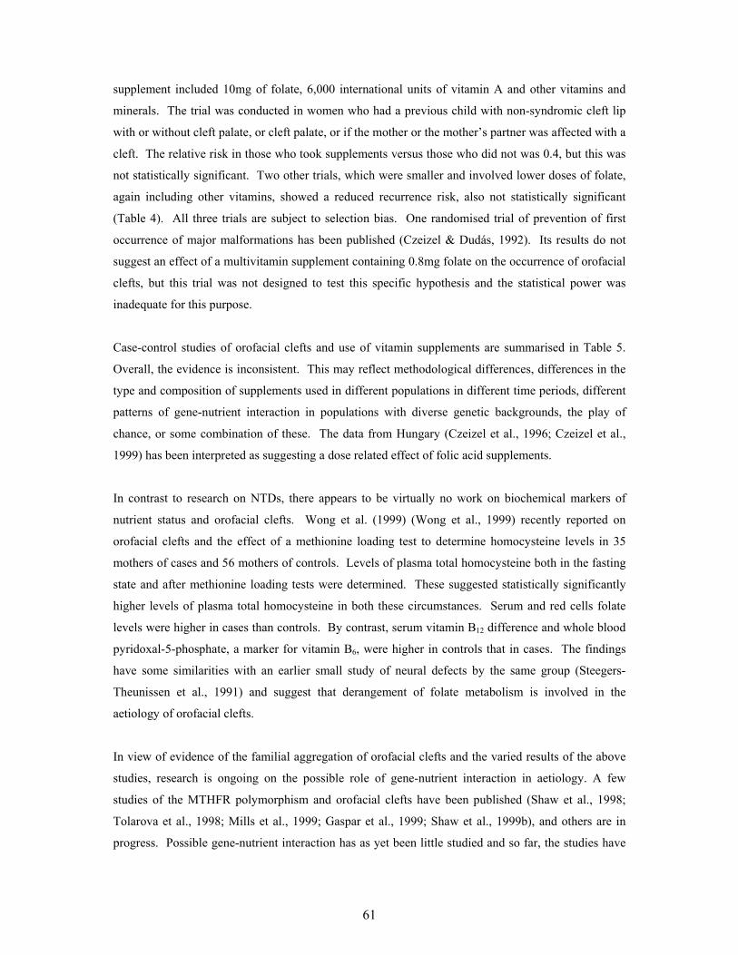
61
supplement included 10mg of folate, 6,000 international units of vitamin A and other vitamins and
minerals. The trial was conducted in women who had a previous child with non-syndromic cleft lip
with or without cleft palate, or cleft palate, or if the mother or the mother’s partner was affected with a
cleft. The relative risk in those who took supplements versus those who did not was 0.4, but this was
not statistically significant. Two other trials, which were smaller and involved lower doses of folate,
again including other vitamins, showed a reduced recurrence risk, also not statistically significant
(Table 4). All three trials are subject to selection bias. One randomised trial of prevention of first
occurrence of major malformations has been published (Czeizel & Dudás, 1992). Its results do not
suggest an effect of a multivitamin supplement containing 0.8mg folate on the occurrence of orofacial
clefts, but this trial was not designed to test this specific hypothesis and the statistical power was
inadequate for this purpose.
Case-control studies of orofacial clefts and use of vitamin supplements are summarised in Table 5.
Overall, the evidence is inconsistent. This may reflect methodological differences, differences in the
type and composition of supplements used in different populations in different time periods, different
patterns of gene-nutrient interaction in populations with diverse genetic backgrounds, the play of
chance, or some combination of these. The data from Hungary (Czeizel et al., 1996; Czeizel et al.,
1999) has been interpreted as suggesting a dose related effect of folic acid supplements.
In contrast to research on NTDs, there appears to be virtually no work on biochemical markers of
nutrient status and orofacial clefts. Wong et al. (1999) (Wong et al., 1999) recently reported on
orofacial clefts and the effect of a methionine loading test to determine homocysteine levels in 35
mothers of cases and 56 mothers of controls. Levels of plasma total homocysteine both in the fasting
state and after methionine loading tests were determined. These suggested statistically significantly
higher levels of plasma total homocysteine in both these circumstances. Serum and red cells folate
levels were higher in cases than controls. By contrast, serum vitamin B12 difference and whole blood
pyridoxal-5-phosphate, a marker for vitamin B6, were higher in controls that in cases. The findings
have some similarities with an earlier small study of neural defects by the same group (Steegers-
Theunissen et al., 1991) and suggest that derangement of folate metabolism is involved in the
aetiology of orofacial clefts.
In view of evidence of the familial aggregation of orofacial clefts and the varied results of the above
studies, research is ongoing on the possible role of gene-nutrient interaction in aetiology. A few
studies of the MTHFR polymorphism and orofacial clefts have been published (Shaw et al., 1998;
Tolarova et al., 1998; Mills et al., 1999; Gaspar et al., 1999; Shaw et al., 1999b), and others are in
progress. Possible gene-nutrient interaction has as yet been little studied and so far, the studies have

62
lacked statistical power to detect departures from multiplicative interaction. Work also is ongoing on
biochemical markers of maternal folate status.
In a comparison of data on 1962 infants with oral clefts and 8387 infants with malformation other
than oral clefts and neural tube, cardiovascular, urinary tract or limb reduction defects the relative risk
of oral clefts associated with maternal use of folic acid antagonist of any type was 2.1 (95% CI 1.4-
3.2; (Hernández-Díaz et al., 2000)). In regard to antiepileptic drugs, the inverse risk appeared to be
specific for cleft lip ± palate.
Vitamin A has been investigated in relation to the aetiology of clefts primarily in the context of
anomalies of structures derived from the neural crest cells. There has been some controversy about
the relationship between these anomalies and use of supplements containing vitamin A. One study of
anomalies of structures derived from these cells suggested that high levels of dietary and
supplemental retinol intake may increase the risk (Rothman et al., 1995). Data specifically on
orofacial clefts and use of supplements containing vitamin A are inconsistent (Table 6). No clear
relationship with maternal blood retinol or vitamin A levels has been found. However, blood retinol
level is under tight homeostatic control (Hunter, 1990), does not clearly reflect dietary intake, and the
metabolic factors controlling it are unclear. In the trial of prevention of recurrence of oral clefts in the
Czech Republic (Tolarova & Harris, 1995) (Table 4), the multivitamin supplement included 6000 IU
of vitamin A. The recurrence risk was reduced in those who took the supplement.
There appears to be no association between oral clefts and reported maternal intake of coffee or
caffeine (Golding, 1995).
The relationships between oral clefts and maternal body mass index, or height, or weight gain in
pregnancy has been analysed only in one investigation (Shaw et al., 2000). No associations were
apparent.
2.6 Hypospadias In a cohort study of diet and pregnancy outcome in the Bristol area of the UK, there was positive
association with maternal vegetarianism in pregnancy (OR 3.53, 95% CI 1.56-7.98), with use of iron
tablets in the first 18 weeks of gestation (OR 1.87, 95% CI 1.02-3.46) and with consumption of pulses
during pregnancy (OR for use 4 or more times a week compared with no use 7.56, 95% CI 2.25-25-
42) (North et al., 2000). In the Hungarian case-control study, the OR for hypospadias associated with
maternal folic acid supplementation at any time during pregnancy was 0.88 (95% CI 0.78-1.01), and

63
that associated with use of supplements containing vitamin A was 0.89 (95% CI 0.8-1.0) (Czeizel et
al., 1996; Czeizel & Rockenbauer, 1998).
2.7 Down Syndrome
DNA hypomethylation has been associated with abnormal chromosome segregation, and methyl
deficiency can result in DNA hypomethylation (Rosenblatt, 1999). This has led to investigations of
the MTHFR C677T polymorphism and folate metabolism in mothers of children with Down
Syndrome. About a two-fold increase in the risk of Down Syndrome in mothers who were
homozygous or heterozygous for the C677T-variant was observed both in an initial study (James et
al., 1999) and in an expansion of this study (Hobbs, 2000). In the initial study, a significant increase
in plasma homocysteine concentrations and lymphocyte methotrexate cytotoxicity was observed in
the mothers of children with Down Syndrome. In the subsequent study, homozygosity for the
methionine synthase reductase (MTRR) A66G polymorphism was associated with an increased risk of
a having child with Down Syndrome. In a study in Ireland, there was no significant association
between maternal MTHFR C677T genotype and Down Syndrome in the offspring, but there was a
positive association with homozygosity and heterozygosity for the MTRR A66G variant (O'Leary et
al., 2002). The MTRR variant was not associated with increased plasma homocysteine.
In a large case-control study in California, maternal consumption of four or more cups of coffee per
day was inversely associated with the risk of Down Syndrome (OR 0.63, 95% CI 0.41-0.96) (Torfs &
Christianson, 2000). This was interpreted as suggesting that high levels of caffeine intake may result
in miscarriage of a fetus with Down Syndrome. Case and control mothers who had high levels of
coffee consumption had previous miscarriage rates that were about 50% higher than mothers who
consumed less coffee.
2.8 Urinary tract malformations In a randomised trial comparing multi-vitamin supplement taken during the periconceptional period
with a supplement containing trace elements only in Hungary in Hungary, urinary tract abnormalities
appeared to contribute to a reduction in the prevalence at birth of congenital anomalies of all types
(Czeizel, 1996). Two cases of congenital anomalies of the urinary tract occurred in the multi-vitamin
group, compared with 9 in the trace element group (relative risk 0.22, 95% CI 0.05-0.99). This
difference appeared to be due mainly to obstructive defects of the urinary system. In a case-control
study in Hungary, the ORs of renal agenesis associated with folic acid supplementation any time
during pregnancy was 0.53 (95% CI 0.23-1.26) and for cystic kidney 0.68 (95% CI 0.35-1.31)

64
(Czeizel et al., 1996). The OR of cystic kidney associated with maternal vitamin A use during
pregnancy was 0.70 (95% CI 0.3-1.5) (Czeizel & Rockenbauer, 1998). In two case-control studies in
the United States, there was an inverse association between urinary tract defects and maternal
multivitamin use in early pregnancy (Werler et al., 1999).
In a comparison of data in 1100 infants with urinary tract malformations and 8387 infants with
malformations other than urinary tract, neural tube, cardiovascular or limb reduction defects, or oral
clefts, the relative risk of urinary tract malformations associated with maternal use of a folic acid
antagonist of any type was 2.1 (95% CI 1.2-3.7; (Hernández-Díaz et al., 2000)). Very few subjects
were exposed to dihydrofolate reductase inhibitors. Maternal supplementation with multivitamins
containing folic acid did not appear to reduce the risk associated with antiepileptics.
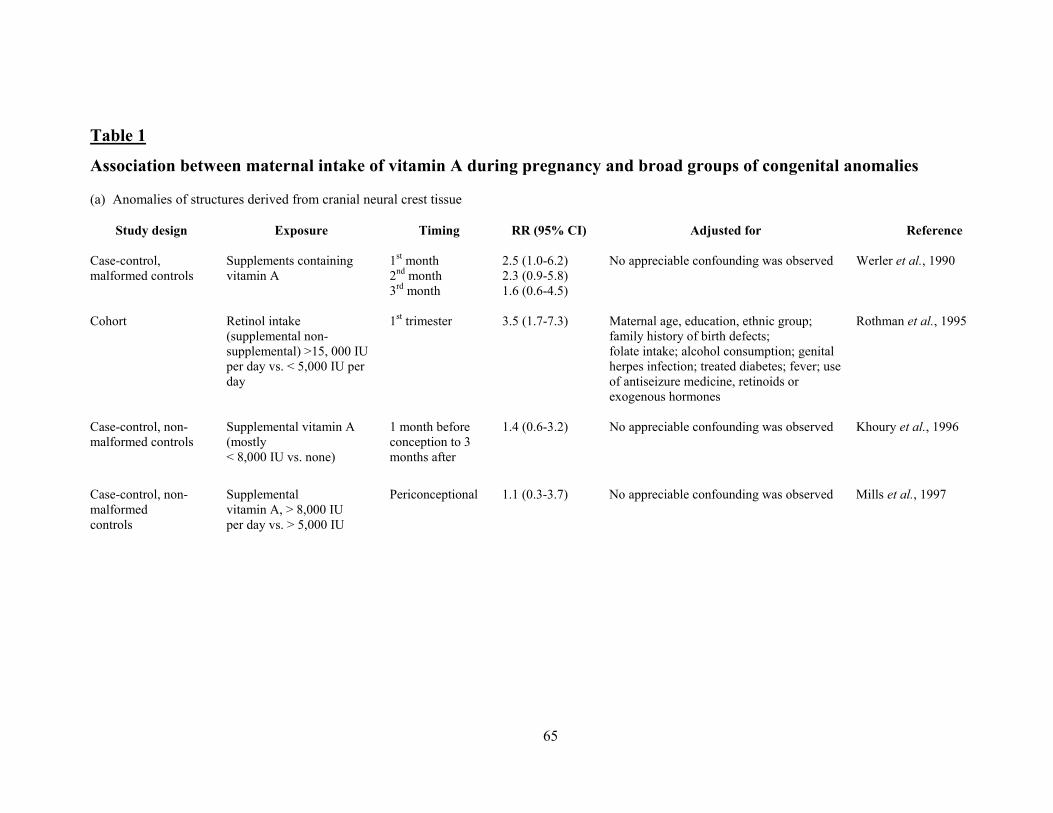
65
Table 1
Association between maternal intake of vitamin A during pregnancy and broad groups of congenital anomalies (a) Anomalies of structures derived from cranial neural crest tissue
Study design Exposure Timing RR (95% CI) Adjusted for Reference
Case-control, malformed controls
Supplements containing vitamin A
1st month 2nd month 3rd month
2.5 (1.0-6.2) 2.3 (0.9-5.8) 1.6 (0.6-4.5)
No appreciable confounding was observed Werler et al., 1990
Cohort Retinol intake
(supplemental non-supplemental) >15, 000 IU per day vs. < 5,000 IU per day
1st trimester 3.5 (1.7-7.3) Maternal age, education, ethnic group; family history of birth defects; folate intake; alcohol consumption; genital herpes infection; treated diabetes; fever; use of antiseizure medicine, retinoids or exogenous hormones
Rothman et al., 1995
Case-control, non-malformed controls
Supplemental vitamin A (mostly < 8,000 IU vs. none)
1 month before conception to 3 months after
1.4 (0.6-3.2) No appreciable confounding was observed Khoury et al., 1996
Case-control, non-malformed controls
Supplemental vitamin A, > 8,000 IU per day vs. > 5,000 IU
Periconceptional 1.1 (0.3-3.7) No appreciable confounding was observed Mills et al., 1997
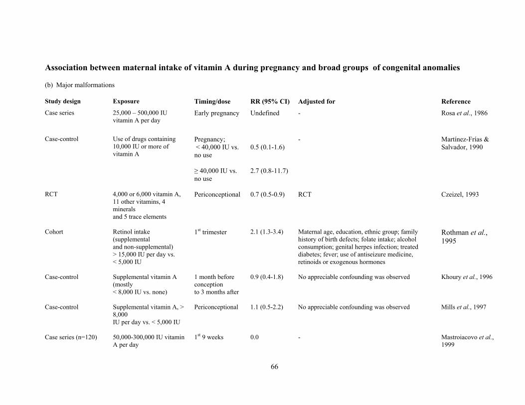
66
Association between maternal intake of vitamin A during pregnancy and broad groups of congenital anomalies (b) Major malformations Study design Exposure Timing/dose RR (95% CI) Adjusted for Reference
Case series 25,000 – 500,000 IU vitamin A per day
Early pregnancy Undefined - Rosa et al., 1986
Case-control Use of drugs containing 10,000 IU or more of vitamin A
Pregnancy; < 40,000 IU vs. no use ≥ 40,000 IU vs. no use
0.5 (0.1-1.6) 2.7 (0.8-11.7)
- Martínez-Frías & Salvador, 1990
RCT 4,000 or 6,000 vitamin A, 11 other vitamins, 4 minerals and 5 trace elements
Periconceptional 0.7 (0.5-0.9) RCT Czeizel, 1993
Cohort Retinol intake (supplemental and non-supplemental) > 15,000 IU per day vs. < 5,000 IU
1st trimester 2.1 (1.3-3.4) Maternal age, education, ethnic group; family history of birth defects; folate intake; alcohol consumption; genital herpes infection; treated diabetes; fever; use of antiseizure medicine, retinoids or exogenous hormones
Rothman et al., 1995
Case-control Supplemental vitamin A (mostly < 8,000 IU vs. none)
1 month before conception to 3 months after
0.9 (0.4-1.8) No appreciable confounding was observed Khoury et al., 1996
Case-control Supplemental vitamin A, > 8,000 IU per day vs. < 5,000 IU
Periconceptional 1.1 (0.5-2.2) No appreciable confounding was observed Mills et al., 1997
Case series (n=120) 50,000-300,000 IU vitamin A per day
1st 9 weeks 0.0 - Mastroiacovo et al., 1999
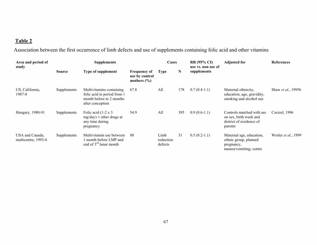
67
Table 2
Association between the first occurrence of limb defects and use of supplements containing folic acid and other vitamins
Supplements
Cases Adjusted for References Area and period of study
Source Type of supplement Frequency of use by control mothers (%)
Type N
RR (95% CI) use vs. non use of supplements
US, California, 1987-8
Supplements Multivitamins containing folic acid in period from 1 month before to 2 months after conception
67.8 All 178 0.7 (0.4-1.1) Maternal ethnicity, education, age, gravidity, smoking and alcohol use
Shaw et al., 1995b
Hungary, 1980-91 Supplements Folic acid (1-2 x 3 mg/day) ± other drugs at any time during pregnancy
54.9 All 395 0.9 (0.6-1.1) Controls matched with are on sex, birth week and district of residence of parents
Czeizel, 1996
USA and Canada, multicentre, 1993-6
Supplements Multivitamin use between 1 month before LMP and end of 3rd lunar month
88 Limb reduction defects
31 0.5 (0.2-1.1) Maternal age, education, ethnic group, planned pregnancy, nausea/vomiting; centre
Werler et al., 1999

68
Table 3
Association between the first occurrence of congenital heart defects and use of supplements containing folic acid and other vitamins
Supplements
Cases Adjusted for References Area and period of study
Source Type of supplement Frequency of use by control mothers (%)
Type N
RR (95% CI) use vs. non use of supplements
USA, Atlanta, 1968-80
Supplements Multivitamin use from 3 months before pregnancy to end of 3rd month of pregnancy
14.2 Non-syndromic cardiac defects
958 0.8 (0.6-1.0) Maternal ethnic group, maternal diabetes, period of birth
Botto et al., 2000b
US, California, 1987-8
Supplements Multivitamins containing folic acid in period from 1 month before to 2 months after conception
67.8 Conotruncal heart defects
207 0.5 (.03-0.9) Maternal ethnicity, education, age, gravidity, smoking and alcohol use
Shaw et al., 1995b
Hungary, 1980-91 Supplements Folic acid (1-2 x 3 mg/day) ± other drugs at any time during pregnancy
54.9 All types 2976 0.9 (0.8-1.0) Controls matched with cases on sex, birth week and district of residence of parents
Czeizel, 1996
USA and Canada, multicentre, 1993-6
Supplements Multivitamin use between 1 month before LMP and end of 2nd lunar month
88 Conotruncal defects VSD
157 186
1.0 (0.7-1.5) 1.2 (0.8-1.8)
Maternal age, education, ethnic group, planned pregnancy, nausea/vomiting and centre
Werler et al., 1999
USA, Maryland, Washington, DC, N Virginia 1987-9
Supplements Multivitamins containing folic acid (≥ 400 mg vs. < 400 mg per day) in year before conception
17% (≥ 400 mg)
Non-syndromic outflow tract defects
126 1.0 (0.6-1.6) Parity, marital status, drug use, daily use of ≥ 10,000 IU of supplemental vitamin A
Scanlon et al., 1998
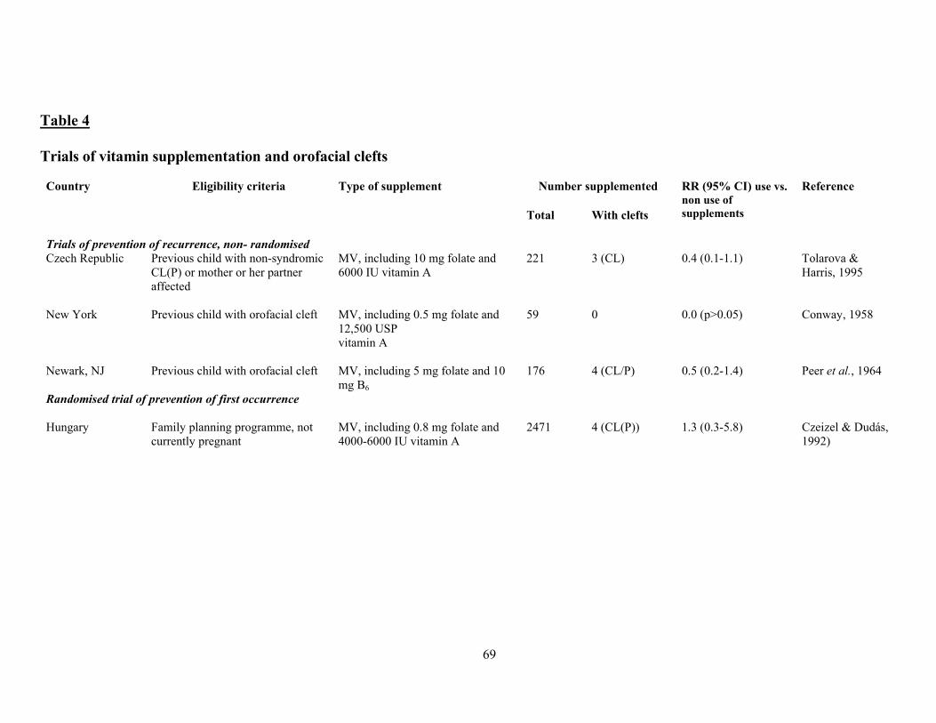
69
Table 4 Trials of vitamin supplementation and orofacial clefts
Country Eligibility criteria Type of supplement Number supplemented
Reference
Total With clefts
RR (95% CI) use vs. non use of supplements
Trials of prevention of recurrence, non- randomised Czech Republic Previous child with non-syndromic
CL(P) or mother or her partner affected
MV, including 10 mg folate and 6000 IU vitamin A
221 3 (CL) 0.4 (0.1-1.1) Tolarova & Harris, 1995
New York Previous child with orofacial cleft MV, including 0.5 mg folate and 12,500 USP vitamin A
59 0 0.0 (p>0.05) Conway, 1958
Newark, NJ Previous child with orofacial cleft
MV, including 5 mg folate and 10 mg B6
176 4 (CL/P) 0.5 (0.2-1.4) Peer et al., 1964
Randomised trial of prevention of first occurrence
Hungary Family planning programme, not currently pregnant
MV, including 0.8 mg folate and 4000-6000 IU vitamin A
2471 4 (CL(P)) 1.3 (0.3-5.8) Czeizel & Dudás, 1992)
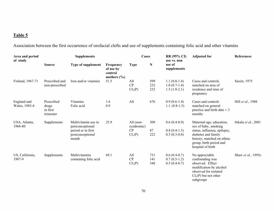
70
Table 5 Association between the first occurrence of orofacial clefts and use of supplements containing folic acid and other vitamins
Supplements
Cases References Area and period of study
Source Type of supplement Frequency of use by control mothers (%)
Type N
RR (95% CI) use vs. non use of supplements
Adjusted for
Finland, 1967-71 Prescribed and non-prescribed
Iron and/or vitamins 51.5 All CP CL(P)
599 232 232
1.1 (0.8-1.4) 1.0 (0.7-1.4) 1.5 (1.0-2.1)
Cases and controls matched on area of residence and time of pregnancy
Saxén, 1975
England and Wales, 1983-4
Prescribed drugs in first trimester
Vitamins Folic acid
1.6 0.9
All 676 0.9 (0.6-1.4) 1.1 (0.8-1.5)
Cases and controls matched on general practice and birth date ± 3 months
Hill et al., 1988
USA, Atlanta, 1968-80
Supplements Multivitamin use in periconceptional period or in first postconceptional month
25.9 All (non-syndromic) CP CL(P)
309 87 222
0.6 (0.4-0.9) 0.8 (0.4-1.5) 0.5 (0.3-0.8)
Maternal age, education, sex of baby, smoking status, influenza, epilepsy, diabetes and family history; matched on ethnic group, birth period and hospital of birth
Itikala et al., 2001
US, California, 1987-9
Supplements Multivitamins containing folic acid
69.1 All CP CL(P)
731 141 348
0.6 (0.4-0.7) 0.7 (0.5-1.2) 0.5 (0.4-0.7)
No appreciable confounding was observed. Effect modification by alcohol observed for isolated CL(P) but not other subgroups
Shaw et al., 1995c
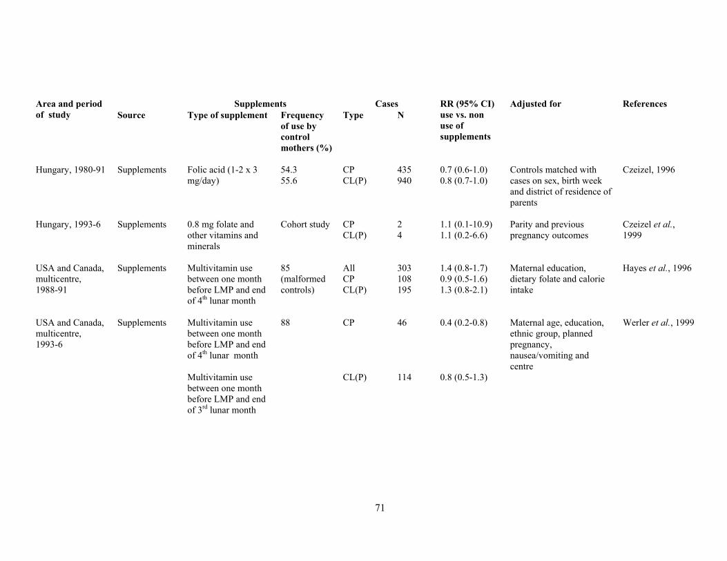
71
Supplements Cases Adjusted for References Area and period
of study Source Type of supplement Frequency of use by control mothers (%)
Type N RR (95% CI) use vs. non use of supplements
Hungary, 1980-91 Supplements Folic acid (1-2 x 3 mg/day)
54.3 55.6
CP CL(P)
435 940
0.7 (0.6-1.0) 0.8 (0.7-1.0)
Controls matched with cases on sex, birth week and district of residence of parents
Czeizel, 1996
Hungary, 1993-6 Supplements 0.8 mg folate and other vitamins and minerals
Cohort study CP CL(P)
2 4
1.1 (0.1-10.9) 1.1 (0.2-6.6)
Parity and previous pregnancy outcomes
Czeizel et al., 1999
USA and Canada, multicentre, 1988-91
Supplements Multivitamin use between one month before LMP and end of 4th lunar month
85 (malformed controls)
All CP CL(P)
303 108 195
1.4 (0.8-1.7) 0.9 (0.5-1.6) 1.3 (0.8-2.1)
Maternal education, dietary folate and calorie intake
Hayes et al., 1996
USA and Canada, multicentre, 1993-6
Supplements Multivitamin use between one month before LMP and end of 4th lunar month Multivitamin use between one month before LMP and end of 3rd lunar month
88 CP CL(P)
46 114
0.4 (0.2-0.8) 0.8 (0.5-1.3)
Maternal age, education, ethnic group, planned pregnancy, nausea/vomiting and centre
Werler et al., 1999
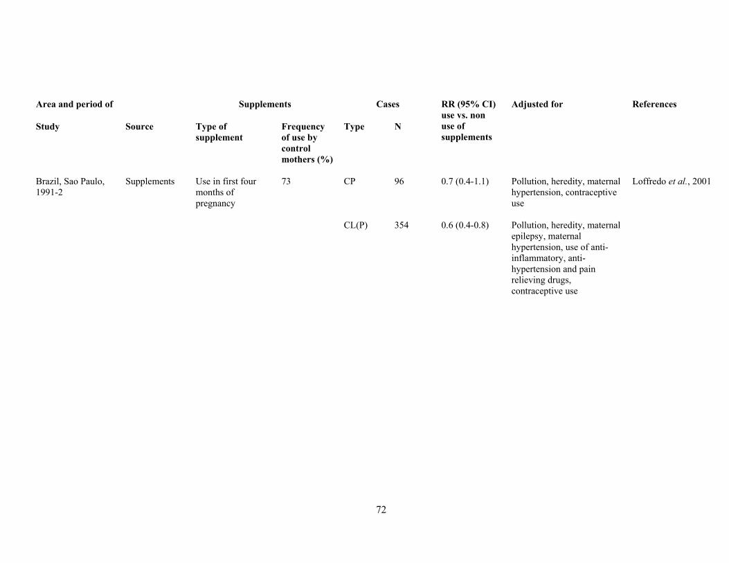
72
Area and period of
Supplements Cases Adjusted for References
Study Source Type of supplement
Frequency of use by control mothers (%)
Type N
RR (95% CI) use vs. non use of supplements
Brazil, Sao Paulo, 1991-2
Supplements Use in first four months of pregnancy
73 CP CL(P)
96 354
0.7 (0.4-1.1) 0.6 (0.4-0.8)
Pollution, heredity, maternal hypertension, contraceptive use Pollution, heredity, maternal epilepsy, maternal hypertension, use of anti-inflammatory, anti-hypertension and pain relieving drugs, contraceptive use
Loffredo et al., 2001

73
Table 6 Association between maternal intake of vitamin A during pregnancy and orofacial clefts
Study design Number of
cases Nature of exposure
Timing or dose RR (95% CI) Adjusted for Ref
Case-control, malformed controls
1332 Supplements containing vitamin A
1st month 2nd month 3rd month
2.3 (0.8-6.6) 2.0 (0.6-6.0) 1.3 (0.4-4.6)
No appreciable confounding was observed
Werler et al., 1990
Case-control, non-malformed controls
731 Supplements containing vitamin A
1 month before conception to 3 months after
0.6 (0.2-1.5) - Knox, 1972
Case-control, non-malformed controls
CL±P 954 CP 433
Vitamin A supplements
1st trimester CL±P 1.0 (0.6-1.8) CP 0.3 (0.1-1.4)
Controls were matched with cases on sex, birth week and district of residence of parents
Czeizel & Rockenbauer, 1998
Case-control, non-malformed controls
59 Maternal serum retinol
Postnatal Similar between mothers of cases and controls
- Niebyl et al., 1985
Case-control, non-malformed controls
141 Maternal serum retinol
Postnatal Lower in mothers of cases than controls
- Munger et al., 1996
Nested case-control, non-malformed controls
CL±P 14 Maternal plasma vitamin A
Early pregnancy Similar between mothers of cases and controls
- Stoll et al., 1999
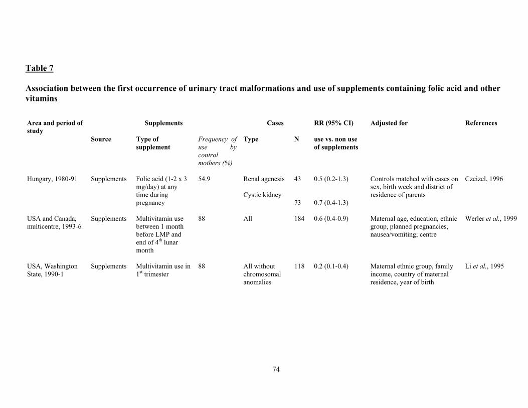
74
Table 7 Association between the first occurrence of urinary tract malformations and use of supplements containing folic acid and other vitamins
Supplements
Cases RR (95% CI) Adjusted for References Area and period of study
Source Type of supplement
Frequency of use by control mothers (%)
Type N use vs. non use of supplements
Hungary, 1980-91 Supplements Folic acid (1-2 x 3 mg/day) at any time during pregnancy
54.9 Renal agenesis Cystic kidney
43 73
0.5 (0.2-1.3) 0.7 (0.4-1.3)
Controls matched with cases on sex, birth week and district of residence of parents
Czeizel, 1996
USA and Canada, multicentre, 1993-6
Supplements Multivitamin use between 1 month before LMP and end of 4th lunar month
88 All 184
0.6 (0.4-0.9) Maternal age, education, ethnic group, planned pregnancies, nausea/vomiting; centre
Werler et al., 1999
USA, Washington State, 1990-1
Supplements Multivitamin use in 1st trimester
88 All without chromosomal anomalies
118 0.2 (0.1-0.4) Maternal ethnic group, family income, country of maternal residence, year of birth
Li et al., 1995
REFERENCES
Berry RJ, Li Z, Erickson JD, Li S, Moore CA, Wang H, Mulinare J, Zhao P, Wong LYC, Gindler J, Hong SX & Correa A (1999) Prevention of neural-tube defects with folic acid in China. New England Journal of Medicine 341, 1485-1490
Boneva RS, Moore CA, Botto L, Wong LY & Erickson JD (1999) Nausea during pregnancy and congenital heart defects: a population-based case-control study. American Journal of Epidemiology 149, 717-725
Botto LD, Erickson JD & Mulinare J (2000a) Neural-tube defects. New England Journal of Medicine 342, 1136
Botto LD, Mulinare J & Erickson JD (2000b) Occurrence of congenital heart defects in relation to maternal multivitamin use. American Journal of Epidemiology 151, 878-884
Botto LD, Loffredo C, Scanlon KS, Ferencz C, Khoury MJ, Wilson PD & Correa A (2001) Vitamin A and cardiac outflow tract defects. Epidemiology 12, 491-496
Botto D & Yang Q (2000) 5,10 methylenetetrahydrofolate reductase gene variants and congenital anomalies: a HuGE review. American Journal of Epidemiology 151, 862-877
Bower C & Stanley FJ (1989) Dietary folate as a risk factor for neural-tube defects: evidence from a case-control study in Western Australia. Medical Journal of Australia 150, 613-619
Centers for Disease Control (2000) Folate status in women of childbearing age - United States, 1999. Morbidity and Mortality Weekly Reports 49, 962-965
Clayton D & McKeigue PM (2001) Epidemiological methods for studying genes and environmental factors in complex diseases. Lancet 358, 1356-1360
Cohlan SQ (1953) Excessive intake of vitamin A as a cause of congenital anomalies in rat. Science 117, 535-536
Conway H (1958) Effect of supplemental vitamin therapy on the limitation of incidence of cleft lip and cleft palate in humans. Plastic and Reconstructive Surgery 22, 450-453
Cornel MC & Erickson JD (1997) Comparison of national policies on periconceptional use of folic acid to prevent spina bifida and anencephaly (SBA). Teratology 55, 134-137
Croen LA, Todoroff K & Shaw GM (2001) Maternal exposure to nitrate from drinking water and diet and risk for neural tube defects. American Journal of Epidemiology 153, 325-331
Czeizel A (1996) Reduction in urinary tract and cardiovascular defects by periconceptional multivitamin supplementation. American Journal of Medical Genetics 62, 179-183
Czeizel AE (1993) Prevention of congenital abnormalities by periconceptional multivitamin supplementation. British Medical Journal 306, 1645-1648
Czeizel AE & Dudás I (1992) Prevention of the first occurrence of neural-tube defects by periconceptional vitamin supplementation. New England Journal of Medicine 327, 1832-1835
Czeizel AE & Rockenbauer M. (1998) Prevention of congenital abnormalities by vitamin A. International Journal for Vitamin and Nutritional Research 68, 219-231

76
Czeizel AE, Tímár L & Sárközi A (1999) Dose-dependent effect of folic acid on the prevention of orofacial clefts . Pediatrics 104, 66
Czeizel AE, Tóth M & Rockenbauer M (1996) Population-based case control study of folic acid supplementation during pregnancy. Teratology 53, 345-351
Daly LE, Kirke PN, Molloy A, Weir DG & Scott JM (1995) Folate levels and neural tube defects: Implications for prevention. Journal of the American Medical Association 274, 1698-1702
De Walle HEK, van der Pal KM, de Jong-van den Berg LTW, Jeeninga W, Schouten JSAG, de Rover CM, Buitendijk SE & Cornel MC (1999) Effect of mass media campaign to reduce socio-economic differences in women's awareness and behaviour concerning use of folic acid: cross sectional study. British Medical Journal 319, 291-292
Dolk HM, Nau H, Hummler H & Barlow SM (1999) Dietary vitamin A and teratogenic risk: European Teratology Society discussion paper. European Journal of Obstetrics, Gynecology, and Reproductive Biology 83, 31-36
Elwood M, Little J & Elwood H (1992a) Conclusions. In: Epidemiology and Control of Neural Tube Defects, (Monographs in Epidemiology and Biostatistics). edited by Elwood JM, Little J & Elwood JH, Oxford University Press, Oxford. p757-777
Elwood JM, Elwood JH & Little J (1992b) Diet. In: Epidemiology and Control of Neural Tube Defects, edited by Elwood JM, Little J & Elwood JH. Oxford University Press, Oxford. p521-603
Elwood M & Little J (1992) Secular trends. In: Epidemiology and Control of Neural Tube Defects, (Monographs in Epidemiology and Biostatistics) edited by Elwood JM, Little J & Elwood JH. Oxford University Press, Oxford. p168-194
Friel JK, Frecker M & Fraser FC (1995) Nutritional patterns of mothers of children with neural tube defects in Newfoundland. American Journal of Medical Genetics 55, 195-199
Gaspar DA, Pavanello RC, Zatz M, Passos-Bueno MR, André M, Steman S, Wyszynski DF & Matiolli SR (1999) Role of the C677T polymorphism at the MTHFR gene on risk to nonsyndromic cleft lip with/without cleft palate: Results from a case-control study in Brazil. American Journal of Medical Genetics 87, 197-199
Geelen JA (1979) Hypervitaminosis A induced teratogenesis. CRC Critical Reviews in Toxicology 6, 351-375
Golding J (1995) Reproduction and caffeine consumption: A literature review. Early Human Development 43, 1-14
Hallberg L, Sandström B, Ralph A & Arthur J (2000) Iron, zinc and other trace elements. In: Human Nutrition and Dietetics, 10th Ed, edited by Garrow JS, James WPT & Ralph A, Churchill, Livingstone. P177-209
Hayes C, Werler MM, Willett WC & Mitchell AA (1996) Case-control study of periconceptional folic acid supplementation and oral clefts. American Journal of Epidemiology 143, 1229-1234
Hendricks KA, Nuno OM, Suarez L & Larsen R (2001) Effects of hyperinsulinemia and obesity on risk of neural tube defects among Mexican Americans. Epidemiology 12, 630-635

77
Hernández-Diaz S, Werler MM, Walker AM & Mitchell AA (2001) Neural tube defects in relation to use of folic acid antagonists during pregnancy. American Journal of Epidemiology 153, 961-968
Hernández-Díaz S, Werler MM, Walker AM & Mitchell AA (2000) Folic acid antagonists during pregnancy and the risk of birth defects. New England Journal of Medicine 343, 1608-1614
Hill L, Murphy M, McDowall M & Paul AH (1988) Maternal drug histories and congenital malformations: limb reduction defects and oral clefts. Journal of Epidemiology and Community Health 42, 1-7
Hobbs CA (2000) Polymorphisms in genes involvedin folate metabolism as maternal risk factors for Down syndrome. American Journal of Human Genetics 67, 623-630
Honein MA, Paulozzi LJ, Mathews TJ, Erickson JD & Wong LYC (2001) Impact of folic acid fortification of the US food supply on the occurrence of neural tube defects. Journal of the American Medical Association 285, 2981-3023
Hunter D (1990) Biochemical indicators of dietary intake. In: Nutritional Epidemiology, edited by Willett W, Oxford University Press, New York. p143-216
Hurley LS & Swenerton H (1966) Congenital malformations resulting from zinc deficiency in rats. Proceedings of the Society for Experimental Biology & Medicine 123, 692-696
Itikala PR, Watkins ML, Mulinare J, Moore CA & Liu Y (2001) Maternal multivitamin use and orofacial clefts in offspring. Teratology 63, 79-86
James SJ, Pogribna M, Pogribny IP, Melnyk S , Hine RJ, Gibson JB, Yi P, Tafoya DL, Swenson DH, Wilson VL & Gaylor DW (1999) Abnormal folate metabolism and mutation in the methylenetetrahydrofolate reductase gene may be maternal risk factors for Down syndrome. American Journal of Clinical Nutrition 70, 495-501
Junker R, Kotthoff S, Vielhaber H, Halimeh S, Kosch A, Koch HG, Kassenbohmer R, Heineking B & Nowak-Gottl U (2001) Infant methylenetetrahydrofolate reductase 677TT genotype is a risk factor for congenital heart disease. Cardiovascular Research 51, 251-254
Källén K (1998) Maternal smoking, body mass index, and neural tube defects. American Journal of Epidemiology 147, 1103-1111
Khoury MJ, Moore CA & Mulinare J (1996) Vitamin A and birth defects. Lancet 347, 321-322
Kirke PN, Daly LE & Elwood JH (1992) A randomised trial of low dose folic acid to prevent neural tube defects. Archives of Disease in Childhood 67 , 1442-1446
Kirke PN, Molloy AM, Daly LE, Burke H, Weir DG & Scott JM (1993) Maternal plasma folate and vitamin B12 are independent risk factors for neural tube defects. Quarterly Journal of Medicine 86, 703-708
Knox EG (1972) Anencephalus and dietary intakes. British Journal of Preventive and Social Medicine 26, 219-223
Lammer EJ, Chen DT, Hoar RM, Agnish ND, Benke PJ, Braun JT, Curry CJ, Fernhoff PM, Grix AW, Lott IT, Richard JM & Sun SC (1985) Retinoic acid embryopathy. New England Journal of Medicine 313, 837-841
Lancet (1985) Vitamin A and teratogenesis. Lancet i, 319-320

78
Laurence KM, James N, Miller MH, Tennant GB & Campbell H (1981) Double-blind randomised controlled trial of folate treatment before conception to prevent recurrence of neural-tube defects. British Medical Journal 282, 1509-1511
Lenke RL & Levy HL (1980) Maternal phenylketonuria and hyperphenylalaninemia: an international survey of the outcome of untreated and treated pregnancies. New England Journal of Medicine 303, 1202-1208
Levy HL, Guldberg P, Guttler F, Hanley WB, Matalon R, Rouse BM, Trefz F, Azen C, Allred EN, de la Cruz F & Koch R (2001) Congenital heart disease in maternal phenylketonuria: report from the maternal PKU collaborative study. Pediatric Research 49, 636-642
Li DK, Daling JR, Mueller BA, Hickok DE, Fantel AG & Weiss NS (1995) Periconceptonal multivitamin use in relation to the risk of congenital urinary tract anomalies. Epidemiology 6, 212-218
Little J (1995) Is folic acid pluripotent? A review of the associations with congenital anomalies, cancer and other diseases. In: Drugs, Diet and Disease. Volume 1. Mechanistic Approaches to Cancer, edited by Ioannides C & Lewis DFV. Ellis Horwood, New York. p259-308
Little J (1999) Problems and approaches in investigating the role of micronutrients in the aetiology of cancer in humans. British Medical Bulletin 55, 600-618
Little J & Elwood JM (1992) Geographical variation. In: Epidemiology and Control of Neural Tube Defects, edited by Elwood JM, Little J & Elwood JH. Oxford University Press, Oxford. p96-145
Loffredo LCM, Souza JMP, Freitas JAS & Mossey PA (2001) Oral clefts and vitamin supplementation. Cleft Palate-Craniofacial Journal 38, 76-83
Lucock M (2000) Folic acid: nutritional biochemistry, molecular biology, and role in disease processes. Molecular Genetics and Metabolism 71, 121-138
Lumley J, Watson L, Watson M & Bower C (2000) Periconceptional supplementation with folate and/or multivitamins for preventing neural tube defects. Cochrane Database of Systematic Reviews [computer file] CD001056
Martínez-Frías ML & Salvador J (1990) Epidemiological aspects of prenatal exposure to high doses of vitamin A in Spain. European Journal of Epidemiology 6, 118-123
Mastroiacovo P, Mazzone T, Addis A, Elephant E, Carlier P, Vial T, Garbis H, Robert E, Bonati M, Ornoy A, Finardi A, Schaffer C, Caramelli L, Rodriguez-Pinilla E & Clementi M (1999) High vitamin A intake in early pregnancy and major malformations: a multicenter prospective controlled study. Teratology 59, 7-11
McKeigue PM, Lamm SH, Linn S & Kutcher JS (1994) Bendectin and birth defects: I. a meta-analysis of the epidemiologic studies. Teratology 50, 27-37
Miller RK, Hendrickx AG, Mills JL, Hummler H & Wiegand UW (1998) Periconceptional vitamin A use: how much is teratogenic? Reproductive Toxicology 12, 75-88
Mills JL & England L (2001) Food Fortification to prevent neural tube defects: Is it working? Journal of the American Medical Association 285, 3022-3023
Mills JL, Kirke PN, Molloy AM, Burke H, Conley MR, Lee YJ, Mayne PD, Weir DG & Scott JM (1999) Methylenetetrahydrofolate reductase thermolabile variant and oral clefts. American Journal of Medical Genetics 86, 71-74

79
Mills JL, McPartlin JM, Kirke PN, Lee YJ, Conley MR, Weir DG & Scott JM (1995) Homocysteine metabolism in pregnancies complicated by neural-tube defects. Lancet 345, 149-151
Mills JL, Rhoads GG, Simpson JL, Cunningham GC, Conley MR, Lassman MR, Walden ME, Depp OR, Hoffman HJ & The National Institute of Child Health and Human Development Neural Tube Defects Study Group (1989) The absence of a relation between the periconceptional use of vitamins and neural-tube defects. New England Journal of Medicine 321, 430-435
Mills JL, Simpson JL, Cunningham GC, Conley MR & Rhoads GG (1997) Vitamin A and birth defects. American Journal of Obstetrics and Gynecology 177, 31-36
Milunsky A, Jick H, Jick SS, Bruell CL, MacLaughlin DS, Rothman KJ & Willett W (1989) Multivitamin/folic acid supplementation in early pregnancy reduces the prevalence of neural tube defects. Journal of the American Medical Association 262, 2847-2852
Molloy AM, Kirke P, Hillary I, Weir DG & Scott JM (1985) Maternal serum folate and vitamin B12 concentrations in pregnancies associated with neural tube defects. Archives of Disease in Childhood 60, 660-665
Moore LL (2001) Is the jury still out on folic acid and congenital anomalies? Epidemiology 12, 141-144
MRC Vitamin Study Research Group (1991) Prevention of neural tube defects: results of the Medical Research Council Vitamin Study. Lancet 338, 131-137
Mulinare J, Cordero JF, Erickson JD & Berry RJ (1988) Periconceptional use of multivitamins and the occurrence of neural tube defects. Journal of the American Medical Association 260, 3141-3145
Munger R, Sauberlich H, Murray J, Layda E, Villaneuva E & Magee W (1996) Abnormal folate and retinol levels in Filipino mothers of children with orofactial clefts. FASEB Journal 10, (Abstract)
Nau H, Chahoud I, Dencker L, Lammer EJ & Scott WJ (1994) Teratogenicity of vitamin A and retinoids. In: Vitamin A in Health and Disease, edited by Blomhoff R, Marcel Dekker, New York. p615-663
Niebyl JR, Blake DA, Rocco LE, Baumgardner R & Mellits ED (1985) Lack of maternal metabolic, endocrine, and environmental influences in the etiology of cleft lip with or without cleft palate. Cleft Palate Journal 22, 20-28
North K, Golding J & The ALSPAC Study Team (2000) A maternal vegetarian diet in pregnancy is associated with hypospadias. BJU International 85, 107-113
O'Leary VB, Parle-McDermott A, Molloy AM, Kirke PN, Johnson Z, Conley M, Scott JM & Mills JL (2002) MTRR and MTHFR polymorphism: link to Down Syndrome? American Journal of Medical Genetics 107, 151-155
Oakley GP & Erickson JD (1995) Vitamin A and birth defects. New England Journal of Medicine 333, 1414-1415
Peer LA, Gordon HW & Bernhard WG (1964) Effect of vitamins on human teratology. Plastic and Reconstructive Surgery 34, 358-361
Prentice A & Goldberg G (1996) Maternal obesity increases congenital malformations. Nutrition Reviews 54, 146-150
Preston-Martin S, Pogoda JM, Mueller BA, Lubin F, Holly EA, Filippini G, Cordier S, Peris-Bonet R, Choi W, Little J & Arslan A (1998) Results from an international case-control study of childhood
80
brain tumors: the role of prenatal vitamin supplementation. Environmental Health Perspectives 106, 887-892
Queisser-Luft A, Kieninger-Baum D, Menger H, Stolz G, Schlaefer K & Merz E (1998) Does maternal obesity increase the risk of fetal abnormalities? analysis of 20,248 newborn infants of the Mainz Birth Register for detecting congenital anomalies. Ultraschall Medicine 19, 40-44
Rosa FW (1983) Teratogenicity of isotretinoin. Lancet ii, 513
Rosa FW, Wilk AL & Kelsey FO (1986) Teratogen update: vitamin A congeners. Teratology 33, 355-364
Rosenblatt DS (1999) Folate and homocysteine metabolism and gene polymorphisms in the etiology of Down syndrome. American Journal of Clinical Nutrition 70, 429-430
Rothman KJ, Moore LL, Singer MR, Nguyen UDT, Mannino S & Milunsky A (1995) Teratogenicity of high vitamin A intake. New England Journal of Medicine 333, 1369-1373
Saxén I (1975) Associations between oral clefts and drugs taken during pregnancy. International Journal of Epidemiology 4, 37-44
Scanlon KS, Ferencz C, Loffredo CA, Wilson PD, Correa-Villaseñor A, Khoury MJ, Willett WC & Baltimore-Washington Infant Study Group (1998) Preconceptional folate intake and malformations of the cardiac outflow tract. Epidemiology 9, 95-98
Schorah CJ, Wild J, Hartley R, Sheppard S & Smithells RW (1983) The effect of periconceptional supplementation on blood vitamin concentrations in women at recurrence risk for neural tube defect. British Journal of Nutrition 49, 203-211
Shah D & Sachdev HPS (2001) Effect of gestational zinc deficiency on pregnancy outcomes: Summary of observation studies and zinc supplementation trials. British Journal of Nutrition 85, S101-S108
Shaw GM, Schaffer D, Velie EM, Morland K & Harris JA (1995a) Periconceptional vitamin use, dietary folate, and the occurrence of neural tube defects. Epidemiology 6, 219-226
Shaw GM, O'Malley CD, Wasserman CR, Tolarova MM & Lammer EJ (1995b) Maternal periconceptional use of multivitamins and reduced risk for conotruncal heart defects and limb deficiencies among offspring. American Journal of Medical Genetics 59, 536-545
Shaw GM, Lammer EJ, Wasserman CR, O'Malley CD & Tolarova MM (1995c) Risks of orofacial clefts in children born to women using multivitamins containing folic acid periconceptionally. Lancet 346, 393-396
Shaw GM, Rozen R, Finnell RH, Todoroff K & Lammer EJ (1998) Infant C677T mutation in MTHFR, maternal periconceptional vitamin use, and cleft lip. American Journal of Medical Genetics 80, 196-198
Shaw GM, Todoroff K, Carmichael SL, Schaffer DM & Selvin S (2001) Lowered weight gain during pregnancy and risk of neural tube defects among offspring. International Journal of Epidemiology 30, 60-65
Shaw GM, Todoroff K, Schaffer DM & Selvin S (1999a) Periconceptional nutrient intake and risk for neural tube defect-affected pregnancies. Epidemiology 10, 711-716
Shaw GM, Todoroff K, Finnell RH, Rozen R & Lammer EJ (1999b) Maternal vitamin use, infant C677T mutation in MTHFR, and isolated cleft palate risk. American Journal of Medical Genetics 85, 84-85

81
Shaw GM, Todoroff K, Schaffer DM & Selvin S (2000) Maternal height and prepregnancy body mass index as risk factors for selected congenital anomalies. Paediatric and Perinatal Epidemiology 14, 234-239
Shaw GM, Wasserman CR, Block G & Lammer EJ (1996) High maternal vitamin A intake and risk of anomalies of structures with a cranial neural crest cell contribution. Lancet 347, 899-900
Shenefelt RE (1972) Morphogenesis of malformations in hamsters caused by retinoic acid: relation to dose and stage at treatment. Teratology 5, 103-118
Smithells RJ, Sheppard S & Schorah CJ (1976) Vitamin deficiencies and neural tube defects. Archives of Disease in Childhood 51, 944-950
Smithells RW, Seller MJ, Harris R, Fielding DW, Schorah CJ, Nevin NC, Sheppard S, Read AP, Walker S & Wild J (1983) Further experience of vitamin supplementation for prevention of neural tube defect recurrences. Lancet i, 1027-1031
Smithells RW, Sheppard S, Schorah CJ, Seller MJ, Nevin NC, Harris R, Read AP & Fielding DW (1980) Possible prevention of neural-tube defects by periconceptional vitamin supplementation. Lancet i, 339-340
Steegers-Theunissen RPM, Boers GHJ & Trijbels FJM (1994) Maternal homocysteinemia: a risk factor for neural-tube defects? Metabolism 43, 1475-1480
Steegers-Theunissen RPM, Boers GHJ, Trijbels FJM & Eskes TKAB (1991) Neural tube defects and derangement of homocysteine metabolism. New England Journal of Medicine 324, 199-200
Stoll C, Dott B, Alembik Y & Koehl C (1999) Maternal trace elements, vitamin B12, vitamin A, folic acid and fetal malformations. Reproductive Toxicology 13, 53-57
Suarez L, Hendricks KA, Cooper SP, Sweeney AM, Hardy RJ & Larsen RD (2000) Neural tube defects among Mexican Americans living on the US-Mexico border: effects of folic acid and dietary folate. American Journal of Epidemiology 152, 1017-1023
Tamura T & Goldenberg RL (1996) Zinc nutriture and pregnancy outcome. Nutrition Research 16, 139-181
Tamura T, Johnston KE, Freeberg LE, Perkins LL & Goldenberg RL (1994) Refrigeration of blood samples prior to separation is essential for the accurate determination of plasma or serum zinc concentrations. Biological Trace Element Research 41, 165-173
Teratology Society (1987) Position paper: Recommendations for vitamin A use during pregnancy. Teratology 35, 269-275
Tolarova M & Harris J (1995) Reduced recurrence of orofacial clefts after periconceptional supplementation with high-dose folic acid and multivitamins. Teratology 51, 71-78
Tolarova MM, Van Rooij LM, Pastor M, van der Put NMJ, Goldberg AC, Hol F, Capozzi A, Thomas MG, Pastor L, Mosby T, Ferrari C, Eskes TKAB & Steegers-Theunissen RPM (1998) A common mutation in the MTHFR gene is a risk factor for nonsyndromic cleft lip and palate anomalies. American Journal of Human Genetics 63,
Torfs CP & Christianson RE (2000) Effect of maternal smoking and coffee consumption on the risk of having a recognized Down Syndrom pregnancy. American Journal of Epidemiology 152, 1185-1191
82
Torfs CP, Lam PK, Schaffer DM & Brand RJ (1998) Association between mothers' nutrient intake and their offspring's risk of gastroschisis. Teratology 58, 241-250
Turner LA, Morrison H & Prabhakaran VM (2001) Do we need another randomized controlled trial of folic acid alone? Epidemiology 12, 262-265
Velie EM, Block G, Shaw GM, Samuels SJ, Schaffer DM & Kulldorff M (1999) Maternal supplemental and dietary zinc intake and the occurrence of neural tube defects in California. American Journal of Epidemiology 150, 605-616
Verkerk PH, van Spronsen FJ, Smit GPA, Cornel MC, Kulpers JRG & Verloove-Vanhorick SP (1991) Prevalence of congenital heart disease in patients with phenylketonuria. Clinical and Laboratory Observations 119, 282-283
Wald NJ, Hackshaw AK, Stone R & Sourial NA (1996) Blood folic acid and vitamin B12 in relation to neural tube defects. British Journal of Obstetrics and Gynaecology 103, 319-324
Waller DK, Mills JL, Simpson JL, Cunningham GC, Conley MR, Lassman MR & Rhoads GG (1994) Are obese women at higher risk for producing malformed offspring? American Journal of Obstetrics and Gynecology 170, 541-548
Watkins ML & Botto LD (2001) Maternal prepregnancy weight and congenital heart defects in the offspring. Epidemiology 11, 439-446
Wenstrom KD, Johanning GL, Johnston KE & DuBard M (2001) Association of the C677T methylenetetrahydrofolate reductase mutation and elevated homocysteine levels with congenital cardiac malformations. American Journal of Obstetrics and Gynecology 184, 806-812
Werler MM, Hayes C, Louik C, Shapiro S & Mitchell AA (1999) Multivitamin supplementation and risk of birth defects. American Journal of Epidemiology 150, 675-682
Werler MM, Lammer EJ, Rosenberg L & Mitchell AA (1990) Maternal vitamin A supplementation in relation to selected birth defects. Teratology 42, 497-503
Werler MM, Shapiro S & Mitchell AA (1993) Periconceptional folic acid exposure and risk of occurrent neural tube defects. Journal of the American Medical Association 269, 1257-1261
Wild J, Sutcliffe M, Schorah CJ & Levene MI (1997) Prevention of neural-tube defects. Lancet 350, 30-31
Willett WC (1990) Total energy intake and nutrient composition: dietary recommendations for epidemiologists. International Journal of Cancer 46, 770-771
Winship KA, Cahal DA, Weber JCP & Griffin JP (1984) Maternal drug histories and central nervous system anomalies. Archives of Disease in Childhood 59, 1052-1060
Wong WY, Eskes TKAB, Kuijpers-Jagtman AM, Spauwen PHM, Steegers EAP, Thomas CMG, Hamel BCJ, Blom HJ & Steegers-Theunissen RPM (1999) Nonsyndromic orofacial clefts: association with maternal hyperhomocysteinemia. Teratology 60, 253-257
Yang Q, Khoury MJ, Olney RS & Mulinare J (1997) Does periconceptional multivitamin use reduce the risk for limb deficiency in offspring? Epidemiology 8, 157-161
Yates JRW, Fergusson-Smith MA, Shenkin A, Guzman-Rodriguez R, White M & Clark BJ (1987) Is disordered folate metabolism the basis for the genetic predisposition to neural tube defects? Clinical Genetics 31, 279-287

83
CHAPTER II.2 SMOKING Julian Little, May 2002
1. Limitations of epidemiologic studies Issues in the interpretation of associations between smoking and congenital anomalies include potential
recall bias, confounding, and the interpretation of dose-response relationships.
There has been considerable discussion about the possible existence of recall bias, but few studies have
attempted to demonstrate or to quantify it. In a comparison of prospective and retrospective responses
between 27 mothers of cases of sudden infant death syndrome and 25 mothers of control infants, Gibbons
et al, (1993) (Gibbons et al., 1993) found good agreement regarding parental smoking habits for both
cases and controls. Verkerk et al. (1994) (Verkerk et al., 1994) compared prospective and retrospective
responses for 2806 mothers, and compared recall of smoking during pregnancy between mothers of (a)
infants with congenital anomalies, (b) stillborn infants, (c) infants small for gestational age, (d) infants
born pre-term, (e) infants of low birth weight and (f) control infants. The only statistically significant
evidence of recall bias was for infants small for gestational age. Wong and Koren (2001) (Wong &
Koren, 2001) found that, compared with information recorded during pregnancy, the mothers of 25
infants with fetal distress decreased their reports of smoking after delivery, whereas the mothers of 95
women who had uneventful deliveries did not. More generally, the available evidence comparing
information collected retrospectively with that collected before the pregnancy outcome was known does
not demonstrate any severe bias for those who have had an adverse pregnancy outcome compared with
those who have had a normal birth (Little, 1992). Furthermore, it has been shown that recall bias can
lead to spurious inferences only under extreme conditions (Drews & Greenland, 1990; Swan et al., 1992;
Khoury et al., 1994).
As smoking is related to socio-economic status (Whitehead, 1988), and to other aspects of lifestyle,
potential confounding is an issue. Potential confounding has been assessed in most studies. However,
the identification of new possible risk factors such as maternal obesity and use of vitamin supplements
for some types of congenital anomalies raises the question about the adequacy of control for confounding
in earlier work.
It is possible that different levels of exposure may lead to different reproductive problems. For example,
low levels of intra-uterine exposure might result in a malformed birth, whereas a higher level of exposure
to the same toxicant could result in a fetal death. In these circumstances, a non-traditional dose-response
relationship might be observed (Selevan & Lemasters, 1987).
84
2. Neural tube defects Except in a few small studies and/or studies in which potential confounding by social class was not
adequately controlled, no positive association between NTDs and smoking has been observed (Elwood et
al., 1992; Källén, 1998).
3. Limb defects Early studies suggested a positive association with maternal smoking which did not reach statistical
significance (Källén, 1997a). In a large case-control study in Hungary, the OR for total limb defects
between categories of never, 1-9 cigarettes per day and 10 or more cigarettes per day was 1.68 (95% CI
1.26-2.24) adjusted for education and birth order (Czeizel et al., 1994). In a record linkage based study
in Sweden, the OR of limb reduction defects associated with maternal smoking was 1.26 (95% CI 1.06-
1.50), adjusted for year of birth, maternal age and parity (Källén, 1997a). For the subset of infants born
in 1986 or 1991, additional adjustment for maternal socio-economic index was possible but had little
impact on the OR observed, suggesting that the socio-economic status was not a major confounder. A
dose-response relationship was apparent. In an analysis of birth defects in risk factor surveillance data
from the northern Netherlands, in which combinations of 32 diagnostic categories and 77 risk factors
recorded in a birth defects registry were analysed, a positive association between reduction deformities of
the upper and lower limbs and maternal smoking was observed, with an OR of 1.73 (95% CI 1.04-2.78)
(Cornel et al., 1997). This association was checked against data from the Atlanta area of the USA, and
the corresponding OR was 1.31 (95% CI 0.92-1.84). In a study in California, an increased risk of limb
reduction defects associated with maternal smoking was not observed in the absence of paternal smoking
(Wasserman et al., 1996).
In the study in the Netherlands already described, there was a positive association between maternal
smoking and deformities of the foot (OR 1.73, 95% CI 1.27-2.34), which was supported by data from the
Atlanta area (OR 1.26, 95% CI 1.04-1.52) (Cornel et al., 1997). In Washington State, the OR for club
foot associated with maternal smoking was 2.6 (95% CI 1.6-4.0) for boys and 1.4 (95% CI 0.6-3.2) for
girls (Alderman et al., 1991).
4. Cardiac anomalies The association between maternal smoking and congenital cardiovascular anomalies is inconsistent
(Wasserman et al., 1996). In some analyses, the association with conotruncal heart defects specifically
has been assessed, but again the results seem to be inconsistent.

85
5. Gastroschisis In four out of five studies of maternal smoking and gastroschisis, a positive association has been
observed (Haddow et al., 1993; Torfs et al., 1994; Curry et al., 2000). In a combined analysis of three of
the studies, the OR associated with maternal smoking was 1.6 (95% CI 1.2-2.2) (Haddow et al., 1993).
In the study with the largest number of cases, the association with maternal smoking did not persist after
adjustment for maternal education, annual family income, marital status, maternal recreational drug use,
and factors associated with the mother’s childhood (Torfs et al., 1994).
6. Orofacial clefts A meta-analysis of the association between maternal cigarette smoking and oral clefts was carried out by
(Wyszynski et al., 1997), based on 11 studies. These authors reported combined ORs for both cleft lip
and cleft palate of about 1.3, based on 11 studies. In addition, an increased risk of similar magnitude has
been observed in more recent studies (Shaw et al., 1996; Beaty et al., 1997; Källén, 1997b; Lieff et al.,
1999; Romitti et al., 1999; Lorente et al., 2000; Chung et al., 2000; Beaty et al., 2001).
7. Hypospadias In a large study in Sweden, maternal smoking was not associated with an increased risk of hypospadias
(Akre et al., 1999).
8. Down syndrome The results of studies of the association between maternal smoking and Down Syndrome are inconsistent
(Little & Vainio, 1994; Chen et al., 1999). A number of earlier studies, mostly based on small numbers
of infants, reported an inverse association with smoking (Källén, 1997c; Chen et al., 1999). In a meta-
analysis published in 1993, a combined OR of 0.84 (95% CI 0.71-0.99) was calculated (Kline et al.,
1993). The authors noted that publication bias was a potential explanation for this observation. In recent
large studies in Sweden, Washington State and California, no overall association between Down
Syndrome overall and smoking was observed (Källén, 1997c; Chen et al., 1999; Torfs & Christianson,
2000). In the study in Washington State, an inverse association was apparent when broad categories of
maternal age were adjusted for, but no association was apparent when exact year of maternal age in
conjunction with ethnic group and parity were adjusted for (Chen et al., 1999). This suggests that there
was substantial potential for residual confounding by maternal age in studies of maternal smoking and
Down Syndrome.

86
REFERENCES
Akre O, Lipworth L, Cnattingius S, Sparén P & Ekbom A (1999) Risk factor patterns for cryptorchidism
and hypospadias. Epidemiology 10, 364-369
Alderman BW, Takahshi ER & LeMier MK. (1991) Risk indicators for talipes equinovarus in Washington State, 1987-1989. Epidemiology 2, 289-292
Beaty TH, Maestri NE, Hetmanski JB, Wyszynski DF, Vanderkolk CA, Simpson JC , McIntosh I, Smith EA, Zeiger JS, Raymond GV, Panny SR, Tifft CJ, Lewanda AF, Cristion CA & Wulfsberg EA (1997) Testing for interaction between maternal smoking and TGFA genotype among oral cleft cases born in Maryland 1992-1996. Cleft Palate-Craniofacial Journal 34, 447-454
Beaty TH, Wang H, Hetmanski JB, Fan YT, Zeiger JS, Liang KY, Chiu YF, Vanderkolk CA, Seifert KC, Wulfsberg EA, Raymond G, Panny SR & McIntosh I (2001) A case-control study of nonsyndromic oral clefts in Maryland. Annals of Epidemiology 11, 434-442
Chen CL, Gilbert TJ & Daling JR (1999) Maternal smoking and Down syndrome: the confounding effect of maternal age. American Journal of Epidemiology 149, 442-446
Chung KC, Kowalski CP, Kim HM & Buchman SR (2000) Maternal cigarette smoking during pregnancy and the risk of having a child with cleft lip/palate. Plastic and Reconstructive Surgery 105, 485-491
Cornel MC, Erickson JD, Khoury MJ, James LM & Liu Y (1997) Population-based birth-defect and risk-factor surveillance: Data from the Northern Netherlands. International Journal of Risk and Safety in Medicine 8, 197-209
Curry JI, McKinney P, Thornton JG & Stringer MD (2000) The aetiology of gastroschisis. British Journal of Obstetrics and Gynaecology 107, 1339-1346
Czeizel AE, Kodaj I & Lenz W (1994) Smoking during pregnancy and congenital limb deficiency. British Medical Journal 308, 1473-1476
Drews CD & Greenland S (1990) The impact of differential recall on the results of case-control studies. International Journal of Epidemiology 19, 1107-1112
Elwood JM, Little J & Elwood JH (1992) Maternal illness and drug use during pregnancy. Epidemiology and Control of Neural Tube Defects, (Monographs in Epidemiology and Biostatistics), Oxford University Press, Oxford, p415-455
Gibbons LE, Ponsonby AL & Dwyer T (1993) A comparison of prospective and retrospective responses on sudden infant death syndrome by case and control mothers. American Journal of Epidemiology 137, 654-659
Haddow JE, Palomaki GE & Holman MS (1993) Young maternal age and smoking during pregnancy as risk factors for gastroschisis. Teratology 47, 225-228
Källén K (1997c) Down's syndrome and maternal smoking in early pregnancy. Genetic Epidemiology 14, 77-84
Källén K (1997a) Maternal smoking during pregnancy and limb reduction malformations in Sweden. American Journal of Public Health 87, 29-32
Källén K (1997b) Maternal smoking and orofacial clefts. Cleft Palate-Craniofacial Journal 34, 11-16

87
Källén K (1998) Maternal smoking, body mass index, and neural tube defects. American Journal of Epidemiology 147, 1103-1111
Khoury MJ, James LM & Erickson JD (1994) On the use of affected controls to address recall bias in case-control studies of birth defects. Teratology 49, 273-281
Kline J, Levin B, Stein Z, Warburton D & Hindin R (1993) Cigarette smoking and trisomy 21 at amniocentesis. Genetic Epidemiology 10, 35-42
Lieff S, Olshan AF, Werler M, Strauss RP, Smith J & Mitchell A (1999) Maternal cigarette smoking during pregnancy and risk of oral clefts in newborns. American Journal of Epidemiology 150, 683-694
Little J (1992) Ascertainment, registration and assessment of exposure. In: Epidemiology and Control of Neural Tube Defects, edited by Elwood JM, Little J & Elwood JH, Oxford University Press, Oxford, p37-95
Little J & Vainio H (1994) Mutagenic lifestyles? A review of evidence of associations between germ-cell mutations in humans and smoking, alcohol consumption and use of 'recreational' drugs. Mutation Research 313, 131-151
Lorente C, Cordier S, Goujard J, Aymé S, Bianchi F, Calzolari E, De Walle HE, Knill-Jones R & The Occupational Exposure and Congenital Malformation Working Group (2000) Tobacco and alcohol use during pregnancy and risk of oral clefts. American Journal of Public Health 90, 415-419
Romitti PA, Lidral AC, Munger RG, Daack-Hirsch S, Burns TL & Murray JC (1999) Candidate genes for nonsyndromic cleft lip and palate and maternal cigarette smoking and alcohol comsumption: evaluation of genotype-environment interactions from a population-based case-control study of orofacial clefts. Teratology 59, 39-50
Selevan SG & Lemasters GK (1987) The dose-response fallacy in human reproductive studies of toxic exposures. Journal of Occupational Medicine 29, 451-454
Shaw GM, Wasserman CR, Lammer EJ, O'Malley CD, Murray JC, Basart AM & Tolarova MM (1996) Orofacial clefts, parental cigarette smoking, and transforming growth factor-alpha gene variants. American Journal of Human Genetics 58, 551-561
Swan SH, Shaw GM & Schulman J (1992) Reporting and selection bias in case-control studies of congenital malformations. Epidemiology 3, 356-363
Torfs CP & Christianson RE (2000) Effect of maternal smoking and coffee consumption on the risk of having a recognized Down Syndrom pregnancy. American Journal of Epidemiology 152, 1185-1191
Torfs CP, Velie EM, Oechsli FW, Bateson TF & Curry CJR (1994) A population-based study of gastroschisis: Demographic, pregnancy, and lifestyle risk factors. Teratology 50, 44-53
Verkerk PH, Buitendijk SE & Verloove-Vanhorick SP (1994) Differential misclassification of alcohol and cigarette consumption by pregnancy outcome. International Journal of Epidemiology 23, 1218-1225
Wasserman CR, Shaw GM, O'Malley CD, Tolarova MM & Lammer EJ (1996) Parental cigarette smoking and risk for congenital anomalies of the heart, neural tube, or limb. Teratology 53, 261-267
Whitehead M (1988) The Health Divide. 290-292

88
Wong M & Koren G (2001) Bias in maternal reports of smoking during pregnancy associated with fetal distress. Canadian Journal of Public Health 92, 109-112
Wyszynski DF, Duffy DL & Beaty TH (1997) Maternal cigarette smoking and oral clefts: a meta-analysis. Cleft Palate-Craniofacial Journal 34, 206-210
89
CHAPTER II.3 IONISING RADIATION Pat Doyle, February 2003
Review of the known causes of congenital malformation Ionising radiation (IR) includes X rays, gamma rays, and alpha and beta high energy particles. All have
the ability to break atomic bonds and alter molecular structures within cells, including DNA. Human
populations are exposed to IR from a variety of sources and exposures (in order of proportion of total
dose) for the UK population include radon gas from the ground (50%), gamma rays from the ground and
buildings (14%), medical sources including x rays (14%), food and drink (12%), cosmic rays (10%),
occupational sources (0.3%), fallout from nuclear tests (0.2%), and very small amounts from nuclear
discharges and consumer products. For any individual their cumulative dose will depend on age,
calendar period, area of residence, medical treatment, and type of work.
Congenital malformations following in utero exposure Ionising radiation at high dose is a known teratogen (Committee on the Biological Effects of Ionising
Radiations 1990). Animal work has clearly demonstrated a relationship between increasing dose and an
increased likelihood of malformation and fetal death (Mole 1992). In humans, large doses of IR were
used to induce therapeutic abortion in the 1920's and 1930's and the high exposures received by pregnant
Japanese women as a result of the atomic bombs resulted in a variety of adverse pregnancy outcomes,
including stillbirth, microcephaly, mental retardation, retarded growth and altered sex ratio (Neel &
Schull 1956; Otake & Schull 1984; Yamazaki & Schull 1990). This work demonstrated that the fetal
nervous system is particularly susceptible to damage from IR between 8 and 15 weeks gestation (Mole
1992; Neel & Schull 1956; Otake & Schull 1984; Yamazaki & Schull 1990).
The teratogenic effects of exposure to IR at low levels, usually taken to be cumulative doses less than
100mSv, is less well understood. There has been a considerable amount of epidemiological research on
cancer and it is generally accepted that in-utero doses in the order of 10mSv is associated with an
increased risk of cancer in childhood (Doll & Wakeford 1997). Theoretically, radiation-induced damage
to the fetus may also lead to fetal death, or to an excess of surviving pregnancies with chromosomal
defects or other malformations, but the epidemiological evidence for such effects is equivocal
(Committee on the Biological Effects of Ionising Radiations 1990; Sever 1991; Kline et al 1989).
In the 1960's it became possible to examine the chromosome constitution of fetal material using
karyotyping. Since it was already know from animal studies that IR could lead to chromosomal re-
arrangement in vitro, the observation that spontaneously aborted fetuses had an increased proportion of

90
chromosomal anomalies led to speculation that IR exposure of the mother before or during pregnancy
could lead to genetic damage of the fetus. Although some studies showed an association between
maternal x-rays during pregnancy and increased risk of Downs syndrome and other trisomies in offspring
(Creasy et al 1976; Boue et al 1975), no clear pattern has emerged (Kline et al 1989). Studies on other
anomalies in relation to X- ray exposure have not demonstrated any consistent association with doses
below 5 mSv (Brent 1989; NRPB 1993).
The Chernobyl incident of 1986 stimulated renewed interest in possible effects of IR exposure during
pregnancy. An increased frequency of trisomy 21 in the former West Berlin, and increases in the
frequency of neural tube defects in several small studies in Turkey, have not been confirmed in larger
epidemiological studies within Western Europe (Little 1999; Dolk & Nichols 1999). Overall there is no
strong evidence to date that the low-level exposures received by pregnant women living in Western
Europe at the time of the incident resulted in an increase in prevalence of congenital malformations in
their children.
Congenital malformations following pre-conceptual exposure It is known from animal work that IR at high dose is a powerful mutagen (Committee on the Biological
Effects of Ionising Radiations 1990). Since many congenital malformations are thought to have a
genetic component it is possible that exposure to IR before conception could result in increased risk of
malformation in subsequent offspring. However human evidence for a genetic effect of pre-conceptual
irradiation is mixed, and generally negative.
Early studies of the offspring of survivors of the Hiroshima and Nagasaki A bombs did not demonstrate
an increase in poor perinatal outcome (Neel & Schull 1956). A more recent re-analysis of "untoward"
pregnancy outcome (pregnancy ending in child with a major congenital anomaly and/or stillbirth and/or
death within 14 days of birth) in over 70,000 pregnancies over 20 weeks gestation to women in
Hiroshima and Nagasaki found a small but non-significant trend of increasing risk with increasing
maternal dose before conception (Otake et al 1990).
Another group of women with fairly high exposure consists of those treated with IR for cancer or other
conditions in childhood and early adulthood. Studies of reproductive outcome in these cohorts have so
far been equivocal, the majority finding no convincing evidence of a link between maternal
preconceptual dose and major congenital malformation in offspring (Byrne 1999; Goldberg et al 1998;
Kallen et al; Hawkins 1991). However, an increase in fetal deaths observed in two of these studies
should be noted (Goldberg et al 1998; Kallen et al; Hawkins 1991).
91
Most occupational epidemiological research has focussed on the relation between paternal exposure to IR
and cancer in their children (Gardner et al 1990; Beral et al 1993; Draper et al 1997; Roman et al 1999;
McLaughlin et al 1993; Roman et al 1996). Evidence for a genetic effect of low-dose preconceptional
exposure in fathers on the risk of cancer, in particular childhood leukaemia, in their offspring caused
considerable concern at the time of publication (Gardner et al 1990). But several other studies since then
have not confirmed this finding (Beral et al 1993; Draper et al 1997; Roman et al 1999; McLaughlin et al
1993; Roman et al 1996).
There have been notably fewer studies investigating congenital malformations and occupational exposure
to IR, especially for exposure in men, reflecting the difficulty of obtaining data on congenital
malformations in a systematic way. A case-control study of congenital malformations conducted in the
1980s in USA examined parental occupational exposure to low-level ionising radiation at the Hanford
nuclear facility (Sever 1988). Neural tube defects showed a statistically significant association with both
paternal and combined maternal and paternal pre-conceptual radiation exposure but eleven other defects,
including Down’s syndrome, showed no evidence of such an association. The authors did not interpret
the neural tube finding as causal. A further case-control study used record linkage to identify congenital
anomalies among liveborn children of workers in Canada’s largest nuclear electrical generating company
(Green et al 1997). Congenital malformation risk was not increased for children whose parents had been
occupationally exposed to ionising radiation before their conception. Similarly, a large study of the
health of offspring of nuclear workers in the UK did not find evidence of a link between congenital
malformations following maternal or paternal preconceptual exposure to IR (Doyle et al 2000).
However the latter study did report an increased risk of late fetal loss for offspring of women exposed to
IR before conception, a finding which is currently being investigated by the study team.
Conclusion Although IR is a know terotogen and mutagen at high dose, most epidemiological studies to date do not
support a hypothesis linking in-utero or parental preconceptual exposure at levels normally encountered
in medical, occupational or environmental settings and congenital malformations in offspring.

92
REFERENCES
Alberman E, Polani PE, Fraser Roberts JA, Spicer CC, Elliott M, Armstrong E, Dhadial RK (1972) Parental X-irradiation and chromosome constitution in their spontaneously aborted foetuses. Ann Hum Genet; 36: 185-194
Beral V, Roman E & Bobrow M (eds) (1993) Childhood cancer and nuclear installations. BMJ Publishing, London
Boue J, Boue A & Lazar P (1975) Retrospective and prospective epidemiological studies of 1500 karyotyped spontaneous human abortions. Teratology; 12: 11-26
Brent RL (1989) The effect of embryonic and fetal exposure to x-ray, microwaves, and ultrasound:
counseling the pregnant and nonpregnant patient about these risks. Semin Oncol; Oct;16(5): 347-368
Byrne J (1999) Long-term genetic and reproductive effects of ionising radiation and chemotheraputic
agents on cancer patients and their offspring. Teratology; 59: 210-215
Committee on the biological Effects of Ionising Radiations: Health Effects of Exposure to low levels of ionising radiation (1990) (BIER V) National Academy Press, Washington
Creasy MR, Crolla JA & Alberman ED (1976) A cytogenetic study of human spontaneous abortions using banding techniques. Hum Genet; 31(2):177-196
Dolk H & Nichols R (1999) Evaluation of the impact of Chernobyl on the prevalence of congenital anomalies in 16 regions of Europe. EUROCAT Working Group. Int J Epidemiol, Oct; 28(5): 941-948
Doll R & Wakeford R (1997) Risk of childhood cancer from fetal irradiation. Br J Radiol; 70:130-9
Doyle P, Maconochie N, Roman E, Davies G, Smith PG & Beral V (2000) Fetal death and congenital malformation in babies born to nuclear industry employees: report from the nuclear industry family study. Lancet; 356 (9238): 1293-1299
Draper GJ, Little MP, Sorahan T, Kinlen LJ, Bunch KJ, Conquest AJ, Kendell GM, Kneale GW, Lancashire RJ, Muirhead CR, O’Connor CM & Vincent TJ (1997) Cancer in the offspring of radiation workers: a record linkage study. BMJ; 315: 1181-1188
Gardner M, Snee M, Hall A, Powell C, Downes S & Terrell J (1990) Results of case-control study of leukaemia and lymphoma among young people near Sellafield nuclear plant in West Cumbria. BMJ; 300: 423-429
Goldberg MS, Mayo NE, Levy AR, Scott SC & Poitras B (1998) Adverse reproductive outcomes among women exposed to low levels of ionising radiation from diagnostic radiography for adolescent idiopathic scoliosis. Epidemiology; 9: 271-278
Green LM, Dodds L, Miller AB, Tomkins DJ, Li J & Escobar M (1997) Risk of congenital anomalies in children of parents occupationally exposed to low level ionising radiation. Occup Environ Med; 54 (9): 629-635
Hawkins MM (1991) Is there evidence of a therapy-related increase in germ cell mutation among childhood cancer survivors? J Natl Cancer Inst, Nov 20; 83 (22): 1643-1650
Kallen B, Karlsson P, Lundell M, Wallgren A & Holm LE (1998) Outcome of reproduction in women irradiated for skin hemangioma in infancy. Radiat Res; 149 (2): 202-208
Kline J, Stein Z & Susser M (1989) Conception to Birth Oxford University Press, NewYork, London Little J (1993) The Chernobyl accident, congenital anomalies and other reproductive outcomes. Paediatr
Perinat Epidemiol; Apr; 7(2): 121-151
McLaughlin JR, King WD, Anderson TW, Clarke EA & Ashmore JP (1993) Paternal radiation exposure and leukaemia in offspring: the Ontario case-control study. BMJ, Oct 16; 307(6910): 959-966

93
Mole RH (1992) Expectation of Malformation after Irradiation of the Developing Human in utero: the
Experimental basis for Predictions. Advances in Radiation Biology, 15: 217- 301
Neel JV & Schull WJ (1956) The Effect of exposure to the Atomic Bombs on Pregnancy Termination in Hiroshima and Nagasaki. Washington, National Academy of Sciences-National Research Research Council
NRPB. (1993) Board Statement on Diagnostic Medical Exposure to Ionising Radiation during pregnancy. Volume 4
Otake M & Schull WJ (1984) In utero exposure to A bomb radiation and mental retardation: a reassessment. Brit. J. Radiol; 52: 409
Otake M, Schull WJ & Neel JV (1990) Congenital malformations, stillbirths, and early mortality among the children of atomic bomb survivors: a reanalysis. Radiat Res; 122(1): 1-11
Roman E, Doyle P, Ansell P, Bull D & Beral V (1996) Health of children born to medical radiographers. Occup Environ Med, 53(2): 73-79
Roman E, Doyle P, Maconochie N, Davies G, Smith PG & Beral V (1999) Cancer in children of nuclear industry employees: a report on offspring under 25 years from the Nuclear Industry Family Study. BMJ; 318: 1443-1450
Sever LE (1991) Parental radiation exposure and children's health: are there effects on the second
generation? Occup Med; 6 (4):613-627
Sever LE, Gilbert ES, Hessol NA & McIntyre JM (1988) A case-control study of congenital malformations and occupational exposure to low-level ionizing radiation. Am J Epidemiol.; 127 (2): 226-242
Yamazaki JN & Schull WJ (1990) Perinatal loss and neurological abnormalities among children of the atomic bomb. Nagasaki and Hiroshima revisited, 1949 to 1989. JAMA; 264 (5): 605-609
94
CHAPTER II.4 MATERNAL DIABETES
Ester Garne, January 2004
In 1989 the St Vincent declaration (World Health Organisation, 1990) set as a goal that “the outcome of
diabetic pregnancy should approximate that of the non-diabetic pregnancy”.
Maternal diabetes is a known risk factor for congenital malformations (CM). It has also been known for
many years that good metabolic control in the preconceptional period decreases the risk of CM (Greene,
1999). Maternal hyperglycemia is a non-specific teratogen imposing the same risk of CM to pregnant
women with both type-1 and type-2 diabetes (Schwartz et al, 2000; Farrel et al, 2002).
The prevalence of pregestational diabetic pregnancies among live and stillbirths is reported from 0.28%
(Sheffield et al, 2002) to 0.35% (Savona-Ventura et al, 2003) and 0.38% (type 1 diabetes) (Penny et al,
2003). During the last 10 years an increasing number of infants and children are diagnosed with type 1
diabetes and it seems as if the increase is related to birth populations after 1985 (Feltbower et al, 2003;
Svensson et al, 1985; Gale, 2002). Therefore the number of pregnant women with pregestational
diabetes will increase over the next decades. Further with the increasing number of obese women the
rate of type-2 diabetes in pregnancy is also expected to increase within in the next decades.
Data from four recent studies on the risk of CM in pregnancies with pre-existing diabetes is presented in
Table 1. The risk of CM in these four populations of diabetic women were 6.0% - 9.7%. The study from
Liverpool (Casson et al, 1997) with the highest risk was from a region without written guidelines for the
management of pregnancy in diabetic women. Older studies have shown both higher and lower risk of
CM (Greene, 1999). It is important to include data from terminations of pregnancy in studies on
malformation risk in diabetic pregnancies, as these pregnancies are considered high-risk pregnancies and
therefore may be offered more intense prenatal ultrasound investigations to search for CM.
As the definition of CM and the follow-up period for diagnosis vary between studies it is difficult to
compare the percentages directly. On the other hand there is no doubt, that the risk of CM for diabetic
pregnancies even with optimal metabolic control is at least twice as high as for non-diabetic pregnancies.
Further, the relationship between maternal hyperglycemia in early pregnancy and the frequency of CM
seems to be linear without any threshold level (Greene, 1999; Schwartz et al, 2000).
The spectrum of CM in diabetic pregnancies differs from non-diabetic pregnancies with a higher
proportion of malformations affecting the central nervous system including neural tube defects, the heart
95
and the kidneys (Schwartz et al, 2000; Sheffield et al, 2002; Becerra et al, 1990). Further the very rare
malformation caudal regression syndrome has a very strong relation to maternal diabetes (Greene, 1999;
Schwartz et al, 2000; Becerra et al, 1990).
A recent prospective, population based study showed a prevalence of cardiac malformations of 36 pr
1000 births in pregestational diabetic pregnancies compared to 7.4 pr 1000 in the non-diabetic
background population (Wren et al, 2003). The spectrum of cardiac malformations was dominated by
malformations of the outflow tract and this observation has also been described by others (Becerra et al,
1990; Myer-Wittkopf et al, 1996; Lofredo et al, 2001). For the prenatal ultrasound screening it is
important to know, that the malformations of the outflow tract may not be seen by the four-chamber
view. Detailed assessment of the outflow tract is necessary and therefore prenatal echocardiography may
be considered if prenatal screening for cardiac malformations in the diabetic pregnancies is requested.
One of the problems in achieving good preconceptional diabetic control is the proportion of unplanned
pregnancies. Differences in the proportion of unplanned diabetic pregnancies makes it difficult to
compare the outcome of diabetic pregnancies between regions and countries. The proportion of planned
pregnancies in the European countries varies from 10-20% to 85% (EUROCAT Working Group, 2003).
An American study with interview of 85 women with diabetes showed that despite the majority of
women were aware of the importance of optimising preconceptional glucose levels only 41% of the
women planned their pregnancy (Holing et al, 1998). This difference between regions and countries in
the proportion of planned diabetic pregnancies will affect the prevalence of CM in the diabetic
pregnancies.
Besides the increased risk of congenital malformations in diabetic pregnancies, these women also have
an increased risk of spontaneous abortions (Greene, 1999). Later in pregnancy there is an increased risk
of preterm birth or birth complications related to macrosomia. Perinatal mortality and neonatal morbidity
is significantly increased for infants from diabetic pregnancies (Greene, 1999; Schwartz et al, 2000). All
these pregnancy complications are related to the level of diabetic control during pregnancy.
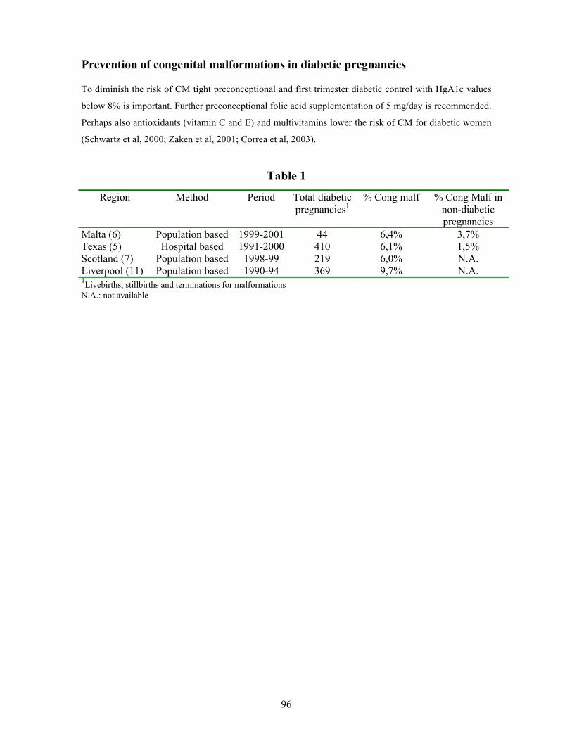
96
Prevention of congenital malformations in diabetic pregnancies To diminish the risk of CM tight preconceptional and first trimester diabetic control with HgA1c values
below 8% is important. Further preconceptional folic acid supplementation of 5 mg/day is recommended.
Perhaps also antioxidants (vitamin C and E) and multivitamins lower the risk of CM for diabetic women
(Schwartz et al, 2000; Zaken et al, 2001; Correa et al, 2003).
Table 1
Region Method Period Total diabetic pregnancies1
% Cong malf % Cong Malf in non-diabetic pregnancies
Malta (6) Population based 1999-2001 44 6,4% 3,7% Texas (5) Hospital based 1991-2000 410 6,1% 1,5% Scotland (7) Population based 1998-99 219 6,0% N.A. Liverpool (11) Population based 1990-94 369 9,7% N.A. 1Livebirths, stillbirths and terminations for malformations N.A.: not available
97
REFERENCES
Becerra JE, Khoury MJ, Cordero JF, Erickson D (1990) Diabetes Mellitus during Pregnancy and the Risks for Specific Birth Defects: A Population-Based Case-Control Study. Pediatric, 85, 1-9
Casson IF, Clarke CA, Howard CV et al (1997) Outcomes of pregnancy in insulin dependent diabetic
women: results of a five year population cohort study. British Medical Journal, 315, 275-278
Correa A, Botto L, Liu Y, Mulinare J, Erickson JD (2003) Do Multivitamin Supplements Attenuate the Risk for Diabetes-Associated Birth Defects? Paediatrics, 111, 1146-1151
EUROCAT Working Group (2003) Special Report: Prevention of Neural Tube Defects by
Periconceptional Folic Acid Supplementation in Europe. University of Ulster, 23
Farrel T, Neale L, Cundy T (2002) Congenital anomalies in the offspring of women with Type 1, Type 2 and gestational diabetes. Diabetic Medicine, 19, 322-326
Feltbower RG, McKinney PA, Parslow RC et al (2003) Type 1 diabetes in Yorkshire, UK: time trends in
0-14 and 15-29-year-olds, age at onset and age-period-cohort modelling. Diab Med, 20, 437-441
Gale EA (2002) The rise of childhood type 1 diabetes in the 20.th century. Diabetes, 51, 3353-3361 Greene MF (1999) Spontaneous abortions and Major Malformations in women with diabetes Mellitus.
Seminars in Reproductive Endocrinology, 17, 127-136 Holing EV, Brown ZA, Beyer CS, Connel FA (1998) Why Don’t Women With Diabetes Plan Their
Pregnancies? Diabetes Care, 21, 889-895
Lofredo CA, Wilson PD, Ferencz C (2001) Maternal Diabetes: An Independent Risk Factor for Major Cardiovascular Malformations with Increased Mortality of Affected Infants. Terat, 64, 98-106
Meyer-Wittkopf M, Simpson JM, Sharland GK (1996) Incidence of congenital heart defects in fetuses of
diabetic mothers: a retrospective study of 326 cases. Ultrasound Obstetric Gynaecology, 8, 8-10 Penny CP, Mair G, Pearson DWM (2003) Outcomes of pregnancies in women with type 1 diabetes in
Scotland: a national population-based study. British Journal of Gynaecology, 110, 315-318 Savona-Ventura C, Ellul A, Chircop M (2003) The outcome of diabetic pregnancies in Malta.
International Journal of Gynaecology & Obstetrics, 82, 217-218 Schwartz R, Teramo KA (2000) Effects of Diabetic Pregnancy on the Fetus and Newborn. Seminars in
Perinatology 2000, 24, 120-135
Sheffield JS, Butler-Koster EL, Casey BM et al (2002) Maternal diabetes mellitus and infant malformations. Obstetrics & Gynaecology 2002, 100, 925-930
Svensson J, Carstensen B, Molbak A et al (2002) Increased risk of childhood type 1 diabetes in children
born after 1985. Diabetes Care, 25, 2197-2201
Wren C, Birrell G, Hawthorne G (2003) Cardiovascular malformations in infants of diabetic mothers. Heart, 89, 1217-1220
World Health Organisation (Europe), International Diabetes Federation (Europe) (1990) Diabetes Care
and Research in Europe: the St Vincent Declaration. Diabetic Medicine, 34, 655-661 Zaken V, Kohen R, Ornoy A (2001) Vitamins C and E improve rat embryonic antioxidant mechanism in
diabetic culture medium. Teratology, 64, 33-44

98
CHAPTER II.5 MATERNAL EPILEPSY
Elisabeth Robert, January 2004 Environmental Causes of Congenital Anomalies: Maternal Epilepsy and Anti-epileptics Approximately 20% of individuals with epilepsy are of childbearing potential and about 3 to 5 births per
thousand will be to women with epilepsy. It has been known for several decades that the risk of major
and minor malformations in infants of epileptic mothers is twice higher than in the general population.
This is partly attributable to the teratogenicity of the drugs used to treat epilepsy, but the disease in itself
most probably increases the risk, while no epidemiological technique is ideal to separate the effects of the
disease and the drugs on the fetus. Very few epileptic patients can live without treatment, and those who
can might be questioned as whether or not they actually are epileptic. Studies have shown a marked
increase in malformations amongst infants exposed to first trimester seizures (Lindhout & Omtzigt,
1992). However, most investigators have found that maternal seizures during pregnancy had no impact
on the frequency of malformations, or on the development of infant epilepsy or febrile convulsions
(Dansky & Finnell, 1991; Canger et al, 1999). There have been reports of maternal seizures during
pregnancy being associated, though, with an increased risk of miscarriage, preterm labor, intracranial
hemorrhage, fetal hypoxia with bradycardia and possible developmental or learning difficulties. In a
recent study 57 children, whose mothers did have a history of epilepsy, but did not have any antiepileptic
medication or any seizures during their pregnancies, were evaluated. Compared with a matched control
group there was no difference between the two groups of children in either IQ scores or physical features
(facial dysmorphia or digit hypoplasia) (Holmes et al, 2000).
Several dysmorphic syndromes have been described, related to classical anti-epileptics, i.e. phenytoin,
valproic acid, trimethadion, carbamazepine, phenobarbital and primidone. The developmental
anomalies occur in about 1/4 of cases, they resemble each other and may be referred to as an
anticonvulsant- or epilepsy- syndrome : epicanthal folds, hypertelorism, hirsute foreheads, broad flat
nasal bridges, upturned nasal tip, straight thin upper lips, and also distal digital hypoplasia. These
children tend to be small for dates, and have an increased risk for psychomotor retardation (Rudd &
Freedom, 1979; Bethenod & Frederich, no date).
Whereas maternal epilepsy induces a two to three-fold increase in the risk of any kind of major
malformations, four main types of defects are overrepresented in infants of epileptic mothers : orofacial
clefts, cardiovascular defects, spina bifida and hypospadias. Preferential associations have been found in
99
different studies between specific anticonvulsants and malformations. A markedly increased risk is
observed after combination therapy with two or more anticonvulsants (Lindhout & Omtzigt, 1992;
Kaneko et al, 1999; Samrén et al, 1999). What must be considered in these cases, however, is that the
mothers had a particularly severe form of epilepsy.
Phenobarbital and primidone are effective in focal epilepsy and grand-mal seizures. In two recent
studies, observed rates of major malformation were about 5% for phenobarbital monotherapy (Canger et
al, 1999; Kaneko et al, 1999). They are mainly congenital heart diseases and oral clefts. Van der Pol (Van
der Pol et al, 1991) reported that cognitive development of children who had been exposed in utero to
phenobarbital alone or in combination with carbamazepine was significantly impaired in comparison
with children of non-epileptic mothers.
Diazepam and clonazepam have proven themselves as anti-epileptics. They are never used in
monotherapy to treat epilepsy, but benzodiazepine therapy does not seem to have as high a teratogenic
risk as the other anticonvulsants.
Phenytoin is the most widely used hydantoin derivative with an anti-epileptic effect. Its teratogenic
potential has been known since 1964 (Janz & Fuchs, 1964). Originally, the anomalies observed were
called ”fetal hydantoin syndrome” (Hanson & Smith, 1975) : craniofacial dysmorphia, anomalies of the
distal phalanges, pre and post-natal growth retardation, and cardiac defects. Limitations in cognitive
development have frequently been observed (Vanoverloop et al, 1992; Scolnik et al, 1994). The most
prominent of the observed major malformations are ptosis, iris coloboma, cleft lip and palate,
microcephaly, short neck, hypoplasia of nails and distal phalanges in fingers and toes, finger-like thumbs
and hip dysplasia (Lu et al, 2000). However, in most cases of affected children only a few of these
mentioned anomalies are present. The differences in susceptibility to specific malformations is expressed
by genetically diverse siblings who shared approximately comparable uterine environments (Phelan et al,
1982; Raymond et al, 1995). Moreover, Buehler and colleagues (Buehler et al, 1990) demonstrated that
infants with low detoxification capacities that were exposed to phenytoin in utero were at increased risks
for the fetal hydantoin syndrome, compared with similarly exposed individuals with normal epoxide
hydrolase activity.
Carbamazepine is used for grand-mal epilepsy, focal and psychomotor seizures as well as for trigeminal
neuralgia. It is teratogenic in human beings. Carbamazepine increases the risk of spina bifida about 10-
fold, that is, the malformation occurs in about 1% of exposed newborns. Apart from this there have been
reports of hypospadias, microcephaly, urinary tract anomalies, and some facial dysmorphic features

100
(Jones et al, 1989; Rosa, 1991; Källén, 1994; Robert & Källén, 1994; Matalon et al, 2002). The risk of
cognitive disturbances with carbamazepine monotherapy was retrieved in subsequent studies (Ornoy &
Cohen, 1996), and observed in particular among children who also have facial dysmorphia.
Ethosuximide is effective with petit-mal seizures. There are a few reports only of ethosuximide therapy
during pregnancy. No typical pattern of birth defects has been observed in the infants of 57 treated
women (Ornoy & Cohen, 1996). In a series of 18 fetuses exposed in the first trimester, there were no
birth defects (Rosa, cited in Briggs et al, 1998). Although the available reports are in no way sufficient
for a differentiated risk assessment, there does not seem to be a teratogenic risk comparable to the other
anticonvulsants.
Valproic acid is effective for different forms of epilepsy. Typical for valproic acid is an approximate 20-
fold increased risk of spina bifida or other neural tube defects, such that the observed incidence of these
defects among exposed children is 1-2% (Robert & Guibaud, 1982). A dose response relationship was
found in several recent studies. Women receiving daily doses of 1000 mg or more were at a significantly
increased risk for having a child with a major malformation (Kaneko et al, 1999; Samrén et al, 1999).
Several cases of radial ray anomalies have been reported after fetal valproate exposure (Seip, 1976;
Jaeger-Roman et al, 1986). Children exposed to valproate in utero also appear to be at greater risk for
perinatal distress (43 %) and low Apgar scores (28 %), postnatal growth deficiency and microcephaly
(Seip, 1976; Robert & Jouk, 1992). Also craniosynostosis, especially trigonocephaly, has been
specifically associated with maternal valproic acid treatment (Ardinger et al, 1988). Other major
malformations have been associated with valproic acid, but only as case reports (eg gastroschisis, septo-
optic dysplasia). A clinical study was reported, describing 57 children with anticonvulsant syndrome, 46
of which having been exposed to valproic acid in utero. Eighty per cent of these children had behavioral
problems, including attention deficit and hyperactivity disorder, autistic features or Asperger's syndrome
(Lajeunie et al, 2001). Other studies seem to confirm these findings (Moore et al, 2000; Bescoby-
Chambers et al, 2001; Williams et al, 2001; Adab et al, 2001). Examining neonatal behavior and later
neurologic functions in a study of 40 children, exposed in utero to a single AED, those exposed to
valproic acid were most compromised. Valproic acid serum concentrations at birth correlated with the
degree of neonatal hyperexcitability and neurologic dysfunction at the age of 6 (Adab et al, 2001).
A number of novel antiepileptic drugs have been marketed in the 90s : vigabatrin, lamotrigine,
felbamate, gabapentin, sultiame, tiagabine, topiramate, levetiracetam, oxcarbamazepine and zonisamide.
While some of these compounds have been available for some time, the clinical experience with pregnant
women is extremely limited. Existing experience is insufficient to estimate if any teratogenic risk,
comparable to that of the classic anticonvulsants, does exist. The available, still unpublished (Dean et al,
101
2002), prospective case observations of the manufacturer on 302 prospective reports of pregnancies with
first trimester exposure to lamotrigine as monotherapy. Of these pregnancies, nine children had
congenital malformations, for a rate of 3%, ie a rate lower than the average frequency of birth defects in
women with epilepsy using monotherapy (Samrén et al, 1999). Moreover, prospective and retrospective
data collected from 39 women exposed to gabapentin during pregnancy did not suggest a teratogenic risk
(Glaxo Wellcome). The limited number of exposed cases, the fact that most of them did not receive
monotherapy but a combination with another anticonvulsant, and the spontaneous recording of the
exposed pregnant women do not allow for a differentiated risk assessment.
Folic acid deficiency has been discussed as a possible teratogenic pathway for anticonvulsants
(Gabapentin, 2003). A study on folic acid antagonists (including anticonvulsants) and the risk of birth
defects (Koch et al, 1996) did not show a protective effect of periconceptional folic acid
supplementation, but as this supplementation is now recommended for any woman, epileptic patients
should be given 4 mg per day, at least through the 8th week of pregnancy.
102
REFERENCES
Adab N, Jacoby A, Smith D & Chadwick D (2001) Additional educational needs in children born to mothers with epilepsy. J Neurol Neurosurg Psychiatry, 70, 15-21
Ardinger HH, Atkin JF, Blackston RD, Elsas LJ, Clarren SK, Livingstone S, Flannery DB, Pellock JM,
Harrod MJ & Lammer EJ (1988) Verification of the fetal valproate syndrome phenotype. American Journal of Medical Genetics, 29, 171-185
Bescoby-Chambers N, Forster P & Bates G (2001) Foetal valproate syndrome and autism: additional
evidence of an association. Dev Med Child Neurol, 43, 847 Bethenod M, Frederich A (no date) Les enfants des antiépileptiques. Pédiatrie, 75, 227-230 Briggs GG, Freeman RK & Yaffe SJ (1998) Drugs in Pregnancy and Lactation, 5th Ed, Baltimore,
Williams & Wilkins, p119-120 Buehler BA, Delimont D, van Waes M & Finnell RH (1990) Prenatal prediction of risk of the fetal
hydantoin syndrome. New England Journal of Medicine, 322, 1567-1572
Canger R, Battino D, Canevini MP, Fumarola C, Guidolin L, Vignoli A, Mamoli D, Palmieri C, Molteni F, Granata T, Hassibi P, Zamperini P, Pardi G, Avanzini G. (1999) Malformations in offspring of women with epilepsy: a prospective study. Epilepsia, 40(9), 1231-1236
Dansky LV, Finnell RH (1991) Parental epilepsy, anticonvulsant drugs, and reproductive outcome:
epidemiologic and experimental findings spanning three decades; 2: Human studies. Reproductive Toxicology, 5, 301-35
Dean JC, Hailey H, Moore SJ, Lloyd DJ, Turnpenny PD & Little J (2002) Long term health and neurodevelopment in children exposed to antiepileptic drugs before birth. Journal of Medical Genetics, 39, 251-259
Glaxo Wellcome. Lamotrigine pregnancy registry. Interim report, 1 September 1992 through 31 March
2003
Hanson JW, Smith DW (1975) The fetal hydantoin syndrome. Journal of Pediatrics, 87, 285-290
Hernandez-Diaz S, Werler MM, Walker AM & Mitchell AA (2000) Folic acid antagonists and the risk of birth defects. New England Journal of Medicine, 343, 1608-1614
Holmes LB, Rosenberger PB, Harvey EA, Khoshbin S, Ryan L (2000) Intelligence and physical features
of children of women with epilepsy. Teratology, 61, 196-202 Jaeger-Roman E, Deichl A, Jakob S, et al. Fetal growth, major malformations, and minor anomalies in
infants born to women receiving valproic acid. J Pediatr 1986;108:997-1004 Janz D, Fuchs U (1964) Are antiepileptic drugs harmful when given during pregnancy? German
Medicine Monthly, 9, 20–22
Jones KL, Lacro RV, Johnson KA & Adams J (1989) Pattern of malformations in the children of women treated with carbamazepine during pregnancy. New England Journel of Medicine, 320, 1661-1666
Källén B (1994) Maternal carbamazepine and infant spina bifida. Reproductive Toxicology, 8, 203-205
103
Kaneko S, Battino D, Anderman E et al (1999) Congenital malformations due to antiepileptic drugs. Epilepsy Research, 33, 145-158
Koch S, Jager-Roman E, Losche G, Nau H, Rating D & Helge H (1996) Antiepileptic drug treatment in
pregnancy: drug side effects in the neonate and neurological outcome. Acta Paediatr, 85, 739-746
Lajeunie E, Barcik U, Thorne JA, Ghouzzi VE, Bourgeois M & Renier D (2001) Craniosynostosis and fetal exposure to sodium valproate. J Neurosurg, 95, 778-782
Lindhout D, Omtzigt JG (1992) Pregnancy and the risk of teratogenicity. Epilepsia, 33 Suppl 4, 41-48
Lu MCK, Sammel MD, Cleveland RH, Ryan LM & Holmes LB (2000) Digit effects produced by prenatal exposure to antiepileptic drugs. Teratology, 61, 277-283
Matalon S, Schechtman S, Goldzweig G &Ornoy A (2002) The teratogenic effect of carbamazepine: a meta-analysis of 1255 exposures. Reproductive Toxicology, 16, 9-17
Montouris G Gabapentin (2003) Exposure in human pregnancy: results from the Gabapentin Pregnancy
Registry. Epilepsy Behav, 4, 310-317
Moore SJ, Turnpenny P, Quinn A, Glover S, Lloyd DJ, Montgomery T & Dean JCS. (2000) A clinical study of 57 children with fetal anticonvulsant syndrome. Journal of Medical Genetics 2000, 37, 489-497
Ornoy A & Cohen E (1996) Outcome of children born to epileptic mothers treated with carbamazepine during pregnancy. Arch Dis Child, 75, 517-520
Phelan MC, Pellock JM & Nance WE. (1982) Discordant expression of fetal hydantoin syndrome in heteropaternal dizygotic twins. New England Journal of Medicine, 307, 99-101
Raymond GV, Buehler BA, Finnell RH & Holmes LB (1995) Anticonvulsant teratogenesis: 3. Possible
metabolic basis [letter] Teratology, 51, 55-56 Robert E & Guibaud P (1982) Maternal Valproic acid and congenital neural tube defects. Lancet, 2, 937
Robert E & Jouk PS (1992) Preaxial limb defects after valproic acid exposure during pregnancy.
Proceedings of the First International Meeting of the Genetic and Reproductive Epidemiology Research Society (GRERS). P.Mastroiacovo, B.Källén & E.Castilla eds. Ghedini Editore, Rome, p101-105
Robert E & Källén B (1994) In utero exposure to carbamazepine: effects on the fetus. Issues and Reviews
in Teratology, 7, 37-55 Rosa FW (1991) Spina bifida in infants of women treated with carbamazepine during pregnancy. New
England Journal of Medicine, 324, 674-677 Rudd NL, Freedom RM (1979) A possible primidone embryopathy. Journal of Pediatrics, 94, 835–837 Samrén EB, van Duijn CM, Christiaens GCM, Hofman A, Lindhout D (1999) Antiepileptic drug
regimens and major congenital abnormalities in the offspring. Ann Neurol, 46, 739-746 Scolnik D, Nulman I, Rovet J, Gladstone D, Czuchta D, Gardner HA, Gladstone R, Ashby P, Weksberg
R & Einarson T (1994) Neurodevelopment of children exposed in utero to phenytoin and carbamazepine monotherapy program JAMA, 271, 767-770

104
Seip M. (1976) Growth retardation, dysmorhic facies and minor malformations following massive exposure to phenobarbital in utero. Acta Paediatr. Scand, 65, 617–621
Van der Pol MC, Hadders-Algra M, Huisjes HJ & Touwen BC (1991) Antiepileptic medication in pregnancy: late effects on the children's central nervous system development. American Journal of Obstetrics & Gynecology, 164, 121-128
Vanoverloop D, Schnell RR, Harvey EA, Holmes LB (1992) The effects of prenatal exposure to phenytoin and other anticonvulsants on intellectual function at 4 to 8 years of age. Neurotoxicol Teratol, 14, 329-335
Williams G, King J, Cunningham M, Stephan M, Kerr B & Hersh JH (2001) Fetal valproate syndrome
and autism: additional evidence of an association. Dev Med Child Neurol, 43, 202-206