Embed Size (px)
Citation preview
Earn2 CE creditsThis course was
written for dentists, dental hygienists,
and assistants.
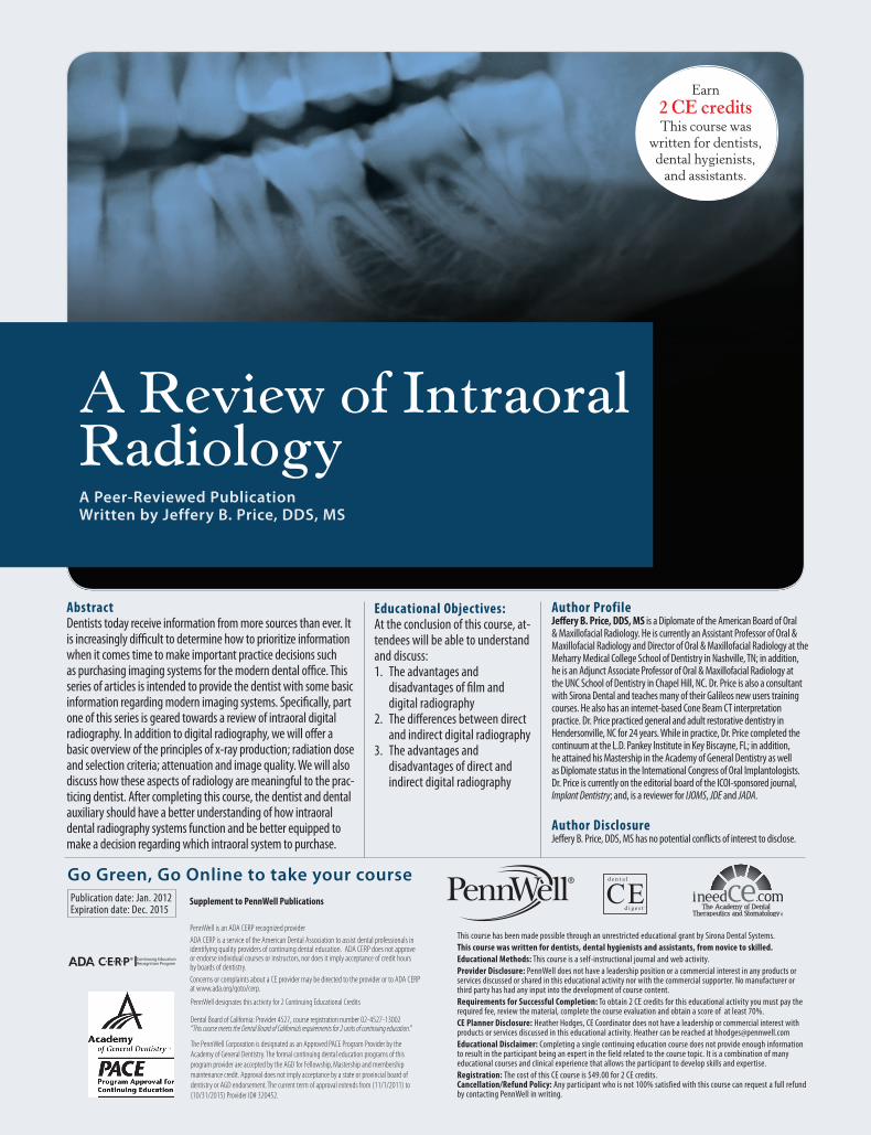
A Review of Intraoral RadiologyA Peer-Reviewed Publication Written by Jeffery B. Price, DDS, MS
AbstractDentists today receive information from more sources than ever. It is increasingly difficult to determine how to prioritize information when it comes time to make important practice decisions such as purchasing imaging systems for the modern dental office. This series of articles is intended to provide the dentist with some basic information regarding modern imaging systems. Specifically, part one of this series is geared towards a review of intraoral digital radiography. In addition to digital radiography, we will offer a basic overview of the principles of x-ray production; radiation dose and selection criteria; attenuation and image quality. We will also discuss how these aspects of radiology are meaningful to the prac-ticing dentist. After completing this course, the dentist and dental auxiliary should have a better understanding of how intraoral dental radiography systems function and be better equipped to make a decision regarding which intraoral system to purchase.
Educational Objectives:At the conclusion of this course, at-tendees will be able to understand and discuss:1. The advantages and
disadvantages of film and digital radiography
2. The differences between direct and indirect digital radiography
3. The advantages and disadvantages of direct and indirect digital radiography
Author ProfileJeffery B. Price, DDS, MS is a Diplomate of the American Board of Oral & Maxillofacial Radiology. He is currently an Assistant Professor of Oral & Maxillofacial Radiology and Director of Oral & Maxillofacial Radiology at the Meharry Medical College School of Dentistry in Nashville, TN; in addition, he is an Adjunct Associate Professor of Oral & Maxillofacial Radiology at the UNC School of Dentistry in Chapel Hill, NC. Dr. Price is also a consultant with Sirona Dental and teaches many of their Galileos new users training courses. He also has an internet-based Cone Beam CT interpretation practice. Dr. Price practiced general and adult restorative dentistry in Hendersonville, NC for 24 years. While in practice, Dr. Price completed the continuum at the L.D. Pankey Institute in Key Biscayne, FL; in addition, he attained his Mastership in the Academy of General Dentistry as well as Diplomate status in the International Congress of Oral Implantologists. Dr. Price is currently on the editorial board of the ICOI-sponsored journal, Implant Dentistry; and, is a reviewer for IJOMS, JDE and JADA.
Author DisclosureJeffery B. Price, DDS, MS has no potential conflicts of interest to disclose.
Publication date: Jan. 2012 Expiration date: Dec. 2015
This course has been made possible through an unrestricted educational grant by Sirona Dental Systems.This course was written for dentists, dental hygienists and assistants, from novice to skilled. Educational Methods: This course is a self-instructional journal and web activity. Provider Disclosure: PennWell does not have a leadership position or a commercial interest in any products or services discussed or shared in this educational activity nor with the commercial supporter. No manufacturer or third party has had any input into the development of course content.Requirements for Successful Completion: To obtain 2 CE credits for this educational activity you must pay the required fee, review the material, complete the course evaluation and obtain a score of at least 70%.CE Planner Disclosure: Heather Hodges, CE Coordinator does not have a leadership or commercial interest with products or services discussed in this educational activity. Heather can be reached at [email protected] Disclaimer: Completing a single continuing education course does not provide enough information to result in the participant being an expert in the field related to the course topic. It is a combination of many educational courses and clinical experience that allows the participant to develop skills and expertise.Registration: The cost of this CE course is $49.00 for 2 CE credits. Cancellation/Refund Policy: Any participant who is not 100% satisfied with this course can request a full refund by contacting PennWell in writing.
Supplement to PennWell Publications
PennWell designates this activity for 2 Continuing Educational Credits
Dental Board of California: Provider 4527, course registration number 02-4527-13002“This course meets the Dental Board of California’s requirements for 2 units of continuing education.”
The PennWell Corporation is designated as an Approved PACE Program Provider by the Academy of General Dentistry. The formal continuing dental education programs of this program provider are accepted by the AGD for Fellowship, Mastership and membership maintenance credit. Approval does not imply acceptance by a state or provincial board of dentistry or AGD endorsement. The current term of approval extends from (11/1/2011) to (10/31/2015) Provider ID# 320452.
Go Green, Go Online to take your course
2 www.ineedce.com
Educational ObjectivesAt the conclusion of this course, attendees will be able to understand and discuss:1. The advantages and disadvantages of film and digital
radiography2. The differences between direct and indirect digital
radiography3. The advantages and disadvantages of direct and indirect
digital radiography
AbstractDentists today receive information from more sources than ever. It is increasingly difficult to determine how to prioritize information when it comes time to make important practice decisions such as purchasing imaging systems for the mod-ern dental office. This series of articles is intended to provide the dentist with some basic information regarding modern imaging systems. Specifically, part one of this series is geared towards a review of intraoral digital radiography. In addition to digital radiography, we will offer a basic overview of the principles of x-ray production; radiation dose and selection criteria; attenuation and image quality. We will also discuss how these aspects of radiology are meaningful to the prac-ticing dentist. After completing this course, the dentist and dental auxiliary should have a better understanding of how intraoral dental radiography systems function and be bet-ter equipped to make a decision regarding which intraoral system to purchase.
Throughout this continuing education course, we will look at the major choices in radiographic systems available to dentists today as they contemplate either building a new dental office or upgrading an existing facility. There are two premises that the reader should assume: first, the course is taught with the assumption that for the foreseeable future, there will be a place in dentistry for intraoral radiography; and secondly, as dentists build or upgrade new facilities, the major emphasis will be on digital receptors, not film-based receptors. Dentists need to focus on deciding when to switch to a digital radiography system, and not whether to switch to digital. For these reasons, the main focus of this course will be intraoral digital radiography.
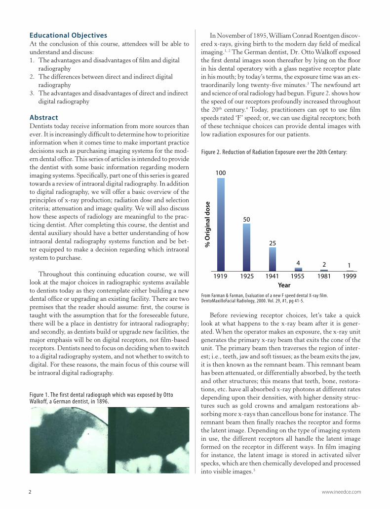
In November of 1895, William Conrad Roentgen discov-ered x-rays, giving birth to the modern day field of medical imaging.1, 2 The German dentist, Dr. Otto Walkoff exposed the first dental images soon thereafter by lying on the floor in his dental operatory with a glass negative receptor plate in his mouth; by today’s terms, the exposure time was an ex-traordinarily long twenty-five minutes.3 The newfound art and science of oral radiology had begun. Figure 2. shows how the speed of our receptors profoundly increased throughout the 20th century.4 Today, practitioners can opt to use film speeds rated ‘F’ speed; or, we can use digital receptors; both of these technique choices can provide dental images with low radiation exposures for our patients.
Before reviewing receptor choices, let’s take a quick look at what happens to the x-ray beam after it is gener-ated. When the operator makes an exposure, the x-ray unit generates the primary x-ray beam that exits the cone of the unit. The primary beam then traverses the region of inter-est; i.e., teeth, jaw and soft tissues; as the beam exits the jaw, it is then known as the remnant beam. This remnant beam has been attenuated, or differentially absorbed, by the teeth and other structures; this means that teeth, bone, restora-tions, etc. have all absorbed x-ray photons at different rates depending upon their densities, with higher density struc-tures such as gold crowns and amalgam restorations ab-sorbing more x-rays than cancellous bone for instance. The remnant beam then finally reaches the receptor and forms the latent image. Depending on the type of imaging system in use, the different receptors all handle the latent image formed on the receptor in different ways. In film imaging for instance, the latent image is stored in activated silver specks, which are then chemically developed and processed into visible images.5
Figure 2. Reduction of Radiation Exposure over the 20th Century:Reduction of radiation exposure
in dental radiography in the 20th century
1919 1925 1941 1955 1981 1999
50
25
4 2 1
Year
% O
rigi
nal d
ose
100
Figure 1. The first dental radiograph which was exposed by Otto Walkoff, a German dentist, in 1896.
From Farman & Farman, Evaluation of a new F speed dental X-ray film. DentoMaxilloFacial Radiology, 2000. Vol. 29, #1, pg 41-5.
www.ineedce.com 3
Basics of radiographyDigital radiography is obviously different than film radi-ography; but, what exactly, is digital radiography and what makes it different than film? This seems such a basic ques-tion, but one that is often overlooked. Before we answer that question, let’s look at an even more basic question. How does a radiographic image give us diagnostic informa-tion about the patient? The useful information obtained from radiographic images directly relates to the shades of gray in the image.
If the remnant beam doesn’t contain many x-ray pho-tons, that means that a dense object, like a restoration, tooth enamel, or thick cortical bone, absorbed a lot of the photons and the image is bright or white. On the other hand, if the remnant beam has a lot of x-ray photons, the x-rays passed through only thin soft tissues or the airway, and that area of the image is dark. In film images, these shades of gray correspond to the amount of silver on the film—dark areas have a lot of silver, and bright or white ar-eas have little or no silver.1 Now, you can understand why a dental radiograph is so vitally important in making the correct diagnosis in areas that the dentist can not visualize; i.e., periapical regions, periodontal bone, implant sites, etc. Dental radiographs are truly the foundation of the diagnosis; and, as we know, without the correct diagnosis even the best dentist cannot provide proper treatment for the patient.
Radiation doseMany of our patients today are concerned about the ra-diation dose of the radiographs that we recommend in the dental office. Here are a few things to consider if you want to decrease your patients’ radiation burden in your office. One of the most efficacious ways to reduce the radiation exposure to your patients is to eliminate unnecessary radio-graphs by instituting office policies that say we only take necessary radiographs that have reasonable expectations of producing clinically useful information.6 In 2004 the FDA and the ADA published an updated set of Selection Crite-ria guidelines.7 They provide professional guidance when making recommendations for radiographs while allowing plenty of room for clinical judgment.
By following these guidelines and making individual decisions regarding the need for radiographs instead of having an office policy for all patients, your patients will appreciate the individual attention to their health. Second, eliminate D-speed film from your dental practice; F-speed film is twice as fast as D-speed film, so if for some reason, you still want to use film, switch to F-speed film.4, 8 Third, consider switching to rectangular collimation. Rectangular collimation re-shapes the beam from a round x-ray beam to a rectangular x-ray beam; and, thereby eliminates approxi-mately two thirds of the unused radiation in the primary x-ray beam.9
What does digital mean?According to the Oxford English Dictionary, the word digital means data expressed as a series of the digits 0 and 1.10 Com-puters work in the discrete world of zeroes and ones and storage units of bits, bytes, megabytes, etc. As described earlier, the film image remains on the physical piece of ra-diographic film in analog form unless the film is scanned and transformed into a digital image. The basic difference then between film and digital radiography is that digital images have their basic image data stored as discrete zeroes and ones in a computer while film image data is stored within the silver particles on the physical piece of film. This digital image data is stored in 2D arrays of picture elements, more commonly known as ‘pixels’.11 We will explore other differences later in the course; but, for now, lets look at how digital images are acquired.
There are two basic types of digital technology—direct and indirect. Most of us are familiar with digital receptors that have USB cables plugged into a computer; these are examples of ‘direct digital’ technology. In other words, the latent image is formed in the electronics of the receptor and
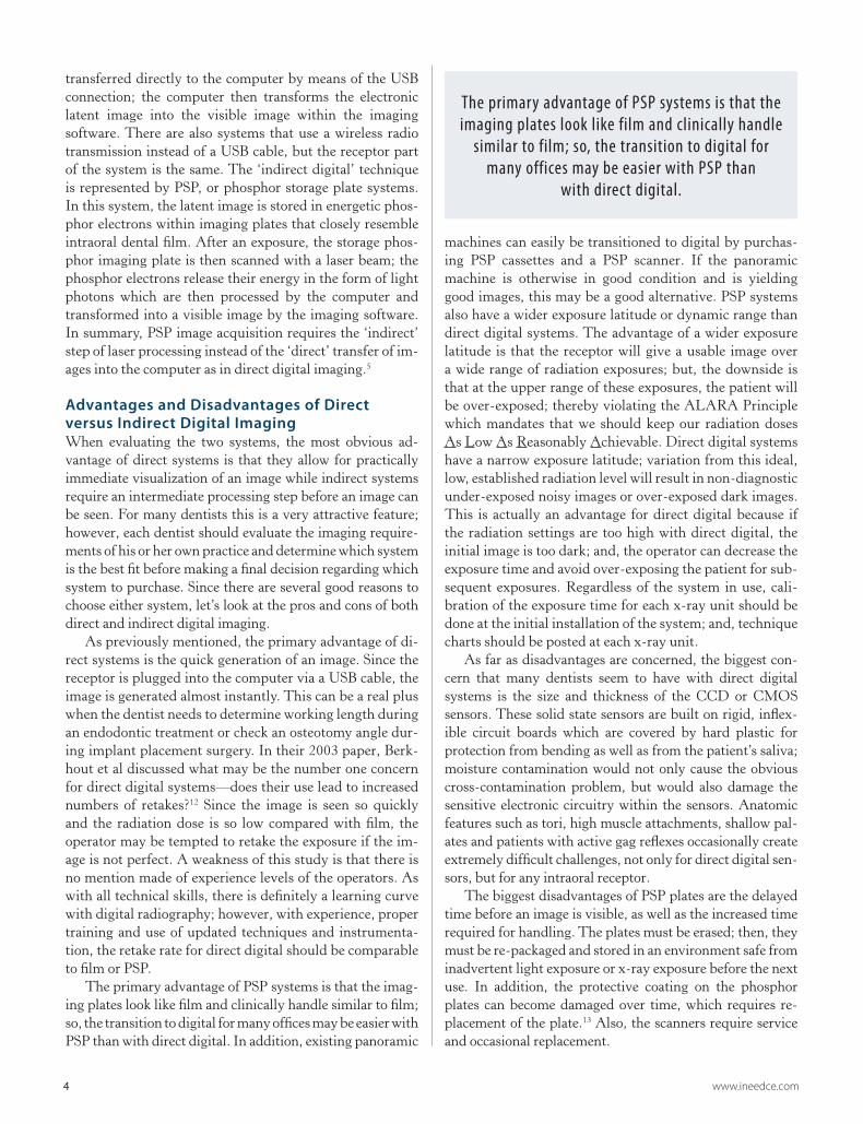
Figure 3. An intraoral dental x-ray unit which was placed into service in 1989. Note the timer which is a dial that controlled the radiation in 1/60th of a second ‘impulses’.
Figure 4. Close-up of the timer.
4 www.ineedce.com
transferred directly to the computer by means of the USB connection; the computer then transforms the electronic latent image into the visible image within the imaging software. There are also systems that use a wireless radio transmission instead of a USB cable, but the receptor part of the system is the same. The ‘indirect digital’ technique is represented by PSP, or phosphor storage plate systems. In this system, the latent image is stored in energetic phos-phor electrons within imaging plates that closely resemble intraoral dental film. After an exposure, the storage phos-phor imaging plate is then scanned with a laser beam; the phosphor electrons release their energy in the form of light photons which are then processed by the computer and transformed into a visible image by the imaging software. In summary, PSP image acquisition requires the ‘indirect’ step of laser processing instead of the ‘direct’ transfer of im-ages into the computer as in direct digital imaging.5
Advantages and Disadvantages of Direct versus Indirect Digital ImagingWhen evaluating the two systems, the most obvious ad-vantage of direct systems is that they allow for practically immediate visualization of an image while indirect systems require an intermediate processing step before an image can be seen. For many dentists this is a very attractive feature; however, each dentist should evaluate the imaging require-ments of his or her own practice and determine which system is the best fit before making a final decision regarding which system to purchase. Since there are several good reasons to choose either system, let’s look at the pros and cons of both direct and indirect digital imaging.
As previously mentioned, the primary advantage of di-rect systems is the quick generation of an image. Since the receptor is plugged into the computer via a USB cable, the image is generated almost instantly. This can be a real plus when the dentist needs to determine working length during an endodontic treatment or check an osteotomy angle dur-ing implant placement surgery. In their 2003 paper, Berk-hout et al discussed what may be the number one concern for direct digital systems—does their use lead to increased numbers of retakes?12 Since the image is seen so quickly and the radiation dose is so low compared with film, the operator may be tempted to retake the exposure if the im-age is not perfect. A weakness of this study is that there is no mention made of experience levels of the operators. As with all technical skills, there is definitely a learning curve with digital radiography; however, with experience, proper training and use of updated techniques and instrumenta-tion, the retake rate for direct digital should be comparable to film or PSP.
The primary advantage of PSP systems is that the imag-ing plates look like film and clinically handle similar to film; so, the transition to digital for many offices may be easier with PSP than with direct digital. In addition, existing panoramic
machines can easily be transitioned to digital by purchas-ing PSP cassettes and a PSP scanner. If the panoramic machine is otherwise in good condition and is yielding good images, this may be a good alternative. PSP systems also have a wider exposure latitude or dynamic range than direct digital systems. The advantage of a wider exposure latitude is that the receptor will give a usable image over a wide range of radiation exposures; but, the downside is that at the upper range of these exposures, the patient will be over-exposed; thereby violating the ALARA Principle which mandates that we should keep our radiation doses As Low As Reasonably Achievable. Direct digital systems have a narrow exposure latitude; variation from this ideal, low, established radiation level will result in non-diagnostic under-exposed noisy images or over-exposed dark images. This is actually an advantage for direct digital because if the radiation settings are too high with direct digital, the initial image is too dark; and, the operator can decrease the exposure time and avoid over-exposing the patient for sub-sequent exposures. Regardless of the system in use, cali-bration of the exposure time for each x-ray unit should be done at the initial installation of the system; and, technique charts should be posted at each x-ray unit.
As far as disadvantages are concerned, the biggest con-cern that many dentists seem to have with direct digital systems is the size and thickness of the CCD or CMOS sensors. These solid state sensors are built on rigid, inflex-ible circuit boards which are covered by hard plastic for protection from bending as well as from the patient’s saliva; moisture contamination would not only cause the obvious cross-contamination problem, but would also damage the sensitive electronic circuitry within the sensors. Anatomic features such as tori, high muscle attachments, shallow pal-ates and patients with active gag reflexes occasionally create extremely difficult challenges, not only for direct digital sen-sors, but for any intraoral receptor.
The biggest disadvantages of PSP plates are the delayed time before an image is visible, as well as the increased time required for handling. The plates must be erased; then, they must be re-packaged and stored in an environment safe from inadvertent light exposure or x-ray exposure before the next use. In addition, the protective coating on the phosphor plates can become damaged over time, which requires re-placement of the plate.13 Also, the scanners require service and occasional replacement.
The primary advantage of PSP systems is that the imaging plates look like film and clinically handle
similar to film; so, the transition to digital for many offices may be easier with PSP than
with direct digital.
www.ineedce.com 5
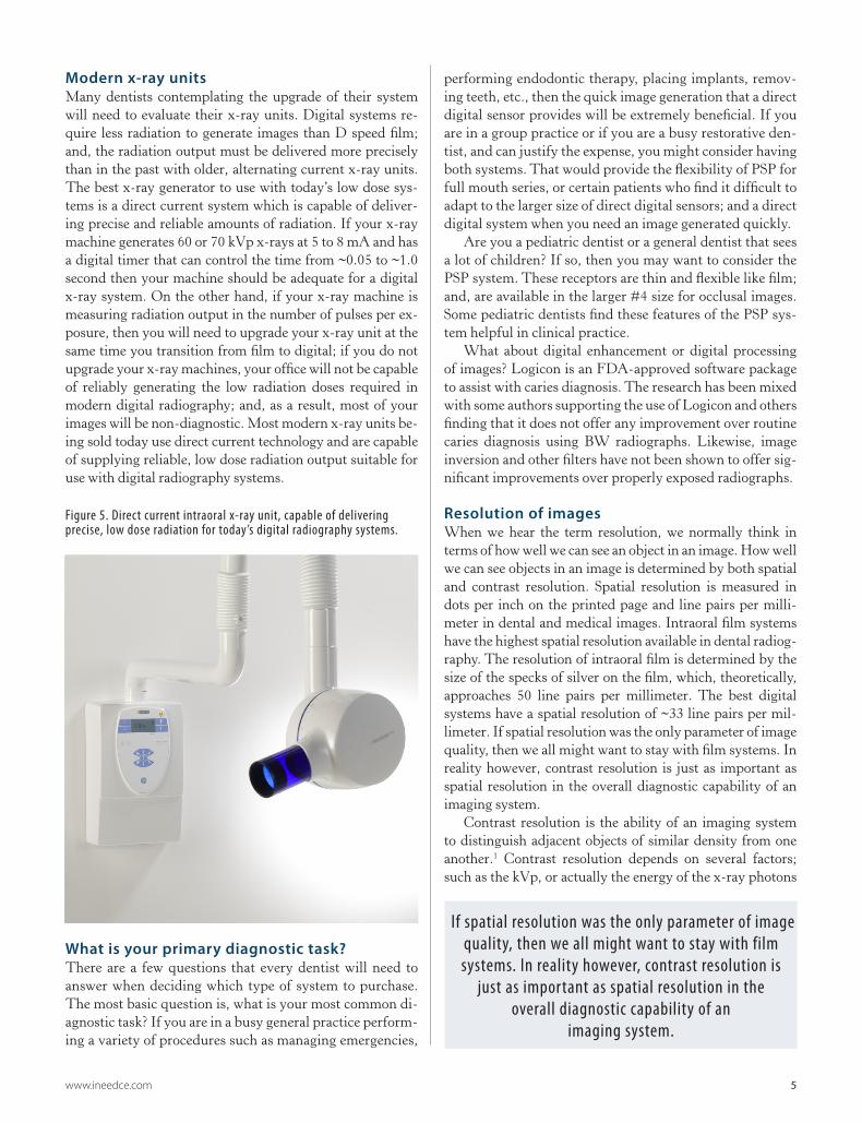
Modern x-ray unitsMany dentists contemplating the upgrade of their system will need to evaluate their x-ray units. Digital systems re-quire less radiation to generate images than D speed film; and, the radiation output must be delivered more precisely than in the past with older, alternating current x-ray units. The best x-ray generator to use with today’s low dose sys-tems is a direct current system which is capable of deliver-ing precise and reliable amounts of radiation. If your x-ray machine generates 60 or 70 kVp x-rays at 5 to 8 mA and has a digital timer that can control the time from ~0.05 to ~1.0 second then your machine should be adequate for a digital x-ray system. On the other hand, if your x-ray machine is measuring radiation output in the number of pulses per ex-posure, then you will need to upgrade your x-ray unit at the same time you transition from film to digital; if you do not upgrade your x-ray machines, your office will not be capable of reliably generating the low radiation doses required in modern digital radiography; and, as a result, most of your images will be non-diagnostic. Most modern x-ray units be-ing sold today use direct current technology and are capable of supplying reliable, low dose radiation output suitable for use with digital radiography systems.
What is your primary diagnostic task?There are a few questions that every dentist will need to answer when deciding which type of system to purchase. The most basic question is, what is your most common di-agnostic task? If you are in a busy general practice perform-ing a variety of procedures such as managing emergencies,
performing endodontic therapy, placing implants, remov-ing teeth, etc., then the quick image generation that a direct digital sensor provides will be extremely beneficial. If you are in a group practice or if you are a busy restorative den-tist, and can justify the expense, you might consider having both systems. That would provide the flexibility of PSP for full mouth series, or certain patients who find it difficult to adapt to the larger size of direct digital sensors; and a direct digital system when you need an image generated quickly.
Are you a pediatric dentist or a general dentist that sees a lot of children? If so, then you may want to consider the PSP system. These receptors are thin and flexible like film; and, are available in the larger #4 size for occlusal images. Some pediatric dentists find these features of the PSP sys-tem helpful in clinical practice.
What about digital enhancement or digital processing of images? Logicon is an FDA-approved software package to assist with caries diagnosis. The research has been mixed with some authors supporting the use of Logicon and others finding that it does not offer any improvement over routine caries diagnosis using BW radiographs. Likewise, image inversion and other filters have not been shown to offer sig-nificant improvements over properly exposed radiographs.
Resolution of imagesWhen we hear the term resolution, we normally think in terms of how well we can see an object in an image. How well we can see objects in an image is determined by both spatial and contrast resolution. Spatial resolution is measured in dots per inch on the printed page and line pairs per milli-meter in dental and medical images. Intraoral film systems have the highest spatial resolution available in dental radiog-raphy. The resolution of intraoral film is determined by the size of the specks of silver on the film, which, theoretically, approaches 50 line pairs per millimeter. The best digital systems have a spatial resolution of ~33 line pairs per mil-limeter. If spatial resolution was the only parameter of image quality, then we all might want to stay with film systems. In reality however, contrast resolution is just as important as spatial resolution in the overall diagnostic capability of an imaging system.
Contrast resolution is the ability of an imaging system to distinguish adjacent objects of similar density from one another.1 Contrast resolution depends on several factors; such as the kVp, or actually the energy of the x-ray photons
Figure 5. Direct current intraoral x-ray unit, capable of delivering precise, low dose radiation for today’s digital radiography systems.
If spatial resolution was the only parameter of image quality, then we all might want to stay with film systems. In reality however, contrast resolution is
just as important as spatial resolution in the overall diagnostic capability of an
imaging system.
6 www.ineedce.com
used for the exposure; speed of the receptor; the number of shades of gray the system can display; and, the techni-cal characteristics of the computer and monitor used for viewing. There is very little that can be done to film images after they have been exposed, while there are post-exposure processing techniques that can be applied to digital images. These processing techniques include brightness, contrast and gamma adjustments. In addition, before the image is even seen on the screen, the proprietary imaging software package applies certain processing algorithms from ‘look-up tables’ to improve image quality. These processes automati-cally improve edge sharpness and contrast among other im-age features. The end result of these image manipulations is a digital image that can be enhanced and optimized to meet the diagnostic task at hand.
Overall computer system issuesIf you are ‘going digital’, there are several scenarios re-garding practice management and computer systems in the marketplace that are available for your practice.14 The most important technical decision you will make is how you will backup your image database. You will need to have an onsite backup as well as an offsite backup; which need to be reliable and tested so you are confident they work. The best approach is to make your backup system as automated as possible and check it on a regular basis. You should have a local computer tech person that you can trust to help you with your technical needs. You will prob-ably have a technical relationship with one of the national practice management systems such as EagleSoft, Dentrix, etc.; but, unless you are located in a metropolitan area, you will also want a local computer tech who can keep you op-erating in the event of a technical emergency. What you do not want to hear is ‘We will see you tomorrow morning,’ when your entire digital radiology system is down due to a computer problem.
There is a very real serendipity effect when a dental office begins using digital radiography. Not only do the dentist and professional staff benefit from the digital im-ages but, so do the patients. Now that the images are on a computer screen and are large enough to be easily seen, take the opportunity to show them to the patient and educate the him or her—perhaps even allow the patient to ‘self-diagnose.’ Patients may never get truly excited about needing endodontic treatment, but if they can at least see a periapical lesion, perhaps they will ‘own the problem’ and be more motivated to seek treatment.
Next, let’s look at just two computer specifications—the graphics, or video, card and the monitor. If all you will ever do with the imaging computer is look at routine 2D images, almost any modern graphics card should be adequate; however, if you will also use the computer for viewing CBCT images, you will need a video card with one gigabyte of dedicated memory. As far as the monitor goes, almost any modern flat panel monitor will be ad-equate for viewing 2D images; but, again, if you will use the computer for viewing CBCT (cone beam computed tomography) images, you will need to spend a little more money and purchase a higher quality monitor capable of rendering 0.10 mm and higher resolution pixel images.
Most offices are sending radiographs electronically, but there will be a need to occasionally print your radiographs. The simplest and least expensive way to print diagnostic x-rays is to use semi-gloss or matte photographic paper with an ink jet printer—using a laser printer with plain paper yields a printed x-ray of poor quality. There are other, more expensive printers that hospital or dental ra-diography departments use; but, these printers and print media are very expensive and exceed the budget for most dental offices.
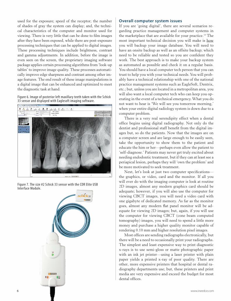
Figure 6. Image of posterior left maxillary teeth taken with the Schick 33 sensor and displayed with Eaglesoft imaging software.
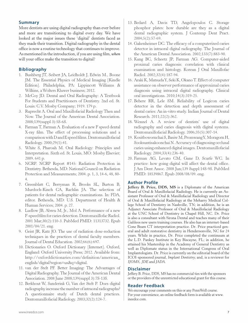
Figure 7. The size #2 Schick 33 sensor with the CDR Elite USB Interface Module.
www.ineedce.com 7
SummaryMore dentists are using digital radiography than ever before and more are transitioning to digital every day. We have looked at the major issues these ‘digital’ dentists faced as they made their transition. Digital radiography in the dental office is now a routine technology that continues to improve. As mentioned in the introduction, if you are using film, when will your office make the transition to digital?
Bibliography1. Bushberg JT, Seibert JA, Leidholdt J, Edwin M., Boone
JM. The Essential Physics of Medical Imaging [Kindle Edition]. Philadelphia, PA: Lippincott Williams & Wilkins, a Wolters Kluwer business; 2012.
2. McCoy JD. Dental and Oral Radiography: A Textbook For Students and Practitioners of Dentistry. 2nd ed. St. Louis: C.V. Mosby Company; 1919. 179 p.
3. Ruprecht A. Oral and Maxillofacial Radiology Then and Now. The Journal of the American Dental Association. 2008;139(suppl 3):5S-6S.
4. Farman T, Farman A. Evaluation of a new F speed dental X-ray film. The effect of processing solutions and a comparison with D and E speed films. Dentomaxillofacial Radiology. 2000;29(1):41.
5. White S, Pharoah M. Oral Radiology: Principles and Interpretation. Sixth ed. St. Louis, MO: Mosby Elsevier; 2009. 641 p.
6. NCRP. NCRP Report #145: Radiation Protection in Dentistry. Bethesda, MD: National Council on Radiation Protection and Measurements; 2004. p. 1, 3, 14-6, 48, 80-4.
7. Greenblatt C, Berryman R, Brooks SL, Burton B, Murdoch-Kinch CA, Rachlin JA. The selection of patients for dental radiographic examinations. In: FDA, editor. Bethesda, MD: U.S. Department of Health & Human Services; 2004. p. 27.
8. Ludlow JB, Abreu M, Jr., Mol A. Performance of a new F-speed film for caries detection. Dentomaxillofac Radiol. 2001 Mar;30(2):110-3. PubMed PMID: 11313732. Epub 2001/04/21. eng.
9. Geist JR, Katz JO. The use of radiation dose-reduction techniques in the practices of dental faculty members. Journal of Dental Education. 2002;66(6):697.
10. Dictionaries O. Oxford Dictionary [Internet]. Oxford, England: Oxford University Press; 2012. Available from: http://oxforddictionaries.com/definition/american_english/digital?region=us&q=digital.
11. van der Stelt PF. Better Imaging: The Advantages of Digital Radiography. The Journal of the American Dental Association. 2008 June 2008;139(suppl 3):7S-13S.
12. Berkhout W, Sanderink G, Van der Stelt P. Does digital radiography increase the number of intraoral radiographs? A questionnaire study of Dutch dental practices. Dentomaxillofacial Radiology. 2003;32(2):124-7.
13. Bedard A, Davis TD, Angelopoulos C. Storage phosphor plates: how durable are they as a digital dental radiographic system. J Contemp Dent Pract. 2004;5(2):57-69.
14. Gakenheimer DC. The efficacy of a computerized caries detector in intraoral digital radiography. The Journal of the American Dental Association. 2002;133(7):883-90.
15. Kang BC, Scheetz JP, Farman AG. Computer-aided proximal caries diagnosis: correlation with clinical examination and histology. Korean J Oral Maxillofac Radiol. 2002;32(4):187-94.
16. Araki K, Matsuda Y, Seki K, Okano T. Effect of computer assistance on observer performance of approximal caries diagnosis using intraoral digital radiography. Clinical oral investigations. 2010;14(3):319-25.
17. Behere RR, Lele SM. Reliability of Logicon caries detector in the detection and depth assessment of dental caries: An in-vitro study. Indian Journal of Dental Research. 2011;22(2):362.
18. Wenzel A. A review of dentists’ use of digital radiography and caries diagnosis with digital systems. Dentomaxillofacial Radiology. 2006;35(5):307-14.
19. Kositbowornchai S, Basiw M, Promwang Y, Moragorn H, Sooksuntisakoonchai N. Accuracy of diagnosing occlusal caries using enhanced digital images. Dentomaxillofacial Radiology. 2004;33(4):236-40.
20. Farman AG, Levato CM, Gane D, Scarfe WC. In practice: how going digital will affect the dental office. J Am Dent Assoc. 2008 Jun;139 Suppl:14S-9S. PubMed PMID: 18539867. Epub 2008/08/09. eng.
Author ProfileJeffery B. Price, DDS, MS is a Diplomate of the American Board of Oral & Maxillofacial Radiology. He is currently an As-sistant Professor of Oral & Maxillofacial Radiology and Director of Oral & Maxillofacial Radiology at the Meharry Medical Col-lege School of Dentistry in Nashville, TN; in addition, he is an Adjunct Associate Professor of Oral & Maxillofacial Radiology at the UNC School of Dentistry in Chapel Hill, NC. Dr. Price is also a consultant with Sirona Dental and teaches many of their Galileos new users training courses. He also has an internet-based Cone Beam CT interpretation practice. Dr. Price practiced gen-eral and adult restorative dentistry in Hendersonville, NC for 24 years. While in practice, Dr. Price completed the continuum at the L.D. Pankey Institute in Key Biscayne, FL; in addition, he attained his Mastership in the Academy of General Dentistry as well as Diplomate status in the International Congress of Oral Implantologists. Dr. Price is currently on the editorial board of the ICOI-sponsored journal, Implant Dentistry; and, is a reviewer for IJOMS, JDE and JADA.
DisclaimerJeffery B. Price, DDS, MS has no commercial ties with the sponsors or the providers of the unrestricted educational grant for this course.
Reader FeedbackWe encourage your comments on this or any PennWell course. For your convenience, an online feedback form is available at www.ineedce.com.

8 www.ineedce.com
Notes

www.ineedce.com 9
Notes

10 www.ineedce.com
Questions
Online CompletionUse this page to review the questions and answers. Return to www.ineedce.com and sign in. If you have not previously purchased the program select it from the “Online Courses” listing and complete the online purchase. Once purchased the exam will be added to your Archives page where a Take Exam link will be provided. Click on the “Take Exam” link, complete all the program questions and submit your answers. An immediate grade report will be provided and upon receiving a passing grade your “Verification Form” will be provided immediately for viewing and/or printing. Verification Forms can be viewed and/or printed anytime in the future by returning to the site, sign in and return to your Archives Page.
1. Wilhelm Conrad Roentgen discovered __________ in 1895?a. Gamma raysb. X-rays, or ionizing radiationc. Alpha particlesd. The Law of Gravity
2. The field of oral radiology began in 1896 with a German dentist. How long was the first dental exposure?a. 10 secondsb. 1 minutec. 5 minutesd. 25 minutes
3. The first receptor used in oral radiology was made of _______?a. Polyesterb. Blue plasticc. Glassd. Acetate
4. Film speeds improved a great deal through-out the 20th century. Using the graph from the article, approximately how much faster is F-speed film introduced in 1999 than film used in 1919?a. 100 times fasterb. 25 times fasterc. 10 times fasterd. Twice as fast
5. F-speed film is faster than D-speed film; how much faster is it?a. 100 times fasterb. 25 times fasterc. 10 times fasterd. Twice as fast
6. Which of the following phrases describes the remnant x-ray beam?a. The x-ray beam that leaves the x-ray machine but
before it enters the object of interest (the jaw)b. The x-ray beam while it is inside the object of interest
(the jaw)c. The x-ray beam that leaves the object of interest (the
jaw) but before it reaches the receptord. The x-ray beam that is scattered within the x-ray
machine that never gets out of the machine
7. Which of the following phrases describes the primary x-ray beam?a. The x-ray beam that leaves the x-ray machine but
before it enters the object of interest (the jaw)b. The x-ray beam while it is inside the object of interest
(the jaw)c. The x-ray beam that leaves the object of interest (the
jaw) but before it reaches the receptord. The x-ray beam that is scattered within the x-ray
machine that never gets out of the machine
8. Why do more dense objects, such as gold restorations, appear brighter or whiter on dental radiographs than do less dense objects like the soft tissues of the cheek?a. A lot of x-ray photons pass through dense objects
and cause the radiograph to be bright from high x-ray exposure
b. Not many x-ray photons pass through dense objects; therefore, not many x-ray photons are available to stimulate the receptor and the radiograph is white
c. Gold and metal restorations reflect light onto the receptors
d. Density has nothing to do with gray values on an x-ray image
9. The shades of gray in a dental radiograph correspond with ___________?a. The density of the objects being imagedb. The amount of humidity in the dental office atmospherec. The type of collimation used for the exposured. There is no correlation with the object being imaged
10. How does instituting ‘Selection Criteria’ in the dental office decrease radiation exposure?a. By using rectangular collimation on all patientsb. By switching to F-speed filmc. By ‘going digital’d. By taking radiographs only when there is a reasonable
expectation of producing clinically useful information11. Switching from round collimation to
rectangular collimation for full mouth series will reduce the patient’s radiation dose by approximately how much?a. 10%b. 25%c. 35%d. 66%
12. What is the basic difference between digital imaging and film imaging?a. The type of x-ray collimatorb. The way the latent image is captured—digital uses bits
and bytes while film uses silver particlesc. The way the lead aprons are positionedd. The distance the operator must stand from the patient
13. A pixel is a ______?a. 2D picture element, or the smallest visible part of a
digital imageb. 3D picture element, part of a CBCT imagec. One half of an x-ray photond. Special type of rectangular collimator
14. The two types of digital radiography systems are known as _________?a. Pixels and voxelsb. Photons and muonsc. Direct and indirectd. Film and filmless
15. The latent image in direct digital systems (wired digital sensors) is stored in ___________?a. Activated silver particlesb. Energetic phosphor electronsc. Solid state electronic circuitsd. Developer solution
16. The latent image in indirect digital systems (storage phosphor imaging plates) is stored in _____________?a. Activated silver particlesb. Energetic phosphor electronsc. Solid state electronic circuitsd. Developer solution
17. Which of the following systems allows for the quickest viewing time of an image?a. Intraoral F-speed filmb. Wired direct digital sensorsc. PSP indirect receptorsd. Screen/film panoramic cassette systems
18. One potential weakness of direct digital systems is _________?a. Slow image acquisitionb. Damage to the phosphor surfacec. Increased retake ratesd. Wide exposure latitude
19. The ALARA Principle is a way to minimize excessive radiation exposures for our patients. What does ALARA mean?a. About Less As Reasonably Attainableb. As Low As Reasonably Achievablec. Approximately Level Amounts of Radiation Altitudesd. About As Low As Roentgen Achieved
20. The primary advantage of PSP systems is ___________?a. Handles similar to filmb. Fast image acquisitionc. Quick turnaround of imaging platesd. The imaging plates are disposable
21. Direct digital systems can be especially beneficial with all the following clinical tasks except for which one?a. Endodontic therapiesb. Implant osteotomiesc. Checking for sequential root tip removal during a surgeryd. Routine full mouth series
22. Wide exposure latitude refers to the ability of an imaging system to ____________?a. Respond to different values of AC/DC settingsb. Respond to a wide range of radiation exposures and still
yield a diagnostic imagec. Yield images with a wide range of gray scale values
within a normal latituded. Produce normal gray value histograms over a wide
exposure range23. Older x-ray units often are inadequate for
use with digital radiography systems. What machine feature prevents older machines from being used with digital radiography?a. Older systems have square collimatorsb. Older systems have lead paint and can’t be used in
modern officesc. Older systems have weak anodes and aren’t powerful enoughd. Older systems use alternating current and do not
produce reliably consistent radiation doses that are low enough to use with digital systems
24. Which of the following systems has the best resolution?a. Screen/film systems such as panoramic cassette systemsb. Intraoral D-speed filmc. Direct digital systemsd. PSP systems
25. Spatial resolution for dental radiography is measured in what units?a. Dots per inchb. Line pairs per millimeterc. Pixels per inchd. Voxels per inch
26. Contrast resolution is defined as ________?a. The ability to resolve contrast between two objectsb. The ability of an imaging system to distinguish two
adjacent objects of similar density from one anotherc. The ability of an imaging system to distinguish a white
pixel from an adjacent black pixeld. The ability of an imaging system to resolve adjacent
pixels that are the same density27. Common image processing tools include all
the following except which one?a. Brightness adjustmentb. Contrast adjustmentc. Edge sharpnessd. Surface polishing
28. When designing your digital radiography system, don’t forget to include a _____ system.a. Portable radiographyb. Backupc. Retina displayd. Thumbprint identification security
29. Which of the following printers should most dental offices use for printing diagnos-tic quality dental x-rays?a. Dye sublimation printersb. Laser printersc. Inkjet or deskjet printersd. Dot matrix printers
30. What role do digital radiography systems have in dental offices presently and in the future?a. The number of digital systems continue to grow and
will keep growing into the futureb. Dentists are unhappy with digital and are going back to
film systemsc. Most offices will have both a film and a digital system to
use simultaneously as a backup for each otherd. Film systems will always be the most popular
radiography system in dentistry
Customer Service 216.398.7822
For IMMEDIATE results, go to www.ineedce.com to take tests online.
Answer sheets can be faxed with credit card payment to (440) 845-3447, (216) 398-7922, or (216) 255-6619.
Payment of $49.00 is enclosed. (Checks and credit cards are accepted.)
If paying by credit card, please complete the following: MC Visa AmEx Discover
Acct. Number: ______________________________
Exp. Date: _____________________
Charges on your statement will show up as PennWell
If not taking online, mail completed answer sheet to
Academy of Dental Therapeutics and Stomatology,A Division of PennWell Corp.
P.O. Box 116, Chesterland, OH 44026 or fax to: (440) 845-3447
INTRARAD0113IMP
PLEASE PHOTOCOPY ANSWER SHEET FOR ADDITIONAL PARTICIPANTS.COURSE EVALUATION and PARTICIPANT FEEDBACK
We encourage participant feedback pertaining to all courses. Please be sure to complete the survey included with the course. Please e-mail all questions to: [email protected].
INSTRUCTIONSAll questions should have only one answer. Grading of this examination is done manually. Participants will receive confirmation of passing by receipt of a verification form. Verification of Participation forms will be mailed within two weeks after taking an examination.
COURSE CREDITS/COSTAll participants scoring at least 70% on the examination will receive a verification form verifying 2 CE credits. The formal continuing education program of this sponsor is accepted by the AGD for Fellowship/Mastership credit. Please contact PennWell for current term of acceptance. Participants are urged to contact their state dental boards for continuing education requirements. PennWell is a California Provider. The California Provider number is 4527. The cost for courses ranges from $20.00 to $110.00.
PROVIDER INFORMATIONPennWell is an ADA CERP Recognized Provider. ADA CERP is a service of the American Dental Association to assist dental professionals in identifying quality providers of continuing dental education. ADA CERP does not approve or endorse individual courses or instructors, nor does it imply acceptance of credit hours by boards of dentistry.
Concerns or complaints about a CE Provider may be directed to the provider or to ADA CERP at www.ada.org/cotocerp/.
The PennWell Corporation is designated as an Approved PACE Program Provider by the Academy of General Dentistry. The formal continuing dental education programs of this program provider are accepted by the AGD for Fellowship, Mastership and membership maintenance credit. Approval does not imply acceptance by a state or provincial board of dentistry or AGD endorsement. The current term of approval extends from (11/1/2011) to (10/31/2015) Provider ID# 320452.
RECORD KEEPINGPennWell maintains records of your successful completion of any exam for a minimum of six years. Please contact our offices for a copy of your continuing education credits report. This report, which will list all credits earned to date, will be generated and mailed to you within five business days of receipt.
Completing a single continuing education course does not provide enough information to give the participant the feeling that s/he is an expert in the field related to the course topic. It is a combination of many educational courses and clinical experience that allows the participant to develop skills and expertise.
CANCELLATION/REFUND POLICYAny participant who is not 100% satisfied with this course can request a full refund by contacting PennWell in writing.
© 2012 by the Academy of Dental Therapeutics and Stomatology, a division of PennWell
Educational Objectives1. The advantages and disadvantages of film and digital radiography
2. The differences between direct and indirect digital radiography
3. The advantages and disadvantages of direct and indirect digital radiography
Course Evaluation1. Were the individual course objectives met? Objective #1: Yes No Objective #3: Yes No
Objective #2: Yes No
Please evaluate this course by responding to the following statements, using a scale of Excellent = 5 to Poor = 0.
2. To what extent were the course objectives accomplished overall? 5 4 3 2 1 0
3. Please rate your personal mastery of the course objectives. 5 4 3 2 1 0
4. How would you rate the objectives and educational methods? 5 4 3 2 1 0
5. How do you rate the author’s grasp of the topic? 5 4 3 2 1 0
6. Please rate the instructor’s effectiveness. 5 4 3 2 1 0
7. Was the overall administration of the course effective? 5 4 3 2 1 0
8. Please rate the usefulness and clinical applicability of this course. 5 4 3 2 1 0
9. Please rate the usefulness of the supplemental webliography. 5 4 3 2 1 0
10. Do you feel that the references were adequate? Yes No
11. Would you participate in a similar program on a different topic? Yes No
12. If any of the continuing education questions were unclear or ambiguous, please list them. ___________________________________________________________________
13. Was there any subject matter you found confusing? Please describe. ___________________________________________________________________ ___________________________________________________________________
14. How long did it take you to complete this course? ___________________________________________________________________ ___________________________________________________________________
15. What additional continuing dental education topics would you like to see? ___________________________________________________________________ ___________________________________________________________________
ANSWER SHEET
A Review of Intraoral Radiology
Name: Title: Specialty:
Address: E-mail:
City: State: ZIP: Country:
Telephone: Home ( ) Office ( ) Lic. Renewal Date:
Requirements for successful completion of the course and to obtain dental continuing education credits: 1) Read the entire course. 2) Complete all information above. 3) Complete answer sheets in either pen or pencil. 4) Mark only one answer for each question. 5) A score of 70% on this test will earn you 2 CE credits. 6) Complete the Course Evaluation below. 7) Make check payable to PennWell Corp. For Questions Call 216.398.7822
AGD Code 165