Embed Size (px)
Citation preview
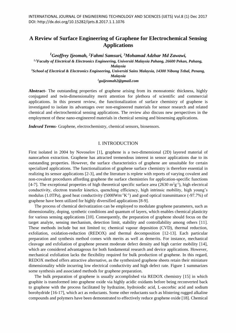
INTERNATIONAL JOURNAL OF ENGINEERING TECHNOLOGY AND SCIENCES (IJETS) Vol.8 (1) Dec 2017 DOI: http://dx.doi.org/10.15282/ijets.8.2017.1.1.1076
A Review of Surface Engineering of Graphene for Electrochemical Sensing
Applications
1Geoffrey Ijeomah, 2Fahmi Samsuri, 3Mohamad Adzhar Md Zawawi, 1,2Faculty of Electrical & Electronics Engineering, Universiti Malaysia Pahang, 26600 Pekan, Pahang,
Malaysia 3School of Electrical & Electronics Engineering, Universiti Sains Malaysia, 14300 Nibong Tebal, Penang,
Malaysia [email protected]
Abstract- The outstanding properties of graphene arising from its monoatomic thickness, highly
conjugated and twin-dimensionality merit attention for plethora of scientific and commercial
applications. In this present review, the functionalization of surface chemistry of graphene is
investigated to isolate its advantages over non-engineered materials for sensor research and related
chemical and electrochemical sensing applications. The review also discuss new perspectives in the
employment of these nano-engineered materials in chemical sensing and biosensing applications.
Indexed Terms- Graphene, electrochemistry, chemical sensors, biosensors.
I. INTRODUCTION
First isolated in 2004 by Novoselov [1], graphene is a two-dimensional (2D) layered material of
nanocarbon extraction. Graphene has attracted tremendous interest in sensor applications due to its
outstanding properties. However, the surface characteristics of graphene are unsuitable for certain
specialized applications. The functionalization of graphene surface chemistry is therefore essential in
realizing its sensor applications [2-3], and the literature is replete with reports of varying covalent and
non-covalent procedures affording graphene the surface chemistries for application-specific functions
[4-7]. The exceptional properties of high theoretical specific surface area (2630 m2g-1), high electrical
conductivity, electron transfer kinetics, quenching efficiency, high intrinsic mobility, high young’s
modulus (1.0TPa), good heat conductivity (5000Wm-1K-1) and good optical transmittance (-97.7%) of
graphene have been utilized for highly diversified applications [8-9].
The process of chemical derivatization can be employed to modulate graphene parameters, such as
dimensionality, doping, synthetic conditions and quantum of layers, which enables chemical plasticity
for various sensing applications [10]. Consequently, the preparation of graphene should focus on the
target analyte, sensing mechanism, detection limit, stability and controllability among others [11].
These methods include but not limited to; chemical vapour deposition (CVD), thermal reduction,
exfoliation, oxidation-reduction (REDOX) and thermal decomposition [12-13]. Each particular
preparation and synthesis method comes with merits as well as demerits. For instance, mechanical
cleavage and exfoliation of graphene present moderate defect density and high carrier mobility [14],
which are considered advantageous for both fundamental research and device applications. However,
mechanical exfoliation lacks the flexibility required for bulk production of graphene. In this regard,
REDOX method offers attractive alternative, as the synthesized graphene sheets retain their miniature
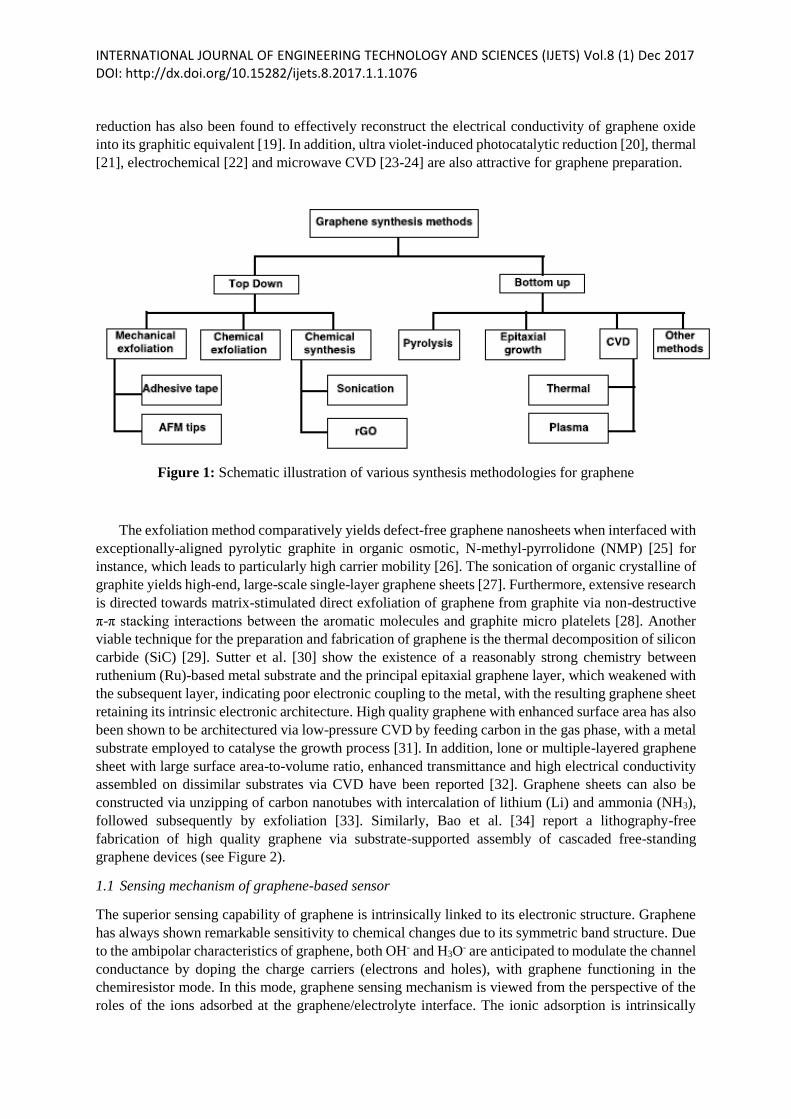
dimensionality while incurring low electrical conductivity and high defect rate. Figure 1 summarizes
some synthesis and associated methods for graphene preparation.
The bulk preparation of graphene is usually accomplished via REDOX chemistry [15] in which
graphite is transformed into graphene oxide via highly acidic oxidants before being reconverted back
to graphene with the process facilitated by hydrazine, hydroiodic acid, L-ascorbic acid and sodium
borohydride [16-17], which act as reductants. Some other reductants such as blistering rugged alkaline
compounds and polymers have been demonstrated to effectively reduce graphene oxide [18]. Chemical
INTERNATIONAL JOURNAL OF ENGINEERING TECHNOLOGY AND SCIENCES (IJETS) Vol.8 (1) Dec 2017 DOI: http://dx.doi.org/10.15282/ijets.8.2017.1.1.1076 reduction has also been found to effectively reconstruct the electrical conductivity of graphene oxide
into its graphitic equivalent [19]. In addition, ultra violet-induced photocatalytic reduction [20], thermal
[21], electrochemical [22] and microwave CVD [23-24] are also attractive for graphene preparation.
Figure 1: Schematic illustration of various synthesis methodologies for graphene
The exfoliation method comparatively yields defect-free graphene nanosheets when interfaced with
exceptionally-aligned pyrolytic graphite in organic osmotic, N-methyl-pyrrolidone (NMP) [25] for
instance, which leads to particularly high carrier mobility [26]. The sonication of organic crystalline of
graphite yields high-end, large-scale single-layer graphene sheets [27]. Furthermore, extensive research
is directed towards matrix-stimulated direct exfoliation of graphene from graphite via non-destructive
π-π stacking interactions between the aromatic molecules and graphite micro platelets [28]. Another
viable technique for the preparation and fabrication of graphene is the thermal decomposition of silicon
carbide (SiC) [29]. Sutter et al. [30] show the existence of a reasonably strong chemistry between
ruthenium (Ru)-based metal substrate and the principal epitaxial graphene layer, which weakened with
the subsequent layer, indicating poor electronic coupling to the metal, with the resulting graphene sheet
retaining its intrinsic electronic architecture. High quality graphene with enhanced surface area has also
been shown to be architectured via low-pressure CVD by feeding carbon in the gas phase, with a metal
substrate employed to catalyse the growth process [31]. In addition, lone or multiple-layered graphene
sheet with large surface area-to-volume ratio, enhanced transmittance and high electrical conductivity
assembled on dissimilar substrates via CVD have been reported [32]. Graphene sheets can also be
constructed via unzipping of carbon nanotubes with intercalation of lithium (Li) and ammonia (NH3),
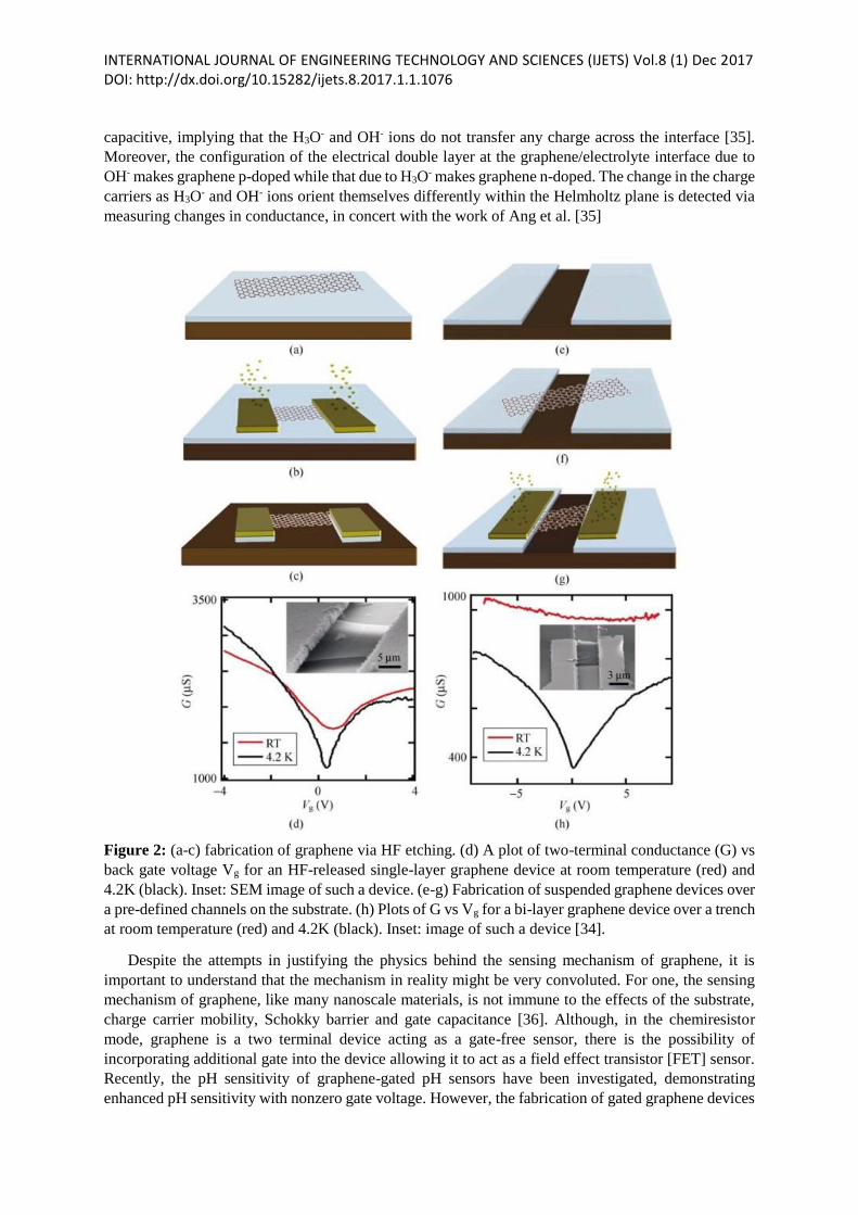
followed subsequently by exfoliation [33]. Similarly, Bao et al. [34] report a lithography-free
fabrication of high quality graphene via substrate-supported assembly of cascaded free-standing
graphene devices (see Figure 2).
1.1 Sensing mechanism of graphene-based sensor
The superior sensing capability of graphene is intrinsically linked to its electronic structure. Graphene
has always shown remarkable sensitivity to chemical changes due to its symmetric band structure. Due
to the ambipolar characteristics of graphene, both OH- and H3O- are anticipated to modulate the channel
conductance by doping the charge carriers (electrons and holes), with graphene functioning in the
chemiresistor mode. In this mode, graphene sensing mechanism is viewed from the perspective of the
roles of the ions adsorbed at the graphene/electrolyte interface. The ionic adsorption is intrinsically
INTERNATIONAL JOURNAL OF ENGINEERING TECHNOLOGY AND SCIENCES (IJETS) Vol.8 (1) Dec 2017 DOI: http://dx.doi.org/10.15282/ijets.8.2017.1.1.1076 capacitive, implying that the H3O- and OH- ions do not transfer any charge across the interface [35].
Moreover, the configuration of the electrical double layer at the graphene/electrolyte interface due to
OH- makes graphene p-doped while that due to H3O- makes graphene n-doped. The change in the charge
carriers as H3O- and OH- ions orient themselves differently within the Helmholtz plane is detected via
measuring changes in conductance, in concert with the work of Ang et al. [35]
Figure 2: (a-c) fabrication of graphene via HF etching. (d) A plot of two-terminal conductance (G) vs
back gate voltage Vg for an HF-released single-layer graphene device at room temperature (red) and
4.2K (black). Inset: SEM image of such a device. (e-g) Fabrication of suspended graphene devices over
a pre-defined channels on the substrate. (h) Plots of G vs Vg for a bi-layer graphene device over a trench
at room temperature (red) and 4.2K (black). Inset: image of such a device [34].
Despite the attempts in justifying the physics behind the sensing mechanism of graphene, it is
important to understand that the mechanism in reality might be very convoluted. For one, the sensing
mechanism of graphene, like many nanoscale materials, is not immune to the effects of the substrate,
charge carrier mobility, Schokky barrier and gate capacitance [36]. Although, in the chemiresistor
mode, graphene is a two terminal device acting as a gate-free sensor, there is the possibility of
incorporating additional gate into the device allowing it to act as a field effect transistor [FET] sensor.
Recently, the pH sensitivity of graphene-gated pH sensors have been investigated, demonstrating
enhanced pH sensitivity with nonzero gate voltage. However, the fabrication of gated graphene devices
INTERNATIONAL JOURNAL OF ENGINEERING TECHNOLOGY AND SCIENCES (IJETS) Vol.8 (1) Dec 2017 DOI: http://dx.doi.org/10.15282/ijets.8.2017.1.1.1076 is very elaborate and convoluted procedure, involving CVD and epitaxy-grown graphene samples.
Comparatively, the planar graphene pH chemiresistor offers a more simplistic design, due in part to its
two terminal architecture and focused ion beam (FIB), making it the simplest reusable graphene-based
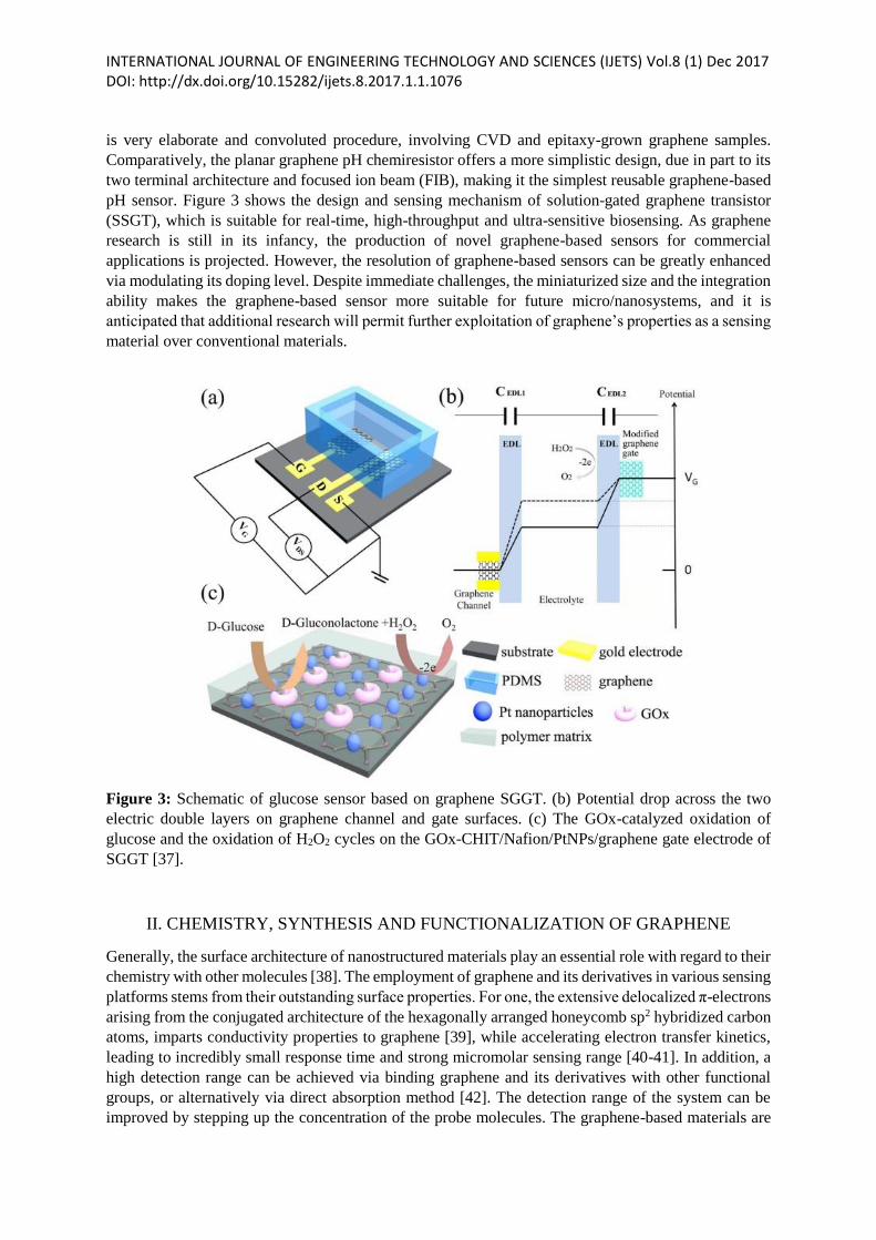
pH sensor. Figure 3 shows the design and sensing mechanism of solution-gated graphene transistor
(SSGT), which is suitable for real-time, high-throughput and ultra-sensitive biosensing. As graphene
research is still in its infancy, the production of novel graphene-based sensors for commercial
applications is projected. However, the resolution of graphene-based sensors can be greatly enhanced
via modulating its doping level. Despite immediate challenges, the miniaturized size and the integration
ability makes the graphene-based sensor more suitable for future micro/nanosystems, and it is
anticipated that additional research will permit further exploitation of graphene’s properties as a sensing
material over conventional materials.
Figure 3: Schematic of glucose sensor based on graphene SGGT. (b) Potential drop across the two
electric double layers on graphene channel and gate surfaces. (c) The GOx-catalyzed oxidation of
glucose and the oxidation of H2O2 cycles on the GOx-CHIT/Nafion/PtNPs/graphene gate electrode of
SGGT [37].
II. CHEMISTRY, SYNTHESIS AND FUNCTIONALIZATION OF GRAPHENE
Generally, the surface architecture of nanostructured materials play an essential role with regard to their
chemistry with other molecules [38]. The employment of graphene and its derivatives in various sensing
platforms stems from their outstanding surface properties. For one, the extensive delocalized π-electrons
arising from the conjugated architecture of the hexagonally arranged honeycomb sp2 hybridized carbon
atoms, imparts conductivity properties to graphene [39], while accelerating electron transfer kinetics,
leading to incredibly small response time and strong micromolar sensing range [40-41]. In addition, a
high detection range can be achieved via binding graphene and its derivatives with other functional
groups, or alternatively via direct absorption method [42]. The detection range of the system can be
improved by stepping up the concentration of the probe molecules. The graphene-based materials are
INTERNATIONAL JOURNAL OF ENGINEERING TECHNOLOGY AND SCIENCES (IJETS) Vol.8 (1) Dec 2017 DOI: http://dx.doi.org/10.15282/ijets.8.2017.1.1.1076 found to be strongly enriched with functional groups at the intersection of the edges following the
application of a smart processing method utilizing heteroatom doping and oxidation techniques
resulting in molecular level adjustment and fabrication of hybrid sensing platforms [43]. Therefore, the
modification of surface architecture of graphene materials may be the most fundamental and significant
step towards redesigning graphene derivatives for varying purposes. The ability of functionalized
graphene materials to conjugate with various recognition molecules, in addition to incorporating
additional functional media is paramount for electrochemical bioanalysis [44].
Before the isolation of the molecular allotropes of carbon – fullerenes, carbon nanotubes (CNTs),
and more recently, 2-D layer graphene, research into carbon was only limited to the fundamental
materials of graphite and diamond. However, graphene has upstaged other carbonaceous materials in
winning massive research interest owing to its classic architectural features and exceptional
performance. As the surface of pristine graphene is unsuitable for sensing purposes, there has been
increasing interest in investigating various facets, particularly the modification of graphene exterior.
Therefore, the functionalization and dispersion of graphene sheets are paramount in this aspect. Further
processing of chemically functionalized graphene can be demonstrated via solvent-facilitated methods,
such as filtration, spin-coating and layer-by-layer assembly. The problem of assemblage of lone layer
of graphene during reduction in osmotic phase still remains. However, this can be shot down via
appropriate chemical functionalization, while retaining graphene’s intrinsic properties. Graphene oxide
(GO) has been widely employed as a veritable precursor material for the growth and synthesis of
processable graphene. The modified Hummers technique is very effective in preparing graphene oxide
from pristine graphite, although a host of other preparation methods exist [45]. The highly oxygenated
nature of graphene oxide exterior coupled with the presence of carboxyl functional groups has the
potential to form a complex with van der waals forces, leading to the formation of a range of soluble
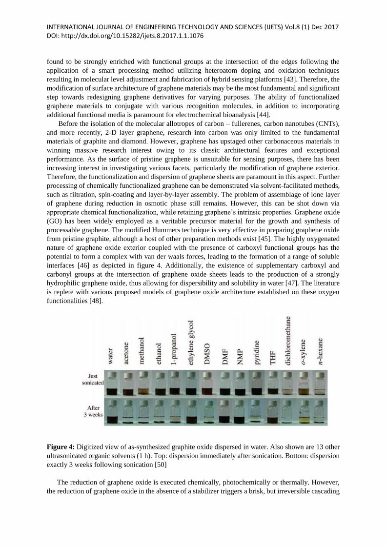
interfaces [46] as depicted in figure 4. Additionally, the existence of supplementary carboxyl and
carbonyl groups at the intersection of graphene oxide sheets leads to the production of a strongly
hydrophilic graphene oxide, thus allowing for dispersibility and solubility in water [47]. The literature
is replete with various proposed models of graphene oxide architecture established on these oxygen
functionalities [48].
Figure 4: Digitized view of as-synthesized graphite oxide dispersed in water. Also shown are 13 other
ultrasonicated organic solvents (1 h). Top: dispersion immediately after sonication. Bottom: dispersion
exactly 3 weeks following sonication [50]
The reduction of graphene oxide is executed chemically, photochemically or thermally. However,
the reduction of graphene oxide in the absence of a stabilizer triggers a brisk, but irreversible cascading
INTERNATIONAL JOURNAL OF ENGINEERING TECHNOLOGY AND SCIENCES (IJETS) Vol.8 (1) Dec 2017 DOI: http://dx.doi.org/10.15282/ijets.8.2017.1.1.1076 of graphene sheets, leading to precipitation of graphene particles. Therefore, the surface
functionalization of graphene oxide is necessary prior to the reduction process, and this can be executed
covalently or non-covalently, followed by reduction [49]. We now know that the reduction of
alkylamine-adjusted graphene oxide creates stable dispersions of functionalized graphene sheets in
biotic solvents. Moreso, the induction of carboxylic or sulfonate ensembles on graphene basal planes
can foster the formation of water-dispersible graphene sheets [51]. Dispersible graphene can be
synthesized directly from natural graphite as reported in [52]. Graphene transformed by ionic liquid and
assembled electrochemically from natural graphite has also been demonstrated [52]. Several other
methods for high yield assembling of stable un-functionalized graphene are reported elsewhere [53].
2.1 Covalent modification
Here, the covalent functionalization of graphene framework is discussed. The architectural
transformations can occur at the edge of the graphene sheets or on its exterior. The re-hybridization of
one or more sp2 carbon atoms of the carbon complex into sp3 layout followed by simultaneous loss of
electronic conjugation constitutes a fundamental step towards surface functionalization of graphene
[54]. The covalent alteration of graphene can be accomplished via any of the following methods;
condensation, nucleophilic substitution, addition and electrophilic addition [55-79]. The varying genre
of covalent modification of graphene oxide with their associated modifying agents, electrical
conductivity, dispersibility and dispersion stability in various osmotic is shown in table 1.
Covalent interaction is vital for graphene functionalization in sensor application [80]. The process is
facilitated via covalent bond formation, which can be architectured at the intersection of the edges or
on the basal planes. This can be accomplished through the chemical interaction of unsaturated π-bonds
of graphene with any of organic functional moieties, oxygen on graphene oxide [80], and heteroatom
doping.
The implementation of C-C bonds couplings of graphene oxide on cascades of diazonium salts has
led to the production of free radicals with exceptional affinity for addition reaction, and having
capability for surface-based vertical immobilization of motley aryl-addends [93]. These engineered
chemical interactions because the sp2 hybridized carbon atoms to transform to its sp3 hybridized
equivalent such that the graphene layer assumes quasi-conducting and unconducting regions [110]. In
addition, certain compounds of dienophile extraction such as azomethine ylide [69], aryne [79] and
nitrene [111] have been found to form excellent complexes when interfaced with graphene, yielding
adaptable varieties which are excellent starting point for further transformation and functionalization of
graphene nano-materials. Before now, several studies have focused on the covalent bond interaction
between other functional groups and oxygen moieties that is known to emerge from graphene oxide
[112]. Using the well-established carbodiimide mechanism, the graphene oxide is subsequently
interfaced with N,N-dicyclohexylcarbodiimide or 1-ethyl-3-(3-dimethylaminopropyl) carbodiimide in
the presence of N-hydroxysuccinimide to generate a stable organic ester, which transforms to an amide
bond when reacted with a target molecule containing amine group. To this end, graphene and its
derivatives present excellent electrochemical sensing platforms when covalently interfaced with poly-
L-lysine [113], DNA [114], beta-aminocyclodextrin [115], and protein [115]. The redesign of
carboxylates to acyl chlorides constitutes another technique for graphene functionalization [116]. This
allows the activation of graphene oxide with thionyl chloride (SOCl2) leading to the production of a
derivative of graphene, acyl chloride which can go into chemical association with amino or hydroxyl
groups. On its part, heteroatom doping plays crucial role in engineering the electronic properties of
graphene nano-materials [117]. In this process, graphene realized via heteroatom doping is synthesized
using trivalent or pentavalent atoms as they share similar architecture with elemental carbon, and are
able to accept or donate electrons [118]. While the incorporation of a pentavalent atom into graphene
basal plane leads to the formation of n-type carrier, the integration of a trivalent atom results in a p-type
material. Using a pentavalent dopant, nitrogen (N) for instance, results in the formation of tripartite
INTERNATIONAL JOURNAL OF ENGINEERING TECHNOLOGY AND SCIENCES (IJETS) Vol.8 (1) Dec 2017 DOI: http://dx.doi.org/10.15282/ijets.8.2017.1.1.1076 bonding layout in the graphitic basal plane, which could be active site for reduction-oxidation action
[119]. In fact, N-based dopants have been demonstrated to enhance sensitivity and biocompatibility,
improve binding potential, electron transfer kinetics, and electrical conductivity which are essential
parameters for sensing applications [120].
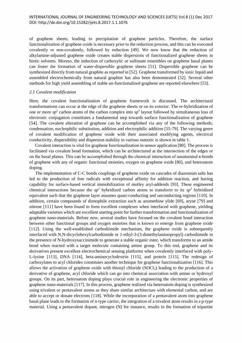
Table 1: Types of covalent modification of graphene oxide using varying modifying agent,
dispersibility, electrical conductivity and dispersion stability in varying solvents.
Modification
methods
Modifying agents Dispersium medium Dispersibility
(mg ml-1)
Elect.
Cond.
(Sm-1)
Refs.
Condensation
Nuecleohilic
substitution
Addition
Electronic
substitution
Organic diisocyanate
Adenine, cysteine,
nicotamide, OVA
TMEDA
A-CD, β-CD, γ-CD
Organic isocyanate
ODA
β-CD
TPP-NH2
PEG-NH2
PVA
TPAPAM
CS
Alkyl amine/amino acid
4-aminobenzene
sulphonic acid
4,4-diaminodiphenyl ester
PDA
Alkylamine
APTS
IL-NH2
PLL
Dopamine
Polyglycerol
Poly(norepinephrine)
Cyclopropanated
malonate
POA
Aryne
Polyacethylene
NMP
4-bromo aniline
ANS
Sulfanilic acid
DMF
H2O
THF
H2O, ethanol, DMF, DMSO
DMF, NMF, NMP, PC, THF
THF, CCl4, 1,2-
dichloroethane
Water, acetone, DMF
DMF
Water
Water, DMSO
TFH
Water
CHCl3, THF, DCM, toluene
H2O
Xylene, methanol
THF
Water, DMF
H2O, ethanol, DMF, DMSO
Water, DMF, DMSO
Water
Water
Water
H2O, NMP, methanol,
acetone, DMF, THF
Tolune, O-DCB, DMF,
DCM
THF
DMF, O-DCB
Ortho dichlorobenzene
Ethanol, DMF, NMP, PC,
THF
DMF
H2O
H2O
-
0.1
0.2
>2.5
1.0(DMF)
0.5(THF)
1.0(DMF)
-
1.0
-
-
2.0
-
0.2
0.1
0.2
1.55
0.5
0.5
0.5
0.05
3.0
-
0.5
0.2
0.4
0.1
0.2 – 1.4
0.02
3.0
2.0
-
-
-
-
-
-
-
-
-
-
-
-
-
-
-
-
-
-
-
-
-
-
-
-
-
-
-
21600
-
145
1250
[81]
[82]
[83]
[84]
[85]
[86]
[87]
[88]
[89]
[90]
[91]
[92]
[93]
[94]
[94]
[95]
[96]
[97[
[98]
[99]
[100]
[101]
[102]
[103]
[95]
[104]
[105]
[106]
[107]
[108]
[109]
2.2 Noncovalent functionalization
Noncovalent modification is fundamentally concerned with hydrophobic, electrostatic, π-π stacking and
van der waals forces, and requires the material adsorption of appropriate molecules on the graphene
exterior. This is achieved via adsorption of aromatic molecules or surfactants, interaction with
biomolecules and peptides and deoxyribonucleic acid (DNA), and polymer wrapping. The literature is
replete with reports of extensive employment of noncovalent functionalization in the surface
INTERNATIONAL JOURNAL OF ENGINEERING TECHNOLOGY AND SCIENCES (IJETS) Vol.8 (1) Dec 2017 DOI: http://dx.doi.org/10.15282/ijets.8.2017.1.1.1076 transformation of the sp2 hybridized CNT ensembles [121]. Research indicates that similar result is
possible using the same approach, but with the application of varying forms of organic modifiers on the
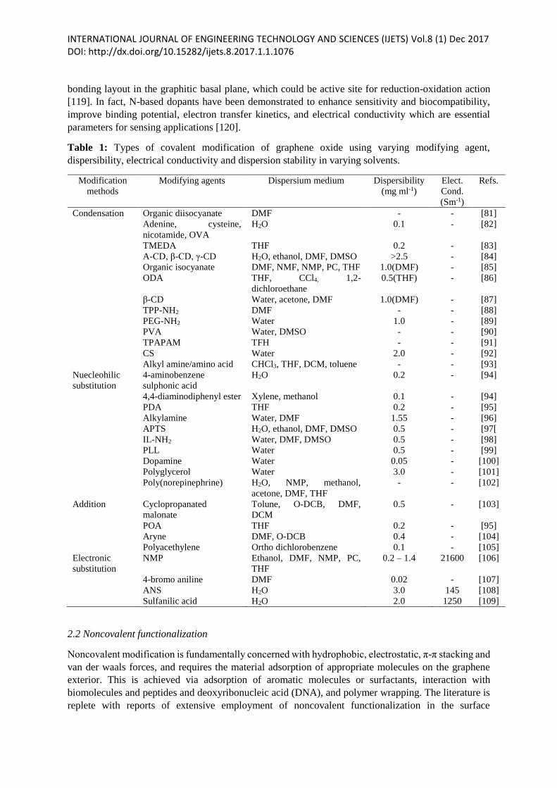
graphene substrate [122-124]. Table 2 shows varying genre of noncovalent modification of graphene
oxide with their associated modifying agents, electrical conductivity, dispersibility, dispersion stability
in various osmotic.
Table 2: Types of noncovalent modification of graphene oxide using varying modifying agent,
dispersibility, electrical conductivity and dispersion stability in varying solvents
Modifying agents Dispersium medium Dispersibility
(mg ml-1)
Elect. Cond.
(Sm-1)
Refs
Poly(propyleneimine
dendrimers)
SLS, SCMS, HPC-Pv
Amino terminated
polymer
Caronene derivative
Porphyrin
SPANI
PYR-NJS
PNIPAAM
SDBS
PIL
PSS
MG
PSS-g-PPY
PBA
PPE-SO3-
Water
Water
1,3-dimethyl-2-imidazolidinone, γ-
butyrolactone, 1-propanol, ethanol,
ethylene, glycol, DMF
Water
Water
Water
Water
Water
Water
Water
Water
Water
Water
Water
Water
-
(0.6-2.0)
0.4
0.15
0.02
>1.0
-
-
1.0
1.5
1.0
0.1
3.0
0.1
0.25
-
-
1500
-
370 Ωcm
30
-
-
80 Ω
3600
-
-
-
200
-
[125]
[126]
[127]
[128]
[129]
[130]
[124]
[131]
[131]
[132]
[133]
[134]
[135]
[136]
[137]
Noncovalent functionalization is frequently exploited to redesign graphene nanostructures. In this
process, the autochthonous sp2-conjugated architecture of graphene remains unchanged in contrast to
covalent modification, with graphene retaining its towering electrical conductivity. This presents
positive implications in sensor applications where unbroken graphene is critical for optimal
functionality.
The binding of readily dissolvable or dispersible pyrene derivatives to graphene sheets is usually
exploited in the fabrication of graphene-based sensors [136], and this is a typical instance of π-π
stacking. In addition, π-π stacking can be exploited to directly attach single stranded DNA (ssDNA) to
graphene exterior [138]. Similarly, different classes of DNA detectable platforms can result from
graphene and nucleic acid bases [139]. Amphiphilic compounds are also found to be excellent drivers
for the dispersion of graphene materials in aqueous solution, with the hydrophobic edge linked to the
graphene exterior. Here, hydrophilic polymers can be employed to decorate graphene oxide in order to
circumvent the problem of aggregation in aqueous solution. For instance, polyetherimide [140],
polyvinyl alcohol [141], polyvinylpyrrolidone [142] and poly(diallyldimethylammonium chloride)
[143] have been extensively employed as dispersants owing to their exceptional hydrophilic chemistry
with and without electrostatic interaction.
2.3 Other decoration methods for graphene
In recent times, inorganic molecules have been exploited to modify graphene sheets, inducing ancillary
electrochemical catalysis that may be a recipe for functionalization [144]. Research effort is directed
towards a mosaic of solution-based metallic nanoparticles and graphene due to their perceived
potentials for scientific and electrochemical sensing applications. Different methodologies have been
devised to synthesize graphene metal composites, especially for noble metals. Hassan et al. [145] report
INTERNATIONAL JOURNAL OF ENGINEERING TECHNOLOGY AND SCIENCES (IJETS) Vol.8 (1) Dec 2017 DOI: http://dx.doi.org/10.15282/ijets.8.2017.1.1.1076 the preparation of metal nanoparticle based on microwave, and dispersed on the graphene exterior in
oleic acid and oleylamine. The combined techniques permit the synchronous reduction of assorted metal
salts and graphene oxide, with the production of nano-catalyst supported on a massive surface exterior
of heat-tolerant graphene. Guo et al. [146] demonstrate a facile technique for the synthesis of platinum
(Pt)-on-palladium (Pd) nanodendrites, and Pt nanoparticles linked in aqueous phase to graphene sheets
via wet chemistry. The other direct and convenient procedure for fabrication of metal graphene
nanocomposites in the absence of any additives or capping agents is the clean-reduction of Pt, Pd and
gold (Au) precursor by graphene oxide [147].
Again, high quality metal graphene hybrids with novel nanoparticle characteristics can be fabricated
via self-assembly method [148]. This is possible through the synthesis of metallic nanoparticles via a
methodological approach exploiting the different dimensionalities, configurations and building blocks
of the nanoparticle. Zhu et al. [149] show the preparation of a hybrid 3-D nanocomposite films via
assembling in alternation, Pt nanoparticles and ionic liquid modified graphene nanosheets. This method
utilizes ionic liquid-functionalized graphene based on imidazolium salts, and prepared via covalent
bonding of 1-(3-aminopropyl)-3-methylimidazolium bromide to graphene nanosheets. The introduction
of ionic liquid based on imidazolium onto the exterior of graphene nanosheets leads to positive-
polarized graphene dispersible in aqueous solution. The formation of as-functionalized multiple layered
film is highly unvarying, thanks to methodical and facile self-assembly. In addition, the selection of
varying sequences in self-assembly can lead to an efficient route for the construction of electrochemical
nanodevices when the electrochemical activity of graphene films is finely engineered. An inclusive and
general approach for the decoration and reduction of graphene oxide exploiting ‘fraction V’ or bovine
serum albumin has been demonstrated by Deng et al. [150]. The metal graphene hybrid is assembled
by establishing a complex via appropriate interface methodology between the bovine serum albumin
and thiol & imidazole, noble metals & amine groups. This lone-stage decoration/reduction technique
aims to prepare nanoparticle materials with tunable surface architecture, configuration, dimensionality
and composition which are highly beneficial for the functionalization of graphene-based materials.
Interestingly, many oxide nanomaterials particularly metallic and quasi-metallic oxide materials
have received considerable research attention in electrochemistry. These oxides exhibit increased
current density and reduced overpotential when transformed to conductive graphene materials owing to
the low electrical conductivity, although their utilization may present fantastic platform for further
functionalization of graphene materials. Graphene oxide is known to bond very well with iron (III)
oxide (Fe2O3), manganese (VI) oxide (MnO2) and cobalt (II) oxide/cobalt (III) hydroxide
(Co(OH)2/Co(OH)3 [151]. Yang et al. [152] assemble graphene oxide decorated with mesoporous silica
(SiO2) using wet chemistry. In particular, the electrostatic adsorption and self-assembling onto the
alkaline, highly negatively-polarized graphene oxide exterior is catalysed via cationic surfecants,
cetyltrimethyl ammonium bromide, for instance. In consequence, the single-layer graphene oxide
exterior becomes capped with mesoporous silica. The injection of the cationic surfecant aims to resolve
the problem of aggregation and mismatch between inorganic particles and graphene oxide, as well as
usher in molecular platform for the development of mesoporous silica and guided nucleation on the
exterior of graphene oxide sheets. Additionally, Dong et al. [151] demonstrate the synthesis of Co(OH)3
based nanowires on 3D graphene foam, taking advantage of the familiar hydrothermal mechanism.
Their study show the formation of a stable, high specific capacitance functionalized graphene at
operating current density of 10 Ag-1 with capability to detect glucose at a sensitivity of 3.39 mAmM-
1cm-2, in addition to having a small detection limit of approximately 25 nM. However, the towering
temperature and pressure inherent in the hydrothermal process limits its usefulness in experimental
research.
INTERNATIONAL JOURNAL OF ENGINEERING TECHNOLOGY AND SCIENCES (IJETS) Vol.8 (1) Dec 2017 DOI: http://dx.doi.org/10.15282/ijets.8.2017.1.1.1076
III. GRAPHENE-BASED ELECTRODE FOR BIOMOLECULE DETECTION
Electrochemical mechanisms are appealing for detection and sensing of biomolecules owing to their
easy of assembly and noncomplication. Such mechanisms depend fundamentally on capacitance
computation, AC conductivity, voltammetry, amperometry, coulometry and potentiometry for
performance evaluation. Generally, biomolecule detection for most graphene-based electrochemical
biosensing is executed amperometrically. In this section, we discuss important select graphene-based
electrodes for small biomolecules.
3.1 Dopamine
An important neurotransmitter, dopamine is reputed for its role in regulating the central nervous,
hormonal, renal and cardiovascular systems [153]. Dopamine detection has generated significant
attention and intense research interest. Ultra-sensitive and fast electrochemical mechanisms are
encouraging in the detection of neurotransmission. However, at the conventional solid electrode,
dopamine and its mutual kind uric acid and ascorbic acid exhibit volumetric feedback overlap leading
to high detection limit and poor selectivity. This explains the complexity in establishing the nuance
surrounding the chemistry of dopamine, uric acid and ascorbic acid in a biotic medium.
Shang et al. [153] report the synthesis of a dopamine-detectable electrode based on multiple layered
graphene nanoflakes via uncatalyzed microwave plasma facilitated chemical vapour deposition (CVD).
The multiple layered graphene nanoflakes were found to be able to resolve and discriminate the
chemical nexus between dopamine, uric acid and ascorbic acid, with dopamine detected at a limit of
0.17 µM. The ability of the defects/plane sites occurring tangential to the terminal of the perpendicular
graphene nanoflakes to nanoconnect, and electrically transport the charge carriers (in this case,
electrons) to the underneath substrate imparts high-end biosensing capability to the electrode [153]. In
addition, graphene is reported to exhibit improved sensing capability towards dopamine, with added
capacity to effectively discriminate dopamine, ascorbic acid and serotonin relative to its precursor
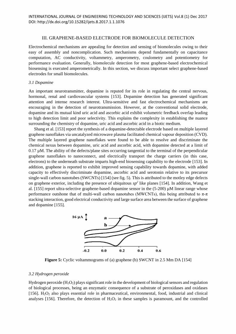
single-wall carbon nanotubes (SWCNTs) [154] (see fig. 5). This is attributed to the motley edge defects
on graphene exterior, including the presence of ubiquitous sp2 like planes [154]. In addition, Wang et
al. [155] report ultra-selective graphene-based dopamine sensor in the (5-200) µM linear range whose
performance outshone that of multi-wall carbon nanotubes (MWCNTs), this being attributed to π-π
stacking interaction, good electrical conductivity and large surface area between the surface of graphene
and dopamine [155].
Figure 5: Cyclic voltammograms of (a) graphene (b) SWCNT in 2.5 Mm DA [154]
3.2 Hydrogen peroxide
Hydrogen peroxide (H2O2) plays significant role in the development of biological sensors and regulation
of biological processes, being an enzymatic consequence of a substrate of peroxidases and oxidases
[156]. H2O2 also plays essential role in pharmaceutical, environmental, food, industrial and clinical
analyses [156]. Therefore, the detection of H2O2 in these samples is paramount, and the controlled
INTERNATIONAL JOURNAL OF ENGINEERING TECHNOLOGY AND SCIENCES (IJETS) Vol.8 (1) Dec 2017 DOI: http://dx.doi.org/10.15282/ijets.8.2017.1.1.1076 reduction of oxidation/reduction overpotentials is the most significant step in this direction. Over the
years, carbon allotropes, such as CNTs [157] have been employed in the fabrication of sensors, and
graphene nanomaterial stands out in this aspect [158-159].
In a study of the electrochemical behaviour of H2O2 on graphene, Zhou et al. [156] observed an
astronomical increase in the electron transfer kinetics relative to graphite/glassy carbon and bare glassy
carbon electrodes (GCEs). The resulting onset potentials for H2O2 oxidized/reduced on graphene/glassy
carbon, graphite/glassy carbon and glassy carbon electrodes (GCEs) are; 0.20/0.10 V, 0.80/-0.35 V and
0.70/-0.25 V respectively, revealing excellent electrocatalytic activity of graphene towards H2O2. In
addition, the linear correlation of H2O2 at -0.2 V overpotential on graphene/GCE is found to show a
mismatch of (0.05-1500) µM in contrast to CNTs [156]. The culpability for this mismatch was placed
on the various active sites for electron transfer to biotic species resulting from high concentration of
edge-plane like defective sites on graphene [160]. Such electrodes could be good starting materials
towards the fabrication of ultra-sensitive and selective electrochemical sensors for the detection of H2O2.
3.3 Nicotinamide adenine dinucleotide
Nicotinamide adenine dinucleotide, which has two active versions – oxidized form (NAD+) and reduced
version (NADH) is responsible for the transfer of charge carriers (electrons) from and/or to active
reaction sites during a reduction/oxidation (REDOX) reaction. These cofactors (NAD+ and NADH) of
numerous dehydrogenases compounds have received considerable attention for several applications
connected with NAD+/NADH reliant hydrogenases, including amperometric biosensing, bioelectronics
devices and fuel cells [161-162]. The anodic signal from the oxidation of NADH faithfully reconstructs
the NAD+ cofactor which is essentially important in biosensing critical substrates such as glucose,
lactate and alcohol [163]. The problem of surface fouling and large NADH oxidation potential
associated with the buildup of reaction products still remained unresolved [163]. However, these
problems can be addressed by incorporating graphene in such reactions.
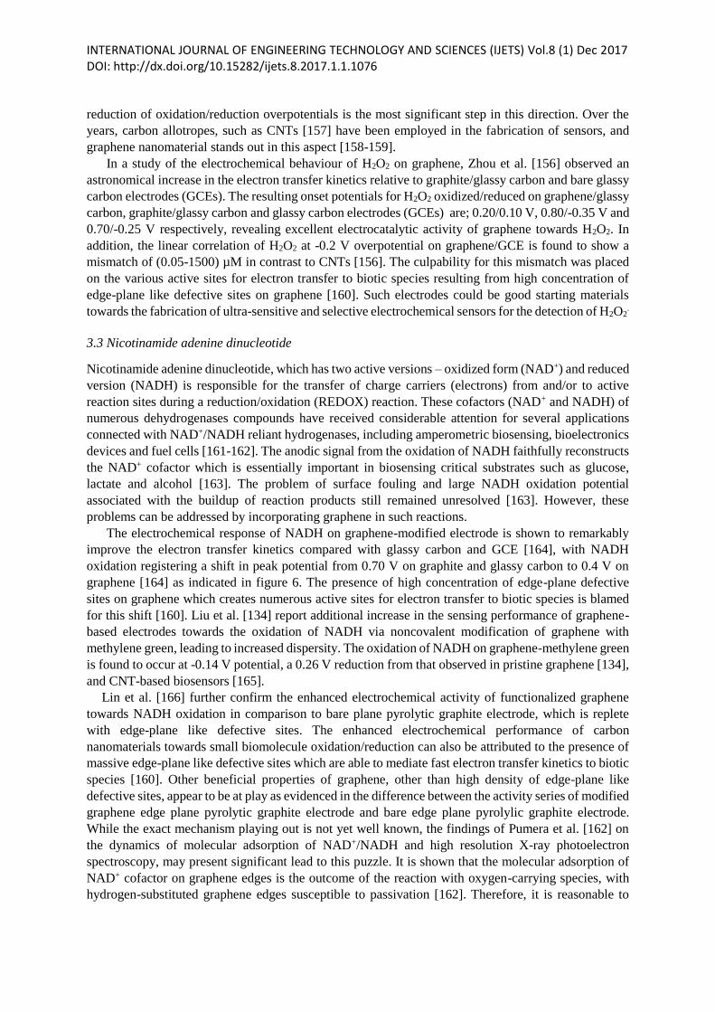
The electrochemical response of NADH on graphene-modified electrode is shown to remarkably
improve the electron transfer kinetics compared with glassy carbon and GCE [164], with NADH
oxidation registering a shift in peak potential from 0.70 V on graphite and glassy carbon to 0.4 V on
graphene [164] as indicated in figure 6. The presence of high concentration of edge-plane defective
sites on graphene which creates numerous active sites for electron transfer to biotic species is blamed
for this shift [160]. Liu et al. [134] report additional increase in the sensing performance of graphene-
based electrodes towards the oxidation of NADH via noncovalent modification of graphene with
methylene green, leading to increased dispersity. The oxidation of NADH on graphene-methylene green
is found to occur at -0.14 V potential, a 0.26 V reduction from that observed in pristine graphene [134],
and CNT-based biosensors [165].
Lin et al. [166] further confirm the enhanced electrochemical activity of functionalized graphene
towards NADH oxidation in comparison to bare plane pyrolytic graphite electrode, which is replete
with edge-plane like defective sites. The enhanced electrochemical performance of carbon
nanomaterials towards small biomolecule oxidation/reduction can also be attributed to the presence of
massive edge-plane like defective sites which are able to mediate fast electron transfer kinetics to biotic
species [160]. Other beneficial properties of graphene, other than high density of edge-plane like
defective sites, appear to be at play as evidenced in the difference between the activity series of modified
graphene edge plane pyrolytic graphite electrode and bare edge plane pyrolylic graphite electrode.
While the exact mechanism playing out is not yet well known, the findings of Pumera et al. [162] on
the dynamics of molecular adsorption of NAD+/NADH and high resolution X-ray photoelectron
spectroscopy, may present significant lead to this puzzle. It is shown that the molecular adsorption of
NAD+ cofactor on graphene edges is the outcome of the reaction with oxygen-carrying species, with
hydrogen-substituted graphene edges susceptible to passivation [162]. Therefore, it is reasonable to
INTERNATIONAL JOURNAL OF ENGINEERING TECHNOLOGY AND SCIENCES (IJETS) Vol.8 (1) Dec 2017 DOI: http://dx.doi.org/10.15282/ijets.8.2017.1.1.1076 conclude that the enhanced activity of graphene may not be unconnected with the presence of oxygen-
carrying groups.
Figure 6: Cyclic voltammograms in 0.1 M pH 6.8 PBS containing 1mM NADH at bare GC (dashed
line) and graphene /GCE (solid line) [164].
IV. ELECTROCHEMICAL SENSORS
Electrochemical sensors possess numerous attractive features which make them suitable for a wide
range of applications for monitoring and detecting analytes in both liquid and gaseous phases.
Generally, electrochemical sensors are available as simple, small, mechanically robust, compact, low
cost and reliable-in-operation devices.
The ability of electrochemical sensors to operate under ambient conditions, without the need for
external perturbations makes them advantageous over their closest competitors. Consequently, the
power requirements of electrochemical sensors is extremely low, although additional power may be
required for extrasensory functions such as alarms, data transmission and recording. In this aspect,
electrochemical sensors are ideally suited to mobile instruments where battery dimension, cost and
power are of utmost concern.
Electrochemical sensors can be broadly categorized into two – potentiometric types which have
reputation for eliciting a voltage response to an analyte, and amperometric types which allow an
electrical current response. In both cases, the sensors contain at least two electrodes, sandwiched by a
solid electrolyte or assembly of ionically conducting liquid. Majority of electrochemical sensors utilize
aqueous solutions of salts, bases and acids as electrolytes. The pages that follow discuss some select
graphene-based electrochemical sensors
4.1 Graphene-based electrochemical sensors
Graphene materials have been utilized as excellent electrodes in electroanalysis owing to its superlative
electrochemical features [163]. Several graphene and graphene composites-based electrochemical
sensors for biological and environmental analysis have been developed [156]. As a promising candidate
in electrochemistry, graphene offers numerous advantages and potential applications relative to CNTs.
The metallic impurities in CNTs which interfere with, and limit their electrochemistry as reported for
H2O2 [167], glucose [168], hydrazine [169], halothane [170], short peptides [171], amino acids [172]
even at levels below 100 ppm [173] are unavailable in graphene. In addition, graphite which is the
starting material for graphene fabrication is both inexpensive and accessible. There has been an
exponential increase in the number of published papers on the employment of graphene for
INTERNATIONAL JOURNAL OF ENGINEERING TECHNOLOGY AND SCIENCES (IJETS) Vol.8 (1) Dec 2017 DOI: http://dx.doi.org/10.15282/ijets.8.2017.1.1.1076 electrochemical sensing and biosensing following the appearance of the first article as reported here
[174]. Graphene has the potential to detect molecules with high REDOX potentials owing to its large
electrochemical potential window [156].
4.1.1 Graphene-based electrochemical DNA biosensors
Electrochemical sensors are critical for the detection of select DNA sequences and mutated conditions
associated with human diseases, offering high sensitivity, low cost and selectivity for diagnostic
interventions [175]. This category of sensors have reputation for allowing device miniaturization for
micro-volume samples [156]. Direct oxidation-based DNA sensors are the simplest kinds of such
sensors [156].
Zhou et al. [156] report an electrochemical sensor based on graphene nanomaterial for DNA
detection. Graphene/glassy carbon is shown to simultaneously detect the four basic free DNA bases,
adenine, thymine, quinine and cytosine as evidenced by the separate current signals of each of the bases
on the graphene/ GCE. This is in direct contrast to individual graphene and glassy carbon which lack
this rare detection capability. This is attributed to the towering electron transfer kinetics for the
oxidation of the bases on graphene/GCE as well as antifouling properties [156], arising from the high
concentration of edge-plane like defective sites and oxygen-rich functional species on graphene, with
active sites that amplify electron exchange between the electrodes and species in solution [160].
Similarly, a well-suited graphene/glassy carbon is able to detect, in both single and double stranded
DNAs (ssDNA and dsDNA) platforms, the four DNA bases which are resistant to oxidation reaction at
physiological pH, eliminating the need for prehydrolysis phase and accelerating the detection of single-
nucleotide polymorphism locus for miniature oligomers having particular progression at the
graphene/GCE in the absence of any labelling phenomena or hybridization. This can be attributed to
the excellent high conductivity, antifouling, lone-sheet, large surface area and towering electron transfer
kinetics of graphene [156].
4.1.2 Graphene-based enzyme biosensors
Graphene could be an outstanding electrode for the detection of glucose oxidase (GOx) given its
superior performance in direct electrochemistry of GOx and good electrocatalytic activity of H2O2.
Several glucose biosensors based on graphene nanomaterial have been reported [176]. Shan et al. [176]
demonstrate the first glucose biosensor based on graphene, and developed via
graphene/polyethylenimine-functionalized ionic liquid nanocomposite transformed electrode with a
linear response of (2-14) mM, R= 0.994, high stability with response current of +4.9 % after a duration
of 7 days, and high reproducibility [176]. Zhou et al. [156] report another glucose biosensor based on
chemically reduced graphene. This sensor was found to exhibit high sensitivity (20.21 µA mM cm-2),
low detection limit (2 µM) and wide linear range (0.01-10) mM. The linear range for the amperometric
detection of glucose spans a wider area than on the other carbon derivative material electrodes, CNTs
[177] and carbon nanofibers [178] are good examples. The limit of detection (LOD) of glucose at
graphene/GOx/GCE is found to pale behind that of sensors based on carbon nanomaterials such as CNT
nanoelectrode [179], exfoliated graphite nanoplatelets [180], carbon nanotube paste [181], carbon
nanotube fiber [178], and extremely ordered mesoporous carbon [182]. The detection of glucose at the
graphene/GOx/GCE was found to be very fast, having a response time of (9±1)s. On the stability
criterion, the electrodes were found to be more stable, being able to fully retain up to 91% of their initial
activity after a duration of 5 h, which makes them suitable for continuous measurement of glucose level
in diabetic patients enabling diagnostic and therapeutic interventions.
4.1.3 Graphene-based electrochemical sensors for heavy metal ions
Graphene-based electrochemical sensors for continuous monitoring and detection of heavy metal ions
such as Pb2+ and Cd2+ in environmentally poor settings have been developed. Segura et al. [183] report
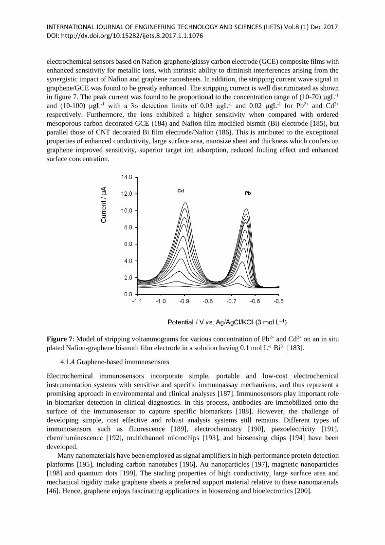
INTERNATIONAL JOURNAL OF ENGINEERING TECHNOLOGY AND SCIENCES (IJETS) Vol.8 (1) Dec 2017 DOI: http://dx.doi.org/10.15282/ijets.8.2017.1.1.1076 electrochemical sensors based on Nafion-graphene/glassy carbon electrode (GCE) composite films with
enhanced sensitivity for metallic ions, with intrinsic ability to diminish interferences arising from the
synergistic impact of Nafion and graphene nanosheets. In addition, the stripping current wave signal in
graphene/GCE was found to be greatly enhanced. The stripping current is well discriminated as shown
in figure 7. The peak current was found to be proportional to the concentration range of (10-70) µgL-1
and (10-100) µgL-1 with a 3σ detection limits of 0.03 µgL-1 and 0.02 µgL-1 for Pb2+ and Cd2+
respectively. Furthermore, the ions exhibited a higher sensitivity when compared with ordered
mesoporous carbon decorated GCE (184) and Nafion film-modified bismth (Bi) electrode [185), but
parallel those of CNT decorated Bi film electrode/Nafion (186). This is attributed to the exceptional
properties of enhanced conductivity, large surface area, nanosize sheet and thickness which confers on
graphene improved sensitivity, superior target ion adsorption, reduced fouling effect and enhanced
surface concentration.
Figure 7: Model of stripping voltammograms for various concentration of Pb2+ and Cd2+ on an in situ
plated Nafion-graphene bismuth film electrode in a solution having 0.1 mol L-1 Bi3+ [183].
4.1.4 Graphene-based immunosensors
Electrochemical immunosensors incorporate simple, portable and low-cost electrochemical
instrumentation systems with sensitive and specific immunoassay mechanisms, and thus represent a
promising approach in environmental and clinical analyses [187]. Immunosensors play important role
in biomarker detection in clinical diagnostics. In this process, antibodies are immobilized onto the
surface of the immunosensor to capture specific biomarkers [188]. However, the challenge of
developing simple, cost effective and robust analysis systems still remains. Different types of
immunosensors such as fluorescence [189], electrochemistry [190], piezoelectricity [191],
chemiluminescence [192], multichannel microchips [193], and biosensing chips [194] have been
developed.
Many nanomaterials have been employed as signal amplifiers in high-performance protein detection
platforms [195], including carbon nanotubes [196], Au nanoparticles [197], magnetic nanoparticles
[198] and quantum dots [199]. The starling properties of high conductivity, large surface area and
mechanical rigidity make graphene sheets a preferred support material relative to these nanomaterials
[46]. Hence, graphene enjoys fascinating applications in biosensing and bioelectronics [200].
INTERNATIONAL JOURNAL OF ENGINEERING TECHNOLOGY AND SCIENCES (IJETS) Vol.8 (1) Dec 2017 DOI: http://dx.doi.org/10.15282/ijets.8.2017.1.1.1076 The immobilization of antibody fragments on the surface of the sensor substrate without
diminishing their binding affinities and binding capacities is paramount. In the physical adsorption
procedure, antibody fragments are stochastically immobilized with the sensor substrate, predominantly
via hydrophilic and hydrophobic interactions. We also note that the chirality of the antibody (Ab) on
the surface of the sensor substrate cannot be controlled. Therefore, the binding activity of the Ab is lost
to the analyte. For the covalent cross-linking technique, the unobstructed amino groups on the Ab can
be stochastically coupled to several reactive moieties on the surface of the sensor substrate.
Consequently, the orientation of the immobilized Ab is also stochastic. Thus, there is increasing need
to develop a methodology to achieve well-oriented immobilization of Ab within a miniscule area
without diminishing their binding affinity for analytes, particularly for miniaturized diagnostic devices.
Chitosan is widely employed as dispersant in preparing graphene sheet-methylene blue (GS-MB)
nanocomposite in order to achieve well-oriented immobilization of antibody due to its attractive
properties such as excellent biocompatibility, permeability and non-toxicity, which also makes it a good
candidate for biomolecule immobilization [201]. When dropped onto the surface of glassy carbon
electrode, the resultant nanocomposite solution formed a stable film rich in hydroxyls and aminos. The
platform for the crosslinking of Ab is usually provided by the high surface area of the graphene sheet
and vast hydroxyls and aminos of chitosin.
4.2 Graphene-based electrical sensors
Following the isolation of first lone-layer graphene via mechanical cleavage of graphite [1], pristine
graphene has enabled the illumination of fundamental properties at condensed matter physics level.
Single layer graphene is quasi-metallic with excellent properties that are particularly exploitable at
nanoscale for sensor design and applications. Due to its zero bandgap, graphene exhibits low intrinsic
noise [202], high carrier mobility [203], ambient temperature hall effect [204], ambipolar field-effect
characteristics and high carrier density [205]. Sensors based on 1D nanomaterials such as CNTs and
nanowires represent a new frontier in sensor technology, opening up assortment of opportunities for
label-free detection, compatibility with lab-on-chip devices and high temporal resolution [206]. Perhaps
silicon nanowires (SiNWs) are the most researched 1D nanomaterial for nanoelectronic sensing [207].
However, their over-reliance on the induced field-effect limits their nanoelectronic applications [208],
making them only sensitive and selective to electrogenic phenomena or polarized analytes [209]. Over
the years, graphene has remained a superstar for various sensing and biosensing applications, thanks to
its superior electrical properties [8].
Graphene is shown to exhibit similar electrical response to varying chemical species as CNTs, as
evidenced in [210]. Therefore, just like CNTs, graphene-based sensors have won considerable research
interest for possible detection of various target species in vapour phase [211]. For the first time,
Novoselov et al. [202] demonstrated a sensor based on graphene with exceptional capability to detect
nitrogen dioxide (NO2). Based on micromechanical cleavage of graphitic material at the exterior
interface of oxidized Si wafers, this sensor has been employed to detect NO2 by taking advantage of
resistance difference of the source-drain. The literature is replete with reports of graphene-based
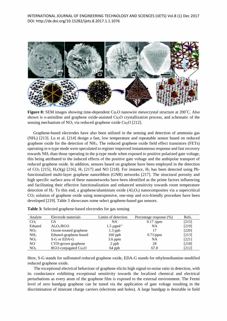
electrodes for the detection of NO2 [211]. A good example is the reduced graphene oxide-copper oxide
nanowire mesocrystals conjugate prepared under hydrothermal conditions via unclassical
crystallization in the presence of graphene oxide and o-anisidine, which are considered attractive for
the detection of NO2 [212]. This composite sensor integrates the rich electrical conductivity of reduced
graphene oxide with the fine interdendritic space, leading to the formation of 3D conducting
architecture. This scenario is well depicted in Figure 8.
INTERNATIONAL JOURNAL OF ENGINEERING TECHNOLOGY AND SCIENCES (IJETS) Vol.8 (1) Dec 2017 DOI: http://dx.doi.org/10.15282/ijets.8.2017.1.1.1076
Figure 8: SEM images showing time-dependent Cu2O nanowire mesocrystal structure at 200°C. Also
shown is o-anisidine and graphene oxide-assisted Cu2O crystallization process, and schematic of the
sensing mechanism of NO2 via reduced graphene oxide Cu2O [212].
Graphene-based electrodes have also been utilized in the sensing and detection of ammonia gas
(NH3) [213]. Lu et al. [214] design a fast, low temperature and repeatable sensor based on reduced
graphene oxide for the detection of NH3. The reduced graphene oxide field effect transistors (FETs)
operating in n-type mode were speculated to register improved instantaneous response and fast recovery
towards NH3 than those operating in the p-type mode when exposed to positive polarized gate voltage,
this being attributed to the induced effects of the positive gate voltage and the ambipolar transport of
reduced graphene oxide. In addition, sensors based on graphene have been employed in the detection
of CO2 [215], H2O(g) [216], H2 [217] and NO [218]. For instance, H2 has been detected using Pb-
functionalized multi-layer graphene nanoribbon (GNR) networks [217]. The structural porosity and
high specific surface area of these nanonetworks have been identified as the prime factors influencing
and facilitating their effective functionalization and enhanced sensitivity towards room temperature
detection of H2. To this end, a graphene/aluminium oxide (Al2O3) nanocomposites via a supercritical
CO2 solution of graphene oxide using nonexpensive, one-step and eco-friendly procedure have been
developed [219]. Table 3 showcases some select graphene-based gas sensors.
Table 3: Selected graphene-based electrodes for gas sensing
Analyte Electrode materials Limits of detection Percentage response (%) Refs.
CO2
Ethanol
NO2
NH3
NO2
NO
NO2
GS
Al2O3/RGO
Ozone-treated graphene
Ethanol-graphene-based
S-G or EDA-G
CVD-grown graphene
RGO-conjugated Cu2O
NA
1.5 µgml-1
1.3 ppb
160 ppb
3.6 ppm
2 ppb
64 ppb
0.17 /ppm
NA
17
0.71/ppm
NA
28
67.8
[215]
[219]
[220]
[213]
[221]
[218]
[212]
Here, S-G stands for sulfonated reduced graphene oxide, EDA-G stands for ethylenediamine-modified
reduced graphene oxide.
The exceptional electrical behaviour of graphene elicits high signal-to-noise ratio in detection, with
its conductance exhibiting exceptional sensitivity towards the localized chemical and electrical
perturbations as every atom of the graphene film is exposed to the external environment. The Fermi
level of zero bandgap graphene can be tuned via the application of gate voltage resulting in the
discrimination of itinerant charge carriers (electrons and holes). A large bandgap is desirable in field
INTERNATIONAL JOURNAL OF ENGINEERING TECHNOLOGY AND SCIENCES (IJETS) Vol.8 (1) Dec 2017 DOI: http://dx.doi.org/10.15282/ijets.8.2017.1.1.1076 effect-induced detection [222]. The opening of graphene bandgap represents a significant step in the
bandgap engineering of graphene, and can be actualized through the introduction of atomic or molecular
dopants [223] or reduction of its dimensions to nanoscale [224]. Graphene, unlike 1D nanostructured
sensing electrodes, exhibits homogeneous exterior for effective and uniform functionalization, and
enhanced detection range [225]. Graphene has the potential to form a sensing complex by interfacing
with flat cell membranes [226]. In addition, the biocompatibility of graphene presents excellent
platform for efficient cell growth and adhesion [227].
One dimensional-based biosensing nanostructured electrodes have been interfaced to living cells to
detect their dynamic activities [228], which include, adipocytokines [229], circulating breast cancer
cells [230], bioelectricity [231], and triggered secretion of proinflammatory cytokines [232]. The
presence of graphene in the nanoelectronic-cell complex increases the dimensionality of the interface
which presents a number of possible applications at device level. Interestingly, cell membranes can also
interface with flat graphene as both have similar dimensionality [233]. This is in direct contrast to cells
interfaced with nanostructures other than graphene and its derivatives, where the intricate interplay is
found to be slack and nonhomogeneous, making the nanotopographic structure-inspired thin cell
membrane-induced local curvature alter the cells chemistry [234]. Therefore, it is reasonable to argue
that any cell activity-induced local chemical and electrical variations in the nanogap between the
exteriors of graphene and cell membrane would significantly cause its conductance to transform, given
the robust chemistry existing in the graphene-cell membrane complex. Lieber et al. [235] demonstrate
the extracellular detection of activity potentials from lone electrogenic cardiomyocytes using graphene
field effect transistors (GFETs). This work exploited mechanically exfoliated graphene to fabricate
devices via electron beam lithography. Here, the sensitivity of GFET was found to be superior to that
of unfunctionalized metallic microelectrodes [236], but was found to parallel those of silicon nanowire
FETs (SiNWFETs) [237]. The field effect-induced device response arising from the fluctuations of the
short-lived nanointerface potential across graphene-membrane exterior is attributed to the current flow
through the membrane ion channels. Although less popular to the field effect of SiNW, GFET showed
a comparable signal-to-noise ratio [238]. This is attributed to the large area spanning the graphene-cell
interface. With its large bandgap, graphene nanorods exhibit high sensitivity due to the combined effects
of enhanced field-effect and exceptional spatial resolution arising from their lateral nanoscale
dimension, thus enabling the sensing of bioelectricity.
Another sensing utilization of graphene due to its superior electrical property is fluorescence
quenching. Fluorescence quenching is a procedure employed to obtain information on the dynamic
changes of protein in complex macromolecular systems. The ability of graphene to quench fluorescence
is highly utilized in the selective detection of biomolecules. The fluorescence quenching of aromatic
molecules by graphene is linked to photoinduced electron transfer. In addition, graphene-initiated
fluorescence quenching of porphyrin and photophysical features of graphene-porphyrin complexes have
been reported [239]. Ramakrishna et al. [240] study fluorescence quenching mechanism in graphene.
Graphene is shown to be excellent fluorescence quencher relative to other metals. The quenching
mechanism of graphene was found to depend on nonradiative decay of the fluorophore, and not due to
charge transfer as previously envisaged. While fluorescence intensity of graphene is distance-
dependent, other contributing factors are substrate doping and roughness, which creates new channels
for the fluorescence intensity to be decayed. However, the mechanism of these effects is not yet well
known, creating room for further investigation.
Recently, Rodrigo et al. [241] reported an ultra-sensitive tunable plasmonic biosensor for
chemically specific label-free detection of protein monolayers, which exploits the rich electro-optical
properties of graphene. It is shown that, in contrast to conventional plasmonic materials such as noble
metals, the feedback mechanism of infrared (IR) is characterized by long shelf life corporative electron
oscillations that can be dynamically tuned via electrostatic gating. In addition, the electromagnetic field
of graphene infrared plasmons is shown to exhibit novel spatial confinement, making them highly
INTERNATIONAL JOURNAL OF ENGINEERING TECHNOLOGY AND SCIENCES (IJETS) Vol.8 (1) Dec 2017 DOI: http://dx.doi.org/10.15282/ijets.8.2017.1.1.1076 suitable for enhanced light-matter and integrated mid-IR photonics, properties with potentials for
biosensing opportunities. The mid-IR range is essential for biosensing actions as it encompasses the
molecular vibrations, which is a unique marker of biomolecules such as DNA, proteins and lipids. The
absorption spectroscopy is powerful enough to provide exquisite biochemical platform for label-free
detection via the accessing of vibrational fingerprints. The large mismatch between biomolecular
dimension and mid-IR wavelengths limits the intensity of the vibrational absorption. However, this can
be overcome by harnessing the robust optical near fields neighbourhood of metallic nanomaterials,
which sacrifices the spectral bandwidth to reduced dimensionality, and itself limited by deficient field
confinement of metals in the mid-infrared.
V. CONCLUSION
As noted, graphene has generated considerable research interest in many fields since its discovery. The
exceptional physical and chemical properties of graphene arising from its unique architecture present a
number of interesting sensing applications. In addition to the applications of graphene herein discussed,
the unusual and exceptional properties of graphene can be utilized in other paradigms such as water and
food safety, sensitive medical analysis, pollution control among others. However, the challenges of
improving functionalization and synthesis methods, extensive understanding of the surface architecture
and graphene engineering, and extending the applications to various paradigms still remain.
The fundamental mechanisms of graphene development which include CVD, reduction of graphite
oxide, mechanical and matrix-assisted exfoliations, unzipping CNTs and thermal decomposition are
still in their developmental phases. The preparation methods may yield outputs with varying properties
which can be tailored to specific desirable performance functionalities. There is increasing momentum
within the scientific community to step up graphene materials and processes, devising novel and
efficient techniques towards the preparation and synthesis of high quality, defect-free and large size
graphene. In this aspect, the challenges of demand-based properties and access to large quantum of high
quality homogeneous graphene have been recognized. Therefore, the near-perfect understanding of the
underlying chemistry and physics of graphene surface engineering, as well as the complex interplay of
chemicals or biomolecules at graphene interface, particularly as nanoscaffold materials in
biosensing/chemical and catalysis, is essentially important. This improved understanding of graphene
interaction at the molecular level could well pave way for the design and fabrication of ultra-sensitive
and selective sensors, and ultimately advance graphene science and engineering.
There remains a development gap in the design and fabrication of graphene-based sensors. The
potential applications of certain graphene materials such as bi-layered graphene or graphene quantum
dots have not been investigated despite the tremendous amount of work done on graphene nanomaterial.
Novel techniques for synthesis and fabrication of graphene with exceptional performance metrics are
continually being invented by scientists. The hybridization or compositing of graphene or its derivatives
with other organic or inorganic materials could further expand the landscape of graphene applications
in sensor development. Improved functionalization techniques are being explored and pursued,
although much work is needed to fully understand and maximize graphene properties in sensor
development and application.
The development of novel techniques for well-modulated processing and synthesis of graphene is
strongly encouraged. As noted, graphene has been developed with varying strategies. However, the
current synthesis methods are uneconomical and do not guarantee high graphene yield. The use of
chemical/thermal reduction of graphene oxide in electrochemical detection applications looks attractive
in this aspect. Graphene electrodes from chemically/thermally reduced graphene oxide are particularly
susceptible to re-stacking during processing and synthesis. So far, a number of strategies to counter the
problem of re-stacking and enhance the dispersion of graphene in the solvent have been adopted. The
literature is replete with reports of graphene produced via electrochemical reduction of graphene oxide,
INTERNATIONAL JOURNAL OF ENGINEERING TECHNOLOGY AND SCIENCES (IJETS) Vol.8 (1) Dec 2017 DOI: http://dx.doi.org/10.15282/ijets.8.2017.1.1.1076 and the electrochemically reduced graphene oxide is found to exhibit enhanced electrochemical activity
relative to the graphene reduced by chemical means, which raises the possibility for large scale and
high yield graphene processing and synthesis.
Another area that requires additional investigation for possible improvement is the doping of
graphene with heteroatoms. The procedure has been shown to greatly improve electrocatalytic activity
in CNTs, and has been well applied in synthesis of graphene-based nanomaterials and theoretical
studies, albeit not applicable in electrochemistry. The doping of graphene with nitrogen can be achieved
at high temperatures which significantly increases the odds of stacking. Therefore, other doping
strategies should be explored.
In conclusion, graphene is an excellent electrode material for electrochemical sensing and
biosensing applications. While much progress has been made in redesigning the surface engineering of
graphene which hitherto had led to numerous applications, there is still much room for further
development particularly at research, material and device levels.
Abbreviations
DMF, N,N-dimethylformide; DMSO, dimethyl sulfoxide; PVA, poly(vinyl alcohol); NMP, N-methyl-
2-pyroolidone; THF, tetrahydrofuran; PBA, 1-pyrenebutyric acid; SLS, static light scattering; PYR-
NHS, pyrenebutanoic acid-succinimidyl ester; TPAPAM, aryl amine terminated trphenylamine-based
polyazomethine; ODA, octadecylamine; APTS, γ-aminopropyl triethoxysilane; PLL, poly-L-lysine;
PSS, poly(styrene sulfonate); β-CD, β-cyclodextrin; α-CD, α-cyclodextrin; γ-CD, γ-cyclodextrin;
TMEDA, tetramethylenediamine; PEG-NH2, amine-functionalized polyethylene glycol; TPP-NH2,
amine-functionalized porphyrin; IL-NH2, amino-terminated ionic liquid; NMF, N-methylformamide;
CS, chondroitin sulfate; OVA, ovalbumin; SLS, sodium lignosulfonate; MG, modified graphene; PC,
phosphatidylcholine; SPANI, sulfonated polyaniline; PNIPAAM, poly(N-isopropylacrylamide); PPE-
SO3-,poly(2,5-bis(3-sulfonatopropoxy)-1,4-ethynyl-phenylene-alt-1,4-ethynylphenylene) sodium salt;
PPY, polypyrrole; PIL, ionic liquid polymers; CCl3 CCl4, carbon tetrachloride; DCB, dichlorobenzene;
DCM, dichloro methane; SDBS, sodium dodecyl benzene sulfonate; ANS, 6-amino-4-hydroxy-2-
naphthalenesulfonic acid; HPC-Py, pyrene-modified hydroxypropyl cellulose; SCMC, sodium
carboxymethyl cellulose; POA, poly(o-anisidine).
REFERENCES
[1] Novoselov K., Geim A., Morozov S., Jiang D., Zhang Y., Dubonos S., Grigorieva I., & Firsov A.
‘‘Electric field effect in atomically thin carbon films’’. Science 306, 666-669, 2004.
[2] Tumer A. P. ‘‘Biosensors: sense and sensibility’’. Chem. Soc. Rev. 42, 3184-3196, 2013.
[3] Yen M. Y., Hsieh C-K., Teng C-C., Hsiao M.-C., Liu P.-L., Ma C-C. M., Tsai M.-C., Tsai C-H.,
Lin Y.-R., & Chou T.-Y. ‘‘Metal-free, nitrogen-doped graphene used as novel catalyst for dye-
sensitized solar cell counter electrodes’’. RSC Adv. 2, 2725-2728, 2012. [cross ref.].
[4] Zhang F., Zhang T., Yang X., Zhang L., Leng K., & Huang Y. C. A. ‘‘high-performance
supercapacitor-battery hybrid energy storage device based on graphene-enhanced electrode
materials with ultrahigh energy density’’. Energy Environ. Sci. 6, 1623-1632, 2013 [cross ref.].
[5] Cao S., Zhang L., Chai Y., & Yuan R. ‘‘Electrochemistry of cholesterol biosensor based on a
novel Pt-Pd bimetallic nanoparticle decorated graphene catalyst’’. Talanta 109,167-172, 2013 [cross
ref.].
[6] Xiao F., Li Y., Zan X., Liao K., Xu R., & Duan H. ‘‘Growth of metal-metal oxide nanostructures
on free standing graphene paper for flexible biosensors’’. Adv. Funct. Mater. 22, 2487-2494, 2012
[cross ref.]
INTERNATIONAL JOURNAL OF ENGINEERING TECHNOLOGY AND SCIENCES (IJETS) Vol.8 (1) Dec 2017 DOI: http://dx.doi.org/10.15282/ijets.8.2017.1.1.1076 [7] Wu Y., Zhang X., Jie J., Xie C., Zhang X., Sun B., Wang Y., & Gao P. ‘‘Graphene transparent
conductive electrodes for highly efficient silicon nanostructures-based hybrid heterojunction solar
cells’’. J. Phys. Chem. C 117, 1027-1036, 2013.
[8] Liu Y., Dong X., & Chen P. ‘‘Biological and chemical sensors based on graphene materials’’. Chem.
Soc. Rev 41, 2283-2307, 2012.
[9] Kuila T., Bose S., Khanra P., Mishra A. K., & Kim N. H., et al. ‘‘Recent advances in graphene based
biosensors’’. Biosens Bioelectron 26: 4637-4648, 2011.
[10] Pumera M., Ambrosi A., Bonanni A., Chng E. L. K., & Poh H. I. ‘‘Graphene for electrochemical
sensing and biosensing’’. Trends Analyst. Chem. 29, 954-965, 2010.
[11] Fang Y., & Wang E. ‘‘Electrochemical biosensors on platforms of graphene’’. Chem. Commun.
49, 9526-9539, 2013 [cross ref.]
[12] Choi W., Lahiri I., Seelaboyina R., Kang Y. S. ‘‘Synthesis of graphene and its applications:
a review’’. Crit. Rev. Solid State 35, 52-71, 2010.
[13] Zhu Y., Murali S., Cai W., Li X., Sul J. W., Potts J. R., & Ruoff, R. S. ‘‘Graphene and graphene
oxide: synthesis, properties and applications, Adv. Mater. 22, 3906-3924, 2010.
[14] Martinez A., Fuse K., & Yamashita S. ‘‘Mechanical exfoliation of graphene for the passive
mode-locking of fiber lasers’’. Appl. Phys. Lett. 99, 2011 [cross ref.].
[15] Stankovich S., Dikin D. A., Piner R. D., Kohlhaas K. A., Kleinhammes A., Jia Y., Wu Y., Nguyen
S.B.T., & Rouff R. S. ‘‘Synthesis of graphene-based nanosheets via chemical reduction of
exfoliated graphite oxide’’. Carbon 45, 1558-1565, 2007.
[16] Pei S., Zhao J., Du J., Ren W., & Cheng H. M. ‘‘Direct reduction of graphene oxide films into
highly conductive and flexible graphene films by hydrohalic acids’’. Carbon 48, 4466-4474, 2010.
[17] Gao J., Liu F., Liu Y., Ma N., Wang Z., Zhang X. ‘‘Environment-friendly method to produce
graphene that employs vitamin C and amino acid’’. Chem. Mater. 22, 2213-2218, 2010.
[18] Glover A. J., Cai M., Overdeep K. R., Kranbuehi D. E., & Schniepp H. C. ‘‘In situ reduction of
graphene oxide in polymers’’. Macromolecules 44, 9821-9829, 2011.
[19] Bourlinos, A. B., Gournis. D., Petridis, D., Szabo, T., Szeri, A., Dekany, I. ‘‘Graphite oxide:
chemical reduction to graphite and surface modification with primary aliphatic amines and amino
acids’’. Langmuir 19, 6050-6055, 2003.
[20] Williams G., Seger B., & Kamat P. V. ‘’TiO2-graphene nanocomposites: UV-assisted
photocatalytic reduction of graphene oxide’’. ACS Nano 2, 1487-1491, 2008.
[21] Zangmeister, C.D. ‘‘Preparation and evaluation of graphite oxide reduced at 220ᵒC’’. Chem.Mater.
22, 5625-5629, 2010.
[22] Xu X., Huang D., Cao K., Wang M., Zakeeruddin S. M., & Gratzel M. ‘‘Electrochemically
reduced graphene oxide multilayer films as efficient counter electrode for dye-sensitized solar
cells’’. sci. Rep. 3, 2013.
[23] Zhu Y., Murali S., Stoller M. D., Velamakanni A., Piner R. D., & Ruoff R. S. ‘‘Microwave
assisted exfoliation and reduction of graphite oxide for ultra-capacitors’’. Carbon 48, 2118-2122,
2010.
[24] Adhikari S., Aryal H. R., Uchida H., Umeno M. ‘‘Catalyst-Free Growth of Graphene by
Microwave Surface Wave Plasma Chemical Vapour Deposition at Low Temperature’’. Journal of
Material Science and Chemical Engineering 4, 10-14, 2016.
[25] Hernandez Y., Nicolosi V., Lotya M., Blighe F. M., Sun Z., De S., McGovern I., Holland B., Byrne
M., & Gun’Ko Y. K. ‘‘High-yield production of graphene by liquid-phase exfoliation of
graphite’’. Nat. Nanotechnol. 3, 563-568, 2008 [cross ref.].
[26] Shukla A., Kumar R., Mazher J., & Balan A. ‘‘Graphene made easy: high quality large-area
samples’’. Solid state Commun. 149, 718-721, 2009.
[27] Li X., Zhang G., Bai X., Sun X., Wang X., Wang E., & Dai H. ‘‘Highly conducting graphene
sheets and Langmuir lodgett films’’. Nat. Nanotechnol. 3, 538-542, 2008.
[28] Artur C., & Paolo S. ‘‘Graphene via sonication assisted liquid-phase exfoliation’’. Chem. Soc.
Rev. 243, 381-398, 2013.
[29] Lin Y. M., Dimitrakopoulos C., Jenkins K. A., Farmer D. B., Chiu H. Y., & Grill A. ‘‘Avouris
100-GHz transistors from wafer-scale epitaxial graphene’’. Science 327, 662, 2010.
INTERNATIONAL JOURNAL OF ENGINEERING TECHNOLOGY AND SCIENCES (IJETS) Vol.8 (1) Dec 2017 DOI: http://dx.doi.org/10.15282/ijets.8.2017.1.1.1076 [30] Sutter P. W., Flege J. I., & Sutter E. A. ‘‘Epitaxial graphene on ruthenium’’. Nat. Mater. 7, 406-
411, 2008 [cross ref.]
[31] De Arco L. G., Zhang Y., Kumar A., & Zhou, C. ‘‘Synthesis, transfer, and devices of single- and
few-layer graphene by chemical vapour deposition’’. Nanotechnology, 8, 135-138, 2009.
[32] Cai W., More A. I., Zhu Y., Li X., Chen S., Shi I., & Ruoff R. S. ‘‘Thermal transport in
suspended and supported monolayer graphene grown by chemical vapour deposition’’. Nano Lett.
10, 1645-1651, 2010.
[33] Cano-Marquez A.G., Rodriquez-Macias F. J., Campos-Delgado J., Espinosa-Gonzalez C. G.,
Tristan-Lopez F., Ramirez-Gonzalez D., Cullen D. A., Smith D. J., Terrones M., Vega-Cantu Y. I.
‘‘Ex-MWCNTs: graphene sheets and ribbons produced by lithium intercalation and exfoliation of
carbon nanotubes’’. Nano Lett. 9, 1527-1533, 2009.
[34] Bao W., Liu G., Zhang H., Yan D., Deshpande A., LeRoy B., & Lau C. N. ‘‘Lithography-Free
Fabrication of High Quality Substrate-Supported and Freestanding Graphene Devices’’. Nano Res
3, 98-102, 2010.
[35] Ang P. K., Chen W., Wee A. T. S., & Loh K. P. ‘‘Solution-gated epitaxial graphene as pH sensor’’.
Journal of the American Chemical Society 130(44), 14392-14393, 2008.
[36] Heller I., Janssens A. M., Mannik J., Minot E. D., Lemay S. G., Dekker C. ‘‘Identifying the
mechanism of Biosensing with Carbon Nanotube Transistors’’. Nano Lett. 8, 591-595, 2008.
[37] Zhang M., Liao C., Mark C. H., You P., Mak C. L., Yan F. ‘‘Highly sensitive glucose sensors
based on enzyme-modified whole-graphene solution-gated transistors’’. Sci. Rep. 5, 8311, 2015.
[38] Verma A., & Stellacci F. ‘‘Effect of surface properties on nanoparticles-cell interactions’’. Small
6, 12-21, 2010.
[39] Yavari F., & Koratkar N. ‘‘Graphene-based chemical sensors’’. J. Phys. Chem. Lett. 3, 1746-
1753, 2012.
[40] Zhang Z., Gu S., Ding Y., Shen M., Jiang L. ‘‘Mild and novel electrochemical preparation of -
cyclodextrin/graphene nanocomposite film for super-sensitive sensing of quercetin’’. Biosens.
Bioelectron. 57, 239-244, 2014 [cross ref.].
[41] Xu Q., Gu S.-X., Jin L., Zhou Y.-E., Yang Z., Wang W., & Hu X. ‘‘Graphene / polyaniline / gold
nanoparticles nanocomposite for the direct electron transfer of glucose oxidase and glucose
biosensing’’.Sens. Actuators B Chem. 190, 562-569, 2014.
[42] Kybert N. J. ‘‘Nano-Bio Hybrid Electronic Sensors for Chemical Detection and Disease
Diagnostics’’. Publicly Accessible Penn Dissertations. Paper 1079, 29-31, 2015.
[43] Gong X., Bi Y., Zhao Y., Liu G., & Teoh W. Y. ‘‘Graphene oxide-based electrochemical sensor:
a platform for ultrasensitive detection of heavy metal ions’’. RSC Adv. 4, 1-2, 2014.
[44] Shao Y., Wang J., Wu H., Liu J., Aksay I. A., & Lin Y. ‘‘Graphene based electrochemical sensors
and biosensors: a review’’. Electroanal 22, 1027-1036, 2010 [cross ref.].
[45] Park S., & Ruoff R. S. ‘‘Chemical methods for the production of graphene’’. Nat. Nanotechnol 4,
217-224, 2009 [cross ref.].
[46] Stankovich S., Dikin D. A., Dommett G. H. B., Kohlhaas K. M., Zimney E. J., Stach E. A., et al.
‘‘Graphene based polymer composites’’. Nature 442, 282-286, 2006 [cross ref.].
[47] Nethravathi C., Rajamathi J. T., Ravishankar N., Shivakumara C., & Rajamathi M. ‘‘Graphite
oxide-intercalated anionic clay and its decomposition to graphene-inorganic material
nanocomposites’’. Langmuir 24, 8240-8244, 2008.
[48] Szabo T., Berkesi O., Forgo P., Josepovits K., Sanakis Y., & Petridis D., et al. ‘‘Evolution of
surface functional groups in a series of progressively oxidized graphite oxides’’. Chem. Mater 18,
2740-2749, 2006.
[49] Fang M., Wang K., Lu H., Yang Y., & Nutt S. ‘‘Single-layer graphene nanosheets with controlled
grafting of polymer chains’’. J. Mater Chem. 20, 1982-1992, 2010.
[50] Paresdes J. I., Villar-Rodil S., Martinez-Alonso A., & Tascon J. M. D. ‘‘Graphene oxide dispersion
in organic solvents’’. Langmuir 24, 4-5, 2008.
INTERNATIONAL JOURNAL OF ENGINEERING TECHNOLOGY AND SCIENCES (IJETS) Vol.8 (1) Dec 2017 DOI: http://dx.doi.org/10.15282/ijets.8.2017.1.1.1076 [51] Kuila T., Bose S., Hong C. E., Uddin M. E., Khanra P., & Kim N. H., et al. ‘‘Preparation of
functionalized graphene / linear low density polyethylene composites by a solution mixing
method’’. Carbon, 49, 1033-1037, 2011.
[52] Liu N., Luo F., Wu H., Liu Y., Zhang C., & Chen J. ‘‘One step ionic-liquid-assisted
electrochemical synthesis of ionic-liquid-functionalized graphene sheets directly from graphene’’.
Adv. Func. Mater 18, 1518-1525, 2008.
[53] Hamilton C. E., Lomeda J. R., Sun Z., Tour J. M., & Barron A. R. ‘‘High yield organic
dispersions of unfunctionalized graphene’’. Nano Lett. 9, 3460-3462, 2009.
[54] Park M. J., Lee J. K., Lee B. S., Lee Y. W., Choi I. S., Lee S. ‘‘Covalent modification of
multiwalled carbon nanotubes with imidazolium-based ionic liquids: effects of anions on
solubility’’. Chem. Mater. 18, 1546-1551, 2006.
[55] Economopoulos S. P., Rotas G., Miyata Y., Shinohara H., & Tagmatarchis N. ‘‘Exfoliation and
chemical modification using microwave irradiation affording highly functionalized graphene’’.
ACS Nano 12, 7499-7507, 2010.
[56] Stankovich S., Dikin D. A., Compton O. C., Dommett G. H. B., Ruoff R. S., & Nguyen S. T.
‘‘Systematic post-assembly modification of graphene oxide paper with primary alkylamines’’.
Chem. Mater 22, 4153-4157, 2010.
[57] Avinash M. B., Subrahmanyam K. S., Sundarayya Y., & Govindaraju T. ‘‘Covalent modification
and exfoliation of graphene oxide using ferrocene’’. Nanoscale 2, 1762-1766 [cross ref.].
[58] Bao H., Pan Y., Ping Y., Sahoo N. G., Wu T., & Li, L., et al. ‘‘Chitosan-functionalized graphene
oxide as a nanocarrier for drug and gene delivery’’. Small 7, 1569-1578, 2011 [cross ref.].
[59] Shen J., Yan B., Shi M., Ma H., Li N., & Ye M. ‘‘Synthesis of graphene oxide-based biocomposites
through diimide-activated amidation’’. J. Colloid Interf Sci. 356, 543-549, 2011.
[60] Pharm V. H., Cuong T. V., Hur S. H., Oh E., & Kim E. J. ‘‘Chemical functionalization of graphene
sheets by solvothermal reduction of a graphene oxide suspension in N-mathyl-2-pyrrolidone’’. J.
Mater Chem 21, 3371-3377 [cross ref.].
[61] Xu X., Luo Q., Lv W., Dong Y., Lin Y., & Yang Q. et al. ‘‘Functionalization of graphene sheets
by polyacetylene: convenient synthesis and enhanced emission’’. Macromol. Chem. Phys. 212,
768-773, 2011 [cross ref.].
[62] Hsiao M. C., Liao S. H., Yen M. Y., Liu P., Pu N. W., & Wang C. A., et al. ‘‘Preparation of
covalently functionalized graphene using residual oxygen-containing functional groups’’. ACS
Appl. Mater Interfaces 2, 3092-3099, 2010.
[63] Zhuang X. D., Chen Y., Liu G., Li P. P., Zhu C. X., & Kang E. T., et al. ‘‘Conjugated-polymer-
functionalized graphene oxide: synthesis and non-volatile rewritable memory effect’’. Adv. Mater
22, 1731-1735, 2010 [cross ref.].
[64] Kang S. M., Park S., Kim D., Park S. Y., Ruoff R. S., & Lee H. ‘‘Simultaneous reduction and
surface functionalization of graphene oxide by mussel-inspired chemistry’’. Adv. Funct. Mater 21,
108-112, 2011.
[65] Cui Y., Kim S. N., Jones S. E., Wissler L. L., Naik R. R., & McAlpine M .C. ‘‘Chemical
functionalization of graphene enabled by phase displayed peptides’’. Nano Lett. 10, 4559-4565,
2010.
[66] Rana V. K., Choi M. C., Kong J. Y., Kim G. Y., Kim M. J., & Kim S. H., et al. ‘‘Synthesis and
drug-delivery behaviour of chitosan functionalized graphene oxide hybrid nanosheets’’.
Macromol. Mater Eng. 296, 131-140, 2011.
[67] Quintana M., Spyrou K., Grzelczak M., Browne W. R., Rudolf P., & Prato M.
‘‘Functionalization of graphene via 1,3-dipolar cycloaddition’’. ACS Nano 4, 3527-3533, 2010.
[68] Zhang Y., Ren L., Wang S., Marathe A., Chaudhuri J., & Li G. ‘‘Functionalization of
graphene sheets through fullerene attachment’’. J. Mater Chem. 21, 5386-5391, 2011 [cross ref.]
[69] Georgakilas V., Bourlinos A. B., Zboril R., Steriotis T. A., Dallas P., & Stubos A. K., et al.
‘‘Organic functionalization of graphenes’’. Chem. Commun. 46, 1766-1768, 2010 [cross ref.]
INTERNATIONAL JOURNAL OF ENGINEERING TECHNOLOGY AND SCIENCES (IJETS) Vol.8 (1) Dec 2017 DOI: http://dx.doi.org/10.15282/ijets.8.2017.1.1.1076 [70] Guo Y., Guo S., Ren J., Zhai Y., Dong S., & Wang E. ‘‘Cyclodextrin functionalized graphene
nanosheets with high supramolecular recognition capability: synthesis and host-guest inclusion for
enhanced electrochemical performance’’. ACS Nano 4, 4001-4010, 2010.
[71] Xu L. Q., Yang W. J., Neoh K. G., Kang E. T., & Fu G. D. ‘‘Dopamine-induced reduction and
functionalization of graphene oxide nanosheets’’. Macromolecules 43, 8336-8339, 2010.
[72] Chen G., Zhai S., Zhai, Y., Zhang K., Yue Q & Wang L. et al. ‘‘Preparation of sulfonic-
functionalized graphene oxide as ion-exchange material and its application into
electrochemiluminescence analysis. Biosens Bioelectron 26, 3136-3141, 2011.
[73] Sun Z., Kohama S. I., Zhang Z., Lomeda J. R., & Tour J. M. ‘‘Soluble graphene through edge
selective functionalization’’. Nano Res. 3, 117-125, 2010.
[74] Kuila T., Khanra P., Bose S., Kim N. H., Ku B. C., & Moon B. et al. ‘‘Preparation of water
dispersible graphene by facile surface modification of graphite oxide’’. Nanotechnology 22,
305710, 2011 [cross ref.].
[75] Xu C., Wang X., Wang J., Hu H., & Wan L. ‘‘Synthesis and photoelectrical properties of
bcyclodextrin functionalized graphene materials with high bio-recognition capability’’. Chem
Phys Lett. 498:162–7, 2010.
[76] Hu H., Wang X., Wang J., Liu F., Zhang M., & Xu C. ‘‘Microwave-assisted covalent
modification of graphene nanosheets with chitosan and its electrorheological characteristics’’.
Appl. Surf Sci. 257:2637–42, 2011.
[77] Shen J., Shi M., Ma H., Yan B., Li N., & Hu Y., et al. ‘‘Synthesis of hydrophilic and
organophilic chemically modified graphene oxide sheets’’. J Colloid Interf Sci. 351:366–70, 2010.
[78] Pham T. A., Kumar N. A., Jeong YT. ‘‘Covalent functionalization of graphene oxide with
polyglycerol and their use as templates for anchoring magnetic nanoparticles’’. Syn Met
160:2028–36, 2010.
[79] Zhong X., Jin J., Li S., Niu Z., Hu W., & Li R. ‘‘Aryne cycloaddition: highly efficient chemical
modification of graphene’’. Chem. Commun. 46, 7340-7342, 2010.
[80] Georgakilas V., Otyepka M., Bourlinos A. B., Chandra V., Kim N., Kemp K. C., Hobza P., Zboril
R., & Kim K. S. ‘‘Functionalization of graphene: covalent and non-covalent approaches,
derivatives and applications’’. Chem. Rev. 112, 6156-6214, 2012.
[81] Huang J.-C. ‘‘Carbon black filled conducting polymers and polymer blends’’. Advances in
Polymer Technology 21, (4), 299-313, 2002.
[82] Eitan A.; Jiang K.; Dukes D.; Andrews R.; & Schadler L. S., ‘‘Surface Modification of Multiwalled
Carbon Nanotubes: Toward the Tailoring of the Interface in Polymer Composites’’. Chemistry of
Materials 15, (16), 3198- 3201, 2003.
[83] Li L.; & James Lee L. ‘‘Effects of a chelating agent - 2,4-pentanedione on low temperature
composite molding of vinyl ester and unsaturated polyester resins’’. Polymer Composites 23, (6),
971-990. 185, 2002.
[85] Muzumdar S. V., & Le L. J. ‘‘Prediction of gel-time in the cure of unsaturated polyester resins:
Phenomenological modelling vs statistical analysis’’. Polymer Engineering & Science 31, 1647-
1656, 1991.
[85] Park K. W., Chowdhury S. R., Park C. C., Kim G. H. ‘‘Effect of dispersion state of organoclay on
cellular foam structure and mechanical properties of ethylene vinyl acetate copolymer/ethylene-1-
butenecopolymer/organoclay nanocomposite foams’’. J Appl Polym Sci 104(6):3879–85, 2007.
[86] Cabrera E., Ko S., Ouyang X., Straus E., Lee L. J., & Castro J. M. ‘‘Technical feasibility of a new
approach to electromagnetic interference (EMI) shielding of injection molded parts using in-mold
coated (IMC) nanopaper’’. Journal of Polymer Engineering 32, 719-811, 2014.
[87] Yang H., & Lee L. J. ‘‘A kinetic model for free-radical crosslinking copolymerization of
styrene/vinylester resin’’. Polymer Composites 22, (5), 668-679, 2001.
[88] Movva S., Ouyang X., Castro J., & Lee L. J. ‘‘Carbon nanofiber paper and its effect on cure
kinetics of low temperature epoxy resin’’. Journal of Applied Polymer Science 125, (3), 2223-
2230, 2012.
INTERNATIONAL JOURNAL OF ENGINEERING TECHNOLOGY AND SCIENCES (IJETS) Vol.8 (1) Dec 2017 DOI: http://dx.doi.org/10.15282/ijets.8.2017.1.1.1076 [89] Tyagi A. K., Choudhary V., Varma I. K. ‘‘Effect of reactive diluents on curing behaviour and
thermal stability of urethane methacrylate’’. Die Angewandte Makromolekulare Chemie 189, (1),
105-115, 1991.
[90] Borisova B., & Kressler J. ‘‘Environmental stress-cracking resistance of LDPE/EVA blends’’.
Macromol Mater Eng. 288(6):509–15, 2003.
[91] Wang F., Hu J. Q., & Tu W. P. ‘‘Study on microstructure of UV-curable polyurethane acrylate
films’’. Progress in Organic Coatings 62, (3), 245-250, 2008.
[92] Mark H. F., & Kroschwitz J. I. ‘‘Nonwoven fabrics to photopolymerization’’. Encyclopedia of
polymer science and engineering 10, 2nd ed. Wiley: New York, 1984.
[93] Lomeda J. R., Doyle C. D., Kosynkin D. V., Hwang W.-F., & Tour J. M. ‘‘Diazonium
Functionalization of Surfecant-Wrapped Chemically Converted Graphene Sheets’’. Journal of the
American Chemical Society 130, (48), 16201-16206, 2008.
[94] Yu D.; & Dai L. ‘‘Self-Assembled Graphene/Carbon Nanotube Hybrid Films for
Supercapacitors’’. The Journal of Physical Chemistry Letters 1, (2), 467-470, 2010.
[95] Kaempgen M., Chan C. K., Ma J., Cui Y., Gruner G. ‘‘Printable Thin Film Supercapacitors Using
Single-Walled Carbon Nanotubes’’. Nano Letters 9, (5), 1872-1876, 2009.
[96] Che G. L., Lakshmi B. B., Fisher E. R., & Martin C. R. ‘‘Carbon nanotubule membranes for
electrochemical energy storage and production’’. Nature 393, (6683), 346-349, 1998.
[97] Reddy A. L. M., Srivastava A., Gowda S. R., Gullapalli H., Dubey M., & Ajayan P. M. ‘‘Synthesis
of Nitrogen-Doped Graphene Films For Lithium Battery Application’’. ACS Nano 4, (11), 6337-
6342, 2010.
[98] Li X., Zhu G., Dordick J. S., & Ajayan P. M. ‘‘Compression-Modulated Tunable Pore Carbon-
Nanotube Membrane Filters’’. Small 3, (4), 595-599, 2007.
[99] Olek M., Ostrander J., Jurga S., Mohwald H., Kotov N., Kempa K., & Giersig M. ‘‘Layer-by-
layer assembled composites from multiwall carbon nanotubes with different morphologies’’. Nano
Letters 4, (10), 1889-1895, 2004.
[100] Li D., Mueller M. B., Gilje S., Kaner R. B., & Wallace G. G. ‘‘Processable aqueous dispersions
of graphene nanosheets’’. Nature Nanotechnology 3, (2), 101-105, 2008.
[101] Bunch J. S., Verbridge S. S., Alden J. S., van der Zande A. M., Parpia J. M., Craighead H. G., &
McEuen P. L. ‘‘Impermeable Atomic Membranes from Graphene Sheets’’. Nano Letters 8, (8),
2458-2462, 2008.
[102] Yamamoto S., Bluhm H., Andersson K., Ketteler G., Ogasawara H., Salmeron M., Nilsson A.
‘‘In situ x-ray photoelectron spectroscopy studies of water on metals and oxides at ambient
conditions’’. Journal of Physics-Condensed Matter 20, 18, 2008.
[103] Qin X., Meng Q., Feng Y., Gao Y. ‘‘Graphene with line defect as a membrane for gas separation:
Design via a first-principles modelling’’. Surface Science 607, 153-158, 2013.
[104] Leenaerts O., Partoens B., & Peeters F. M. ‘‘Graphene: A perfect nanoballoon. Applied Physics
Letters 93, 193107, 2008.
[105] Ahn C. H., Baek Y., Lee C., Kim S. O., Kim S., Lee S., Kim S.-H., Bae S. S., Park J., & Yoon J.
‘‘Carbon nanotube-based membranes: Fabrication and application to desalination’’. Journal of
Industrial and Engineering Chemistry 18, (5), 1551-1559, 2012.
[106] Park S., Suk J. W., An J., Oh J., Lee S., Lee W., Potts J. R., Byun J.-H., & Ruoff R. S. ‘‘The
effect of concentration of graphene nanoplatelets on mechanical and electrical properties of
reduced graphene oxide papers’’. Carbon 50, (12), 4573-4578, 2012.
[107] Eswaraoah V., Sankaranarayanan V., & Ramaprabhu S. ‘‘Functionalized Graphene PVDF Foam
Composites for EMI Shielding’’. Macromolecular Materials and Engineering 296, 894-898,
2011.
[108] Hajian M., Reisi M. R., Koohmareh G. A., & Jam A. R. Z. ‘‘Preparation and characterization of
Polyvinylbutyral/Graphene Nanocomposite’’. J. Polym Res 19, 9966, 2012.
[109] Paloniemi H., Lukkarinen M., Aaritalo T., Areva S., Leiro J., Heinonen M., Haapakka K., &
Lukkari J. ‘‘Layer-by-Layer Electrostatic Self-Assembly of Single-Wall Carbon Nanotube
Polyelectrolytes’’. Langmuir 22, (1), 74-83.DF (458 KB), 2005.)
INTERNATIONAL JOURNAL OF ENGINEERING TECHNOLOGY AND SCIENCES (IJETS) Vol.8 (1) Dec 2017 DOI: http://dx.doi.org/10.15282/ijets.8.2017.1.1.1076 [110] Bekyarova E., Itkis M. E., Ramesh P., Berger C., Sprinkle M., & Her W. A., et al. ‘‘Chemical
modification of epitaxial graphene: spontaneous grafting of aryl groups’’. J. Am Chem. Soc. 131,
1336-1337, 2009.
[111] Vadukumpully S., Gupta J., Zhang Y., Xu G.Q., & Valiyaveettil S. ‘‘Functionalization of
surfecant wrapped graphene nanosheets with alkylazides for enhanced dispersibility’’. Nanoscale
3, 303–308, 2011.
[112] Liu Z.-B., Xu Y.-F., Zhang X.-Y., Zhang X.-L., Chen Y.-S., & Tian J.-G. ‘‘Porphyrin and
fullerene covalently functionalized graphene hybrid materials with large nonlinear optical
properties’’. J. Phys. Chem. B 113, 9681–9686, 2009.
[113] Shan C., Yang H., Han D., Zhang Q., Ivaska A., Niu L. ‘‘Water-soluble graphene covalently
functionalized by biocompatible poly-L-lysine’’. Langmuir 25:12030–12033, 2009.
[114] Wang Q., Su J., Xu J., Xiang Y., Yuan R., & Chai Y. ‘‘Dual amplified, sensitive
electrochemical detection of pathogenic sequences based on biobarcode labels and functional
graphene modified electrode’’. Sens. Actuators B Chem. 163, 267–271, 2012.
[115] Konkena B., & Vasudevan S., ‘‘Covalently linked, water-dispersible cyclodextrin:
reducedgraphene oxide sheets’’. Langmuir 28, 12432-12437, 2012.
[116] Tang X.-Z., Li W., Yu Z.-Z., Rafiee M.A., Rafiee J., Yavari F., & Koratkar N. ‘‘Enhanced
thermal stability in graphene oxide covalently functionalized with 2-amino-4,6-didodecylamino-
1,3,5-triazine’’. Carbon 49, 1258–1265, 2011.
[117] Liu H., Liu Y., & Zhu D. ‘‘Chemical doping of graphene’’. J. Mater. Chem. 21, 3335–3345,
2011.
[118] Xue X., Wei Q., Wu D., Li H., Zhang Y., Feng R., & Du B. ‘‘Determination of methyl
parathion by a molecularly imprinted sensor based on nitrogen doped graphene sheets’’.
Electrochim. Acta 116, 366–371, 2014.
[119] Ye T.N., Lv L.B., Li X.H., Xu M., & Chen J.S. ‘‘Strongly veined carbon nanoleaves as highly
efficient metal-free electrocatalyst’’ Angew. Chem. 6905-6909, 2014
[120] Ju J., & Chen W. ‘‘Synthesis of highly fluorescent nitrogen-doped graphene quantum dots
for sensitive, label-free detection of Fe(III) in aqueous media’’. Biosens. Bioelectron. 58, 219–
225, 2014.
[121] Zhao Y. L., & toddart J. F. ‘‘Noncovalent functionalization of single-walled carbon nanotubes’’.
Acc. Chem. Res. 42, 1161-1171, 2009.
[122] Chen C., Zhai W., Lu D., Zhang H., & Zheng W. ‘‘A facile method to prepare stable
noncovalent functionalized graphene solution by using thionine’’. Mater Res Bull. 46:583–7,
2011.
[123] Kamada S., Nomoto H., Fukuda K., Fukawa T., Shirai H., & Kimura M. ‘‘Noncovalent
wrapping of chemically modified graphene with p-conjugated disk-like molecules’’. Colloid
Polym. Sci. 289:925–32, 2011.
[124] Kodali V. K., Scrimgeour J., Kim S., Hankinson J. H., Carroll K. M., de & Heer W. A., et al.
‘‘Nonperturbative chemical modification of graphene for protein micropatterning’’. Langmuir
27:863–865, 2011.
[125] Yoon S., & In I. ‘‘Solubilization of reduced graphene in water through noncovalent
interaction with dendrimers’’. Chem Lett. 39:1160, 2010.
[126] Yang Q., Pan X., Huang F., & Li K. ‘‘Fabrication of high-concentration and stable aqueous
suspensions of graphene nanosheets by noncovalent functionalization with lignin and cellulose
derivatives’’. J Phys Chem C. 114:3811–6, 2010.
[127] Choi E. Y., Han TH., Hong J., Kim J E., Lee S.H., & Kim H. W., et al. ‘‘Noncovalent
functionalization of graphene with end-functional polymers’’. J. Mater Chem, 20:1907–12, 2010.
[128] Ghosh A., Rao K. V., George S. J., & Rao C. N. R. ‘‘Noncovalent functionalization, exfoliation,
and solubilization of graphene in water by employing a fluorescent coronene carboxylate’’. Chem
Eur J. 16:2700–4, 2010.
INTERNATIONAL JOURNAL OF ENGINEERING TECHNOLOGY AND SCIENCES (IJETS) Vol.8 (1) Dec 2017 DOI: http://dx.doi.org/10.15282/ijets.8.2017.1.1.1076 [129] Geng, J., & Jung, H. T. ‘‘Porphyrin functionalized graphene sheets in aqueous suspensions:
from the preparation of graphene sheets to highly conductive graphene films’’. J Phys Chem C.
114:8227–34, 2010.
[130] Bai H., Xu Y., Zhao L., Li C., & Shi G. ‘‘Non-covalent functionalization of graphene sheets
by sulfonated polyaniline’’. Chem Commun: 1667–9, 2009.
[131] Pan Y., Bao H., Sahoo N. G., Wu T., & Li L. ‘‘Water-soluble poly(N-isopropylacrylamide)-
graphene sheets synthesized via click chemistry for drug delivery’’. Adv. Funct. Mater. 21:2754–
63, 2011.
[132] Chang H., Wang G., Yang A., Tao X., Liu X., & Shen Y., et al. ‘‘A transparent, flexible, low
temperature, and solution-processible graphene composite electrode’’. Adv Funct Mater.
20:2893-902, 2010.
[133] Stankovich S., Piner R. D., Chen X. Q., Wu N. Q., Nguyen S. T., Ruoff R. S. ‘‘Stable aqeous
dispersions of graphitic nanoplatelets via the reduction of exfoliated graphite oxide in the
presence of poly(sodium 4-styrenesulfonate)’’. J. Mater. Chem. 16, 155-158, 2006.
[134] Liu H., Gao J., Xue M., Zhu N., Zhang M., & Cao T. ‘‘Processing of graphene for electrochemical
application: noncovalently functionalized graphene sheets with water-soluble electroactive
methylene green’’. Langmuir 25:12006–10, 2009.
[135] Zhang J., Lei J., Pan R., Xue Y., & Ju H. ‘‘Highly sensitive electrocatalytic biosensing of
hypoxanthine based on functionalization of graphene sheets with water-soluble conducting graft
copolymer’’. Biosens Bioelectron. 26:371–6, 2010.
[136] Xu Y., Bai H., Lu G., Li C., & Shi G. ‘‘Flexible graphene films via the filtration of water soluble
noncovalent functionalized graphene sheets’’. J Am Chem Soc. 130:5856–7, 2008.
[137] Yang H., Zhang Q., Shan C., Li F., Han D., & Niu L. ‘‘Stable, conductive supramolecular
composite of graphene sheets with conjugated polyelectrolyte’’. Langmuir 26:6708–12, 2010.
[138] Patil A.J., Vickery J.L., Scott T.B., Mann S. ‘‘Aqueous stabilization and self-assembly of
graphene sheets into layered bio nanocomposites using DNA’’. Adv. Mater. 21, 3159–3164,
2009.
[139] He S., Song B., Li D., Zhu C., Qi W., Wen Y., Wang L., Song S., Fang H., & Fan C. ‘‘A
graphene nanoprobe for rapid, sensitive, and multicolor fluorescent DNA analysis’’ Adv. Funct.
Mater.20, 453–459, 2010.
[140] Li B., Olson E., Perugini A., & Zhong W.-H. ‘‘Simultaneous enhancements in damping and
static dissipation capability of polyetherimide composites with organosilane surface modified
graphene nanoplatelets’’. Polymer 52, 5606–5614, 2011.
[141] Liang J., Huang Y., Zhang L., Wang Y., Ma Y., Guo T., Chen Y. ‘‘Molecular-level
dispersion of graphene into poly(vinyl alcohol) and effective reinforcement of their
nanocomposites’’. Adv. Funct. Mater. 19, 2297–2302, 2009.
[142] Bourlinos A.B., Georgakilas V., Zboril R., Steriotis T.A., Stubos A.K., & Trapalis C.
‘‘Aqueous-phase exfoliation of graphite in the presence of polyvinylpyrrolidone for the
production of water-soluble graphenes’’. Solid State Commun. 149, 2172–2176, 2009.
[143] Liu K., Zhang J., Yang G., Wang C., & Zhu J.-J. (2010) ‘‘Direct electrochemistry and
electrocatalysis of hemoglobin based on poly(diallyldimethylammonium chloride)
functionalized graphene sheets/room temperature ionic liquid composite film’’. Electrochem.
Commun. 12, 402–405, 2010.
[144] Bai S., & Shen X. ‘‘Graphene-inorganic nanocomposites’’. RSC Adv. 2, 64–98, 2012.
[145] Hassan H.M., Abdelsayed V., Abd El Rahman S.K., AbouZeid K.M., Terner J., El-Shall M.S.,
AlResayes S.I., & El-Azhary A.A. ‘‘Microwave synthesis of graphene sheets supporting metal
nanocrystals in aqueous and organic media’’. J. Mater. Chem. 19, 3832–3837, 2009.
[146] Guo S., Wen D., Zhai Y., Dong S., & Wang E. ‘‘Platinum nanoparticle ensembleon-graphene
hybrid nanosheet: one-pot, rapid synthesis, and used as new electrode material for
electrochemical sensing’’. ACS Nano 4, 3959–3968, 2010.
INTERNATIONAL JOURNAL OF ENGINEERING TECHNOLOGY AND SCIENCES (IJETS) Vol.8 (1) Dec 2017 DOI: http://dx.doi.org/10.15282/ijets.8.2017.1.1.1076 [147] Yin H., Tang H., Wang D., Gao Y., & Tang Z. ‘‘Facile synthesis of surfactant-free au
cluster/graphene hybrids for high-performance oxygen reduction reaction’’. ACS Nano 6, 8288–
8297, 2012.
[148] Huang K.-J., Niu D.-J., Liu X., Wu Z.-W., Fan Y., Chang Y.-F., & Wu Y.-Y. ‘‘Direct
electrochemistry of catalase at amine-functionalized graphene/gold nanoparticles composite film
for hydrogen peroxide sensor’’ Electrochim. Acta 56, 2947–2953, 2011.
[149] Zhu C., Guo S., Zhai Y., & Dong S. ‘‘Layer-by-layer self-assembly for constructing a
graphene/platinum nanoparticle three-dimensional hybrid nanostructure using ionic liquid as a
linker’’. Langmuir 26, 7614–7618, 2010.
[150] Liu J., Fu S., Yuan B., Li Y., & Deng Z. ‘‘Toward a universal adhesive nanosheet for the
assembly of multiple nanoparticles based on a protein-induced reduction/decoration of graphene
oxide’’. J. Am. Chem. Soc. 132, 7279–7281, 2010.
[151] Dong X.-C., Xu H., Wang X.-W., Huang Y.-X., Chan-Park M.B., Zhang H., Wang L.-H., Huang
W., & Chen P. ‘‘3D graphene–cobalt oxide electrode for high-performance supercapacitor and
enzymeless glucose detection’’. ACS Nano 6, 3206–3213, 2012.
[152] Yang S., Feng X., Wang L., Tang K., Maier J., & Müllen K. ‘‘Graphene-based nanosheets
with a sandwich structure’’. Angew. Chem. Int. Ed. 49, 4795–4799, 2010.
[153] Shang N. G., Papakonstantinou P., McMullan M., Chu M., Stamboulis A., Potenza A., Dhesi
S. S., & Marchetto H. ‘‘Catalyst-free efficient growth, orientation and biosensing properties of
multilayer graphene nanoflake films with sharp edge planes’’. Adv. Funct. Mater. 18, 3506, 2008.
[154] Alwarappan S., Erdem A., Liu C., & Li C. Z. ‘‘Probing the electrochemical properties of
graphene nanosheets for biosensing applications’’. J. Phys. Chem. C 113, 8853, 2009.
[155] Wang Y., Li Y. M., Tang L. H., Lu J., & Li J. H. ‘‘Application of graphene-modified electrode
for selective detection of dopamine’’. Electrochem. Commun. 11, 889, 2009.
[156] Zhou M., Zhai Y. M., & Dong S. J. ‘‘Electrochemical sensing and biosensing platform based
on chemically reduced graphene oxide’’. Anal. Chem. 81, 5603, 2009.
[157] Ding S., Cargill A. A., Das S. R., Medintz I. L., & Claussen J. C. ‘‘Biosensing with Forster
Resonance Energy Transfer Coupling between Fluorophores and Nanocarbon Allotropes.
Sensors’’. 15,14766-14787; doi: 10.3390/s150614766, 2015.
[158]Toda K., Furue R., & Hayami S. ‘‘Recent progress in applications of graphene oxide for gas
sensing: A review’’. Analytica Chimica Acta 878, 43-53, 2015.
[159] Milowska K. Z., & Majewski J. A. ‘‘Graphene-based sensors: Theoretical Study’’. J. Phys.
Chem. C 118, 1-2, 2014.
[160] Banks C. E., Davies T. J., Wildgoose G. G., & Compton R. G. ‘‘Electrocatalysis at graphite
and carbon nanotube modified electrodes: edge-plane sites and tubes ends are the reactive sites’’.
Chem. Commun. 829, 2005.
[161] Reeve H. A., Ash P. A., Park H., Huang A., Posidias M., Tomlinson C., Lenz O., & Vincent K.
A. ‘‘Enzymes as modular catalysts for redox half-reactions in H2-powered chemical synthesis:
from biology to technology’’. Biochem J. 474,215-230, 2017.
[162] Pumera M., Scipioni R., Iwai H., Ohno T., Miyahara Y., & Boero M. ‘‘A mechanism of
adsorption of β-Nicotinamide Adenine Dincleotide on graphene sheets’’. Chem. Eur. J. 15,
10851, 2009.
[163] Wang, J. ‘‘Carbon-nanotube based electrochemical biosensors: A review’’. Electroanalysis
17, 7, 2005.
[164] Tang L. H., Wang Y., Li Y M., Feng H. B., Lu J., & Li J. H. ‘‘Preparation, structure and
electrochemical properties of reduced graphene sheet films’’. Adv. Funct. Mater. 19, 2782, 2009.
[165] Zhang M. G., Smith A., & Gorski W. ‘‘Carbon nanotube-chitosan system for electrochemical
sensing based on dehydrogenase enzymes’’. Anal. Chem. 76, 5045, 2004.
[166] Lin W. J., Liao C. S., Jhang J. H., & Tsai Y. C. ‘‘Graphene modified basal and edge plane
pyrolytic graphite electrodes for electrocatalytic oxidation of hydrogen peroxide and β-
nicotinamide adenine dinucleotide’’. Electrochem. Commun. 11, 2153, 2009.
INTERNATIONAL JOURNAL OF ENGINEERING TECHNOLOGY AND SCIENCES (IJETS) Vol.8 (1) Dec 2017 DOI: http://dx.doi.org/10.15282/ijets.8.2017.1.1.1076 [167] Jiang B.-B., Wei X.-W., Wu F.-H., Wu K.-L., Chen L., Yuan G.-Z., Dong C., & Ye Y. ‘‘A
nonenzymatic hydrogen peroxide sensor based on a glassy carbon electrode modified with
cuprous oxide and nitrogen-doped graphene in a nafion matrix’’. Microchim. Acta 181, 1–8,
2014.
[168] Jiang D., Liu Q., Wang K., Qian J., Dong X., Yang Z., Du X., & Qiu B. ‘‘Enhanced
nonenzymatic glucose sensing based on copper nanoparticles decorated nitrogen-doped
graphene’’. Biosens. Bioelectron. 54, 273–278, 2014.
[169] Pumera M., & Iwai H. ‘‘Multicomponent metallic impurities and their influence upon the
electrochemistry of carbon nanotubes’’. J. Phys. Chem. C 113, 4401–4405, 2009.
[170] Dai X., Wildgoose G. G., & Compton R.G. ‘‘Apparent ‘electrocatalytic' activity of multiwalled
carbon nanotubes in the detection of the anaesthetic halothane: occluded copper nanoparticles’’.
Analyst 131, 901–906, 2006.
[171] Ambrosi A., & Pumera M. ‘‘Regulatory peptides are susceptible to oxidation by metallic
impurities within carbon nanotubes’’. Chem. Eur. J. 16, 1786–1792, 2010.
[172] Pumera M., Iwai H., & Miyahara Y. ‘‘Bimetallic nickel–iron impurities within single-walled
carbon nanotubes exhibit redox activity towards the oxidation of amino acids’’. ChemPhysChem
10, 1770–1773 [cross ref.], 2009.
[173] Pumera M., & Miyahara Y. ‘‘What amount of metallic impurities in carbon nanotubes is
small enough not to dominate their redox properties?’’. Nanoscale 1, 260–265, 2009.
[174] Sundaram R.S., Gómez-Navarro C., Balasubramanian K., Burghard M., & Kern K.
‘‘Electrochemical modification of graphene’’. Adv. Mater. 20, 3050–3053, 2008.
[175] Akhavan O., Ghaderi E., & Rahighi R. ‘‘Towards single-DNA electrochemical biosensing by
graphene nanowalls’’. ACS Nano 6, 1-3, 2012.
[176] Shan C. S., Yang H. F., Song J. F., Han D. X., Ivaska A., & Niu L. ‘‘Direct electrochemistry of
glucose oxidase and biosensing for glucose based on graphene’’. Anal. Chem.81, 2378, 2009.
[177] Liu G. D., & Lin Y. H. ‘‘Amperometric glucose biosensor based on self-assembling glucose
oxidase on carbon nanotubes’’. Electrochem. Commun. 8, 251, 2006.
[178] Wu L., Zhang X. J., & Ju H. X. ‘‘Amperometric glucose sensor based on catalytic reduction
of dissolved oxygen at soluble carbon nanofiber’’. Biosens. Bioelectron. 23, 479, 2007.
[179] Lin Y. H., Lu F., Tu Y., & Ren Z. F. ‘‘Glucose biosensors based on carbon nanotube
nanoelectrode ensembles’’. Nano Lett. 4, 191, 2004.
[180] Lu J., Drzal L. T., Worden R. M., & Lee I. ‘‘Simple fabrication of a highly sensitive glucose
biosensor using enzymes immobilized in exfoliated graphite nanoplatelets nafion membrane’’.
Chem.
Mat.19, 6240, 2007.
[181] Rubianes M. D., & Rivas G. A. ‘‘Carbon nanotube paste electrode. Electrochem’’. Commun.
5, 689, 2003.
[182] Zhou M., Shang L., Li B. L., Huang L. J., & Dong S. J. ‘‘Highly ordered mesoporous carbons
as electrode material for the construction of electrochemical dehydrogenase-and oxidase-based
biosensors’’. Biosens. Bioelectron. 24, 442, 2008.
[183] Segura R. A., Pizarro J. A., Oyarzum M. P., Castillo A. D., Diaz K. J., & Placencio A. B.
‘‘Determination of Lead and Cadmium in Water Samples by Adsorptive Stripping Voltammetry
Using a Bismuth film/1-Nitroso-2-Napthol/Nafion Modified Glassy Carbon Electrode’’. Int. J.
Electrochem. Sci. 11, 1707-1719, 2016.
[184] Zhu L. D., Tian C. Y., Yang R. L., & Zhai J. L. ‘‘Anodic stripping voltammetric determination
of lead in tap water at an ordered mesoporous carbon/Nafion composite film electrode’’.
Electroanalysis 20, 527, 2008.
[185] Kefala G., Economou A., & Voulgaropoulos A. ‘‘A study of Nafion-coated bismuth-film
electrodes for the determination of trace metals by anodic stripping voltammetry’’. Analyst. 129,
1082, 2004.
INTERNATIONAL JOURNAL OF ENGINEERING TECHNOLOGY AND SCIENCES (IJETS) Vol.8 (1) Dec 2017 DOI: http://dx.doi.org/10.15282/ijets.8.2017.1.1.1076 [186] Xu H., Zeng L. P., Xing S. J., Xian Y. Z., & Shi G. Y. ‘‘Ultrasensitive Voltammetric Detection
of Trace Lead (II) and Cadmium (II) using MWCNTs-Nafion/Bismuth Composite electrodes’’.
Electroanalysis. 20, 2655, 2008.
[187] Du D., Wang L., Shao Y., Wang J., Engelhard M. H., & Lin Y. ‘‘Functionalized graphene oxide
as a nanocarrier in a multienzyme labeling amplification strategy for ultrasensitive
electrochemical immunoassay of phosphorylated p53 (S392)’’. Anal. Chem. 83, 746-752, 2011.
[188] Ibii T., Kaieda M., Hatakeyama S., Shiotsuka H., Watanabe H., Umetsu M., Kumagai I., Imamura
T. ‘‘Direct immobilization of gold-binding antibody for immunosensor applications’’. Anal.
Chem. 82, 4229-4235, 2010.
[189] Lee H. U., Shin H. Y., Lee J. Y., Song Y. S., Park C. H., & Kim S. W. ‘‘Quantitative detection
of glyphosphate by simultaneous analysis of UV spectroscopy and fluorescence using DNA-
labeled gold nanoparticles’’. J. Agr. Food Chem. 58, 12096-12100, 2010.
[190] Wan Y., Wang Y., Wu J. J., Zhang D. ‘‘Graphene oxide sheet-mediated silver enhancement for
application to electrochemical biosensors’’. Anal. Chem. 83, 648-653, 2011.
[191] Kholkin A., Amdursky N., Bdikin I., Gazit E., & Rosenman G. ‘‘Strong piezoelectricity in
bioinspired peptide nanotubes’’. ACS Nano 4, 610-614, 2010.
[192] Tian D. Y., Duan C. F., Wang W., & Cui H. ‘‘Ultrasensitive electrochemiluminescence
immunosensor based on luminol functionalized gold nanoparticle labeling. Biosens’’.
Bioelectron. 25, 2290-2295, 2010.
[193] Ramn-Azcn J., Yasukawa T., & Mizutani F. ‘‘Sensitive and spatially multiplexed detection
system based on dielectrophoretic manipulation of DNA-encoded particles used as
immnoreactions platform’’. Anal. Chem. 83, 1053-1060, 2011.
[194] Laboria N., Fragoso A., Kemmner W., Latta D., Nilsson O., Botero M. L., Drese K., & O’Sullivan
C. K. ‘‘Amperometric immunosensor for carcinoembryonic antigen in colon cancer samples
based on monolayers of dendritic bipodal scaffolds’’. Anal. Chem. 82, 1712-1719, 2010.
[195] Rissin D. M., Kan C. W., Campbell T. G., Howes S. C., Fournier D. R., Song L. N., Piech T.,
Patel P. P., Chang L., Rivnak A. J., Ferrel E. P., Randall J. D., Provuncher G. K., Walt D. R., &
Duffy D. C. ‘‘Single-molecule enzyme-linked immunosorbent assay detects serum proteins at
subfemtomolar concentrations’’. Nat. Biotechnol. 28, 595-600, 2010.
[196] Malhotra R., Patel V., Vagu J. P., Gutkind J. S., & Rusling J. F. ‘‘Ultrasensitive electrochemical
immunosensor for oral cancer biomarker IL-6 using carbon nanotube forest electrodes and
multilabel amplification’’. Anal. Chem. 82, 3118-3123, 2010.
[197] Song Z. J., Yuan R., Chai Y. Q., Yin B., Fu P., & Wang J. F. ‘‘Multilayer structured amperometric
immunosensor based on gold nanoparticles and prusian blue nanoparticles/nanocomposite
functionalized interface’’. Electrochim. Acta 55, 1778-1784, 2010.
[198] Lai F., Yan F., Wu J., Leng C., & Ju H. X. ‘‘Ultrasensitive multiplexed immunoassay with
electrochemical stripping analysis of silver nanoparticle catalytically deposited by gold
nanoparticle and enzymatic reaction’’. Anal. Chem. 83, 2726-2732, 2011.
[199] Morarka A., Arawal S., Kale S., Kale A., Ogale S., Paknikar K., & Bodas D. ‘‘Quantum dot based
immunosensor using 3D circular microchannels fabricated in PDMS’’. Biosens. Bioelectron. 26,
3050-3053, 2011.
[200] Cai Y. Y., Li H., Du B., Yang M. H., Li Y., Wu D., Zhao Y. F., Dai Y. X., & Wei Q.
‘‘Ultrasensitive electrochemical immunoassay for BRCA1 using BMIM_BF4 coated SBA-15 as
labels and functionalized graphene as enhancer’’. Biomaterials 32, 2117-2123, 2011.
[201] Kang X., Wang J., Tang Z., Wu H., & Lin Y. ‘‘Direct electrochemistry and electrocatalysis of
horseradish peroxidase immobilized in hybrid organic-inorganic film of chitosan/sol-gel/carbon
nanotubes’’. Talanta 78, 120-125, 2009.
[202] Schedin F., Geim A.K., Morozov S.V., Hill E.W., Blake P., Katsnelson M.I., Novoselov K.S.
‘‘Detection of individual gas molecules adsorbed on graphene’’. Nat. Mater. 6, 652–655, 2007.
[203] Tan Y.-W., Zhang Y., Stormer H.L., & Kim P. ‘‘Temperature dependent electron transport
in graphene’’. Eur. Phys. J. Spec. Top. 148, 15–18, 2007.
INTERNATIONAL JOURNAL OF ENGINEERING TECHNOLOGY AND SCIENCES (IJETS) Vol.8 (1) Dec 2017 DOI: http://dx.doi.org/10.15282/ijets.8.2017.1.1.1076 [204] Zhang Y., Tan Y.-W., Stormer H.L., Kim P. ‘‘Experimental observation of the quantum
Hall effect and Berry’s phase in graphene’’. Nature 438, 201–204, 2005.
[205] Geim A. K., & Novoselov K. S. ‘‘The rise of graphene’’. Nat. Mater. 6, 183-191 [cross ref.],
2007.
[206] Huang Y., & Chen P. ‘‘Nanoelectronic biosensing of dynamic cellular activities based on
nanostructured materials’’. Adv. Mater. 22, 2818–2823, 2010.
[207] Patolsky F., Zheng G., & Lieber C.M. ‘‘Nanowire sensors for medicine and the life sciences’’.
Nanomedicine 1, 51–65, 2006.
[208] Chen K.-I., Li B.-R., & Chen Y.-T. ‘‘Silicon nanowire field-effect transistor based biosensors
for biomedical diagnosis and cellular recording investigation’’. Nano Today 6, 131-154, 2011.
[209] Patolsky F., Zheng G., & Lieber C.M. ‘‘Fabrication of silicon nanowire devices for
ultrasensitive label-free, real-time detection of biological and chemical species’’. Nat. Protoc. 1,
1711–1724, 2006.
[210] Kauffman D.R., & Star A. ‘‘Graphene versus carbon nanotubes for chemical sensor and fuel cell
applications’’. Analyst 135, 2790–2797, 2010.
[211] Jeong H.Y., Lee D.-S., Choi H.K., Lee D.H., Kim J.-E., Lee J.Y., Lee W.J., Kim S.O., & Choi
S.-Y. ‘‘Flexible room-temperature NO2 gas sensors based on carbon nanotubes/reduced graphene
hybrid films’’. Appl. Phys. Lett. 96, 213105–213108, 2010.
[212] Deng S., Tjoa V., Fan H.M., Tan H.R., Sayle D.C., Olivo M., Mhaisalkar S., Wei J., & Sow C.H.
‘‘Reduced graphene oxide conjugated Cu2O nanowire mesocrystals for high-performance NO2
gas sensor’’. J. Am. Chem. Soc. 134, 4905–4917, 2012.
[213] Paul R.K., Badhulika S., Saucedo N.M., & Mulchandani A. ‘‘Graphene nanomesh as highly
sensitive chemiresistor gas sensor’’. Anal. Chem. 84, 8171–8178, 2012.
[214] Ruitao, L. V., Gugang, C., Qing, L., Amber, M., Andres Botello-Mendez. et al. ‘‘Ultrasensitive
gas detection of large-area boron-doped graphene’’. Proc Natl Acad Sci. 112, 14527-14532,
2015.
[215] Yoon H. J., Jun D. H., Yang J.H., Zhou Z., Yang S.S., & Cheng M. M.-C. ‘‘Carbon dioxide gas
sensor using a graphene sheet’’. Sens. Actuators B Chem. 157, 310–313, 2011.
[216] Chen Z., Ren W., Gao L., Liu B., Pei S., & Cheng H.-M. ‘‘Three-dimensional flexible and
conductive interconnected graphene networks grown by chemical vapour deposition’’. Nat.
Mater. 10, 424–428, 2011.
[217] Johnson J. L., Behnam A., Pearton S., & Ural A. ‘‘Hydrogen sensing using Pd-functionalized
multi-layer graphene nanoribbon networks’’. Adv. Mater. 22, 4877–4880, 2010.
[218] Li W., Geng X., Guo Y., Rong J., Gong Y., Wu L., Zhang X., Li P., Xu J., & Cheng G.
‘‘Reduced graphene oxide electrically contacted graphene sensor for highly sensitive nitric oxide
detection’’. ACS Nano 5 6955–6961, 2011.
[219] Jiang Z., Wang J., Meng L., Huang Y., & Liu L. ‘‘A highly efficient chemical sensor material
for ethanol: Al2O3/graphene nanocomposites fabricated from graphene oxide’’. Chem.
Commun. 47, 6350–6352, 2011.
[220] Chung M.G., Kim D.H., Lee H.M., Kim T., Choi J.H., Yoo J.-B., Hong S.-H., Kang T.J., & Kim
Y.H. “Highly sensitive NO2 gas sensor based on ozone treated graphene, Sens”. Actuators B
Chem. 166, 172–176, 2012.
[221] Yuan W., Liu A., Huang L., Li, C., Shi G. ‘‘High-performance NO2 sensors based on
chemically modified graphene’’. Adv. Mater. 25, 766–771, 2012.
[222] Ohno Y., Maehashi K., Yamashiro Y., Matsumoto K. ‘‘Electrolyte-gated graphene field effect
transistors for detecting pH and protein adsorption’’. Nano Lett. 9, 3318–3322, 2009.
[223] Wei D., Liu Y., Wang Y., Zhang H., Huang L., & Yu G. ‘‘Synthesis of N-doped graphene by
chemical vapor deposition and its electrical properties’’. Nano Lett. 9, 1752–1758, 2009.
[224] Barone V., Hod O., & Scuseria G.E. ‘‘Electronic structure and stability of semiconducting
graphene nanoribbons’’. Nano Lett. 6, 2748–2754, 2006.
[225] Dreyer D. R., Park S., Bielawski C. W., & Rouf R.S. ‘‘The chemistry of graphene oxide’’. Chem.
Soc. Rev. 39, 228-240, 2009.
INTERNATIONAL JOURNAL OF ENGINEERING TECHNOLOGY AND SCIENCES (IJETS) Vol.8 (1) Dec 2017 DOI: http://dx.doi.org/10.15282/ijets.8.2017.1.1.1076 [226] Ma J., Zhang J., Xiong Z., Yong Y., Zhao X. ‘‘Preparation, characterization and
antibacterial properties of silver-modified graphene oxide’’. J. Mater. Chem. 21, 3350–3352,
2011.
[227] Kalbacova M., Broz A., Kong J., & Kalbac M. ‘‘Graphene substrates promote adherence of
human osteoblasts and mesenchymal stromal cells’’. Carbon 48, 4323–4329, 2010.
[228] Sudibya H.G., Ma J., Dong X., Ng S., Li L.-J., Liu X.-W., & Chen P. ‘‘Interfacing glycosylated
carbon-nanotube-network devices with living cells to detect dynamic secretion of biomolecules’’.
Angew. Chem. Int. Ed. 48, 2723–2726, 2009.
[229] Pui T.-S., Agarwal A., Ye F., Tou Z.-Q., Huang Y., & Chen P. ‘‘Ultra-sensitive detection of
adipocytokines with CMOS-compatible silicon nanowire arrays’’. Nanoscale 1, 159–163, 2009.
[230] Ning S., Eric W., & Balaji P. ‘‘Nanotube-antibody biosensor arrays for the detection of
circulating breast cancer cells’’. Nanotechnology 19, 465101, 2008.
[231] Pui T.-S., Agarwal A., Ye F., Balasubramanian N., & Chen P. ‘‘CMOS-compatible nanowire
sensor arrays for detection of cellular bioelectricity’’. Small 5, 208–212, 2009.
[232] Pui T.-S., Agarwal A., Ye F., Huang Y., & Chen P. ‘‘Nanoelectronic detection of triggered
secretion of pro-inflammatory cytokines using CMOS compatible silicon nanowires’’. Biosens.
Bioelectron. 26, 2746–2750, 2011.
[233] Wang Y., Li Z., Hu D., Lin C.-T., Li J., Lin Y. ‘‘Aptamer/graphene oxide nanocomplex for
in situ molecular probing in living cells’’. J. Am. Chem. Soc. 132, 9274–9276, 2010.
[234] Zhang J., Fu D., Chan-Park M.B., Li L.-J., & Chen P. ‘‘Nanotopographic carbon nanotube
thin-film substrate freezes lateral motion of secretory vesicles’’. Adv. Mater. 21, 790–793, 2009.
[235] Cohen-Karni T., Qing Q., Li Q., Fang Y., & Lieber C.M. ‘‘Graphene and nanowire transistors for
cellular interfaces and electrical recording’’. Nano Lett. 10, 1098–1102, 2010.
[236] Heer F., Hafizovic S., Ugniwenko T., Frey U., Franks W., Perriard E., Perriard J.-C., Blau A.,
Ziegler C., & Hierlemann, A. ‘‘Single-chip microelectronic system to interface with living
cells’’. Biosens. Bioelectron. 22, 2546–2553, 2007.
[237] Yeung C.K., Ingebrandt S., Krause M., Offenhäusser A., & Knoll W. ‘‘Validation of the use
of field effect transistors for extracellular signal recording in pharmacological bioassays’’. J.
Pharmacol. Toxicol. 45, 207–214, 2001.
[238] Meric I., Han M.Y., Young A.F., Ozyilmaz B., Kim P., & Shepard K.L. ‘‘Current saturation in
zero-bandgap, top-gated graphene field-effect transistors’’. Nat. Nanotechnol. 3, 654–659, 2008.
[239] Xu Y., Zhao L., Bai H., Hong W., Li C., & Shi G. ‘‘Chemically converted graphene induced
molecular flattening of 5,10,15,20-tetrakis(1-methyl-4-pyridinio) porphyrin and its application
for optical detection of cadmium(II) ions’’. Journal of American Chemical Society 131, 13490-
13497, 2009.
[240] Ramakrishna Matte H. S. S., Subrahmanyam K. S., Venkata Rao K., George S. J., & Rao C. N.
R. ‘‘Quenching of fluorescence of aromatic molecules by graphene due to electron transfer’’.
Chemical Physics Letter, 506, 260-264, 2011.
[241] Rodrigo D., Limaj O., Janner D., Etezadi D., Javier Garcia de Abajo F., Pruneri V., & Altug H.
‘‘Mid-infrared plasmonic biosensing with graphene’’. Science 349, 165-168, 2015.