Embed Size (px)
Citation preview
Accepted by B. Halliday: 30 Jan. 2013; published: 14 Mar. 2013
ZOOTAXAISSN 1175-5326 (print edition)
ISSN 1175-5334 (online edition)Copyright © 2013 Magnolia Press
Zootaxa 3626 (3): 301–325 www.mapress.com/zootaxa/ Article
301
http://dx.doi.org/10.11646/zootaxa.3626.3.1http://zoobank.org/urn:lsid:zoobank.org:pub:48BB7EC4-8570-4813-8BB8-7B666B85E718
A revision of the genus Afroheterozercon (Acari: Heterozerconidae)
HANS KLOMPEN1, MILAN AMIN2 & BEVERLY SWAIM GERDEMAN3
1Acarology Collection, Ohio State University, 1315 Kinnear Rd., Columbus, OH 43212, USA. E-mail: [email protected] Biology Program, Ohio State University, Columbus, OH, USA. E-mail: [email protected] 3Northwestern Washington Research and Extension Center, Washington State University, 16650 State Route 536, Mount Vernon, WA 98273, USA. E-mail: [email protected] author
Abstract
Six new species of Afroheterozercon, A. mahsbergi, A. sanghae, A. goodmani, A. gabonensis, A. tanzaniensis and A.madagascariensis, are described from a range of localities in sub-Saharan Africa, bringing the total number of described species in the genus to ten. These mites appear to be common on especially larger millipedes. Distribution data suggest locality, rather than host, specificity. A preliminary analysis of relationships provided relatively weak resolution and no evidence of geographically restricted lineages.
Key words: Diplopoda, Heterozerconidae, taxonomy
Introduction
Mites in the family Heterozerconidae are distributed throughout most tropical and subtropical regions, with the exception of the Australian faunal region. The adults of most species are found attached to medium to large (> 4cm) millipedes, while immatures appear to live off-host either in millipede nests (Gerdeman et al. 2000) or in the litter (Gerdeman & Garcia 2009(2010)). Adults of the genus Amheterozercon Fain 1989 (= ZeteroherconFlechtmann & Johnston 1990) are exceptional in that they are parasitic on snakes (Squamata, Serpentes) and worm lizards (Squamata: Amphisbaenidae). Feeding biology for the adult millipede associates is still not clear, however immatures are predatory and have been observed feeding on immature mites (e.g. Oribatida) and other small arthropods (e.g. Collembola) (Gerdeman et al. 2000; Gerdeman & Garcia 2010). So far, only 14 species have been described in the entire family, but this is clearly an underestimate of actual diversity. The current study was undertaken to address this issue, by describing a wider range of taxa, starting with African material.
The genus Afroheterozercon Fain, 1989 was described to accommodate adults of three species described from the Democratic Republic of Congo, A. spirostreptus (Fain 1988) from Spirostreptus cornutus Attems, A.pachybolus (Fain 1988) from Pachybolus macrosternus Cook, and A. ancoratus Fain 1989 from a termite nest (Cubitermes sp.). The genus is characterised by the presence of series of anchor-shaped spines on the latero-ventral region of the opisthosoma, and a specific pattern of setal modifications on the femur of legs II in the male (Fain 1989). We also include Heterozercon cautus Berlese 1924 (= Afroheterozercon cautus (Berlese) comb. nov.), described from “East Africa”. The brief description of this species (Berlese 1924) is not clear on the shape and degree of development of the lateral spines (he mentions “Margo lateralis ventris scutulis minimus subrotundatis 10 numero, seriatis, ornatus”, which may refer to the lateral spines). The description does specify the presence of a single, massive and curved spine on femur II of the male (“calcari robusto”), another condition that is unique to Afroheterozercon. A more complete analysis of evidence favoring inclusion of H. cautus in Afroheterozercon is presented below. After this change all reported Heterozerconidae from sub-Saharan Africa are assigned to the genus Afroheterozercon.
KOLMPEN ET AL.302 · Zootaxa 3626 (3) © 2013 Magnolia Press
As part of this study eight species were identified, two of which correspond to described species, A.spirostreptus and A. pachybolus, while the remaining six are new. The three species previously described by Fain are (partially) redescribed and figured, along with the six new species, to provide consistent data for comparative studies.
Material and methods
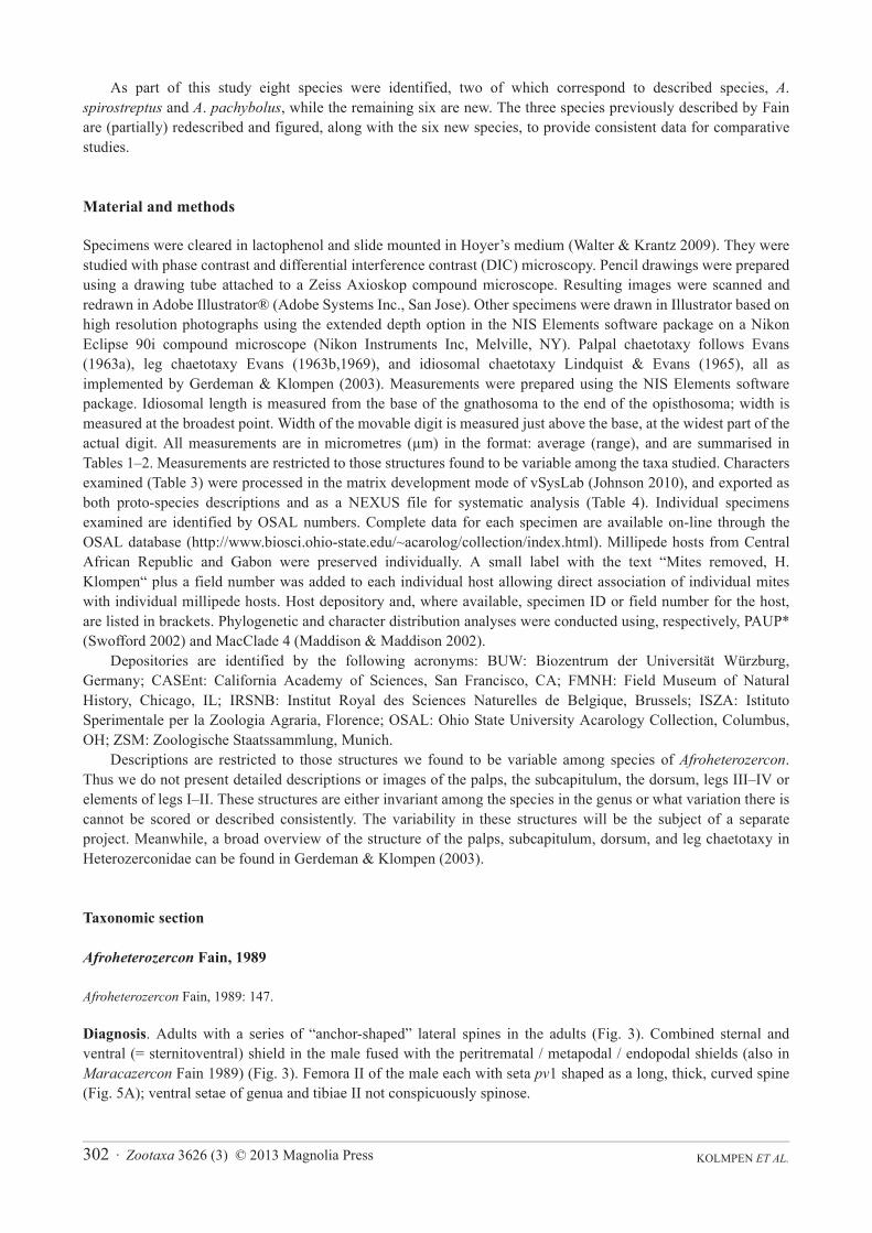
Specimens were cleared in lactophenol and slide mounted in Hoyer’s medium (Walter & Krantz 2009). They were studied with phase contrast and differential interference contrast (DIC) microscopy. Pencil drawings were prepared using a drawing tube attached to a Zeiss Axioskop compound microscope. Resulting images were scanned and redrawn in Adobe Illustrator® (Adobe Systems Inc., San Jose). Other specimens were drawn in Illustrator based on high resolution photographs using the extended depth option in the NIS Elements software package on a Nikon Eclipse 90i compound microscope (Nikon Instruments Inc, Melville, NY). Palpal chaetotaxy follows Evans (1963a), leg chaetotaxy Evans (1963b,1969), and idiosomal chaetotaxy Lindquist & Evans (1965), all as implemented by Gerdeman & Klompen (2003). Measurements were prepared using the NIS Elements software package. Idiosomal length is measured from the base of the gnathosoma to the end of the opisthosoma; width is measured at the broadest point. Width of the movable digit is measured just above the base, at the widest part of the actual digit. All measurements are in micrometres (μm) in the format: average (range), and are summarised in Tables 1–2. Measurements are restricted to those structures found to be variable among the taxa studied. Characters examined (Table 3) were processed in the matrix development mode of vSysLab (Johnson 2010), and exported as both proto-species descriptions and as a NEXUS file for systematic analysis (Table 4). Individual specimens examined are identified by OSAL numbers. Complete data for each specimen are available on-line through the OSAL database (http://www.biosci.ohio-state.edu/~acarolog/collection/index.html). Millipede hosts from Central African Republic and Gabon were preserved individually. A small label with the text “Mites removed, H. Klompen“ plus a field number was added to each individual host allowing direct association of individual mites with individual millipede hosts. Host depository and, where available, specimen ID or field number for the host, are listed in brackets. Phylogenetic and character distribution analyses were conducted using, respectively, PAUP* (Swofford 2002) and MacClade 4 (Maddison & Maddison 2002).
Depositories are identified by the following acronyms: BUW: Biozentrum der Universität Würzburg, Germany; CASEnt: California Academy of Sciences, San Francisco, CA; FMNH: Field Museum of Natural History, Chicago, IL; IRSNB: Institut Royal des Sciences Naturelles de Belgique, Brussels; ISZA: Istituto Sperimentale per la Zoologia Agraria, Florence; OSAL: Ohio State University Acarology Collection, Columbus, OH; ZSM: Zoologische Staatssammlung, Munich.
Descriptions are restricted to those structures we found to be variable among species of Afroheterozercon.Thus we do not present detailed descriptions or images of the palps, the subcapitulum, the dorsum, legs III–IV or elements of legs I–II. These structures are either invariant among the species in the genus or what variation there is cannot be scored or described consistently. The variability in these structures will be the subject of a separate project. Meanwhile, a broad overview of the structure of the palps, subcapitulum, dorsum, and leg chaetotaxy in Heterozerconidae can be found in Gerdeman & Klompen (2003).
Taxonomic section
Afroheterozercon Fain, 1989
Afroheterozercon Fain, 1989: 147.
Diagnosis. Adults with a series of “anchor-shaped” lateral spines in the adults (Fig. 3). Combined sternal and ventral (= sternitoventral) shield in the male fused with the peritrematal / metapodal / endopodal shields (also in Maracazercon Fain 1989) (Fig. 3). Femora II of the male each with seta pv1 shaped as a long, thick, curved spine (Fig. 5A); ventral setae of genua and tibiae II not conspicuously spinose.
Zootaxa 3626 (3) © 2013 Magnolia Press · 303REVISION OF AFROHETEROZERCON
KOLMPEN ET AL.304 · Zootaxa 3626 (3) © 2013 Magnolia Press
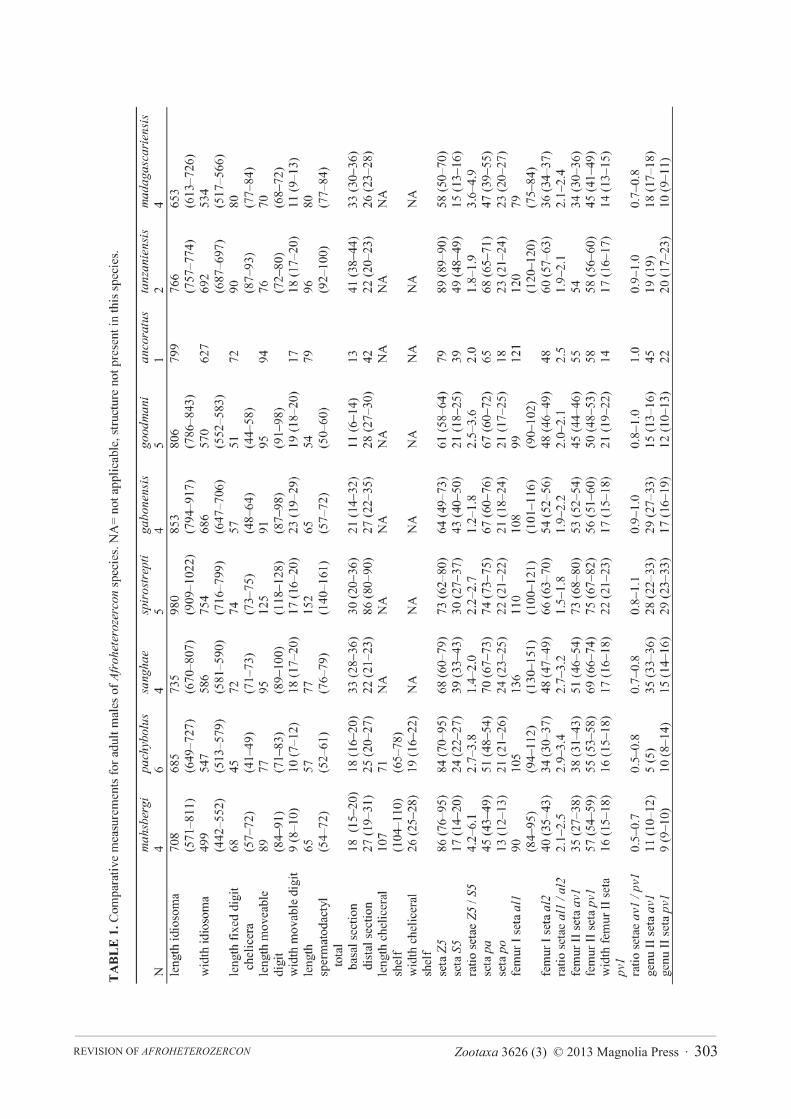
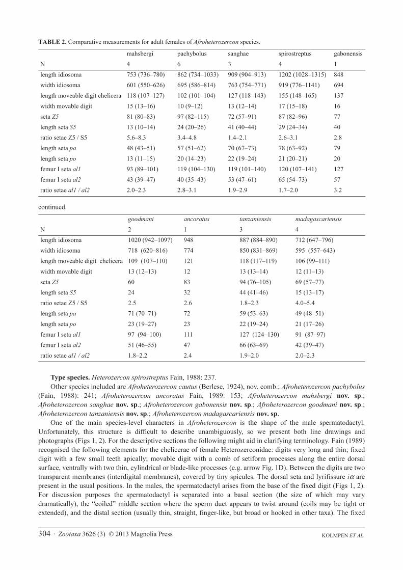
TABLE 2. Comparative measurements for adult females of Afroheterozercon species.
continued.
Type species. Heterozercon spirostreptus Fain, 1988: 237.Other species included are Afroheterozercon cautus (Berlese, 1924), nov. comb.; Afroheterozercon pachybolus
(Fain, 1988): 241; Afroheterozercon ancoratus Fain, 1989: 153; Afroheterozercon mahsbergi nov. sp.;Afroheterozercon sanghae nov. sp.; Afroheterozercon gabonensis nov. sp.; Afroheterozercon goodmani nov. sp.;Afroheterozercon tanzaniensis nov. sp.; Afroheterozercon madagascariensis nov. sp.
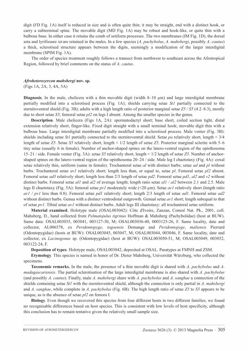
One of the main species-level characters in Afroheterozercon is the shape of the male spermatodactyl. Unfortunately, this structure is difficult to describe unambiguously, so we present both line drawings and photographs (Figs 1, 2). For the descriptive sections the following might aid in clarifying terminology. Fain (1989) recognised the following elements for the chelicerae of female Heterozerconidae: digits very long and thin; fixed digit with a few small teeth apically; movable digit with a comb of setiform processes along the entire dorsal surface, ventrally with two thin, cylindrical or blade-like processes (e.g. arrow Fig. 1D). Between the digits are two transparent membranes (interdigital membranes), covered by tiny spicules. The dorsal seta and lyrifissure iα are present in the usual positions. In the males, the spermatodactyl arises from the base of the fixed digit (Figs 1, 2). For discussion purposes the spermatodactyl is separated into a basal section (the size of which may vary dramatically), the “coiled” middle section where the sperm duct appears to twist around (coils may be tight or extended), and the distal section (usually thin, straight, finger-like, but broad or hooked in other taxa). The fixed
mahsbergi pachybolus sanghae spirostreptus gabonensisN 4 6 3 4 1length idiosoma 753 (736–780) 862 (734–1033) 909 (904–913) 1202 (1028–1315) 848width idiosoma 601 (550–626) 695 (586–814) 763 (754–771) 919 (776–1141) 694length moveable digit chelicera 118 (107–127) 102 (101–104) 127 (118–143) 155 (148–165) 137
width movable digit 15 (13–16) 10 (9–12) 13 (12–14) 17 (15–18) 16seta Z5 81 (80–83) 97 (82–115) 72 (57–91) 87 (82–96) 77length seta S5 13 (10–14) 24 (20–26) 41 (40–44) 29 (24–34) 40ratio setae Z5 / S5 5.6–8.3 3.4–4.8 1.4–2.1 2.6–3.1 2.8length seta pa 48 (43–51) 57 (51–62) 70 (67–73) 78 (63–92) 79
length seta po 13 (11–15) 20 (14–23) 22 (19–24) 21 (20–21) 20femur I seta al1 93 (89–101) 119 (104–130) 119 (101–140) 120 (107–141) 127femur I seta al2 43 (39–47) 40 (35–43) 53 (47–61) 65 (54–73) 57ratio setae al1 / al2 2.0–2.3 2.8–3.1 1.9–2.9 1.7–2.0 3.2
goodmani ancoratus tanzaniensis madagascariensisN 2 1 3 4length idiosoma 1020 (942–1097) 948 887 (884–890) 712 (647–796)width idiosoma 718 (620–816) 774 850 (831–869) 595 (557–643)length moveable digit chelicera 109 (107–110) 121 118 (117–119) 106 (99–111)
width movable digit 13 (12–13) 12 13 (13–14) 12 (11–13)seta Z5 60 83 94 (76–105) 69 (57–77)length seta S5 24 32 44 (41–46) 15 (13–17)ratio setae Z5 / S5 2.5 2.6 1.8–2.3 4.0–5.4length seta pa 71 (70–71) 72 59 (53–63) 49 (48–51)
length seta po 23 (19–27) 23 22 (19–24) 21 (17–26)femur I seta al1 97 (94–100) 111 127 (124–130) 91 (87–97)femur I seta al2 51 (46–55) 47 66 (63–69) 42 (39–47)ratio setae al1 / al2 1.8–2.2 2.4 1.9–2.0 2.0–2.3
Zootaxa 3626 (3) © 2013 Magnolia Press · 305REVISION OF AFROHETEROZERCON
digit (FD Fig. 1A) itself is reduced in size and is often quite thin; it may be straight, end with a distinct hook, or carry a subterminal spine. The movable digit (MD Fig. 1A) may be robust and hook-like, or quite thin with a bulbous base. In either case it retains the comb of setiform processes. The two membranes (IM Fig. 1D), the dorsal seta and lyrifissure iα are retained in the males. In a few species (A. pachybolus, A. mahsbergi, possibly A. cautus)a thick, sclerotised structure appears between the digits, seemingly a modification of the larger interdigital membrane (SPIM Fig. 1A).
The order of species treatment roughly follows a transect from northwest to southeast across the Afrotropical Region, followed by brief comments on the status of A. cautus.
Afroheterozercon mahsbergi nov. sp.(Figs 1A, 2A, 3, 4A, 5A)
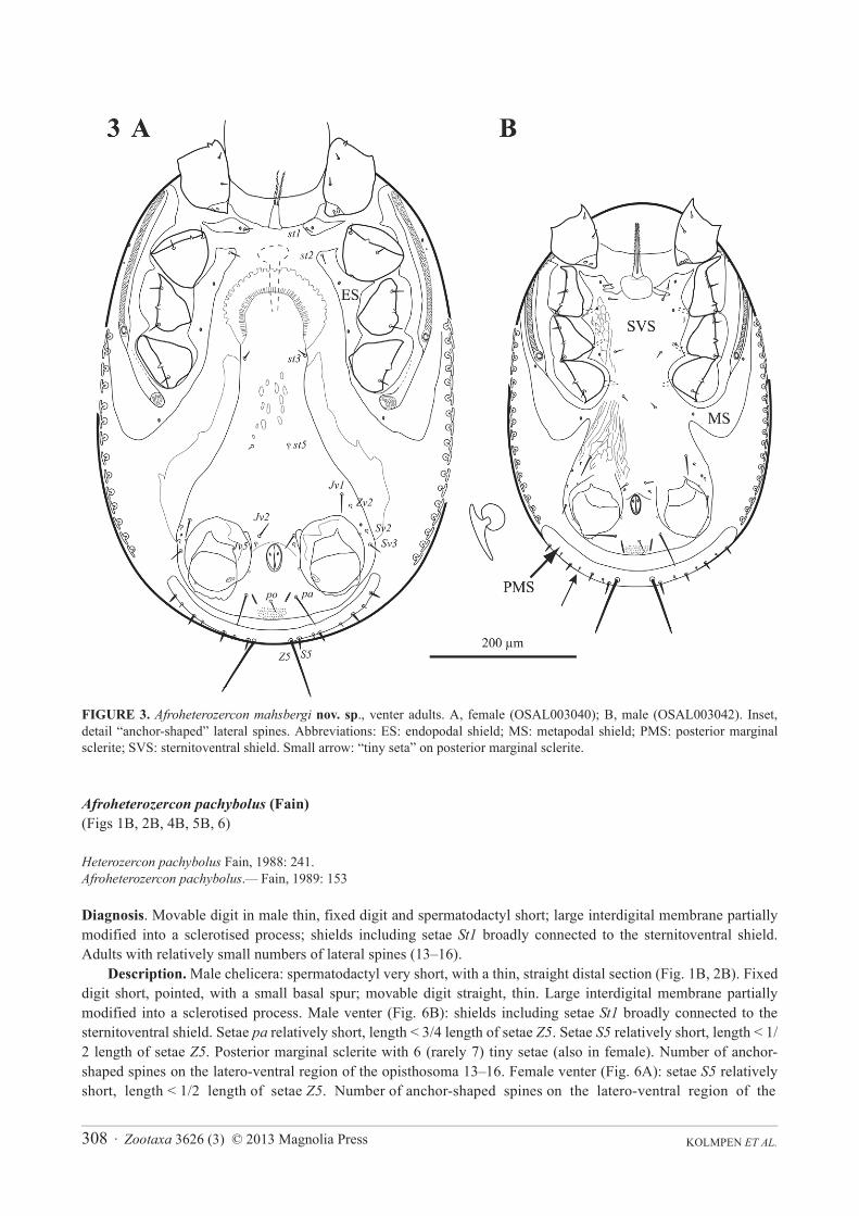
Diagnosis. In the male, chelicera with a thin movable digit (width 8–10 μm) and large interdigital membrane partially modified into a sclerotised process (Fig. 1A); shields carrying setae St1 partially connected to the sternitoventral shield (Fig. 3B); adults with a high length ratio of posterior marginal setae Z5 / S5 (4.2–8.3), mostly due to short setae S5; femoral setae pl2 on legs I absent. Among the smaller species in the genus.
Description. Male chelicera (Figs 1A, 2A): spermatodactyl short; base short, coiled section tight, distal extension relatively short, finger-like. Fixed digit straight with a small terminal hook; movable digit thin with a bulbous base. Large interdigital membrane partially modified into a sclerotised process. Male venter (Fig. 3B): shields including setae St1 partially connected to the sternitoventral shield. Setae pa relatively short, length < 3/4 length of setae Z5. Setae S5 relatively short, length < 1/2 length of setae Z5. Posterior marginal sclerite with 5–6 tiny setae (usually 6 in female). Number of anchor-shaped spines on the latero-ventral region of the opisthosoma 15–21 / side. Female venter (Fig. 3A): setae S5 relatively short, length < 1/2 length of setae Z5. Number of anchor-shaped spines on the latero-ventral region of the opisthosoma 20–24 / side. Male leg I chaetotaxy (Fig. 4A): coxal setae relatively thin, setiform (same in female). Trochanteral setae al with distinct barbs; setae ad and pl without barbs. Trochanteral setae av1 relatively short; length less than, or equal to, setae pl. Femoral setae pl2 absent. Femoral setae ad3 relatively short; length less than 2/3 length of setae pd2. Femoral setae pd1, al2 and v2 without distinct barbs. Femoral setae al1 and al2 of average length; length ratio setae al1 / al2 between 2.1 and 2.5. Male legs II chaetotaxy (Fig. 5A): femoral setae pv1 moderately wide (<20 μm). Setae av1 relatively short (length ratio av1 / pv1 less than 0.8). Femoral setae pd1 relatively short; length 2/3 length of setae ad1. Femoral setae ad2without distinct barbs. Genua with a distinct ventrodistal outgrowth. Genual setae av1 short; length subequal to that of setae pv1. Tibial setae av1 without distinct barbs. Adult legs III chaetotaxy: all trochanteral setae setiform.
Material examined. Holotype male (OSAL003042): Côte d'Ivoire, Zanzan, Comoé Nat. Pk., 2001, coll. Mahsberg, D., hand collected from Pelmatojulus tigrinus Hoffman & Mahsberg (Pachybolidae) (host at BUW). Same data: OSAL003035, 003041, 003127-30, M; OSAL003036-40, 0003125-26, F. Same locality, date and collector, AL006578, ex Peridontopyge, togoensis Demange and Peridontopyge, maliensis Pierrard (Odontopygidae) (hosts at BUW): OSAL003045, 003047, M; OSAL003044, 003046, F. Same locality, date and collector, ex Lacinogonus sp. (Odontopygidae) (host at BUW): OSAL003050-51, M; OSAL003049, 003052, 003122-24, F.
Deposition of types. Holotype male, OSAL003042, deposited at OSAL. Paratypes at FMNH and ZSM.Etymology. This species is named in honor of Dr. Dieter Mahsberg, Universität Würzburg, who collected the
specimens.Taxonomic remarks. In the male, the presence of a thin movable digit is shared with A. pachybolus and A.
madagascariensis. The partial sclerotisation of the large interdigital membrane is also shared with A. pachybolus (and possibly A. cautus). Finally, male A. mahsbergi share with A. pachybolus and A. sanghae a connection of the shields containing setae St1 with the sternitoventral shield, although the connection is only partial in A. mahsbergiand A. sanghae, while complete in A. pachybolus (Fig. 6B). The high length ratio of setae Z5 to S5 appears to be unique, as is the absence of setae pl2 on femora I.
Biology. Even though we recovered this species from four different hosts in two different families, we found no recognisable differences based on host species. This is consistent with low levels of host specificity, although this conclusion has to remain tentative given the relatively small sample size.
KOLMPEN ET AL.306 · Zootaxa 3626 (3) © 2013 Magnolia Press
FIGURE 1. Afroheterozercon spp., chelicerae male. A, A. mahsbergi nov. sp. (OSAL003051); B, A. pachybolus (Fain) (OSAL092084); C, A. sanghae nov. sp. (OSAL053288); D, A. spirostreptus (Fain) (OSAL053950); E, A. gabonensis nov. sp.(OSAL92096); F, A. goodmani nov. sp. (OSAL102681); G, A. tanzaniensis nov. sp. (OSAL53955); H, A. madagascariensisnov. sp. (OSAL053962). Abbreviations: FD: fixed digit; IM: interdigital membrane; MD: movable digit; S: spermatodactyl; SPIM: sclerotised portion interdigital membrane. Arrow to ventral blade-like processes.
Zootaxa 3626 (3) © 2013 Magnolia Press · 307REVISION OF AFROHETEROZERCON
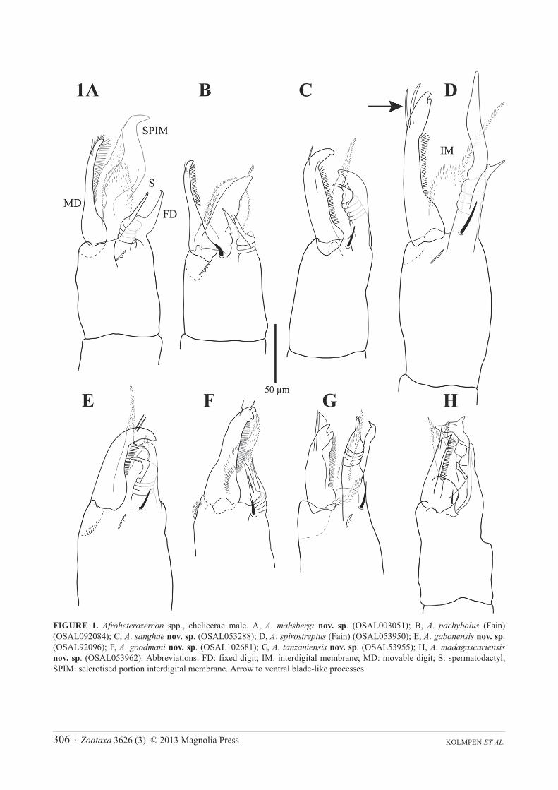
FIGURE 2. Afroheterozercon spp., chelicerae male, photographs. A, A. mahsbergi nov. sp. (OSAL003045); B, A. pachybolus (Fain) (OSAL092070); C, A. sanghae nov. sp. (OSAL053288); D, A. spirostreptus (Fain) (OSAL053952); E, A. gabonensisnov. sp. (OSAL053942); F, A. goodmani nov. sp. (OSAL102681); G, A. tanzaniensis nov. sp. (OSAL053955); H, A.madagascariensis nov. sp. (OSAL053962).
KOLMPEN ET AL.308 · Zootaxa 3626 (3) © 2013 Magnolia Press
FIGURE 3. Afroheterozercon mahsbergi nov. sp., venter adults. A, female (OSAL003040); B, male (OSAL003042). Inset, detail “anchor-shaped” lateral spines. Abbreviations: ES: endopodal shield; MS: metapodal shield; PMS: posterior marginal sclerite; SVS: sternitoventral shield. Small arrow: “tiny seta” on posterior marginal sclerite.
Afroheterozercon pachybolus (Fain) (Figs 1B, 2B, 4B, 5B, 6)
Heterozercon pachybolus Fain, 1988: 241.Afroheterozercon pachybolus.— Fain, 1989: 153
Diagnosis. Movable digit in male thin, fixed digit and spermatodactyl short; large interdigital membrane partially modified into a sclerotised process; shields including setae St1 broadly connected to the sternitoventral shield. Adults with relatively small numbers of lateral spines (13–16).
Description. Male chelicera: spermatodactyl very short, with a thin, straight distal section (Fig. 1B, 2B). Fixed digit short, pointed, with a small basal spur; movable digit straight, thin. Large interdigital membrane partially modified into a sclerotised process. Male venter (Fig. 6B): shields including setae St1 broadly connected to the sternitoventral shield. Setae pa relatively short, length < 3/4 length of setae Z5. Setae S5 relatively short, length < 1/2 length of setae Z5. Posterior marginal sclerite with 6 (rarely 7) tiny setae (also in female). Number of anchor-shaped spines on the latero-ventral region of the opisthosoma 13–16. Female venter (Fig. 6A): setae S5 relatively short, length < 1/2 length of setae Z5. Number of anchor-shaped spines on the latero-ventral region of the
Zootaxa 3626 (3) © 2013 Magnolia Press · 309REVISION OF AFROHETEROZERCON
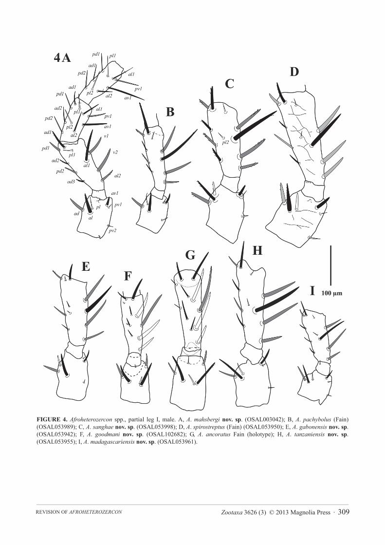
FIGURE 4. Afroheterozercon spp., partial leg I, male. A, A. mahsbergi nov. sp. (OSAL003042); B, A. pachybolus (Fain) (OSAL053989); C, A. sanghae nov. sp. (OSAL053998); D, A. spirostreptus (Fain) (OSAL053950); E, A. gabonensis nov. sp.(OSAL053942); F, A. goodmani nov. sp. (OSAL102682); G, A. ancoratus Fain (holotype); H, A. tanzaniensis nov. sp.(OSAL053955); I, A. madagascariensis nov. sp. (OSAL053961).

KOLMPEN ET AL.310 · Zootaxa 3626 (3) © 2013 Magnolia Press
FIGURE 5. Afroheterozercon spp., partial leg II, male. A, A. mahsbergi nov. sp. (OSAL003042); B, A. pachybolus (Fain) (OSAL053989); C, A. sanghae nov. sp. (OSAL053998); D, A. spirostreptus (Fain) (OSAL053950); E, A. gabonensis nov. sp.(OSAL053942); F, A. goodmani nov. sp. (OSAL102682); G, A. ancoratus Fain (holotype); H, A. tanzaniensis nov. sp.(OSAL053955); I, A. madagascariensis nov. sp. (OSAL053961).
opisthosoma 14–16. Male legs I: chaetotaxy (Fig. 4B): coxae I setae spinose (also in female). Trochanteral setae av1 relatively short; length less than, or equal to, setae pl. Trochanteral setae ad, al and pl without distinct barbs. Femoral setae pl2 present. Femoral setae ad3 length relatively long; length subequal to that of setae pd2. Femoral setae pd1, al2 and v2 without distinct barbs. Femoral setae al2 unusually short, setae al1 moderately long; length ratio setae al1 / al2 more than 2.7. Male legs II, chaetotaxy (Fig. 5B): femoral setae pv1 moderately wide (<20 μm). Setae av1 relatively short (length ratio av1 / pv1 less than 0.8). Femoral setae pd1 distinctly shorter than setae ad1.Femoral setae ad2 without distinct barbs. Genua with a distinct ventrodistal outgrowth. Genual setae av1 very short; length half of that of setae pv1. Tibial setae av1 with distinct barbs. Adult legs III, chaetotaxy: one or more trochanteral setae spine-like.

Zootaxa 3626 (3) © 2013 Magnolia Press · 311REVISION OF AFROHETEROZERCON
Material examined. Democratic Republic of Congo, forest of Kwango river, 5°S 17°E, 19 April 1964, coll. A. Fain, ex Pachybolus macrosternus, male holotype, IRSNB. New material: Central African Republic, Sangha-Mbaéré, Parc National Dzanga-Ndoki, 510 m, Mabéa Bai, 1–7 May 2001, coll. B. L. Fisher, ex pitfall trap, with unidentified millipede (host at CASEnt, field number AL6647): OSAL053989-91, 053994, 053997, male. Same locality, date, collector, ex pitfall trap with unidentified millipede (CASEnt AL6667): OSAL092084, M; OSAL092083, female. Same locality, date, collector, ex pitfall trap with unidentified millipede (CASEnt AL6683): OSAL092069-71, male; OSAL092072-73, 092095, female. Same locality, date, collector, ex pitfall trap with unidentified millipede (CASEnt AL6694): OSAL092087, 092090, males; OSAL092088-89, 092091-92, females.
FIGURE 6. Afroheterozercon pachybolus (Fain), venter adults. A, female (OSAL092091); B, male (OSAL092084).
Deposition of specimens. New material deposited at CASEnt and OSAL.Taxonomic remarks. Our specimens correspond in most available details to the description by Fain (1988,
1989). The one discrepancy concerns the number of anchor-shaped lateral spines, 17–18 according to the description (Fain 1988), but 13–15 in our specimens and in Fain’s figures (1988, 1989). Examination of the male holotype showed 15 lateral spines, suggesting the number in the description by Fain (1988) is inaccurate. The original description was based on one specimen only. The current description adds data on multiple male
KOLMPEN ET AL.312 · Zootaxa 3626 (3) © 2013 Magnolia Press
specimens and includes the first notes on the female. In the male, A. pachybolus resembles A. goodmani by having a short spermatodactyl and short fixed cheliceral digit, and A. mahsbergi by sharing the thin movable digit, the partial sclerotisation of the large interdigital membrane, and the fusion of the shields carrying setae St1 with the sternitoventral shield. It differs from both of those species by the male chelicerae that combine a short spermatodactyl and fixed digit with a thin movable digit, the presence of sclerotisation of the interdigital membrane, plus complete fusion of the shields carrying setae St1 with the sternitoventral shield.
Afroheterozercon sanghae nov. sp.(Figs 1C, 2C, 4C, 5C, 7)
Diagnosis. Male chelicera with a distinct subterminal spine on the fixed digit and a relatively long basal section of the spermatodactyl, shields carrying setae St1 partially connected to the sternitoventral shield, and unusually long setae al1 on femora I. In adults length ratio of setae Z5 to S5 unusually small (1.4–2.1) due to relatively long setae S5 and short setae Z5.
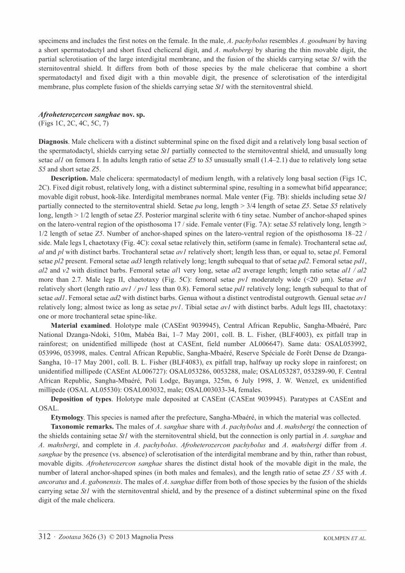
Description. Male chelicera: spermatodactyl of medium length, with a relatively long basal section (Figs 1C, 2C). Fixed digit robust, relatively long, with a distinct subterminal spine, resulting in a somewhat bifid appearance; movable digit robust, hook-like. Interdigital membranes normal. Male venter (Fig. 7B): shields including setae St1partially connected to the sternitoventral shield. Setae pa long, length > 3/4 length of setae Z5. Setae S5 relatively long, length > 1/2 length of setae Z5. Posterior marginal sclerite with 6 tiny setae. Number of anchor-shaped spines on the latero-ventral region of the opisthosoma 17 / side. Female venter (Fig. 7A): setae S5 relatively long, length > 1/2 length of setae Z5. Number of anchor-shaped spines on the latero-ventral region of the opisthosoma 18–22 / side. Male legs I, chaetotaxy (Fig. 4C): coxal setae relatively thin, setiform (same in female). Trochanteral setae ad,al and pl with distinct barbs. Trochanteral setae av1 relatively short; length less than, or equal to, setae pl. Femoral setae pl2 present. Femoral setae ad3 length relatively long; length subequal to that of setae pd2. Femoral setae pd1,al2 and v2 with distinct barbs. Femoral setae al1 very long, setae al2 average length; length ratio setae al1 / al2more than 2.7. Male legs II, chaetotaxy (Fig. 5C): femoral setae pv1 moderately wide (<20 μm). Setae av1relatively short (length ratio av1 / pv1 less than 0.8). Femoral setae pd1 relatively long; length subequal to that of setae ad1. Femoral setae ad2 with distinct barbs. Genua without a distinct ventrodistal outgrowth. Genual setae av1relatively long; almost twice as long as setae pv1. Tibial setae av1 with distinct barbs. Adult legs III, chaetotaxy: one or more trochanteral setae spine-like.
Material examined. Holotype male (CASEnt 9039945), Central African Republic, Sangha-Mbaéré, Parc National Dzanga-Ndoki, 510m, Mabéa Bai, 1–7 May 2001, coll. B. L. Fisher, (BLF4003), ex pitfall trap in rainforest; on unidentified millipede (host at CASEnt, field number AL006647). Same data: OSAL053992, 053996, 053998, males. Central African Republic, Sangha-Mbaéré, Reserve Spéciale de Forêt Dense de Dzanga-Sangha, 10–17 May 2001, coll. B. L. Fisher (BLF4083), ex pitfall trap, halfway up rocky slope in rainforest; on unidentified millipede (CASEnt AL006727): OSAL053286, 0053288, male; OSAL053287, 053289-90, F. Central African Republic, Sangha-Mbaéré, Poli Lodge, Bayanga, 325m, 6 July 1998, J. W. Wenzel, ex unidentified millipede (OSAL AL05530): OSAL003032, male; OSAL003033-34, females.
Deposition of types. Holotype male deposited at CASEnt (CASEnt 9039945). Paratypes at CASEnt and OSAL.
Etymology. This species is named after the prefecture, Sangha-Mbaéré, in which the material was collected.Taxonomic remarks. The males of A. sanghae share with A. pachybolus and A. mahsbergi the connection of
the shields containing setae St1 with the sternitoventral shield, but the connection is only partial in A. sanghae andA. mahsbergi, and complete in A. pachybolus. Afroheterozercon pachybolus and A. mahsbergi differ from A.sanghae by the presence (vs. absence) of sclerotisation of the interdigital membrane and by thin, rather than robust, movable digits. Afroheterozercon sanghae shares the distinct distal hook of the movable digit in the male, the number of lateral anchor-shaped spines (in both males and females), and the length ratio of setae Z5 / S5 with A.ancoratus and A. gabonensis. The males of A. sanghae differ from both of those species by the fusion of the shields carrying setae St1 with the sternitoventral shield, and by the presence of a distinct subterminal spine on the fixed digit of the male chelicera.
Zootaxa 3626 (3) © 2013 Magnolia Press · 313REVISION OF AFROHETEROZERCON
FIGURE 7. Afroheterozercon sanghae nov. sp., venter adults. A, female (OSAL003034; B, male (OSAL053998).
Afroheterozercon spirostreptus (Fain)(Figs 1D, 2D, 4D, 5D)
Heterozercon spirostreptus Fain, 1988: 237.Afroheterozercon spirostreptus.— Fain, 1989: 153.
Diagnosis. Male chelicera with long movable digit (118–128 μm) and a very long spermatodactyl, especially the distal section (80–90 μm); shields carrying setae St1 not connected to the sternitoventral shield; with an unusually small length ratio of femoral setae al1 to al2 (1.6–1.7). Adults always with more than 6 tiny setae on posterior marginal sclerite. In adults with relatively small numbers of lateral spines. The largest species examined.
KOLMPEN ET AL.314 · Zootaxa 3626 (3) © 2013 Magnolia Press
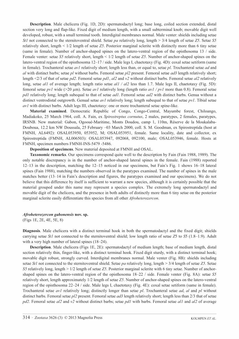
Description. Male chelicera (Fig. 1D, 2D): spermatodactyl long; base long, coiled section extended, distal section very long and flap-like. Fixed digit of medium length, with a small subterminal knob; movable digit well developed, robust, with a small terminal tooth. Interdigital membranes normal. Male venter: shields including setae St1 not connected to the sternitoventral shield. Setae pa relatively long, length > 3/4 length of setae Z5. Setae S5relatively short, length < 1/2 length of setae Z5. Posterior marginal sclerite with distinctly more than 6 tiny setae (same in female). Number of anchor-shaped spines on the latero-ventral region of the opisthosoma 13 / side. Female venter: setae S5 relatively short, length < 1/2 length of setae Z5. Number of anchor-shaped spines on the latero-ventral region of the opisthosoma 12–17 / side. Male legs I, chaetotaxy (Fig. 4D): coxal setae setiform (same in female). Trochanteral setae av1 relatively short; length less than, or equal to, setae pl. Trochanteral setae ad and al with distinct barbs; setae pl without barbs. Femoral setae pl2 present. Femoral setae ad3 length relatively short; length <2/3 of that of setae pd2. Femoral setae pd1, al2 and v2 without distinct barbs. Femoral setae al2 relatively long, setae al1 of average length; length ratio setae al1 / al2 less than 1.7. Male legs II, chaetotaxy (Fig. 5D): femoral setae pv1 wide (>20 μm). Setae av1 relatively long (length ratio av1 / pv1 more than 0.8). Femoral setae pd1 relatively long; length subequal to that of setae ad1. Femoral setae ad2 with distinct barbs. Genua without a distinct ventrodistal outgrowth. Genual setae av1 relatively long; length subequal to that of setae pv1. Tibial setae av1 with distinct barbs. Adult legs III, chaetotaxy: one or more trochanteral setae spine-like.
Material examined. Democratic Republic of Congo, Congo-Central, Mayumbe forest, Chiloango, Madiakoko, 25 March 1964, coll. A. Fain, ex Spirostreptus cornutus, 2 males, paratypes, 2 females, paratypes, IRSNB. New material: Gabon, Ogooué-Maritime, Monts Doudou, camp 1, 110m, Réserve de la Moukalaba-Douboua, 12.2 km NW Doussala, 25 February –03 March 2000, coll. S. M. Goodman, ex Spirostreptida (host at FMNH, AL6492): OSAL053950, 053952, M; OSAL053951, female. Same locality, date and collector, ex Spirostreptida (FMNH, AL006503): OSAL053947, 092068, 092100, male; OSAL053946, female. Hosts at FMNH, specimen numbers FMNH-INS-5479 -5486.
Deposition of specimens. New material deposited at FMNH and OSAL.Taxonomic remarks. Our specimens correspond quite well to the description by Fain (Fain 1988, 1989). The
only notable discrepancy is in the number of anchor-shaped lateral spines in the female. Fain (1988) reported 12–13 in the description, matching the 12–15 noticed in our specimens, but Fain’s Fig. 1 shows 16–18 lateral spines (Fain 1988), matching the numbers observed in the paratypes examined. The number of spines in the male matches better (13–14 in Fain’s description and figures, the paratypes examined and our specimens). We do not believe that this difference by itself is sufficient to warrant a new species, although it is certainly possible that the material grouped under this name may represent a species complex. The extremely long spermatodactyl and movable digit of the chelicera, and the presence in both adults of distinctly more than 6 tiny setae on the posterior marginal sclerite easily differentiate this species from all other Afroheterozercon.
Afroheterozercon gabonensis nov. sp.(Figs 1E, 2E, 4E, 5E, 8)
Diagnosis. Male chelicera with a distinct terminal hook in both the spermatodactyl and the fixed digit; shields carrying setae St1 not connected to the sternitoventral shield; low length ratio of setae Z5 to S5 (1.8–1.9). Adult with a very high number of lateral spines (18–24).
Description. Male chelicera (Figs 1E, 2E): spermatodactyl of medium length; base of medium length, distal section relatively thin, finger-like, with a distinct terminal hook. Fixed digit sturdy, with a distinct terminal hook; movable digit robust, strongly curved. Interdigital membranes normal. Male venter (Fig. 8B): shields including setae St1 not connected to the sternitoventral shield. Setae pa relatively long, length > 3/4 length of setae Z5. Setae S5 relatively long, length > 1/2 length of setae Z5. Posterior marginal sclerite with 6 tiny setae. Number of anchor-shaped spines on the latero-ventral region of the opisthosoma 18–22 / side. Female venter (Fig. 8A): setae S5relatively short, length approximately 1/2 length of setae Z5. Number of anchor-shaped spines on the latero-ventral region of the opisthosoma 22–24 / side. Male legs I, chaetotaxy (Fig. 4E): coxal setae setiform (same in female). Trochanteral setae av1 relatively long; distinctly longer than setae pl. Trochanteral setae ad, al and pl without distinct barbs. Femoral setae pl2 present. Femoral setae ad3 length relatively short; length less than 2/3 that of setae pd2. Femoral setae al2 and v2 without distinct barbs; setae pd1 with barbs. Femoral setae al1 and al2 of average
Zootaxa 3626 (3) © 2013 Magnolia Press · 315REVISION OF AFROHETEROZERCON
length; length ratio setae al1 / al2 between 1.9 and 2.2. Male legs II, chaetotaxy (Fig. 5E): femoral setae pv1moderately wide (<20 μm). Setae av1 relatively long (length ratio av1 / pv1 more than 0.8). Femoral setae pd1relatively long; length subequal to that of setae ad1. Femoral setae ad2 without distinct barbs. Genua without a distinct ventrodistal outgrowth. Genual setae av1 relatively long; length almost twice that of setae pv1. Tibial setae av1 with distinct barbs. Adult legs III, chaetotaxy: one or more trochanteral setae spine-like.
Material examined. Holotype male (OSAL053942): Gabon, Mt. Doudou, camp 3, 585–660 m, Aire d’Exploitation Rationelle de Faune des Monts Doudou, 25.2 km NW Doussala, 52.6 km NW Mourindi, -2.2272, 10.3945, 14–20 March 2000, S. M. Goodman, ex Spirostreptida (host in FMNH, AL6508) in mid-level rainforest. Same data: OSAL003119-21, 053943-44, 092096-99, M; OSAL053941, 053945, females. Hosts at FMNH, specimen numbers FMNH-INS-5477-5478.
Deposition of types. Holotype male, OSAL053942, deposited at FMNH. Paratypes at FMNH and OSAL.Etymology. This species is named after the country in which the material was collected.
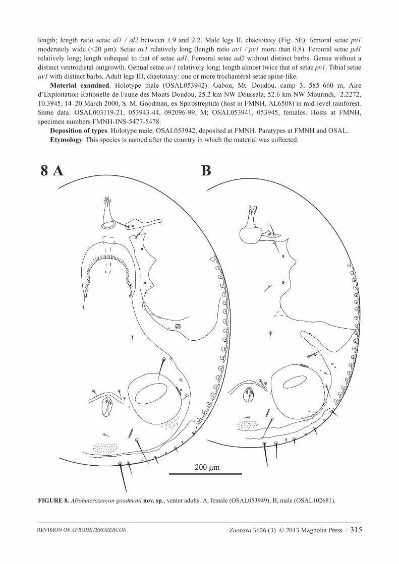
FIGURE 8. Afroheterozercon goodmani nov. sp., venter adults. A, female (OSAL053949); B, male (OSAL102681).
KOLMPEN ET AL.316 · Zootaxa 3626 (3) © 2013 Magnolia Press
Taxonomic remarks. Afroheterozercon gabonensis shares the robust development of the cheliceral digits, the number of lateral anchor-shaped spines in both males and females, and the length ratio of setae Z5 / S5 with A.ancoratus and A. sanghae. However, it differs from A. sanghae by the lack of fusion of the shields carrying seta St1with the sternitoventral shield, and the lack of a subterminal spine on the fixed digit of the male chelicera, and from A. ancoratus by the hooked (not straight) distal portion of both the spermatodactyl and fixed digit, and the smaller length ratio of femur I setae al1 / al2 (1.9–2.2 vs. 2.5).
Afroheterozercon goodmani nov. sp.(Figs 1F, 2F, 4F, 5F, 9)
Diagnosis. Male chelicera with a short spermatodactyl with a very short basal section; shields carrying setae St1not connected to the sternitoventral shield; with a short, but relatively wide, spur on femora II (setae pv1).
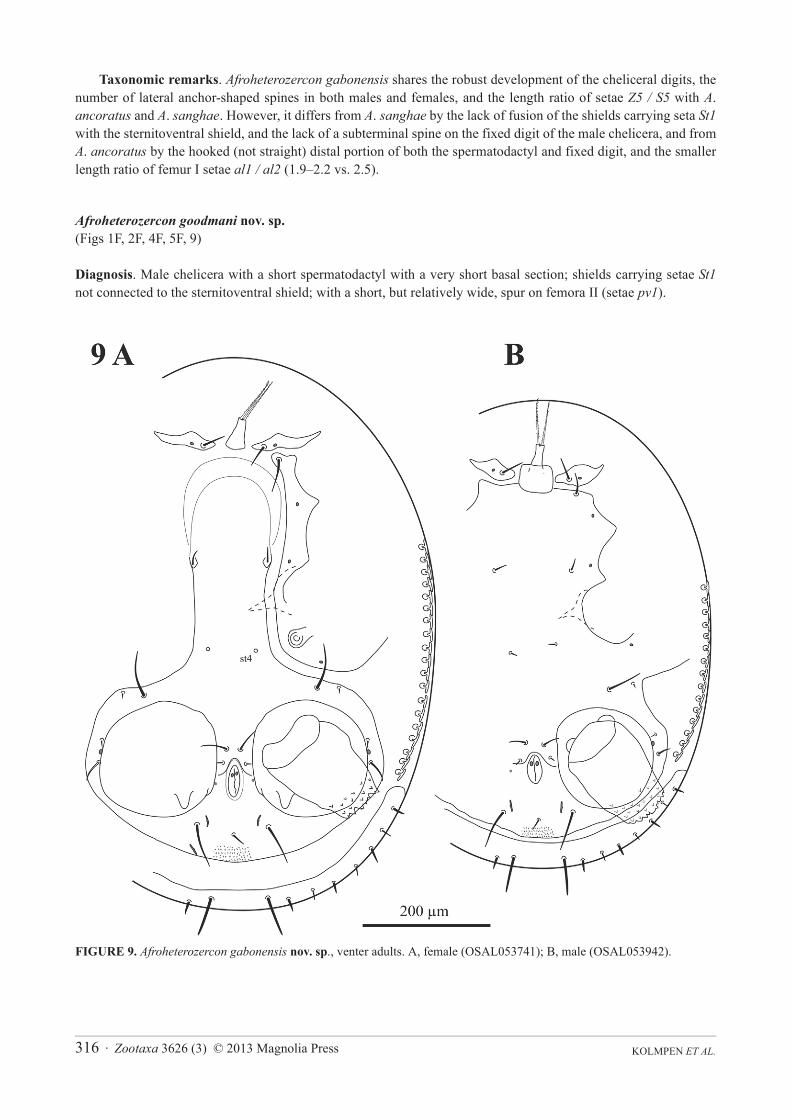
FIGURE 9. Afroheterozercon gabonensis nov. sp., venter adults. A, female (OSAL053741); B, male (OSAL053942).
Zootaxa 3626 (3) © 2013 Magnolia Press · 317REVISION OF AFROHETEROZERCON
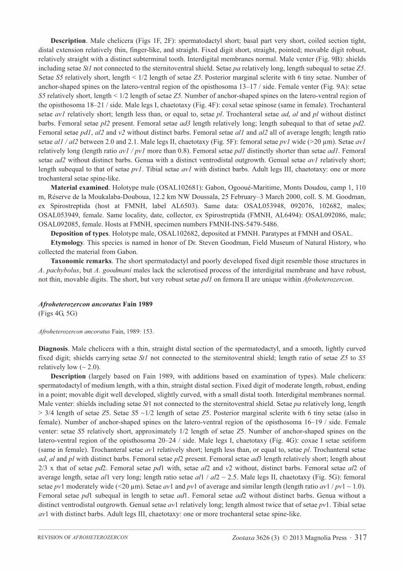
Description. Male chelicera (Figs 1F, 2F): spermatodactyl short; basal part very short, coiled section tight, distal extension relatively thin, finger-like, and straight. Fixed digit short, straight, pointed; movable digit robust, relatively straight with a distinct subterminal tooth. Interdigital membranes normal. Male venter (Fig. 9B): shields including setae St1 not connected to the sternitoventral shield. Setae pa relatively long, length subequal to setae Z5.Setae S5 relatively short, length < 1/2 length of setae Z5. Posterior marginal sclerite with 6 tiny setae. Number of anchor-shaped spines on the latero-ventral region of the opisthosoma 13–17 / side. Female venter (Fig. 9A): setae S5 relatively short, length < 1/2 length of setae Z5. Number of anchor-shaped spines on the latero-ventral region of the opisthosoma 18–21 / side. Male legs I, chaetotaxy (Fig. 4F): coxal setae spinose (same in female). Trochanteral setae av1 relatively short; length less than, or equal to, setae pl. Trochanteral setae ad, al and pl without distinct barbs. Femoral setae pl2 present. Femoral setae ad3 length relatively long; length subequal to that of setae pd2.Femoral setae pd1, al2 and v2 without distinct barbs. Femoral setae al1 and al2 all of average length; length ratio setae al1 / al2 between 2.0 and 2.1. Male legs II, chaetotaxy (Fig. 5F): femoral setae pv1 wide (>20 μm). Setae av1relatively long (length ratio av1 / pv1 more than 0.8). Femoral setae pd1 distinctly shorter than setae ad1. Femoral setae ad2 without distinct barbs. Genua with a distinct ventrodistal outgrowth. Genual setae av1 relatively short; length subequal to that of setae pv1. Tibial setae av1 with distinct barbs. Adult legs III, chaetotaxy: one or more trochanteral setae spine-like.
Material examined. Holotype male (OSAL102681): Gabon, Ogooué-Maritime, Monts Doudou, camp 1, 110 m, Réserve de la Moukalaba-Douboua, 12.2 km NW Doussala, 25 February–3 March 2000, coll. S. M. Goodman, ex Spirostreptida (host at FMNH, label AL6503). Same data: OSAL053948, 092076, 102682, males; OSAL053949, female. Same locality, date, collector, ex Spirostreptida (FMNH, AL6494): OSAL092086, male; OSAL092085, female. Hosts at FMNH, specimen numbers FMNH-INS-5479-5486.
Deposition of types. Holotype male, OSAL102682, deposited at FMNH. Paratypes at FMNH and OSAL.Etymology. This species is named in honor of Dr. Steven Goodman, Field Museum of Natural History, who
collected the material from Gabon.Taxonomic remarks. The short spermatodactyl and poorly developed fixed digit resemble those structures in
A. pachybolus, but A. goodmani males lack the sclerotised process of the interdigital membrane and have robust, not thin, movable digits. The short, but very robust setae pd1 on femora II are unique within Afroheterozercon.
Afroheterozercon ancoratus Fain 1989(Figs 4G, 5G)
Afroheterozercon ancoratus Fain, 1989: 153.
Diagnosis. Male chelicera with a thin, straight distal section of the spermatodactyl, and a smooth, lightly curved fixed digit; shields carrying setae St1 not connected to the sternitoventral shield; length ratio of setae Z5 to S5relatively low (~ 2.0).
Description (largely based on Fain 1989, with additions based on examination of types). Male chelicera: spermatodactyl of medium length, with a thin, straight distal section. Fixed digit of moderate length, robust, ending in a point; movable digit well developed, slightly curved, with a small distal tooth. Interdigital membranes normal. Male venter: shields including setae St1 not connected to the sternitoventral shield. Setae pa relatively long, length > 3/4 length of setae Z5. Setae S5 ~1/2 length of setae Z5. Posterior marginal sclerite with 6 tiny setae (also in female). Number of anchor-shaped spines on the latero-ventral region of the opisthosoma 16–19 / side. Female venter: setae S5 relatively short, approximately 1/2 length of setae Z5. Number of anchor-shaped spines on the latero-ventral region of the opisthosoma 20–24 / side. Male legs I, chaetotaxy (Fig. 4G): coxae I setae setiform (same in female). Trochanteral setae av1 relatively short; length less than, or equal to, setae pl. Trochanteral setae ad, al and pl with distinct barbs. Femoral setae pl2 present. Femoral setae ad3 length relatively short; length about 2/3 x that of setae pd2. Femoral setae pd1 with, setae al2 and v2 without, distinct barbs. Femoral setae al2 of average length, setae al1 very long; length ratio setae al1 / al2 ~ 2.5. Male legs II, chaetotaxy (Fig. 5G): femoral setae pv1 moderately wide (<20 μm). Setae av1 and pv1 of average and similar length (length ratio av1 / pv1 ~ 1.0). Femoral setae pd1 subequal in length to setae ad1. Femoral setae ad2 without distinct barbs. Genua without a distinct ventrodistal outgrowth. Genual setae av1 relatively long; length almost twice that of setae pv1. Tibial setae av1 with distinct barbs. Adult legs III, chaetotaxy: one or more trochanteral setae spine-like.
KOLMPEN ET AL.318 · Zootaxa 3626 (3) © 2013 Magnolia Press
Material examined. Democratic Republic of Congo, Congo-Central, Mayumbe forest, near Luki river, 23 Apr 1965, coll. R. P. Bouillon, ex nest Cubitermes sp. (Isoptera), 1 male, holotype, 1 female, paratype, IRSNB.
Taxonomic remarks. Afroheterozercon ancoratus shares with A. spirostreptus and A. gabonensis the robust male cheliceral digits, and the relative short (relative to setae pd2) setae ad3 on femora I. It differs from A.spirostreptus in the much smaller male chelicera and the small number of tiny setae on the posterior ventral sclerite (among others). It differs from A. gabonensis in the shape of the male spermatodactyl and fixed digit (with very distinct hooks in A. gabonensis, largely straight in A. ancoratus), and shorter (relative to setae pl) setae av1 on trochanter I.
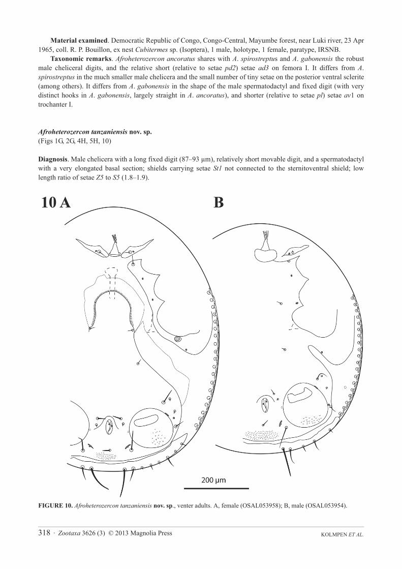
Afroheterozercon tanzaniensis nov. sp.(Figs 1G, 2G, 4H, 5H, 10)
Diagnosis. Male chelicera with a long fixed digit (87–93 μm), relatively short movable digit, and a spermatodactyl with a very elongated basal section; shields carrying setae St1 not connected to the sternitoventral shield; low length ratio of setae Z5 to S5 (1.8–1.9).
FIGURE 10. Afroheterozercon tanzaniensis nov. sp., venter adults. A, female (OSAL053958); B, male (OSAL053954).
Zootaxa 3626 (3) © 2013 Magnolia Press · 319REVISION OF AFROHETEROZERCON
Description. Male chelicera (Figs 1G, 2G): spermatodactyl medium long; base distinctly elongate, coiled section tight, distal extension short, finger-like, straight. Fixed digit strap-like, relatively long, with a small terminal spine; movable digit relatively short, robust, with a strong subterminal knob. Interdigital membranes normal. Male venter (Fig. 10B): shields including setae St1 not connected to the sternitoventral shield. Setae parelatively long, length > 3/4 length of setae Z5. Setae S5 relatively long, length > 1/2 length of setae Z5. Posterior marginal sclerite with 6 tiny setae. Number of anchor-shaped spines on the latero-ventral region of the opisthosoma 16–19 / side. Female venter (Fig. 10A): setae S5 relatively long, approximately 1/2 length of setae Z5. Number of anchor-shaped spines on the latero-ventral region of the opisthosoma 17–19 / side. Male legs I, chaetotaxy (Fig. 4H): coxal setae relatively thin, setiform (same in female). Trochanteral setae av1 relatively long; length similar to, or exceeding that of, setae pl. Trochanteral setae ad, al and pl without distinct barbs. Femoral setae pl2 present. Femoral setae ad3 length relatively long; length subequal to that of setae pd2. Femoral setae pd1, al2 and v2 without distinct barbs. Femoral setae al1 and al2 relatively long; length ratio setae al1 / al2 more between 1.9 and 2.1. Male legs II, chaetotaxy (Fig. 5H): femoral setae pv1 moderately wide (<20 μm). Setae av1 relatively long (length ratio av1 / pv1 more than 0.8). Femoral setae pd1 relatively long; length subequal to that of setae ad1.Femoral setae ad2 without distinct barbs. Genua without a distinct ventrodistal outgrowth. Genual setae av1moderately long; length subequal to setae pv1. Tibial setae av1 with distinct barbs. Adult legs III, chaetotaxy: all trochanteral setae setiform.
Material examined. Holotype male (OSAL053955): Tanzania, Tanga, Mazumbai, 1 January 1981, coll. K. M. Howell, ex “common spirostrepsid” (no data on host depository). Same data: OSAL053954, male, OSAL053956-58, female.
Deposition of types. Holotype male, OSAL053955, deposited at OSAL. Paratypes at OSAL.Etymology. This species is named after the country in which the material was collected.Taxonomic remarks. The movable digit of the male chelicera, robust with a terminal tooth, is shared with A.
goodmani, while the long, thin fixed digit resembles that in A. ancoratus and A. mahsbergi, but the combination of these two characteristics appears to be unique.
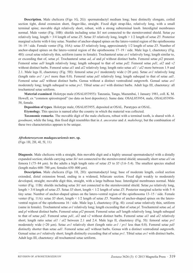
Afroheterozercon madagascariensis nov. sp.(Figs 1H, 2H, 4I, 5I, 11)
Diagnosis. Male chelicera with a straight, thin movable digit and a highly unusual spermatodactyl with a distally expanded section; shields carrying setae St1 not connected to the sternitoventral shield; unusually short setae al1 on femora I (75–84 μm). In the adults a high length ratio of setae Z5 to S5 (3.6–5.4). The smallest species studied (length males 600–700 μm; females 650–800 μm).
Description. Male chelicera (Figs 1H, 2H): spermatodactyl long; base of moderate length, coiled section extended, distal extension broad, ending in a widened, bifurcate section. Fixed digit weakly to moderately developed, straight; movable digit thin, straight, with a large bulbous base. Interdigital membranes normal. Male venter (Fig. 11B): shields including setae St1 not connected to the sternitoventral shield. Setae pa relatively long, length > 3/4 length of setae Z5. Setae S5 short, length < 1/2 length of setae Z5. Posterior marginal sclerite with 5–6 tiny setae. Number of anchor-shaped spines on the latero-ventral region of the opisthosoma 13–15 / side. Female venter (Fig. 11A): setae S5 short, length < 1/2 length of setae Z5. Number of anchor-shaped spines on the latero-ventral region of the opisthosoma 16 / side. Male legs I, chaetotaxy (Fig. 4I): coxal setae relatively thin, setiform (same in female). Trochanteral setae av1 relatively long; length exceeding that of setae pl. Trochanteral setae ad, al and pl without distinct barbs. Femoral setae pl2 present. Femoral setae ad3 length relatively long; length subequal to that of setae pd2. Femoral setae pd1, al2 and v2 without distinct barbs. Femoral setae al1 and al2 relatively short; length ratio setae al1 / al2 between 2.1 and 2.4. Male legs II, chaetotaxy (Fig. 5I): femoral setae pv1moderately wide (<20 μm). Setae av1 relatively short (length ratio av1 / pv1 less than 0.8). Femoral setae pd1distinctly shorter than setae ad1. Femoral setae ad2 without barbs. Genua with a distinct ventrodistal outgrowth. Genual setae av1 relatively short; length distinctly exceeding that of setae pv1. Tibial setae av1 with distinct barbs. Adult legs III, chaetotaxy: all trochanteral setae setiform.

KOLMPEN ET AL.320 · Zootaxa 3626 (3) © 2013 Magnolia Press
Material examined. Holotype male (OSAL053960): Madagascar, Antsiranana, near Andrafiabe cave, 2.6 km E Andrafiabe, Reserve Speciale d'Ankaranana, ~50m, 20–27 January 2001, coll. S. M. Goodman, ex pitfall trap residue with multiple millipedes, in dry deciduous forest at base tsingy, collection no. FMHD#2001-41 (hosts at FMNH, specimen numbers FMNH-INS-3957 (Spirostreptida), FMNH-INS-5443 (Spirobolida), FMNH-INS-3960 (Spirobolida)). Same data: OSAL053959, 053961-63, 053974, male; OSAL 053964-73, 053975-88, 003131, female.
Deposition of types. Holotype, OSAL053960, deposited at FMNH. Paratypes at FMNH and OSAL.Etymology. This species is named after the country in which the material was collected.Taxonomic remarks. The thin moveable digit of the male spermatodactyl is shared with A. pachybolus and A.
mahsbergi, but both of those species show a partial sclerotisation of the interdigital membranes. The shape of the distal part of the spermatodactyl is unique within the genus.
FIGURE 11. Afroheterozercon madagascariensis nov. sp., venter adults. A, female (OSAL053968); B, male (OSAL053961).

Zootaxa 3626 (3) © 2013 Magnolia Press · 321REVISION OF AFROHETEROZERCON
Afroheterozercon cautus (Berlese) com. nov.
Heterozercon cautus Berlese 1924: 251.
Fain (1989) noted “It is possible that H. cautus Berlese, 1924, described from East Africa, also belongs to this genus” (= Afroheterozercon). Re-examination of the type specimen (no. 221/40, Berlese acarotheca at ISZA) did not clarify the issue of presence or absence of lateral spines (the specimen is insufficiently cleared). It did reveal that, in addition to the presence of a single large spine-like seta on femur II of the male (seta pv1 by our designation), the male spermatodactyl has a shape characteristic of Afroheterozercon. The chelicera appear to have the partially sclerotised interdigital membrane observed in A. mahsbergi and A. pachybolus (Figs 1A–B, 2A–B), while the spermatodactyl shows a tight coiled middle, and a short thin distal section. Afroheterozercon cautusdiffers from A. mahsbergi and A. pachybolus by the shape of the distal section of the spermatodactyl, straight in A.mahsbergi and A. pachybolus, while gently curved in A. cautus. Unfortunately, the condition of the type specimen does not allow us to score most characters examined for this study. We therefore have made no attempt to present a full re-description.
Key to the species of the genus Afroheterozercon (males only). Afroheterozercon cautus is excluded due to insufficient data.
1. Posterior marginal sclerite with distinctly more than 6 tiny setae (male and female) (see small arrow Fig. 3B); spermatodactyl strongly elongate (Figs 1D, 2D) . . . . . . . . . . . . . . . . . . . . . . . . . . . . . . . . . . . . . . . . . . . . . . . . . . . . . . . . . . . . . . A. spirostreptus
- Posterior marginal sclerite with only 6 (rarely 5 or 7) tiny setae; spermatodactyl much smaller . . . . . . . . . . . . . . . . . . . . . . . . 22. Shields carrying setae St1 connected with sternitoventral shield . . . . . . . . . . . . . . . . . . . . . . . . . . . . . . . . . . . . . . . . . . . . . . . . 3- Shields carrying setae St1 not connected to sternitoventral shield . . . . . . . . . . . . . . . . . . . . . . . . . . . . . . . . . . . . . . . . . . . . . . . 53. Movable digit of chelicera robust; interdigital membranes not sclerotised (Figs 1C, 2C) . . . . . . . . . . . . . . . . . . . . . . A. sanghae- Movable digit of chelicera thin (< 13 μm); one interdigital membrane partially sclerotised (Figs 1A–B) . . . . . . . . . . . . . . . . . 44. Length ratio of setae Z5 to S5 very high, 4.2–6.1 (long Z5, short S5) (Fig. 3), setae pl2 on femora I absent (males and females)
. . . . . . . . . . . . . . . . . . . . . . . . . . . . . . . . . . . . . . . . . . . . . . . . . . . . . . . . . . . . . . . . . . . . . . . . . . . . . . . . . . . . . . . . . . A. mahsbergi- Length ratio of setae Z5 to S5 smaller, 2.7–3.8 (Fig. 6), setae pl2 on femora I present . . . . . . . . . . . . . . . . . . . . . .A. pachybolus5. Fixed digit and spermatodactyl reduced (Fig. 1F) . . . . . . . . . . . . . . . . . . . . . . . . . . . . . . . . . . . . . . . . . . . . . . . . . . .A. goodmani- Fixed digit chelicera and spermatodactyl well developed . . . . . . . . . . . . . . . . . . . . . . . . . . . . . . . . . . . . . . . . . . . . . . . . . . . . . 66. Distal end of spermatodactyl expanded; movable digit chelicera thin (<13 μm wide) (Figs 1H, 2H) . . . . . A. madagascariensis- Distal end of spermatodactyl tapering, not expanded; movable digit chelicera robust . . . . . . . . . . . . . . . . . . . . . . . . . . . . . . . . 77. Movable digit of chelicera not strongly curved but with a well-differentiated distal tooth; spermatodactyl long, especially in
basal section (Figs 1G, 2G) . . . . . . . . . . . . . . . . . . . . . . . . . . . . . . . . . . . . . . . . . . . . . . . . . . . . . . . . . . . . . . . . . . .A. tanzaniensis- Movable digit chelicera strongly curved, but distal teeth absent or weakly developed . . . . . . . . . . . . . . . . . . . . . . . . . . . . . . . 88. Distal sections of the fixed digit and spermatodactyl straight . . . . . . . . . . . . . . . . . . . . . . . . . . . . . . . . . . . . . . . . . .A. ancoratus- Distal sections of the fixed digit and spermatodactyl with well developed hooks (Figs 1E, 2E) . . . . . . . . . . . . . .A. gabonensis
Discussion
Biology. Most of the material for A. pachybolus and A. sanghae is part of a large collection of millipedes (~100) that was preserved individually, allowing some inferences on incidence (percentage of millipedes infested) and abundance (average number of mites/infested host) at the mite genus level. Unfortunately we have not been able to get identifications of the millipede hosts, limiting us to treatment by relative size, which may reflect either different host species or immature vs. adult hosts. Incidence for "large" millipedes (N=50) was 76% and abundance was 12.0. Clearly these mites are not rare in appropriate circumstances. On the other hand recovery is very poor if hosts are not killed and preserved individually (or if preservative is changed without consideration of associates) because the mites rapidly drop from the host once those hosts are fluid preserved. There is a marked correlation between presence of heterozerconids and the size of the host millipede. Although incidence for "medium" sized millipede hosts (N=26) was comparable to that for "large" ones (72% vs. 76%), abundance was clearly lower (6.8 vs. 12.0). Both measures were much lower for "small" (N=14) millipedes, 29% and 1.3, respectively. This pattern matches that observed for the association of the North American species Narceoheterozercon ohioensis Gerdeman & Klompen 2003 and its millipede host, Narceus annularis (Rafinesque) (Gerdeman 2002). It is also similar to the
KOLMPEN ET AL.322 · Zootaxa 3626 (3) © 2013 Magnolia Press
pattern observed for another group of mesostigmatid associates of the Central African Republic millipedes, members of the similar sized genus Julolaelaps (Iphiopsididae). Numbers for this genus are, respectively, 64% and 37.4, 48% and 15.7, and 7% and 1.0, for the "large", "medium" and "small" millipede size categories. The trends for the two taxa from the Central African Republic are similar with two small differences: (1) incidence, as well as abundance, drops strongly from "large" to "medium" millipedes in Julolaelaps, and (2) the drop in abundance across the host size categories is even more pronounced for Julolaelaps than for Afroheterozercon. A study of mite associates of millipedes in the eastern U.S. also noted the lack of (the relatively large) Mesostigmata on small millipedes (e.g. family Julidae, Blaniulidae). The few records of Mesostigmata in this study were from larger hosts (mostly Polydesmida: Euryuridae) (Farfan & Klompen 2012). Thus our records support the generally accepted notion that medium to large mites require relatively large-bodied hosts.
Males of A. sanghae and A. pachybolus were found on the same individual millipede (field number AL6647). Similarly, we found two species, A. spirostrepti and A. goodmani, on a single millipede in Gabon (field number AL6503). The presence of multiple species of Heterozerconidae on a single host individual has been suggested previously (Kethley, pers. comm.), and is hereby confirmed. Further studies are needed to assess how common this type of association is.
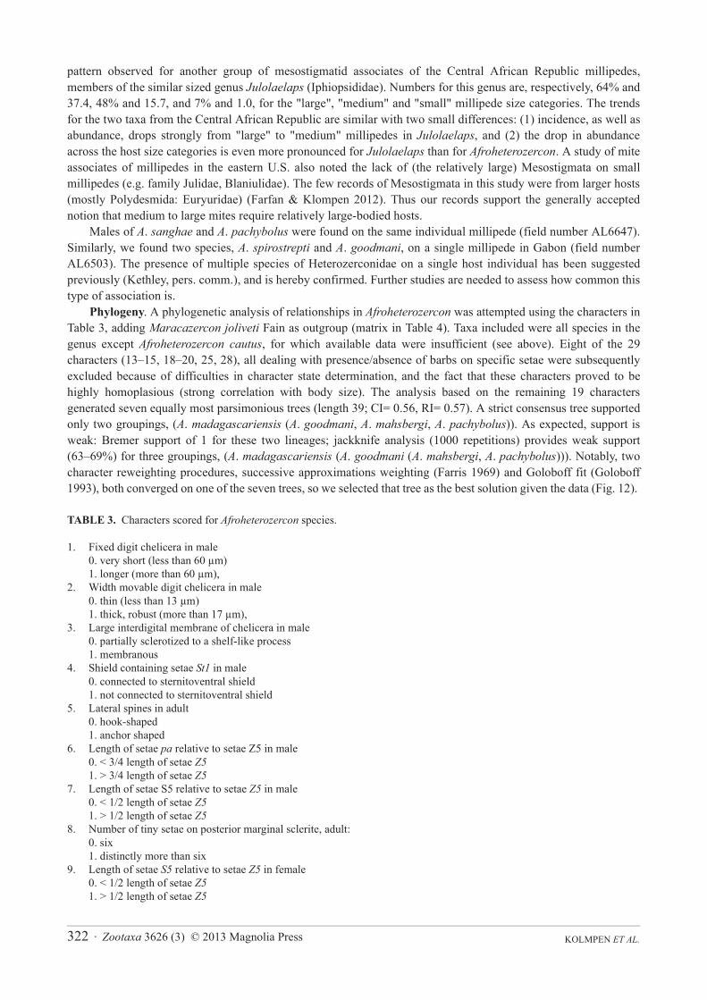
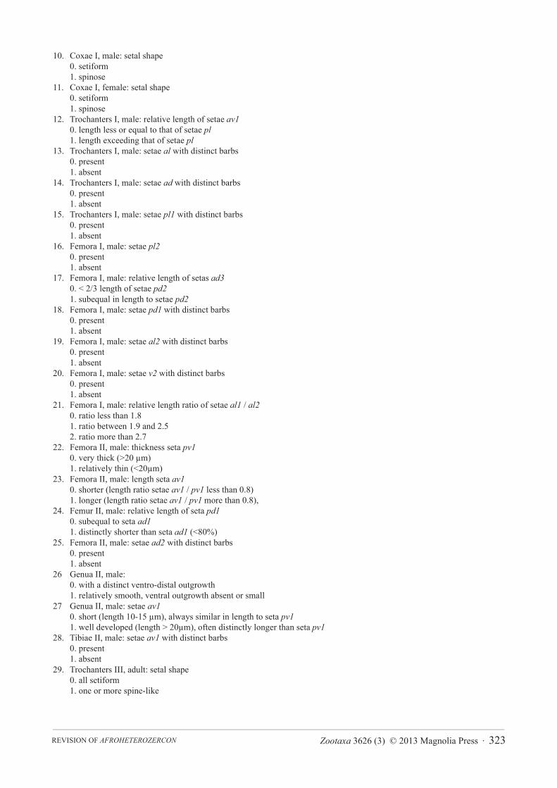
Phylogeny. A phylogenetic analysis of relationships in Afroheterozercon was attempted using the characters in Table 3, adding Maracazercon joliveti Fain as outgroup (matrix in Table 4). Taxa included were all species in the genus except Afroheterozercon cautus, for which available data were insufficient (see above). Eight of the 29 characters (13–15, 18–20, 25, 28), all dealing with presence/absence of barbs on specific setae were subsequently excluded because of difficulties in character state determination, and the fact that these characters proved to be highly homoplasious (strong correlation with body size). The analysis based on the remaining 19 characters generated seven equally most parsimonious trees (length 39; CI= 0.56, RI= 0.57). A strict consensus tree supported only two groupings, (A. madagascariensis (A. goodmani, A. mahsbergi, A. pachybolus)). As expected, support is weak: Bremer support of 1 for these two lineages; jackknife analysis (1000 repetitions) provides weak support (63–69%) for three groupings, (A. madagascariensis (A. goodmani (A. mahsbergi, A. pachybolus))). Notably, two character reweighting procedures, successive approximations weighting (Farris 1969) and Goloboff fit (Goloboff 1993), both converged on one of the seven trees, so we selected that tree as the best solution given the data (Fig. 12).
TABLE 3. Characters scored for Afroheterozercon species.
1. Fixed digit chelicera in male0. very short (less than 60 μm) 1. longer (more than 60 μm),
2. Width movable digit chelicera in male0. thin (less than 13 μm) 1. thick, robust (more than 17 μm),
3. Large interdigital membrane of chelicera in male0. partially sclerotized to a shelf-like process1. membranous
4. Shield containing setae St1 in male0. connected to sternitoventral shield1. not connected to sternitoventral shield
5. Lateral spines in adult0. hook-shaped1. anchor shaped
6. Length of setae pa relative to setae Z5 in male0. < 3/4 length of setae Z51. > 3/4 length of setae Z5
7. Length of setae S5 relative to setae Z5 in male 0. < 1/2 length of setae Z51. > 1/2 length of setae Z5
8. Number of tiny setae on posterior marginal sclerite, adult: 0. six1. distinctly more than six
9. Length of setae S5 relative to setae Z5 in female 0. < 1/2 length of setae Z51. > 1/2 length of setae Z5
Zootaxa 3626 (3) © 2013 Magnolia Press · 323REVISION OF AFROHETEROZERCON
10. Coxae I, male: setal shape0. setiform1. spinose
11. Coxae I, female: setal shape0. setiform1. spinose
12. Trochanters I, male: relative length of setae av10. length less or equal to that of setae pl1. length exceeding that of setae pl
13. Trochanters I, male: setae al with distinct barbs0. present1. absent
14. Trochanters I, male: setae ad with distinct barbs0. present1. absent
15. Trochanters I, male: setae pl1 with distinct barbs0. present1. absent
16. Femora I, male: setae pl20. present1. absent
17. Femora I, male: relative length of setas ad30. < 2/3 length of setae pd21. subequal in length to setae pd2
18. Femora I, male: setae pd1 with distinct barbs0. present1. absent
19. Femora I, male: setae al2 with distinct barbs0. present1. absent
20. Femora I, male: setae v2 with distinct barbs0. present1. absent
21. Femora I, male: relative length ratio of setae al1 / al20. ratio less than 1.8 1. ratio between 1.9 and 2.5 2. ratio more than 2.7
22. Femora II, male: thickness seta pv10. very thick (>20 μm) 1. relatively thin (<20μm)
23. Femora II, male: length seta av10. shorter (length ratio setae av1 / pv1 less than 0.8)1. longer (length ratio setae av1 / pv1 more than 0.8),
24. Femur II, male: relative length of seta pd10. subequal to seta ad11. distinctly shorter than seta ad1 (<80%)
25. Femora II, male: setae ad2 with distinct barbs0. present1. absent
26 Genua II, male: 0. with a distinct ventro-distal outgrowth1. relatively smooth, ventral outgrowth absent or small
27 Genua II, male: setae av10. short (length 10-15 μm), always similar in length to seta pv11. well developed (length > 20μm), often distinctly longer than seta pv1
28. Tibiae II, male: setae av1 with distinct barbs0. present1. absent
29. Trochanters III, adult: setal shape0. all setiform1. one or more spine-like

KOLMPEN ET AL.324 · Zootaxa 3626 (3) © 2013 Magnolia Press
TABLE 4. Character matrix for Afroheterozercon species.
Character number 1 11111 11112 22222 2222 12345 67890 12345 67890 12345 6789
mahsbergi 00001 00000 00011 101?1 11011 0010pachybolus 00001 00001 10111 01111 21011 0001sanghae 11101 1101? ?0000 01000 21000 1101spirostreptus 11111 10100 00011 00111 00100 1101gabonensis 11111 110?0 01111 00011 11101 1101goodmani 01111 00001 10111 01111 10111 0001ancoratus 11111 1?000 00001 00011 11101 1101tanzaniensis 11111 110?? ?1111 01111 11101 1100madagascariensis 10111 1000? ?1111 01111 11011 0100Maracazercon joliveti 01100 11110 00111 00111 11111 1111
FIGURE 12. Phylogenetic tree Afroheterozercon species (see text and tables 3 & 4). Character changes indicated on the branches. Closed boxes indicate apomorphies; open boxes homoplasious changes. Only unambiguous character changes are listed.
The grouping of A. madagascariensis, A. goodmani, A. mahsbergi, and A. pachybolus is supported by the presence of a ventrodistal outgrowth on genu II of the male (character 26: 0). This structure is best developed in the latter three species, which are also characterised by short setae pa (char. 6: 0) and av1 on genua II (char. 27: 0), and a very short fixed cheliceral digit (char. 1: 0). The grouping of A. mahsbergi and A. pachybolus is characterised by the partial sclerotisation of the large interdigital membrane of the chelicerae (char. 3: 0), and partial fusion of the shield containing setae St1 with the sternitoventral shield (char. 4: 0). Given the poor resolution of relationships among the remaining taxa it seems overly speculative to discuss their affinities, other than to note that the topology in Fig. 12 is heavily influenced by the ratio of setae S5 to Z5, in both females (char. 9: 0) and males (char. 7: 0).
The topology presented in Fig. 12 does not match any obvious broad biogeographical patterns. For example, the madagascariensis - pachybolus lineage includes species from the northern, western, and southern extremes of the known range. Additional collections from a broader range of localities in the Afrotropical region, and improved host identification may help improve support for phylogenetic relationships and allow more meaningful analyses of biogeography and host associations.
Zootaxa 3626 (3) © 2013 Magnolia Press · 325REVISION OF AFROHETEROZERCON
Acknowledgements
We would like to thank Brian Fisher, Steven Goodman, Dieter Mahsberg, and John Wenzel for making specimens available, Andrei Bochkov and Wouter Dekoninck, IRSNB, Brussels, for the loan of type material of the Fain species, Sam Bolton for examining the holotype of A. cautus, and Roberto Nannelli, ISZA, Florence, for permission to examine that type.
References
Berlese, A. (1924) Centuria sesta di Acari nuovi. Redia (Firenze), 15, 237–262.Evans, G.O. (1963a) Some observations on the chaetotaxy of the pedipalps on the Mesostigmata (Acari). Annals and Magazine
of Natural History (13th series), London, 6, 513–527. http://dx.doi.org/10.1080/00222936308651393
Evans, G.O. (1963b) Observations on the chaetotaxy of the legs in free-living Gamasina (Acari: Mesostigmata). Bulletin of the British Museum of Natural History, Zoology, 10, 277–303.
Evans, G.O. (1969) Observations on the ontogenetic development of the chaetotaxy of the tarsi of legs II–IV in the Mesostigmata (Acari). In: Evans, G.O. (Ed), Proceedings of the 2nd International Congress of Acarology. Akadémiai Kiadó, Budapest, pp. 195–200.
Fain, A. (1988) Notes on mites associated with Myriapoda. III. Two new species of the genus Heterozercon Berlese, 1888 (Acari, Mesostigmata) from Afrotropical Myriapods. Bulletin et Annales de la Société royale belge d'Entomologie, 124, 237–242.
Fain, A. (1989) Notes on mites associated with Myriapoda. IV. New taxa in the Heterozerconidae (Acari, Mesostigmata). Bulletin de l'Institut royal des Sciences naturelles de Belgique, Entomologie, 59, 145–156.
Farfan, M. & Klompen, H. (2012) Phoretic mite associates of millipedes (Diplopoda: Julidae) in the northern Atlantic region (North America, Europe). International Journal of Myriapodology, 7, 69–91. http://dx.doi.org/10.3897/ijm.7.3064
Farris, J.S. (1969) A successive approximations approach to character weighting. Systematic Zoology, 18, 374–385. http://dx.doi.org/10.2307/2412182
Gerdeman, B.S., Klompen, H. & Tanigoshi, L. (2000) Insights into the biology of a mite - millipede association. Fragmenta Faunistica, Warszawa, 43 (Suppl.), 223–227.
Gerdeman, B.S. (2002) The biology of the Heterozerconidae. Ph.D. Ohio State University, Columbus, Ohio, USA, pp. 119.Gerdeman, B.S. & Klompen, H. (2003) A new North American heterozerconid, Narceoheterozercon ohioensis, n. gen., n. sp.,
with first description of immatures of Heterozerconidae (Acari: Mesostigmata). International Journal of Acarology, 29, 351–370. http://dx.doi.org/10.1080/01647950308684352
Gerdeman, B.S. & Garcia, R. (2010) Heterozerconidae: A comparison between a temperate and a tropical species. In: Sabelis, M.W. & Bruin, J. (Eds), Trends in Acarology, Proceedings of the 12th International Congress. Springer Science, Dordrecht, pp. 93–96.
Goloboff, P.A. (1993) Estimating character weights during tree search. Cladistics, 9, 83–91. http://dx.doi.org/10.1111/j.1096-0031.1993.tb00209.x
Johnson, N.F. (2010) Future taxonomy today: new tools applied to accelerate the taxonomic process. In: Polaszek, A. (Ed), Systema Naturae 250: The Linnaean Ark. CRC Press Taylor & Francis Group, London, pp. 137–147.
Lindquist, E.E. & Evans, G.O. (1965) Taxonomic concepts in the Ascidae, with a modified setal nomenclature for the idiosoma of the Gamasina (Acarina: Mesostigmata). Memoirs of the Entomological Society of Canada, 47, 1–64. http://dx.doi.org/10.4039/entm9747fv
Maddison, D.R. & Maddison, W.P. (2002) MacClade 4: Analysis of phylogeny and character evolution. Vs. 4.05. Sinauer Associates, Inc., Sunderland, MA.
Swofford, D.L. (2002) PAUP*. Phylogenetic Analysis Using Parsimony (*and other methods). Vs. 4.0b.10. Sinauer Associates, Inc., Sunderland, MA.
Walter, D.E. & Krantz, G.W. (2009) Collecting, rearing and preparing specimens. In: Krantz, G.W. & Walter, D.E. (Eds), Amanual of acarology. Texas Tech University Press, Lubbock, TX, pp. 83–95.