Embed Size (px)
Citation preview

All Rights Reserved – ISPI 2008 1
A Scientific Rationale for Treating Lumbar Facet Joint Dysfunction with
Physical Therapy
The ever-increasing number of spine related disorders along with limited funds available to treat spinal
disorders, has lead to the development and implementation of evidence-based medicine. Third party
payers have used evidence-based practice (EBP) to force clinicians to prove the efficacy of their
interventions, if they were to be paid for their interventions.1 This has lead to a hierarchy of evidence,
with randomized controlled trials (RCT’s) and systematic reviews of RCT’s as the “highest” forms of
evidence. In developing a best-evidence approach for the efficacy of physical therapy (PT) in treating
lumbar facet joint dysfunction, EBP would require us to search for the systematic reviews and RCT’s. The
main problem with this is that PT, although utilized successfully in various forms of LBP2-5
, does not
target a specific tissue. Whereas procedures such as medial branch blocks can be very specialized and
precise6, 7
, especially with the visual feedback using fluoroscopy, treatments such as spinal
manipulation3, exercise
8, 9, traction
10 and more have shown efficacy in treating LBP, their effect on a
precise tissue such as intervertebral disc (IVD), facet joint, muscle or ligament is unknown. To develop a
scientific rationale for PT treatment for lumbar facet joints, it is suggested that clinicians view the patho-
anatomy of lumbar facet joints, their clinical presentations and the effect of PT on these pathological
changes and clinical presentations.
The lumbar spine consists of 5 paired, 10-total facet joints. These joints are synovial joints and
anatomically consist of:
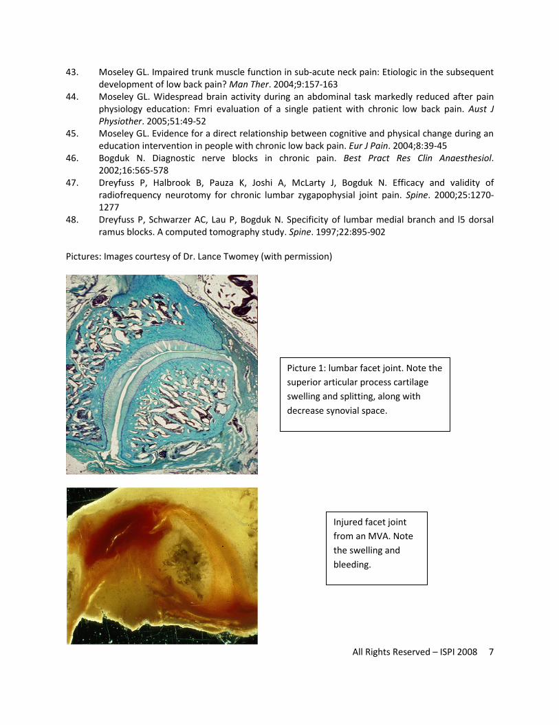
1. Superior and inferior articular processes. These processes are lined with hyaline cartilage, which
serves as load-bearing surfaces. The hyaline cartilage is dependent on movement, more
precisely compression and distraction to maintain adequate nutrition to the cartilage.4, 11
The
articular surfaces are avascular through diffusion, via movement, allows nutrition to flow from
mainly the synovial fluid inside the facet joint to keep the cartilage healthy. Numerous studies
have shown that in the absence of movement, articular cartilage in the lumbar facet joints will
imbibe more fluid, cause the articular cartilage to swell and ultimately lead to articluar splitting
and wearing of the cartilage, potentially exposing the subchondral bone-plate, which is highly
innervated and a potential cause of facet joint pain, especially on weight-bearing. The fact that
articular cartilage is dependent on movement to keep it healthy and hydrated would imply that
All Rights Reserved – ISPI 2008 2
movement-based treatments such as spinal mobilization (level-specific, oscillating compression
and distraction techniques on the facet joints) and exercises aimed at creating movement of the
lumbar spine may be of benefit in treating lumbar facet joints. A recent cadaver study on
patients that did not survive car accidents, by dissecting the lumbar spines, have shown that >
70% of these facet joints showed damage, none of which were shown on standard lumbar X-
rays, pre-dissection12
. These joints showed damage (articluar splitting and swelling) of the facet
joints. It could be argued if these patients survived the accidents, and after subsequent X-rays,
may have been referred to PT. Treatments aimed at allowing normal movement, along with
compression/distraction techniques would more than likely allow the injured cartilage receive
much needed nutrition, which may/may not correlate to decreased pain and increased function.
The facet can be viewed the same as swollen, inflamed knee joint. Best evidence would require
early, gentle movement to a “swollen, hot” knee. The same may apply to lumbar facet joints.
Recent MRI studies have implicated facet joint swelling in LBP populations.13
The authors argue
that increased mechanical loading on facet joints, along with degenerative changes cause
swelling in facet joints, which can be readily detected on MRI, compared to uninjured levels and
normal healthy volunteers.13, 14
These studies would imply that injured and degenerated facet
joints are swollen, which I turn makes an added case for the potential of movement based
strategies such as mobilization and exercise to decrease joint swelling and thus pain.
2. Capsule. The articluar surfaces are surrounded by a thick, fibrous capsule, which encapsulates
the synovial fluid, which is critical to maintain nutrition of the articluar cartilage (see above). The
collagenous capsule allows for normal movement in various planes. As with any other collagen,
it responds well to slow movements as well as sustained movements. The capsule, however,
does not respond well to sudden movement. Sudden movement of the lumbar spine, especially
at end-ROM, may result in tearing of the collagen.12
This could be compared to an ankle sprain,
where sudden movement causes different degrees of collagen damage (grade I, II or III). The
injured tissue (capsule) will go through the normal phases of healing: inflammation, scarring and
remodeling. In ankle sprains, early movement and subsequent progressive load is applied to the
ligament to help the ankle heal. Basic sciences would imply that progressive exercises towards
the end-ROM of the joint (flexion and extension) may help keep the capsule “stretched out” –
which in turn allows normal movement of the joint, adequate synovial fluid flow and thus
healthy cartilage (see above).4 Also interesting to note is that recent studies have implicated
facet joints in the development of lumbar radiculopathy.15
In the past inflammatory properties
of the IVD have been implicated in the pathogenesis of lumbar radiculopathy due to chemical
activation of the dorsal root ganglion.16, 17 Tachihara et al, in discussion of the possible causes of
lumbar radiculopathy due to lumbar facets, implicates (amongst other potential causes), the
facet joint capsule. Movement-based approaches aimed at capsular healing may thus potentially
also have an effect on lumbar radiculopathy due to facet joint irritation.
All Rights Reserved – ISPI 2008 3
a. Multifidus. In the lumbar spine, the facet joint capsule is reinforced by the multifidus
(MF). The lumbar multifidus along with the transverses abdominus (TA) are very
important in the spinal stabilization mechanism.18
In normal, healthy people, MF and TA
fires prior to the use of the upper and lower extremities (feedforward control) to allow a
person to lift/use the extremity without causing unwanted stress on the local spinal
levels.19-23
In LBP populations, this feedforward mechanism is altered (delayed), which
leads to increased stress on the local spinal level. 19-23
MRI studies have shown that MF
atrophy is side and level-specific, as well as non-pathology specific.24-28
This may in
essence implicate the facet joints, based on the fact that the MF receives it’s nerve
supply from the dorsal ramus, which also innervates the local facet joint.29
Spinal stabilization, by virtue of retraining the feed-forward, co-contraction of the TA
and MF may work by abolishing the unwanted stress on the local spinal level, including
facet joint. Numerous studies have shown the benefit of spinal stabilization in treating
LBP.8, 9, 18, 30-32
An analogy to the facet joint capsule laxity/instability would be a medial
collateral ligament (MCL) injury. Injury to the MCL causes the knee to have increased
joint play and valgus, thus causing additional painful excursion and loading of the knee
joint. To help compensate for the MCL and the knee’s lack of stability, PT will often
strengthen the quadriceps, specifically the vastus mediallis (VMO) thus utilizing more
motor control to take unwanted stress off the knee, allowing it to heal and protecting it
from further damage (especially in the healing phase). Could this be the same for MF?
By retraining MF (along with TA), the facet joint’ stability could be enhanced, take
unwanted stress off the pain-sensitive joint, thus helping control and manage the
patient’s LBP.
b. Ligamentum flavum. In the lumbar spine, the part of the facet joint is reinforced by the
ligamentum flavum (LF). The LF consists of a high concentration of elsatin fibers,
allowing it to always be taught, thus not allowing it to “buckle” into the spinal canal,
causing neurovascular compromise.33, 34
Apart from bony changes to the lumbar
vertebrae (bone spurs, vertebral compression) and age-related changes to the lumbar
disc35
the LF has been implicated in the development of degenerative lumbar spinal
stenosis.5, 36
With aging, the LF becomes thicker and less elastic. This allows for a steady
encroachment of the spinal canal – hence degenerative spinal stenosis. Spinal stenosis is
a progressive disorder and clinical presentations may vary from mild to moderate to
severe. Patients with minimal to moderate spinal stenosis often get’s referred to PT for
conservative management. 5 A recent systematic review have shown that PT is very
effective in treating mild to moderate degenerative spinal stenosis:
i. Treadmill or cycling with body weight support37, 38
. It could be argued that
aquatic exercise that mimic such activities, or an underwater treadmill could
potentially provide similar results
ii. Lumbar traction37, 38
iii. Flexion exercises37, 38

All Rights Reserved – ISPI 2008 4
iv. Manual therapy38
3. Nerve supply. A final potential mechanism for pain relief from PT for facet joint pain may be the
same nerve supply that implicates the utilization of medial branch blocks and radiofrequency.
The lumbar facet joint receives innervation from the medial branch of the dorsal ramus.29
All of
the research into spinal stabilization, feedforward control and the immediate effect of pain on
the size and contraction of the MF had lead researchers to ask if treatments aimed at a spinal
level may have a neuro-physiolgical effect on the local muscles and thus create a mechanism of
pain control.21, 26, 39
A recent pilot study examined the immediate effect of spinal manipulation
(high-velocity, small amplitude) technique on the local stabilizing muscles utilizing diagnostic
ultrasound. This preliminary study showed that immediately post-manipulation, that the TA had
an immediate statistically significant increase in size in 8 out of 9 patients when asked to
perform a deep corset control maneuver.39
This study showed an immediate neuro-muscular
effect of a spinal manipulation, which led to a better quality contraction of the stabilizing
muscles, associated with protecting the local facet joint.
The previous passages are aimed at developing a theoretical model for the efficacy of treating facet joint
based pain with movement-based approaches utilized in physical therapy. It is interesting to note that
each and all of these mechanisms described above are all inter-related – stabilization, movement, motor
control and pain relief. These “anatomical” and “biomechanical” models described above do not take
into account additional potential benefits of PT intervention on a more cerebral level. New research into
pain science education utilized by physical therapists (aimed at explaining a patient’s pain to them,
decreasing fear, explaining the biology behind their pain) have shown significant changes in regards to
pain beliefs and attitudes 40, 41
, improved cognition and physical performance 42
, increased pain
thresholds and improved outcomes from therapeutic exercise 43, 44
. Improved understanding of pain
science or neurophysiology may also lead to a decrease in the fear and anxiety associated with spinal
surgery, and could potentially result in better outcomes related to decreased pain and improved
function 45
.
The aforementioned passages also make for another case: the importance of the relationship between a
good manual physical therapist and a physician able to help with diagnostic labeling, testing and pain
management. The effect of pain is well-known. Treatments such as radiofrequency and medial branch
blocks may be able to help decrease pain associated with a local facet disorder,46-48
while the patient
(now with less pain) may benefit from physical therapy aimed at restoring normal movement and
function along with decreasing fear associated with the injury.
References
1. Sackett DL. Evidence-based medicine. Spine. 1998;23:1085-1086
2. Waddell G, Burton AK. Concepts of rehabilitation for the management of low back pain. Best
Pract Res Clin Rheumatol. 2005;19:655-670

All Rights Reserved – ISPI 2008 5
3. Childs JD, Fritz JM, Flynn TW, Irrgang JJ, Johnson KK, Majkowski GR, Delitto A. A clinical
prediction rule to identify patients with low back pain most likely to benefit from spinal
manipulation: A validation study. Ann Intern Med. 2004;141:920-928
4. Twomey LT. A rationale for the treatment of back pain and joint pain by manual therapy. Phys
Ther. 1992;72:885-892
5. Fritz JM, Delitto A, Welch WC, Erhard RE. Lumbar spinal stenosis: A review of current concepts in
evaluation, management, and outcome measurements. Arch Phys Med Rehabil. 1998;79:700-
708
6. Bogduk N. International spinal injection society guidelines for the performance of spinal
injection procedures. Part 1: Zygapophysial joint blocks. Clin J Pain. 1997;13:285-302
7. Bogduk N. Diagnostic blocks: A truth serum for malingering. Clin J Pain. 2004;20:409-414
8. O'Sullivan PB, Phyty GD, Twomey LT, Allison GT. Evaluation of specific stabilizing exercise in the
treatment of chronic low back pain with radiologic diagnosis of spondylolysis or
spondylolisthesis. Spine. 1997;22:2959-2967
9. Hodges PW. Core stability exercise in chronic low back pain. Orthop Clin North Am. 2003;34:245-
254
10. Clarke J, van Tulder M, Blomberg S, de Vet H, van der Heijden G, Bronfort G. Traction for low
back pain with or without sciatica: An updated systematic review within the framework of the
cochrane collaboration. Spine. 2006;31:1591-1599
11. Twomey LT, Taylor JR. Sagittal movements of the human lumbar vertebral column: A
quantitative study of the role of the posterior vertebral elements. Arch Phys Med Rehabil.
1983;64:322-325
12. Twomey LT, Taylor JR, Taylor MM. Unsuspected damage to lumbar zygapophyseal (facet) joints
after motor-vehicle accidents. Med J Aust. 1989;151:210-212, 215-217
13. Friedrich KM, Nemec S, Peloschek P, Pinker K, Weber M, Trattnig S. The prevalence of lumbar
facet joint edema in patients with low back pain. Skeletal Radiol. 2007;36:755-760
14. Rihn JA, Lee JY, Khan M, Ulibarri JA, Tannoury C, Donaldson WF, 3rd, Kang JD. Does lumbar facet
fluid detected on magnetic resonance imaging correlate with radiographic instability in patients
with degenerative lumbar disease? Spine. 2007;32:1555-1560
15. Tachihara H, Kikuchi S, Konno S, Sekiguchi M. Does facet joint inflammation induce
radiculopathy?: An investigation using a rat model of lumbar facet joint inflammation. Spine.
2007;32:406-412
16. Ozaktay AC, Kallakuri S, Cavanaugh JM. Phospholipase a2 sensitivity of the dorsal root and
dorsal root ganglion. Spine. 1998;23:1297-1306
17. Takebayashi T, Cavanaugh JM, Cuneyt Ozaktay A, Kallakuri S, Chen C. Effect of nucleus pulposus
on the neural activity of dorsal root ganglion. Spine. 2001;26:940-945
18. Richardson CA, Jull GA. Muscle control-pain control. What exercises would you prescribe? Man
Ther. 1995;1:2-10
19. Hodges P, Cresswell A, Thorstensson A. Preparatory trunk motion accompanies rapid upper limb
movement. Exp Brain Res. 1999;124:69-79
20. Hodges PW. Changes in motor planning of feedforward postural responses of the trunk muscles
in low back pain. Exp Brain Res. 2001;141:261-266
21. Hodges PW, Moseley GL, Gabrielsson A, Gandevia SC. Experimental muscle pain changes
feedforward postural responses of the trunk muscles. Exp Brain Res. 2003;151:262-271
22. Hodges PW, Richardson CA. Feedforward contraction of transversus abdominis is not influenced
by the direction of arm movement. Exp Brain Res. 1997;114:362-370

All Rights Reserved – ISPI 2008 6
23. Tsao H, Hodges PW. Immediate changes in feedforward postural adjustments following
voluntary motor training. Exp Brain Res. 2007;181:537-546
24. Gille O, Jolivet E, Dousset V, Degrise C, Obeid I, Vital JM, Skalli W. Erector spinae muscle changes
on magnetic resonance imaging following lumbar surgery through a posterior approach. Spine.
2007;32:1236-1241
25. Hides JA, Belavy DL, Stanton W, Wilson SJ, Rittweger J, Felsenberg D, Richardson CA. Magnetic
resonance imaging assessment of trunk muscles during prolonged bed rest. Spine.
2007;32:1687-1692
26. Hides JA, Richardson CA, Jull GA. Magnetic resonance imaging and ultrasonography of the
lumbar multifidus muscle. Comparison of two different modalities. Spine. 1995;20:54-58
27. Hides JA, Stokes MJ, Saide M, Jull GA, Cooper DH. Evidence of lumbar multifidus muscle wasting
ipsilateral to symptoms in patients with acute/subacute low back pain. Spine. 1994;19:165-172
28. Hodges P, Holm AK, Hansson T, Holm S. Rapid atrophy of the lumbar multifidus follows
experimental disc or nerve root injury. Spine. 2006;31:2926-2933
29. Bogduk N, Wilson AS, Tynan W. The human lumbar dorsal rami. J Anat. 1982;134:383-397
30. Goldby LJ, Moore AP, Doust J, Trew ME. A randomized controlled trial investigating the
efficiency of musculoskeletal physiotherapy on chronic low back disorder. Spine. 2006;31:1083-
1093
31. Hodges P, Jull G. Does strengthening the abdominal muscles prevent low back pain? J
Rheumatol. 2000;27:2286-2288
32. Jull GA, Richardson CA. Motor control problems in patients with spinal pain: A new direction for
therapeutic exercise. J Manipulative Physiol Ther. 2000;23:115-117
33. Taylor JR, Twomey LT, Corker M. Bone and soft tissue injuries in post-mortem lumbar spines.
Paraplegia. 1990;28:119-129
34. Okada A, Harata S, Takeda Y, Nakamura T, Takagaki K, Endo M. Age-related changes in
proteoglycans of human ligamentum flavum. Spine. 1993;18:2261-2266
35. Twomey LT, Taylor JR. Age changes in lumbar vertebrae and intervertebral discs. Clin Orthop
Relat Res. 1987:97-104
36. Cho DY, Lin HL, Lee WY, Lee HC. Split-spinous process laminotomy and discectomy for
degenerative lumbar spinal stenosis: A preliminary report. J Neurosurg Spine. 2007;6:229-239
37. Pua YH, Cai CC, Lim KC. Treadmill walking with body weight support is no more effective than
cycling when added to an exercise program for lumbar spinal stenosis: A randomised controlled
trial. Aust J Physiother. 2007;53:83-89
38. Whitman JM, Flynn TW, Childs JD, Wainner RS, Gill HE, Ryder MG, Garber MB, Bennett AC, Fritz
JM. A comparison between two physical therapy treatment programs for patients with lumbar
spinal stenosis: A randomized clinical trial. Spine. 2006;31:2541-2549
39. Raney NH, Teyhen DS, Childs JD. Observed changes in lateral abdominal muscle thickness after
spinal manipulation: A case series using rehabilitative ultrasound imaging. J Orthop Sports Phys
Ther. 2007;37:472-479
40. Moseley L. Combined physiotherapy and education is efficacious for chronic low back pain. Aust
J Physiother. 2002;48:297-302
41. Moseley GL, Nicholas MK, Hodges PW. A randomized controlled trial of intensive
neurophysiology education in chronic low back pain. Clin J Pain. 2004;20:324-330
42. Moseley GL, Brhyn L, Ilowiecki M, Solstad K, Hodges PW. The threat of predictable and
unpredictable pain: Differential effects on central nervous system processing? Aust J Physiother.
2003;49:263-267
All Rights Reserved – ISPI 2008 7
43. Moseley GL. Impaired trunk muscle function in sub-acute neck pain: Etiologic in the subsequent
development of low back pain? Man Ther. 2004;9:157-163
44. Moseley GL. Widespread brain activity during an abdominal task markedly reduced after pain
physiology education: Fmri evaluation of a single patient with chronic low back pain. Aust J
Physiother. 2005;51:49-52
45. Moseley GL. Evidence for a direct relationship between cognitive and physical change during an
education intervention in people with chronic low back pain. Eur J Pain. 2004;8:39-45
46. Bogduk N. Diagnostic nerve blocks in chronic pain. Best Pract Res Clin Anaesthesiol.
2002;16:565-578
47. Dreyfuss P, Halbrook B, Pauza K, Joshi A, McLarty J, Bogduk N. Efficacy and validity of
radiofrequency neurotomy for chronic lumbar zygapophysial joint pain. Spine. 2000;25:1270-
1277
48. Dreyfuss P, Schwarzer AC, Lau P, Bogduk N. Specificity of lumbar medial branch and l5 dorsal
ramus blocks. A computed tomography study. Spine. 1997;22:895-902
Pictures: Images courtesy of Dr. Lance Twomey (with permission)
Picture 1: lumbar facet joint. Note the
superior articular process cartilage
swelling and splitting, along with
decrease synovial space.
Injured facet joint
from an MVA. Note
the swelling and
bleeding.
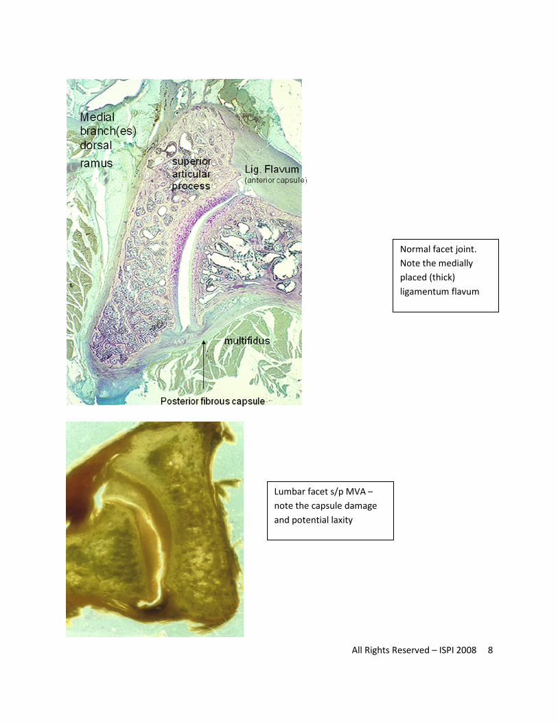
All Rights Reserved – ISPI 2008 8
Normal facet joint.
Note the medially
placed (thick)
ligamentum flavum
Lumbar facet s/p MVA –
note the capsule damage
and potential laxity

![Diagnostic Value of Lumbar Facet Joint Injection: A ...ative LBP are required [33,35,36,37]. The indication for diagnostic FJB by facet joint injection (FJI) using LA is therefore](https://img.pdfslide.net/doc/110x75/60907ffe022acf79d76d2ef9/diagnostic-value-of-lumbar-facet-joint-injection-a-ative-lbp-are-required-33353637.jpg)