Embed Size (px)
DESCRIPTION
Horse pathology
Citation preview
A SELECTIVE REVIEW OF POSTMORTEM LESIONS IN HORSESby
Paul C. Stromberg DVM, PhDDiplomate, American College of Veterinary Pathologists
Professor of Veterinary PathologyOhio State [email protected]
OBJECTIVES
A. Reinforce or broaden your equine pathology experienceB. See some “neat”, common and not-so-common lesions from horsesC. Practice applying the principles of gross pathology interpretationD. Develop appropriate morphologic diagnoses for the observed lesionsE. Learn how to play the “Gross Pathology Game” correctly or
“When is a response not responsive”
CARDIOVASCULAR SYSTEM
1-2 84927B&F Heart with fibrinonecrotic exudate
Mx= Fibrinonecrotic pericarditis
Cause = foreign body penetration of the pericardial sac
3. 87951-13 Heart with myocardial necrosis
Mx= acute myocardial coagulation necrosis
Cause = phenylbutazone; monensin?
4. 79334 Artery with thrombosis
Mx= granulomatous mesenteric arteritis with arteriosclerosis and thrombosis
Ex= verminous arteritis
Cause= Strongylus vulgaris
5. 90-2751-2 Lt ventricular endocardium with opaque white plaques
1
Mx = endocardial mineralization
Cause = 1) Hypervitaminosis D
A. Inappropriate iatrogenic supplementation
B. Vit D containing plants; Cestrum diurnum, Solanum malacoxylon, Trisetum flavescens
2) Renal failure ~ aminoglycoside therapy (Gentocin)
ENDOCRINE SYSTEM
6. G-884 Adrenal gland
Mx = Diffuse Adrenocortical atrophy
Cause = steroid therapy
7. 805280 Adrenal Gland
Mx = pheochromocytoma
8. Thyroid gland
Mx = Thyroid follicular adenoma (Nodular goiter)
9-10. Brain with large pituitary gland
Mx = pars intermedia adenoma of the pituitary gland
Name 4 associated clinical signs
1. Hyperpyrexia 3. PU/PD2. Hyperhidrosis 4. Hirsutism
HEMATOPOIETIC SYSTEM
2
11-12. 97-1518-1,2 Spleen with “petecchia”
Mx = herniated red pulp - normal finding
13-14. 90-1152-3,4 Spleen with hematomas and icterus
Mx = Splenic infarcts with hemorrhage and icterus
Name the disease = neonatal isoerythrolysis
Cause: maternal sensitization to fetal alloantigens leading to immune-mediated hemolysis
* The key here is that you must know this is a neonate.
15. 90-623-3 Spleen with large white nodules
Mx = LSA
16. E-11939-93 Spleen with black nodules
Mx = metastatic “malignant” melanoma
17. 80475C Spleen, X-sec with “raspberry jam” appearance
Mx = Diffuse lymphoid hyperplasia.
*Note also the splenic LN with hemorrhage
Name the Disease = Equine Infectioius Anemia
Cause = Equine lentivirus
18. Spleen with visible stroma
Mx = diffuse lymphoid atrophy
Name the Disease = CID19. 923916 LN, pharyngeal with lost architecture
Mx = LSA
3
* ID of the tissue here is part of the question
20. 87-683-9 LN, mesenteric with miliary foci
Mx = granulomatous lymphadenitis
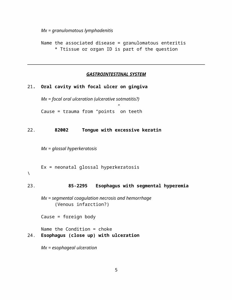
Name the associated disease = granulomatous enteritis* Ttissue or organ ID is part of the question
GASTROINTESTINAL SYSTEM
21. Oral cavity with focal ulcer on gingiva
Mx = focal oral ulceration (ulcerative sotmatitis?)
Cause = trauma from “points” on teeth
22. 82002 Tongue with excessive keratin
Mx = glossal hyperkeratosis
Ex = neonatal glossal hyperkeratosis\
23. 85-2295 Esophagus with segmental hyperemia
Mx = segmental coagulation necrosis and hemorrhage(Venous infarction?)
Cause = foreign body
Name the Condition = choke24. Esophagus (close up) with ulceration
Mx = esophageal ulceration
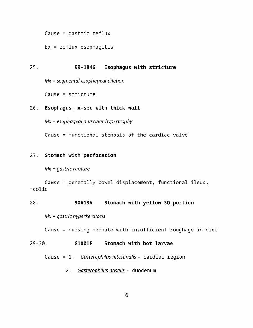
Cause = gastric reflux
Ex = reflux esophagitis
4
25. 99-1846 Esophagus with stricture
Mx = segmental esophageal dilation
Cause = stricture
26. Esophagus, x-sec with thick wall
Mx = esophageal muscular hypertrophy
Cause = functional stenosis of the cardiac valve
27. Stomach with perforation
Mx = gastric rupture
Cause = generally bowel displacement, functional ileus, “colic”
28. 90613A Stomach with yellow SQ portion
Mx = gastric hyperkeratosis
Cause - nursing neonate with insufficient roughage in diet
29-30. G1001F Stomach with bot larvae
Cause = 1. Gasterophilus intestinalis - cardiac region
2. Gasterophilus nasalis - duodenum
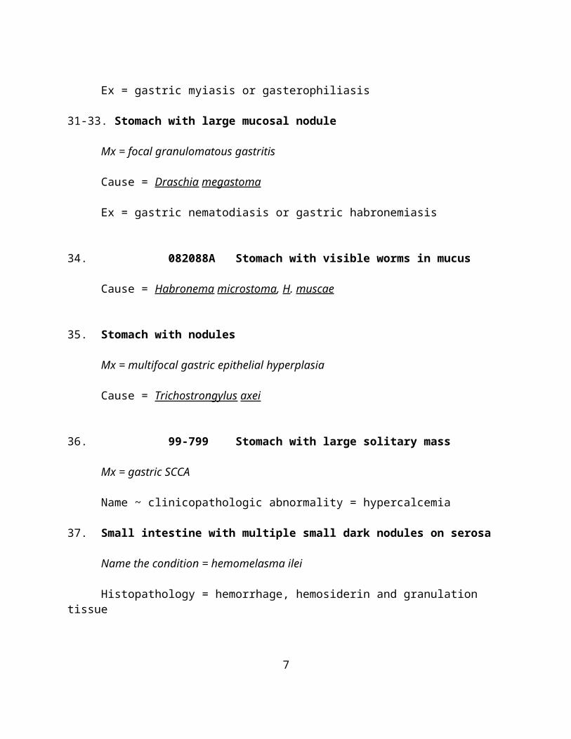
Ex = gastric myiasis or gasterophiliasis
31-33. Stomach with large mucosal nodule
Mx = focal granulomatous gastritis
Cause = Draschia megastoma
Ex = gastric nematodiasis or gastric habronemiasis
5
34. 082088A Stomach with visible worms in mucus
Cause = Habronema microstoma, H. muscae
35. Stomach with nodules
Mx = multifocal gastric epithelial hyperplasia
Cause = Trichostrongylus axei
36. 99-799 Stomach with large solitary mass
Mx = gastric SCCA
Name ~ clinicopathologic abnormality = hypercalcemia
37. Small intestine with multiple small dark nodules on serosa
Name the condition = hemomelasma ilei
Histopathology = hemorrhage, hemosiderin and granulation tissue
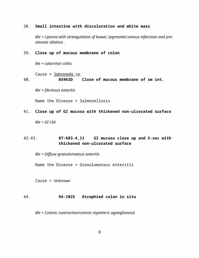
38. Small intestine with discoloration and white mass
Mx = Lipoma with strangulation of bowel, segmental venous infarction and pre-stenotic dilation.
39. Close up of mucous membrane of colon
Mx = catarrhal colitis
Cause = Salmonella sp.40. 85963D Close of mucous membrane of sm int.
Mx = fibrinous enteritis
Name the Disease = Salmonellosis
41. Close up of GI mucosa with thickened non-ulcerated surface
Mx = GI LSA
6
42-43. 87-683-4,11 GI mucosa close up and X-sec with thickened non-ulcerated surface
Mx = Diffuse granulomatous enteritis
Name the Disease = Granulomatous enteritis
Cause = Unknown
44. 94-1025 Atrophied colon in situ
Mx = Colonic contraction/colonic myenteric aganglionosis
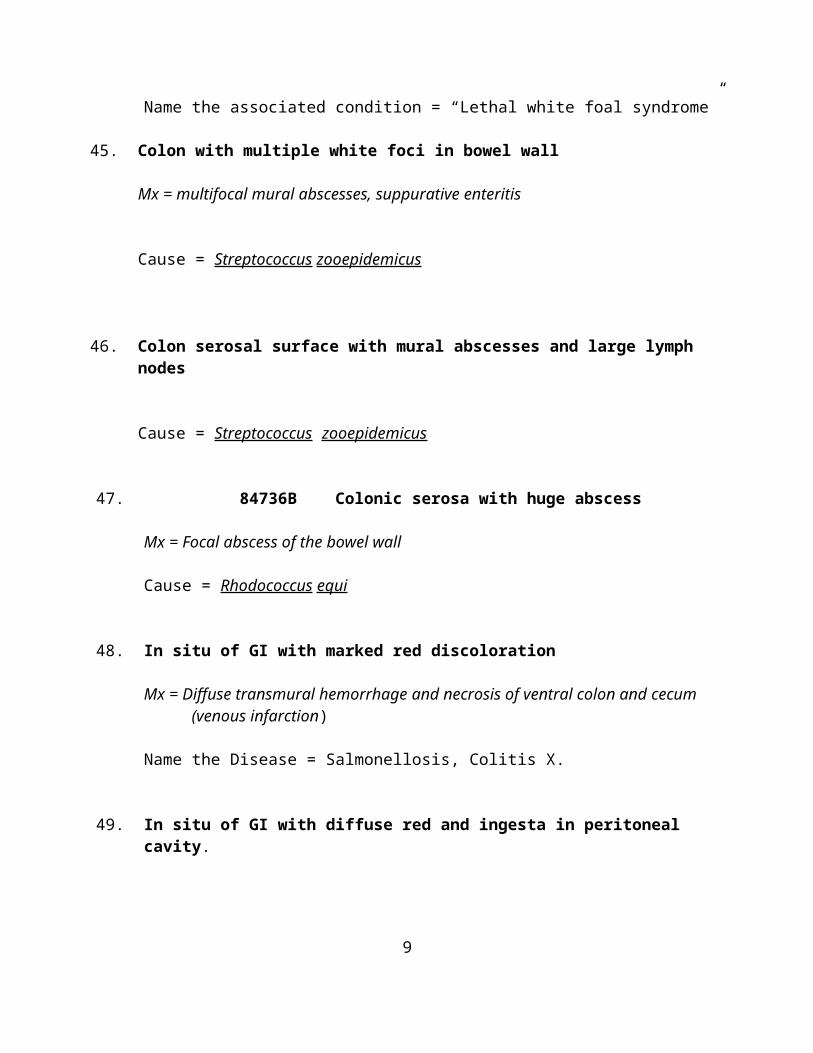
Name the associated condition = “Lethal white foal syndrome”
45. Colon with multiple white foci in bowel wall
Mx = multifocal mural abscesses, suppurative enteritis
Cause = Streptococcus zooepidemicus
46. Colon serosal surface with mural abscesses and large lymph nodes
Cause = Streptococcus zooepidemicus
47. 84736B Colonic serosa with huge abscess
Mx = Focal abscess of the bowel wall
Cause = Rhodococcus equi
48. In situ of GI with marked red discoloration
Mx = Diffuse transmural hemorrhage and necrosis of ventral colon and cecum (venous infarction)
7
Name the Disease = Salmonellosis, Colitis X.
49. In situ of GI with diffuse red and ingesta in peritoneal cavity.
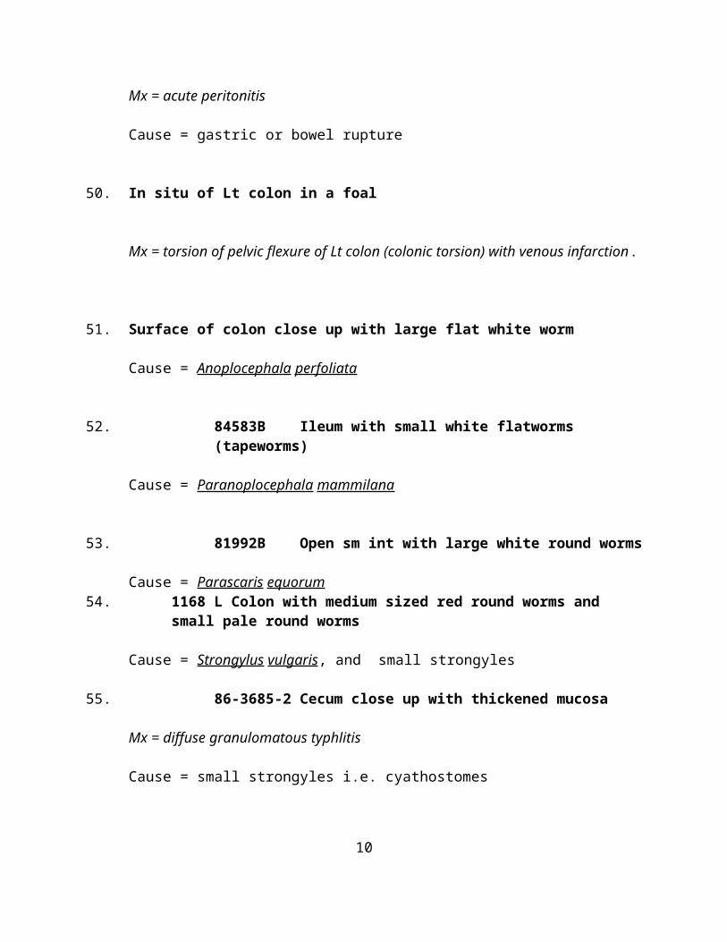
Mx = acute peritonitis
Cause = gastric or bowel rupture
50. In situ of Lt colon in a foal
Mx = torsion of pelvic flexure of Lt colon (colonic torsion) with venous infarction.
51. Surface of colon close up with large flat white worm
Cause = Anoplocephala perfoliata
52. 84583B Ileum with small white flatworms (tapeworms)
Cause = Paranoplocephala mammilana
53. 81992B Open sm int with large white round worms
Cause = Parascaris equorum54. 1168 L Colon with medium sized red round worms and small pale round
worms
Cause = Strongylus vulgaris, and small strongyles
55. 86-3685-2 Cecum close up with thickened mucosa
Mx = diffuse granulomatous typhlitis
Cause = small strongyles i.e. cyathostomes
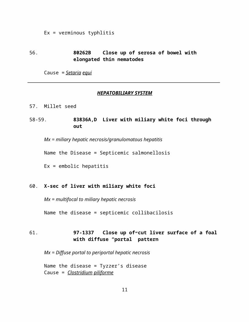
Ex = verminous typhlitis
8
56. 80262B Close up of serosa of bowel with elongated thin nematodes
Cause = Setaria equi
HEPATOBILIARY SYSTEM
57. Millet seed
58-59. 83836A,D Liver with miliary white foci through out
Mx = miliary hepatic necrosis/granulomatous hepatitis
Name the Disease = Septicemic salmonellosis
Ex = embolic hepatitis
60. X-sec of liver with miliary white foci
Mx = multifocal to miliary hepatic necrosis
Name the disease = septicemic collibacilosis
61. 97-1337 Close up of cut liver surface of a foal with diffuse “portal” pattern
Mx = Diffuse portal to periportal hepatic necrosis
Name the disease = Tyzzer’s diseaseCause = Clostridium piliforme
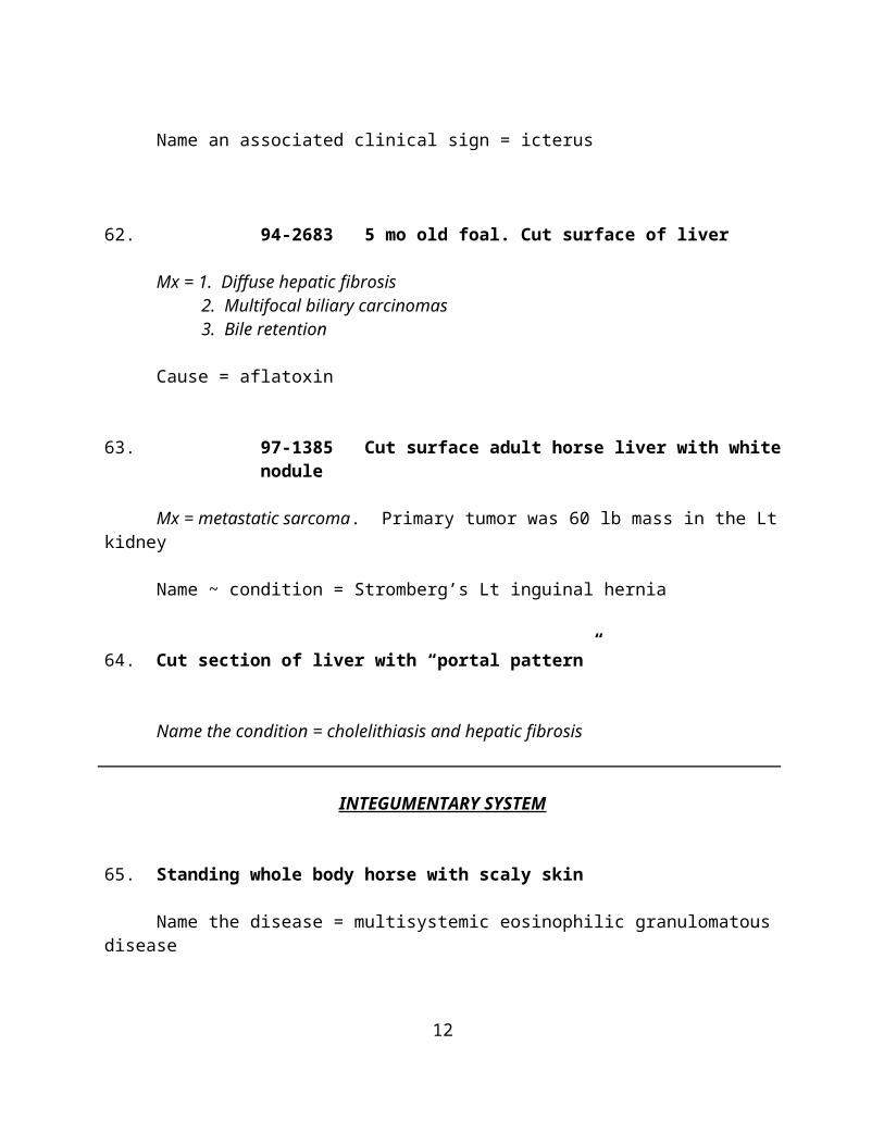
Name an associated clinical sign = icterus
62. 94-2683 5 mo old foal. Cut surface of liver
Mx = 1. Diffuse hepatic fibrosis 2. Multifocal biliary carcinomas 3. Bile retention
Cause = aflatoxin
9
63. 97-1385 Cut surface adult horse liver with white nodule
Mx = metastatic sarcoma. Primary tumor was 60 lb mass in the Lt kidney
Name ~ condition = Stromberg’s Lt inguinal hernia
64. Cut section of liver with “portal pattern”
Name the condition = cholelithiasis and hepatic fibrosis
INTEGUMENTARY SYSTEM
65. Standing whole body horse with scaly skin
Name the disease = multisystemic eosinophilic granulomatous disease
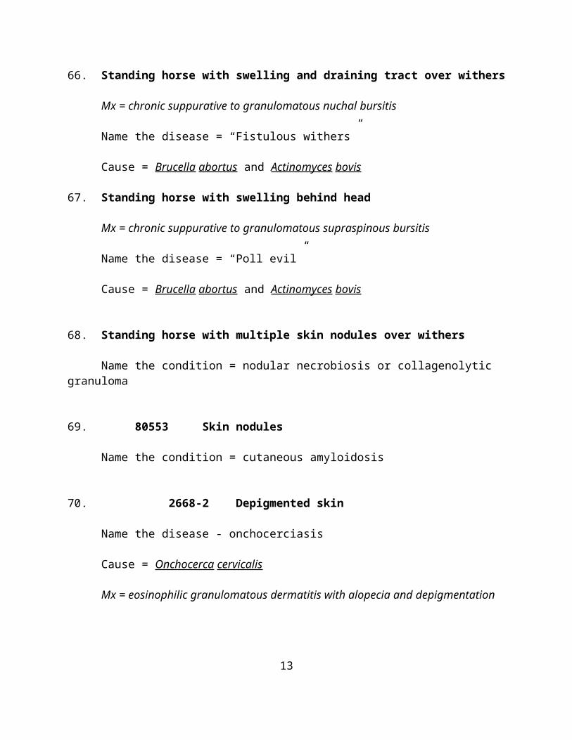
66. Standing horse with swelling and draining tract over withers
Mx = chronic suppurative to granulomatous nuchal bursitis
Name the disease = “Fistulous withers”
Cause = Brucella abortus and Actinomyces bovis
67. Standing horse with swelling behind head
Mx = chronic suppurative to granulomatous supraspinous bursitis
Name the disease = “Poll evil”
Cause = Brucella abortus and Actinomyces bovis
68. Standing horse with multiple skin nodules over withers
Name the condition = nodular necrobiosis or collagenolytic granuloma
69. 80553 Skin nodules
10
Name the condition = cutaneous amyloidosis
70. 2668-2 Depigmented skin
Name the disease - onchocerciasis
Cause = Onchocerca cervicalis
Mx = eosinophilic granulomatous dermatitis with alopecia and depigmentation
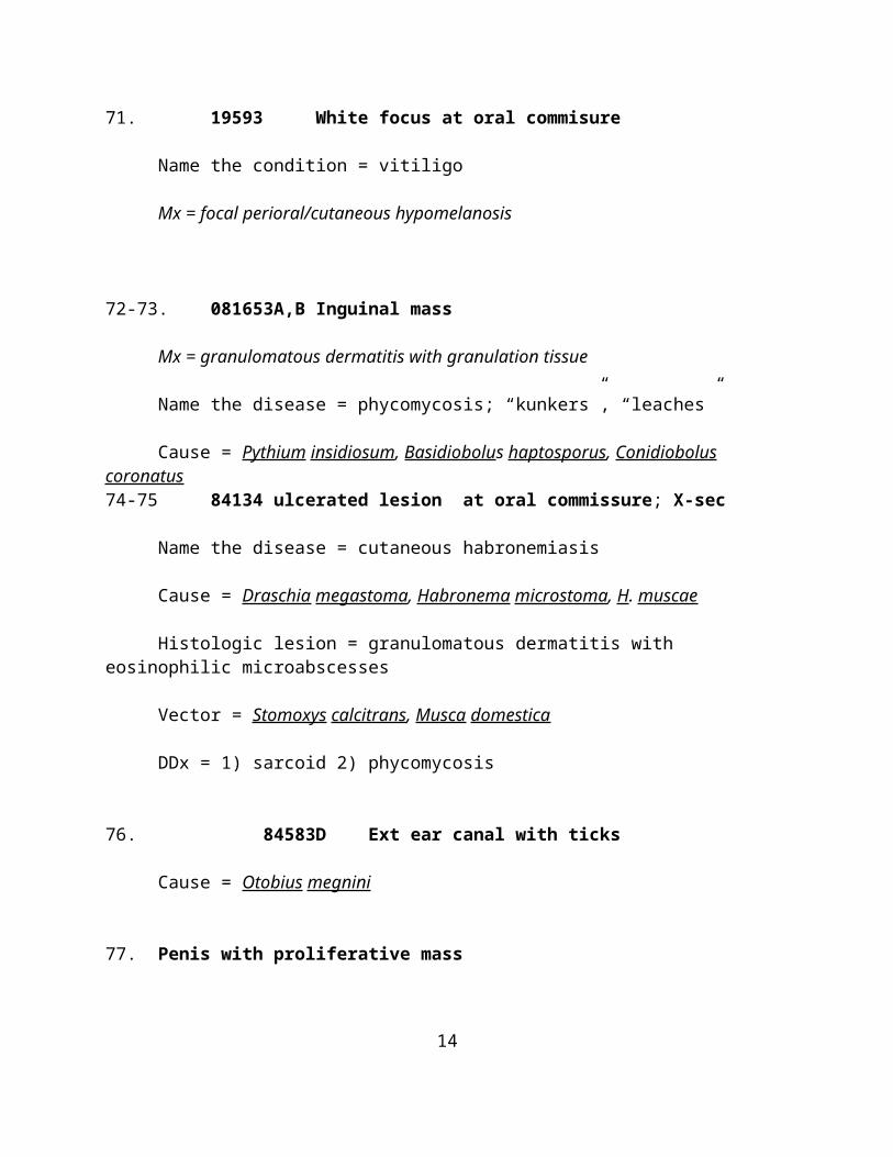
71. 19593 White focus at oral commisure
Name the condition = vitiligo
Mx = focal perioral/cutaneous hypomelanosis
72-73. 081653A,B Inguinal mass
Mx = granulomatous dermatitis with granulation tissue
Name the disease = phycomycosis; “kunkers”, “leaches”
Cause = Pythium insidiosum, Basidiobolus haptosporus, Conidiobolus coronatus74-75 84134 ulcerated lesion at oral commissure; X-sec
Name the disease = cutaneous habronemiasis
Cause = Draschia megastoma, Habronema microstoma, H. muscae
Histologic lesion = granulomatous dermatitis with eosinophilic microabscesses
Vector = Stomoxys calcitrans, Musca domestica
DDx = 1) sarcoid 2) phycomycosis
76. 84583D Ext ear canal with ticks
Cause = Otobius megnini
11
77. Penis with proliferative mass
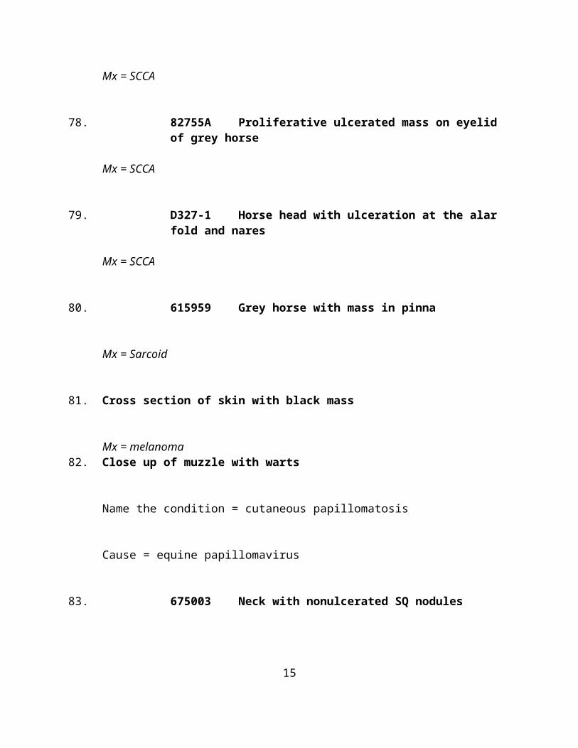
Mx = SCCA
78. 82755A Proliferative ulcerated mass on eyelid of grey horse
Mx = SCCA
79. D327-1 Horse head with ulceration at the alar fold and nares
Mx = SCCA
80. 615959 Grey horse with mass in pinna
Mx = Sarcoid
81. Cross section of skin with black mass
Mx = melanoma82. Close up of muzzle with warts
Name the condition = cutaneous papillomatosis
Cause = equine papillomavirus
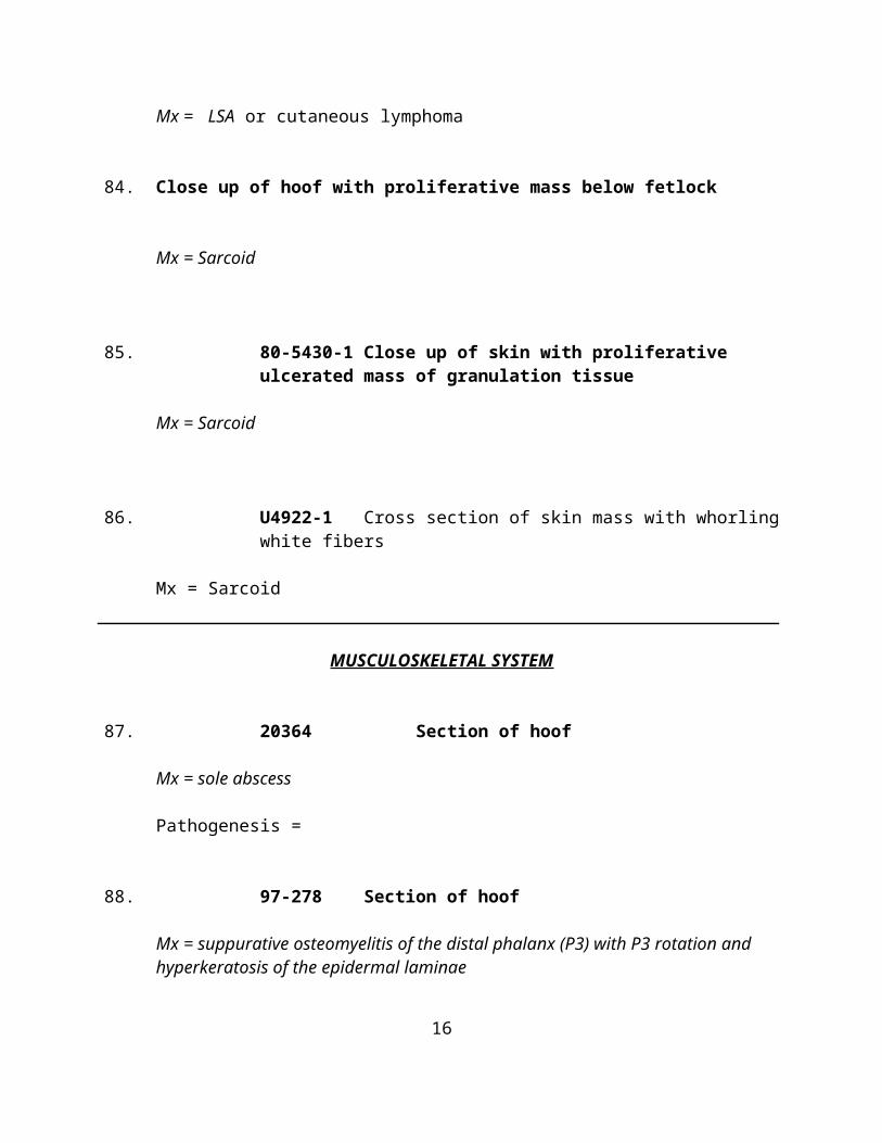
83. 675003 Neck with nonulcerated SQ nodules
Mx = LSA or cutaneous lymphoma
84. Close up of hoof with proliferative mass below fetlock
Mx = Sarcoid
12
85. 80-5430-1 Close up of skin with proliferative ulcerated mass of granulation tissue
Mx = Sarcoid
86. U4922-1 Cross section of skin mass with whorling white fibers
Mx = Sarcoid
MUSCULOSKELETAL SYSTEM
87. 20364 Section of hoof
Mx = sole abscess
Pathogenesis =
88. 97-278 Section of hoof
Mx = suppurative osteomyelitis of the distal phalanx (P3) with P3 rotation and hyperkeratosis of the epidermal laminae
Name the Condition = chronic laminitis
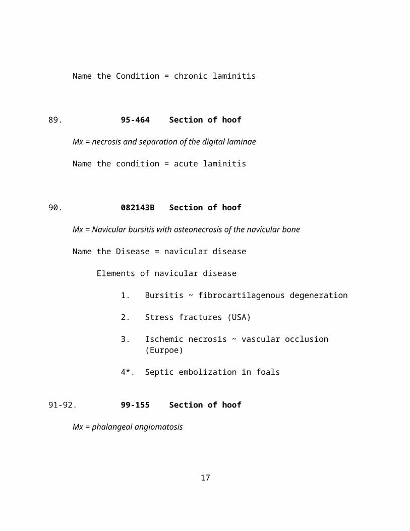
89. 95-464 Section of hoof
Mx = necrosis and separation of the digital laminae
Name the condition = acute laminitis
90. 082143B Section of hoof
13
Mx = Navicular bursitis with osteonecrosis of the navicular bone
Name the Disease = navicular disease
Elements of navicular disease
1. Bursitis ~ fibrocartilagenous degeneration
2. Stress fractures (USA)
3. Ischemic necrosis ~ vascular occlusion (Eurpoe)
4*. Septic embolization in foals
91-92. 99-155 Section of hoof
Mx = phalangeal angiomatosis
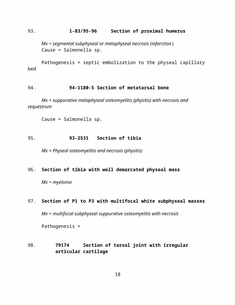
93. 1-83/95-96 Section of proximal humerus
Mx = segmental subphyseal or metaphyseal necrosis (infarction)Cause = Salmonella sp.
Pathogenesis = septic embolization to the physeal capillary bed
94. 94-1180-5 Section of metatarsal bone
Mx = suppurative metaphyseal osteomyelitis (physitis) with necrosis and sequestrum
Cause = Salmonella sp.
95. 93-2531 Section of tibia
Mx = Physeal osteomyelitis and necrosis (physitis)
96. Section of tibia with well demarcated physeal mass
Mx = myeloma
14
97. Section of P1 to P3 with multifocal white subphyseal masses
Mx = multifocal subphyseal suppurative osteomyelitis with necrosis
Pathogenesis =
98. 79174 Section of tarsal joint with irregular articular cartilage
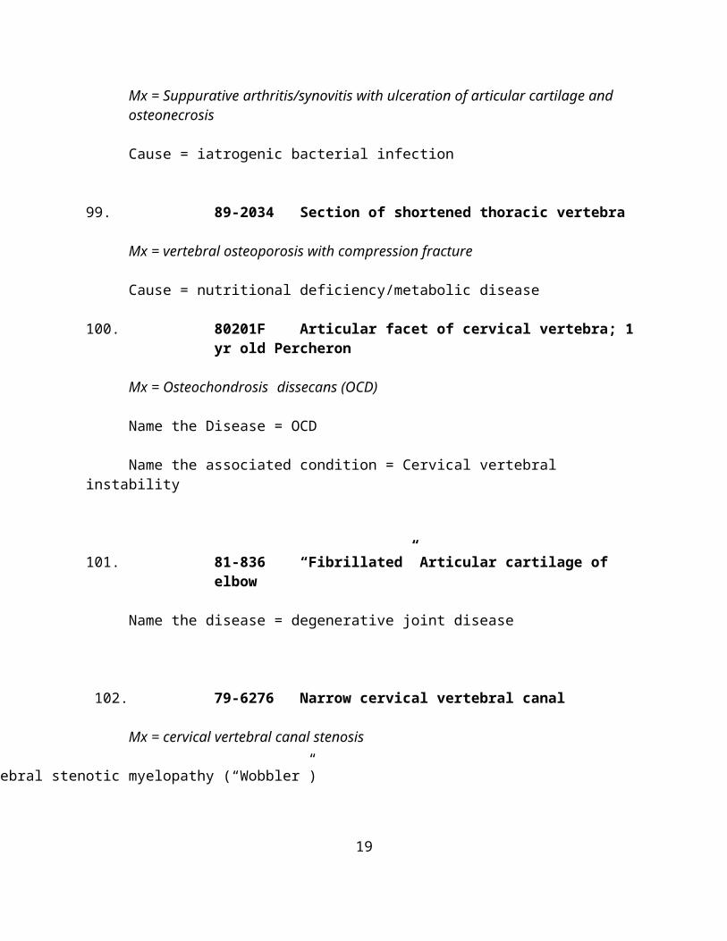
Mx = Suppurative arthritis/synovitis with ulceration of articular cartilage and osteonecrosis
Cause = iatrogenic bacterial infection
99. 89-2034 Section of shortened thoracic vertebra
Mx = vertebral osteoporosis with compression fracture
Cause = nutritional deficiency/metabolic disease
100. 80201F Articular facet of cervical vertebra; 1 yr old Percheron
Mx = Osteochondrosis dissecans (OCD)
Name the Disease = OCD
Name the associated condition = Cervical vertebral instability
101. 81-836 “Fibrillated” Articular cartilage of elbow
Name the disease = degenerative joint disease
102. 79-6276 Narrow cervical vertebral canal
Mx = cervical vertebral canal stenosis
Name the associated condition = cervical vertebral stenotic myelopathy (“Wobbler”)
15
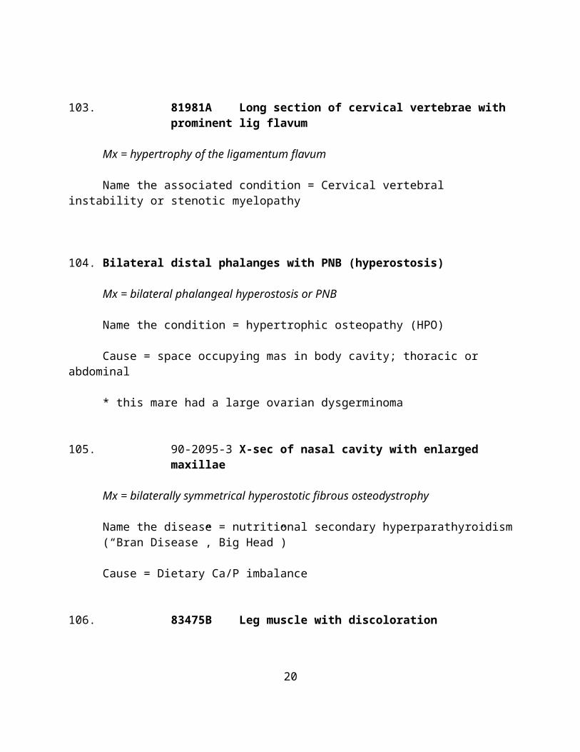
103. 81981A Long section of cervical vertebrae with prominent lig flavum
Mx = hypertrophy of the ligamentum flavum
Name the associated condition = Cervical vertebral instability or stenotic myelopathy
104. Bilateral distal phalanges with PNB (hyperostosis)
Mx = bilateral phalangeal hyperostosis or PNB
Name the condition = hypertrophic osteopathy (HPO)
Cause = space occupying mas in body cavity; thoracic or abdominal
* this mare had a large ovarian dysgerminoma
105. 90-2095-3 X-sec of nasal cavity with enlarged maxillae
Mx = bilaterally symmetrical hyperostotic fibrous osteodystrophy
Name the disease = nutritional secondary hyperparathyroidism (“Bran Disease”, Big Head”)
Cause = Dietary Ca/P imbalance
106. 83475B Leg muscle with discoloration
Mx = myonecrosis with emphysema
Cause = Clostridium novyi
Etiologic Dx = clostridial myositis
NERVOUS SYSTEM
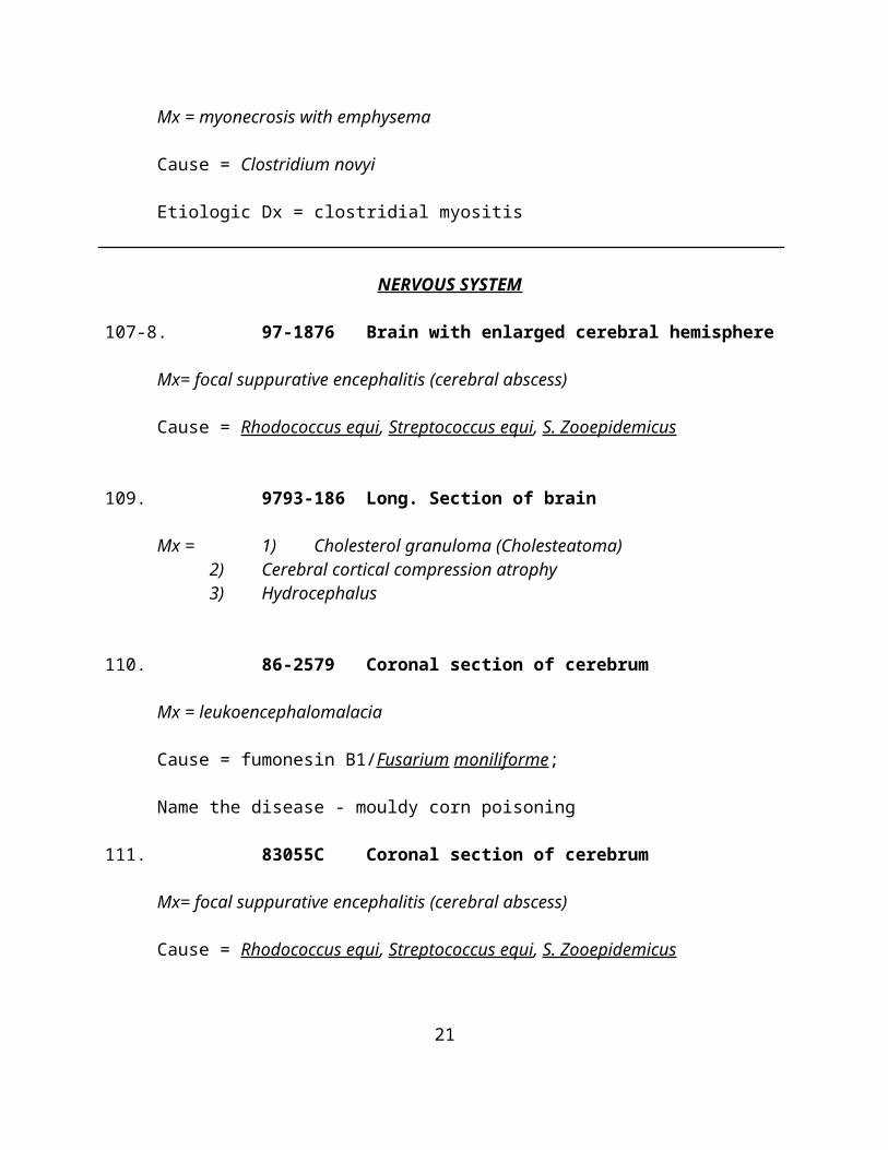
107-8. 97-1876 Brain with enlarged cerebral hemisphere
Mx= focal suppurative encephalitis (cerebral abscess)
16
Cause = Rhodococcus equi, Streptococcus equi, S. Zooepidemicus
109. 9793-186 Long. Section of brain
Mx = 1) Cholesterol granuloma (Cholesteatoma)2) Cerebral cortical compression atrophy3) Hydrocephalus
110. 86-2579 Coronal section of cerebrum
Mx = leukoencephalomalacia
Cause = fumonesin B1/Fusarium moniliforme;
Name the disease - mouldy corn poisoning
111. 83055C Coronal section of cerebrum
Mx= focal suppurative encephalitis (cerebral abscess)
Cause = Rhodococcus equi, Streptococcus equi, S. Zooepidemicus
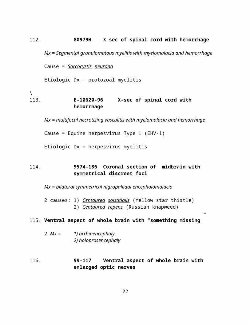
112. 80979H X-sec of spinal cord with hemorrhage
Mx = Segmental granulomatous myelitis with myelomalacia and hemorrhage
Cause = Sarcocystis neurona
Etiologic Dx - protozoal myelitis
\113. E-10620-96 X-sec of spinal cord with hemorrhage
Mx = multifocal necrotizing vasculitis with myelomalacia and hemorrhage
Cause = Equine herpesvirus Type 1 (EHV-1)
Etiologic Dx = herpesvirus myelitis
114. 9574-186 Coronal section of midbrain with symmetrical discreet foci
17
Mx = bilateral symmetrical nigropallidal encephalomalacia
2 causes: 1) Centaurea solstitialis (Yellow star thistle)2) Centaurea repens (Russian knapweed)
115. Ventral aspect of whole brain with “something missing”
2 Mx = 1) arrhinencephaly2) holoprosencephaly
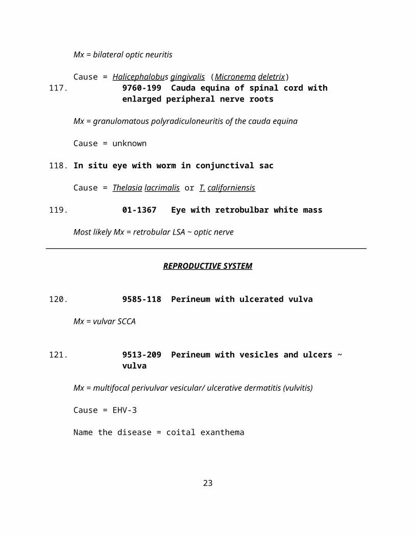
116. 99-117 Ventral aspect of whole brain with enlarged optic nerves
Mx = bilateral optic neuritis
Cause = Halicephalobus gingivalis (Micronema deletrix)117. 9760-199 Cauda equina of spinal cord with enlarged peripheral
nerve roots
Mx = granulomatous polyradiculoneuritis of the cauda equina
Cause = unknown
118. In situ eye with worm in conjunctival sac
Cause = Thelasia lacrimalis or T. californiensis
119. 01-1367 Eye with retrobulbar white mass
Most likely Mx = retrobular LSA ~ optic nerve
REPRODUCTIVE SYSTEM
120. 9585-118 Perineum with ulcerated vulva
Mx = vulvar SCCA
121. 9513-209 Perineum with vesicles and ulcers ~ vulva
Mx = multifocal perivulvar vesicular/ ulcerative dermatitis (vulvitis)
18
Cause = EHV-3
Name the disease = coital exanthema
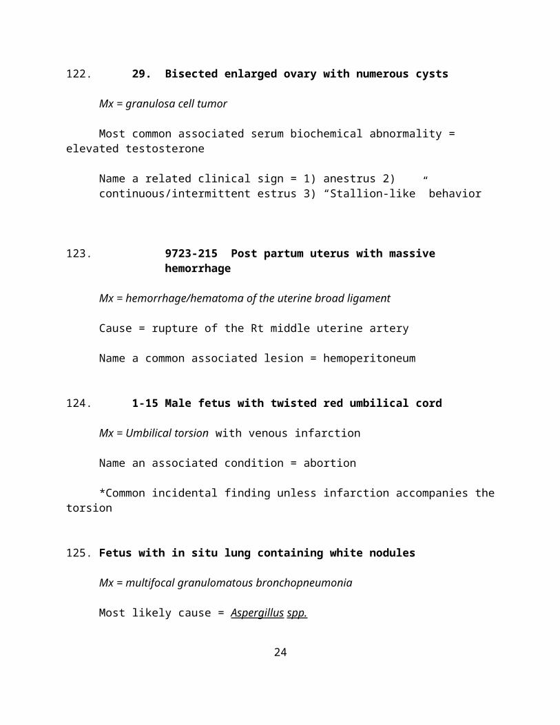
122. 29. Bisected enlarged ovary with numerous cysts
Mx = granulosa cell tumor
Most common associated serum biochemical abnormality = elevated testosterone
Name a related clinical sign = 1) anestrus 2) continuous/intermittent estrus 3) “Stallion-like” behavior
123. 9723-215 Post partum uterus with massive hemorrhage
Mx = hemorrhage/hematoma of the uterine broad ligament
Cause = rupture of the Rt middle uterine artery
Name a common associated lesion = hemoperitoneum
124. 1-15 Male fetus with twisted red umbilical cord
Mx = Umbilical torsion with venous infarction
Name an associated condition = abortion
*Common incidental finding unless infarction accompanies the torsion
125. Fetus with in situ lung containing white nodules
Mx = multifocal granulomatous bronchopneumonia
Most likely cause = Aspergillus spp.
Pathogenesis: open cervix placentitis amniotic fluid spread to fetal airways
126. Twin fetuses, at 7 mo.
19
Name the condition = chronic placental insufficiency (Twin pregnancy)
Features: 1) abortion ~ 7 mo2) one fetus dead, autolyzed3) viable fetus emaciated, pale, smaller than normal
127. 91-3099 Placenta with white stellate area
Name the structure = cervical star
128. 84124 Placenta with linear flat white areas
Name the structure = endometrial cups
When do these occur? 45-150 days
129. 97-601-1 Placenta with rough surface
Mx = fibrinopurulent to necrotizing placentitis
130. 02-1315 Placenta with large white raised masses
Mx = Adenomatous (cystic) dysplasia/hyperplasia of the allantois
131-2 Uterus with pus
Mx = pyometra or suppurative endometritis
RESPIRATORY SYSTEM
133. 79-5391-7 Long. Sec of nasal cavity with white mass at ethmoid
Mx = adenocarcinoma of the ethmoturbinate
20
134. 9752-232 Long sec of nasal cavity with dark red swollen turbinate
Name the condition = progressive nasal hematoma/ethmoidal hematoma
Histopathologic appearance = angiomatosis with hemorrhage, hemosiderosis, granulation tissue, and edema
135. 80301B Coronal section of nasal cavity with white mass in sinus
Mx= maxillary sinus abscess (chronic suppurative sinusitis), periodontitis and FB
136. 86173 Coronal section of nasal cavity with white exudate
Mx = suppurative rhinitis
Cause = Streptococcus equi,
Name the Disease = “Strangles”
Name an associated condition = purpura hemorrhagica
137. 78948 Polyp from the nasal cavity
Mx = mucosal or inflammatory polyp/ granulomatous rhinitis with granulation tissue
Cause = Rhinosporidium seeberi
138. 426 en face view of epiglottis with caudal displacement of epiglottis
Name the condition = entrapment of the epiglottis
Name the underlying condition = epiglottal hypoplasia
139. En face view of the asymmetrical epiglottis
21
Name the condition = laryngeal hemiplegia
140. 86-3472-1 Larynx, asymmetrical pale color
Mx = atrophy of the Lt cricoarytenoideus dorsalis m.
Cause = axonal degeneration of the Lt recurrent laryngeal nerve
141. 78520A Gutteral Pouch with amorphous white “stuff”
Mx = chronic suppurative gutteral diverticulitis (gutteral empyema)“Lufttsackstein”
Cause = Streptococcus equi
142. 84772C Gutteral pouch mucosa, discolored
Mx = granulomatous gutteral diverticulitis
Cause = **Pythium insidiosum, Aspergillus spp.
143. W2188-1 Trachea with yellow cast
Mx = fibrinous tracheitis
Cause -= Equine herpesvirus type 1 (Equine Rhinopneumonitis Virus)
144. Lung, adult with A-V white nodules
Mx = chronic suppurartive bronchopneumonia (pulmonary abscesses)
Cause = Rhodococcus equi
145. 84197D Lung, Arab foal with AV pattern, bulla
Mx = Bronchiolitis/ bronchopneumonia with subpleural emphysema
Cause = equine adenovirus
22
What is the underlying condition = Combined Immunodeficiency Disease
146. Lung, foal with miliary white nodules
Mx = miliary pyogranulomatous pneumonia
Cause = Rhodococcus equi147. 88-306-3 Cut surface of lung, neonate with miliary white foci
Mx = miliary exudative (suppurative) interstitial pneumonia
Cause = E. coli
Etiologic Dx = suppurative (septic) embolic pneumonia
Name the Disease = septicemic collibacillosis
148. 83294B Lung with poorly defined pale foci
Mx = chronic mucoid bronchiolitis
Name the Disease = Chronic Obstructive Pulmonary Disease
Name the associated clinical condition = “Heaves”
149. 82002D Lung with extensive hilar pattern
Mx = Pulmonary edema
150. 99-1583 In situ pleura, foal
Mx = Diffuse fibrinous pleuritis
Cause - E. coli.
151. 81980C Close up of fetal/neonatal lung with yellow foci
Name the condition = mecomium aspiration
23
152. 91-937 Lung with intraluminal bronchial mass
Mx = Granular cell tumor
153. 95-691 Thorax and abdomen, foal, in situ
Mx = diaphragmatic hernia with displacement of the colon into the thorax.
URINARY SYSTEM
154-5. 92-3944 Kidney, whole and section, with white foci
Mx = miliary suppurative nephritis
Etiologic Dx = suppurative embolic nephritis/ septic embolic nephritis
Cause = Actinobacillus equuili
156. RKH10 Section of neonatal kidney colored olive green
Mx = cholemic nephrosis
Name the condition = hepatorenal syndrome
Pathogenesis: Immature or obstructed liver conjugation of bilirubin unconjugated bilirubin hyperbilirubinemia bilirubinuria reabsorption by pct epithelium
157. 7137 Ureter
Mx = hemorrhagic ureteritis
Name the disease: Blister Beetle Poisoning/ cantharidin toxicity
Name an associated lesion in another organ system = 1) myocardial necrosis 2) hemorrhagic gastritis
24
158. Epicauta sp. (Blister Beetles)
159. MAR83-21 Urinary Bladder, open with opaque white material
Mx = chronic mucopurulent to fibrinopurulent cystitis
Cause: 1) Sorghum grass toxicity 2) Sudan Grass toxicity 3) Sarcocystis neurona
Pathogenesis: Damage to lumbar spinal cord or nerve roots bladder atony urine stasis
160. 02-783 Urinary bladder in neonate with rent in dorsal surface
Mx = cystorrhexis (ruptured urinary bladder)
Name a sequella = uroabdomen
Name 3 ~ clinicopathologic abnormalities
1. Hyponatremia2. Hypochloremia3. Hyperkalemia4. High peritoneal fluid/serum Cr ratio
25