Embed Size (px)
Citation preview

lable at ScienceDirect
Biomaterials 32 (2011) 3351e3357
Contents lists avai
Biomaterials
journal homepage: www.elsevier .com/locate/biomater ia ls
A self-assembling hydrophobically modified chitosan capable of reversiblehemostatic action
Matthew B. Dowling a,**, Rakesh Kumar b, Mark A. Keibler b, John R. Hess c, Grant V. Bochicchio d,Srinivasa R. Raghavan a,b,*
a Fischell Department of Bioengineering, University of Maryland, College Park, MD 20742, USAbDepartment of Chemical & Biomolecular Engineering, University of Maryland, College Park, MD 20742, USAcDepartment of Pathology, University of Maryland School of Medicine, Baltimore, MD 21201, USAdDepartment of Surgery, University of Maryland School of Medicine, Baltimore, MD 21201, USA
a r t i c l e i n f o
Article history:Received 29 November 2010Accepted 22 December 2010Available online 5 February 2011
Keywords:ChitosanHemostasisSelf-assemblyCyclodextrinAmphiphilic biopolymer
* Corresponding author. Department of ChemicalUniversity of Maryland, College Park, MD 20742, USA** Corresponding author.
E-mail addresses: [email protected] (M.B. D(S.R. Raghavan).
0142-9612/$ e see front matter � 2010 Elsevier Ltd.doi:10.1016/j.biomaterials.2010.12.033
a b s t r a c t
Blood loss at the site of a wound in mammals is curtailed by the rapid formation of a hemostatic plug, i.e.,a self-assembled network of the protein, fibrin that locally transforms liquid blood into a gelled clot.Here, we report an amphiphilic biopolymer that exhibits a similar ability to rapidly gel blood; moreover,the self-assembly underlying the gelation readily allows for reversibility back into the liquid state viaintroduction of a sugar-based supramolecule. The biopolymer is a hydrophobically modified (hm)derivative of the polysaccharide, chitosan. When hm-chitosan is contacted with heparinized humanblood, it rapidly transforms the liquid into an elastic gel. In contrast, the native chitosan (withouthydrophobes) does not gel blood. Gelation occurs because the hydrophobes on hm-chitosan insert intothe membranes of blood cells and thereby connect the cells into a sample-spanning network. Gelation isreversed by the addition of a-cyclodextrin, a supramolecule having an inner hydrophobic pocket:polymer hydrophobes unbind from blood cells and embed within the cyclodextrins, thereby disruptingthe cell network. We believe that hm-chitosan has the potential to serve as an effective, yet low-costhemostatic dressing for use by trauma centers and the military. Preliminary tests with small and largeanimal injury models show its increased efficacy at achieving hemostasis e e.g., a 90% reduction inbleeding time over controls for femoral vein transections in a rat model.
� 2010 Elsevier Ltd. All rights reserved.
1. Introduction
The blood coagulation cascade is an exquisite example ofa responsive self-assembly process in biology [1,2]. When awound isformed, a cascade of self-assemblyevents occurs inblood at the site ofthe wound. The net outcome is the assembly of the globular protein,fibrinogen, catalyzed by a second protein, thrombin to yield chains offibrin [3,4]. A network of insoluble fibrin chains forms the hemostatic“plug” or clot, which presents a physical barrier to the loss of bloodfromthewound [1,2]. The coagulation cascade is a delicately balancedseries of eventse if it was to occur too easily, blood clots may form inunwanted areas leading to strokes or other complications.
& Biomolecular Engineering,. Tel.: þ1 301 405 8164.
owling), [email protected]
All rights reserved.
Scientists have long sought toharness the clottingpoweroffibrinto create hemostatic dressings or bandages [5e7]. Hemostaticdressings that can staunch the bleeding from serious wounds area pressing need both in civilian trauma centers aswell as formilitarypersonnel. Indeed, uncontrolled hemorrhage from severe injuries isa leading cause of death among young adults (e.g., accident victims)and it is also responsible for themajority of deaths on the battlefield[8e10]. Fibrin-based hemostatic sealants were first-developed inthe 1940s and have proven to be quite effective [6,7]. For example,one form involves a dry powderedmixture of humanfibrinogen andthrombin packed onto a solid bandage backing. When sucha bandage is firmly pressed onto a bleeding injury, a strong fibrinseal quickly forms and bleeding is stopped [6]. However, fibrinbandages have limited practical applicability in trauma medicinebecause human fibrinogen and thrombin are highly expensivemolecules that are scarce in supply [7].
A need thus exists for an inexpensive hemostatic agent based onwidely available materials that could match the blood-clottingability of fibrin. Although a variety of advanced hemostats have
M.B. Dowling et al. / Biomaterials 32 (2011) 3351e33573352
been brought to market [5,11e15], none have shown the efficacy offibrin sealants [11,12]. Several products work simply by absorbingthe blood at the site of the wound rather than by actively coagu-lating the blood [12,14]. Recently, a new approach has been putforward by Ellis-Behnke et al. [16], wherein the self-assembly ofa synthetic peptide into a nano-fibrous network [17] is used toachieve hemostasis independent of the natural coagulationcascade. While this method is promising, the synthetic peptidesemployed are expensive and difficult to synthesize e thereforetheir practical viability is unclear. An additional factor to considerwith hemostats such as the above is the risk of undesired gelationor clotting, i.e., embolization, in parts of the body that are periph-eral to the site of injury [14,18]. To mitigate against such risks, itwould be desirable to have the hemostat disassemble or “unclot” ifand when desired; however, none of the hemostats described inthe literature have been shown to have this ability.
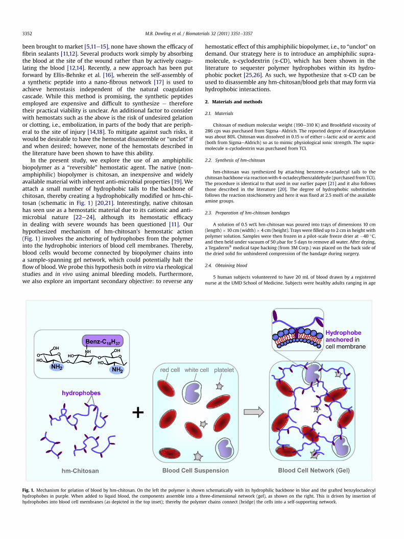
In the present study, we explore the use of an amphiphilicbiopolymer as a “reversible” hemostatic agent. The native (non-amphiphilic) biopolymer is chitosan, an inexpensive and widelyavailable material with inherent anti-microbial properties [19]. Weattach a small number of hydrophobic tails to the backbone ofchitosan, thereby creating a hydrophobically modified or hm-chi-tosan (schematic in Fig. 1) [20,21]. Interestingly, native chitosanhas seen use as a hemostatic material due to its cationic and anti-microbial nature [22e24], although its hemostatic efficacyin dealing with severe wounds has been questioned [11]. Ourhypothesized mechanism of hm-chitosan’s hemostatic action(Fig. 1) involves the anchoring of hydrophobes from the polymerinto the hydrophobic interiors of blood cell membranes. Thereby,blood cells would become connected by biopolymer chains intoa sample-spanning gel network, which could potentially halt theflow of blood. We probe this hypothesis both in vitro via rheologicalstudies and in vivo using animal bleeding models. Furthermore,we also explore an important secondary objective: to reverse any
Fig. 1. Mechanism for gelation of blood by hm-chitosan. On the left the polymer is shownhydrophobes in purple. When added to liquid blood, the components assemble into a thhydrophobes into blood cell membranes (as depicted in the top inset); thereby the polyme
hemostatic effect of this amphiphilic biopolymer, i.e., to “unclot” ondemand. Our strategy here is to introduce an amphiphilic supra-molecule, a-cyclodextrin (a-CD), which has been shown in theliterature to sequester polymer hydrophobes within its hydro-phobic pocket [25,26]. As such, we hypothesize that a-CD can beused to disassemble any hm-chitosan/blood gels that may form viahydrophobic interactions.
2. Materials and methods
2.1. Materials
Chitosan of medium molecular weight (190e310 K) and Brookfield viscosity of286 cps was purchased from SigmaeAldrich. The reported degree of deacetylationwas about 80%. Chitosan was dissolved in 0.15 M of either L-lactic acid or acetic acid(both from SigmaeAldrich) so as to mimic physiological ionic strength. The supra-molecule a-cyclodextrin was purchased from TCI.
2.2. Synthesis of hm-chitosan
hm-chitosan was synthesized by attaching benzene-n-octadecyl tails to thechitosan backbone via reactionwith 4-octadecylbenzaldehyde (purchased fromTCI).The procedure is identical to that used in our earlier paper [21] and it also followsthose described in the literature [20]. The degree of hydrophobic substitutionfollows the reaction stoichiometry and here it was fixed at 2.5 mol% of the availableamine groups.
2.3. Preparation of hm-chitosan bandages
A solution of 0.5 wt% hm-chitosan was poured into trays of dimensions 10 cm(length)� 10 cm (width)� 4 cm (height). Trays were filled up to 2 cm in height withpolymer solution. Samples were then frozen in a pilot-scale freeze drier at �40 �C,and then held under vacuum of 50 mbar for 5 days to remove all water. After drying,a Tegaderm� medical tape backing (from 3M Corp.) was placed on the back side ofthe dried solid for unhindered compression of the bandage during surgery.
2.4. Obtaining blood
5 human subjects volunteered to have 20 mL of blood drawn by a registerednurse at the UMD School of Medicine. Subjects were healthy adults ranging in age
schematically with its hydrophilic backbone in blue and the grafted benzyloctadecylree-dimensional network (gel), as shown on the right. This is driven by insertion ofr chains connect (bridge) the cells into a self-supporting network.
M.B. Dowling et al. / Biomaterials 32 (2011) 3351e3357 3353
from 20 to 40 years (4 males, 1 female). 10 mL intervals of blood were drawn intoBecton Dickinson Vacutainers� containing 143 USP units of sodium heparin. Theprotocol was approved by the Institutional Research Board (IRB) at UMD.
2.5. Rheological experiments
Steady and dynamic rheological experiments were performed on a RheometricsAR2000 stress-controlled rheometer. A cone-and-plate geometry of 40mmdiameterand 4� cone anglewas used and sampleswere run at the physiological temperature of37 �C. Dynamic frequency spectra were obtained in the linear viscoelastic regime ofthe samples, as determined from dynamic strain sweep experiments.
2.6. Rat injury models
Surgical procedures were approved by the Institutional Animal Care and UseCommittee (IACUC) at UMD. 15 fasted male Long-Evans rats (250e275 g, fromHarlan Laboratories) were anesthetized (60 mg/kg ketamine and 7.5 mg/kg xylazinegiven IP) and allowed to breathe air. Animals were maintained under pathogen-freeconditions in 12 h diurnal cycles, with water and food ad libitum. Animal roomswerekept at 21 � 3 �C with several changes of air per hour. All husbandry and animalprocedures were in accordance with humane animal handling practices under theguidance of the Unit for Laboratory Animal Medicine at the UMD School of Medicine.At the end of each procedure, all animals were humanely sacrificed by ketamineadministration.
Using a scalpel, the femoral vein was transected and allowed to bleed for 30 s,after a unilateral groin incision was made over the femoral canal. Exposure andisolation of at least 1 cm of the femoral vein was performed. 1 mL of test materialwas then dispensed onto 5 randomly selected animals via syringe (with a 22 gaugeneedle) after wiping away excess blood from the site of injury via cotton gauze.Bleeding timewasmeasured via stopwatch, with the start time corresponding to theapplication of sample and the end time corresponding to visual observation ofhalted blood flow. Test materials studied were (1) saline buffer, (2) 0.5 wt% chitosansolution and (3) 0.5 wt% hm-chitosan solution.
2.7. Pig injury models
3 Yorkshire crossbred swine, age 2.5 months and weighing 39.6e42.8 kg, wereused following screening by a veterinarian. Animals were allowed free access towater and to commercial food, which was withheld the night before the study. Allanimals were maintained in a facility accredited by the Association for Assessmentand Accreditation of Laboratory Animal Care, and all experiments were performed inaccordance with the National Research Council’s Guide for the Care and Use ofLaboratory Animals. The protocol was approved by the IACUC at UMD. The swinewere anesthetized with 6 mg/kg of telazol and 0.01 mg/kg of glycopyrrolate that
hm-chitosan
chitosan +
Frequency, ω (rad/s)10-1 100 101
G',
G" (
Pa)
100
101
102G”
G’
Dynamic Rheologya
hm-chitosan + blood
G”
G’
chitosan + blood
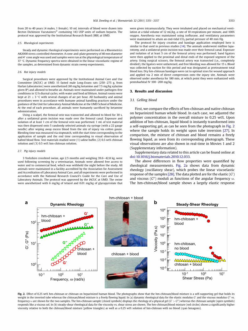
Fig. 2. Effect of 0.25 wt% hm-chitosan or chitosan on heparinized human blood. The photogweight in the inverted tube whereas the chitosan/blood mixture is a freely flowing liquid. Infrequency u are shown for the two samples. The hm-chitosan sample (closed symbols) displaresponds like a viscous sol. In (b) steady-shear rheological data for the viscosity vs. shear streviscosity relative to both the chitosan/blood mixture (yellow triangles) as well as a 0.25 w
were given intramuscularly. They were intubated and placed on mechanical venti-lation at a tidal volume of 12 mL/kg, a rate of 10 respirations per minute, and 100%oxygen. Anesthesia was maintained using isoflurane, and ventilatory parameterswere maintained to attain an end-tidal CO2 partial pressure of 40 mm Hg.
The protocol for injury creation and bandage application employed here issimilar to that used in previous studies [14]. The animals underwent midline lapa-rotomy, and a unilateral groin incision was made over their femoral canal. Exposureand isolation of at least 5 cm of the femoral artery was performed; band ligatorswere then applied to the proximal and distal ends of the exposed segment of theartery. Using surgical scissors, the femoral artery was transected (i.e., completelydivided), the ligators were unfastened, and free bleeding was allowed for 15 s. Bloodwas collected by suction for this period and was designated as pretreatment totalblood loss. Dry hm-chitosan bandages were cut to optimal sizes for wound approachand applied via 2 min of direct compression onto the injury site. Animals wereobserved under anesthesia for 180 min, at which point they were euthanized withpentobarbital IV 100e200 mg/kg.
3. Results and discussion
3.1. Gelling blood
First, we compare the effects of hm-chitosan and native chitosanon heparinized human whole blood. In each case, we adjusted thepolymer concentration in the overall mixture to 0.25 wt%. Uponaddition of hm-chitosan, liquid blood is instantly transformed intoa self-supporting gel, as can be seen from the photograph in Fig. 2where the sample holds its weight upon tube inversion [27]. Incomparison, the mixture of chitosan and blood remains a freelyflowing liquid, as seen from its corresponding photograph. Thesevisual observations are also shown in real-time in Movies 1 and 2(Supplementary information).
Supplementary data related to this article can be found online atdoi:10.1016/j.biomaterials.2010.12.033.
The above differences in flow properties were quantified byrheological measurements. Fig. 2a shows data from dynamicrheology (oscillatory shear), which probes the linear viscoelasticresponse of the samples [28]. The data plotted are for the elastic (G0)and viscous (G00) moduli as functions of the angular frequency u.The hm-chitosan/blood sample shows a largely elastic response
+ blood
blood
b
Shear Stress (Pa)10-1 100 101
Visc
osity
(Pa.
s)
10-3
10-2
10-1
100
101
102
103
104
Steady-Shear Rheology
hm-chitosan + blood
chitosan + blood
hm-chitosan, no blood
raphs show that the hm-chitosan/blood mixture is a self-supporting gel that holds its(a) dynamic rheological data for the elastic modulus G0 and the viscous modulus G00 vs.ys the rheology of a physical gel (G0 > G00) whereas the chitosan sample (open symbols)ss are shown. The hm-chitosan/blood mixture (red circles) shows a significantly highert% solution of hm-chitosan with no blood (cyan hexagons).
M.B. Dowling et al. / Biomaterials 32 (2011) 3351e33573354
typical of a physical gel: note that its G0 exceeds G00 over the entirefrequency range and moreover, both moduli are weakly dependenton frequency. The weak frequency dependence of the moduli isindicative of a sample-spanning network that is able to store theshear deformation [28]; relaxation of this network occurs veryslowly over long time scales. Conversely, the chitosan/blood sampleshows a viscous response: both its moduli decrease sharply withfrequency and its G00 exceeds G0 over the frequency range.
Data on the same samples via steady-shear rheology are shownin Fig. 2b, where the apparent viscosity is plotted as a function ofshear stress. Here, we note that the chitosan/blood sample hasa constant viscosity of about 0.01 Pa.s, which is about 4 times that ofblood alone. A 0.25 wt% solution of hm-chitosan in water hasa viscosity of about 0.07 Pa.s in the low-shear limit, indicating thatthe sample is slightly viscous but far from being a gel. In contrast,the sample of hm-chitosan/blood has a low-shear viscosity around10,000 Pa.s, which is amillion-fold higher than that of blood. Also, inthis case, the steep drop in viscosity around a stress of 2 Pa isindicative of a yield stress [28], meaning that the sample hardlyflows at stresses below this value. This accounts for the gel-likebehavior seen in the photograph where the sample holds its weightand does not flow down in the inverted tube [27,28].
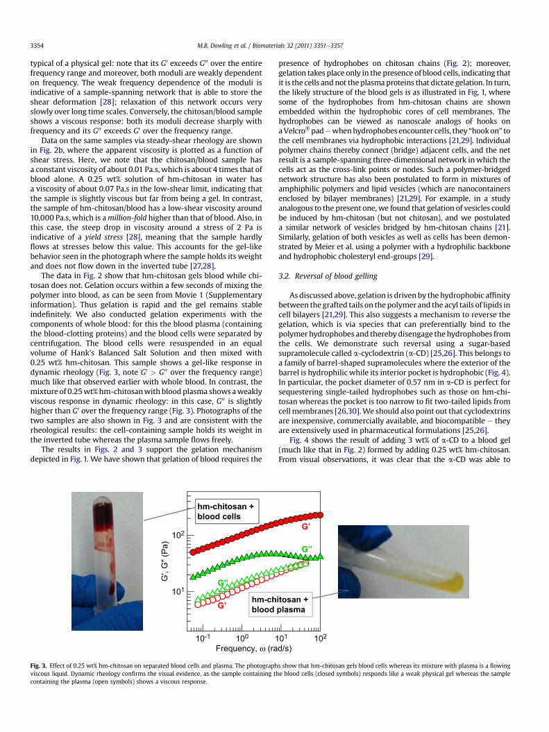
The data in Fig. 2 show that hm-chitosan gels blood while chi-tosan does not. Gelation occurs within a few seconds of mixing thepolymer into blood, as can be seen from Movie 1 (Supplementaryinformation). Thus gelation is rapid and the gel remains stableindefinitely. We also conducted gelation experiments with thecomponents of whole blood: for this the blood plasma (containingthe blood-clotting proteins) and the blood cells were separated bycentrifugation. The blood cells were resuspended in an equalvolume of Hank’s Balanced Salt Solution and then mixed with0.25 wt% hm-chitosan. This sample shows a gel-like response indynamic rheology (Fig. 3, note G0 > G00 over the frequency range)much like that observed earlier with whole blood. In contrast, themixture of 0.25wt%hm-chitosanwith blood plasma shows aweaklyviscous response in dynamic rheology: in this case, G00 is slightlyhigher than G0 over the frequency range (Fig. 3). Photographs of thetwo samples are also shown in Fig. 3 and are consistent with therheological results: the cell-containing sample holds its weight inthe inverted tube whereas the plasma sample flows freely.
The results in Figs. 2 and 3 support the gelation mechanismdepicted in Fig. 1. We have shown that gelation of blood requires the
Frequency, ω (ra10-1 100 1
G',
G" (
Pa)
101
102
G”
G’
hm-ch
blood
hm-chitosan +
blood cells
Fig. 3. Effect of 0.25 wt% hm-chitosan on separated blood cells and plasma. The photographviscous liquid. Dynamic rheology confirms the visual evidence, as the sample containing thcontaining the plasma (open symbols) shows a viscous response.
presence of hydrophobes on chitosan chains (Fig. 2); moreover,gelation takes place only in the presence of blood cells, indicating thatit is the cells andnot the plasmaproteins that dictate gelation. In turn,the likely structure of the blood gels is as illustrated in Fig. 1, wheresome of the hydrophobes from hm-chitosan chains are shownembedded within the hydrophobic cores of cell membranes. Thehydrophobes can be viewed as nanoscale analogs of hooks onaVelcro�padewhenhydrophobes encounter cells, they “hookon” tothe cell membranes via hydrophobic interactions [21,29]. Individualpolymer chains thereby connect (bridge) adjacent cells, and the netresult is a sample-spanning three-dimensional network inwhich thecells act as the cross-link points or nodes. Such a polymer-bridgednetwork structure has also been postulated to form in mixtures ofamphiphilic polymers and lipid vesicles (which are nanocontainersenclosed by bilayer membranes) [21,29]. For example, in a studyanalogous to the present one, we found that gelation of vesicles couldbe induced by hm-chitosan (but not chitosan), and we postulateda similar network of vesicles bridged by hm-chitosan chains [21].Similarly, gelation of both vesicles as well as cells has been demon-strated by Meier et al. using a polymer with a hydrophilic backboneand hydrophobic cholesteryl end-groups [29].
3.2. Reversal of blood gelling
As discussed above, gelation is driven by thehydrophobic affinitybetween thegrafted tails on thepolymerand the acyl tails of lipids incell bilayers [21,29]. This also suggests a mechanism to reverse thegelation, which is via species that can preferentially bind to thepolymerhydrophobes and therebydisengage thehydrophobes fromthe cells. We demonstrate such reversal using a sugar-basedsupramolecule called a-cyclodextrin (a-CD) [25,26]. This belongs toa family of barrel-shaped supramolecules where the exterior of thebarrel is hydrophilic while its interior pocket is hydrophobic (Fig. 4).In particular, the pocket diameter of 0.57 nm in a-CD is perfect forsequestering single-tailed hydrophobes such as those on hm-chi-tosanwhereas the pocket is too narrow to fit two-tailed lipids fromcellmembranes [26,30].We should also point out that cyclodextrinsare inexpensive, commercially available, and biocompatible e theyare extensively used in pharmaceutical formulations [25,26].
Fig. 4 shows the result of adding 3 wt% of a-CD to a blood gel(much like that in Fig. 2) formed by adding 0.25 wt% hm-chitosan.From visual observations, it was clear that the a-CD was able to
d/s)01 102
G”
G’
itosan +
plasma
s show that hm-chitosan gels blood cells whereas its mixture with plasma is a flowinge blood cells (closed symbols) responds like a weak physical gel whereas the sample
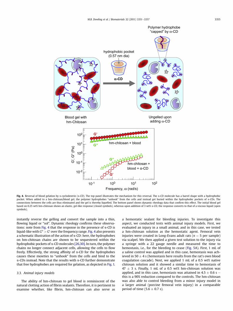
Fig. 4. Reversal of blood gelation by a-cyclodextrin (a-CD). The top panel illustrates the mechanism for this reversal. The a-CD molecule has a barrel shape with a hydrophobicpocket. When added to a hm-chitosan/blood gel, the polymer hydrophobes “unhook” from the cells and instead get buried within the hydrophobic pockets of a-CDs. Theconnections between the cells are thus eliminated and the gel is thereby liquefied. The bottom panel shows dynamic rheology data that confirm this effect. The initial blood gelbased on 0.25 wt% hm-chitosan shows an elastic, gel-like response (closed symbols), whereas upon addition of 3 wt% a-CD, the response converts to that of a viscous liquid (opensymbols).
M.B. Dowling et al. / Biomaterials 32 (2011) 3351e3357 3355
instantly reverse the gelling and convert the sample into a thin,flowing liquid or “sol”. Dynamic rheology confirms these observa-tions: note from Fig. 4 that the response in the presence of a-CD isliquid-likewithG00 > G0 over the frequency range. Fig. 4 also presentsa schematic illustration of the action of a-CD: here, the hydrophobeson hm-chitosan chains are shown to be sequestered within thehydrophobic pockets of a-CDmolecules [26,30]. In turn, the polymerchains no longer connect adjacent cells, allowing the cells to flowfreely. Effectively, the strong affinity of a-CD for the hydrophobescauses these moieties to “unhook” from the cells and bind to thea-CDs instead. Note that the results with a-CD further demonstratethat free hydrophobes are required for gelation, as depicted in Fig. 1.
3.3. Animal injury models
The ability of hm-chitosan to gel blood is reminiscent of thenatural clotting action of fibrin sealants. Therefore, it is pertinent toexamine whether, like fibrin, hm-chitosan can also serve as
a hemostatic sealant for bleeding injuries. To investigate thisaspect, we conducted tests with animal injury models. First, weevaluated an injury in a small animal, and in this case, we testeda hm-chitosan solution as the hemostatic agent. Femoral veininjuries were created in Long-Evans adult rats (n ¼ 5 per sample)via scalpel. We then applied a given test solution to the injury viaa syringe with a 22 gauge needle and measured the time tohemostasis, i.e., for the bleeding to cease (Fig. 5A). First, 1 mL ofa saline control was applied and in this case, hemostasis was ach-ieved in 50 � 4 s (hemostasis here results from the rat’s own bloodcoagulation cascade). Next, we applied 1 mL of a 0.5 wt% nativechitosan solution and it showed a similar time to hemostasis of47 � 3 s. Finally, 1 mL of a 0.5 wt% hm-chitosan solution wasapplied, and in this case, hemostasis was attained in 4.5 � 0.6 s ethis is a 90% reduction compared to the controls. The hm-chitosanwas also able to control bleeding from a minor injury model ina larger animal (porcine femoral vein injury) in a comparableperiod of time (5.6 � 0.7 s).
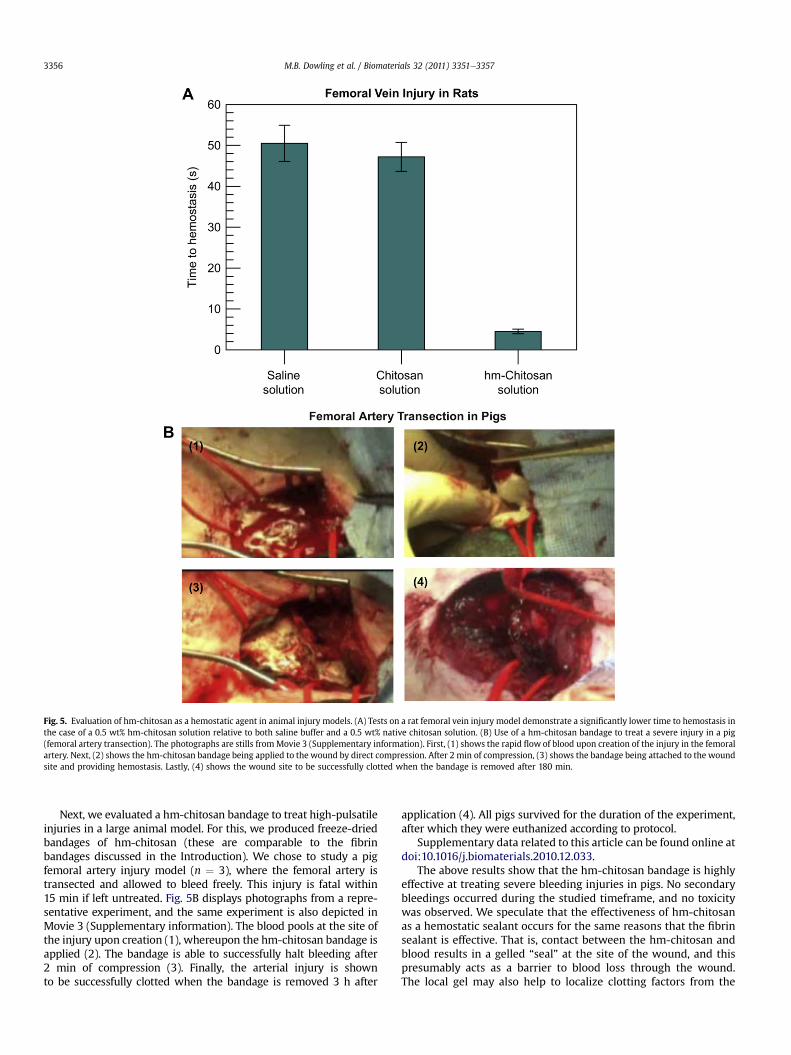
Fig. 5. Evaluation of hm-chitosan as a hemostatic agent in animal injury models. (A) Tests on a rat femoral vein injury model demonstrate a significantly lower time to hemostasis inthe case of a 0.5 wt% hm-chitosan solution relative to both saline buffer and a 0.5 wt% native chitosan solution. (B) Use of a hm-chitosan bandage to treat a severe injury in a pig(femoral artery transection). The photographs are stills fromMovie 3 (Supplementary information). First, (1) shows the rapid flow of blood upon creation of the injury in the femoralartery. Next, (2) shows the hm-chitosan bandage being applied to the wound by direct compression. After 2 min of compression, (3) shows the bandage being attached to the woundsite and providing hemostasis. Lastly, (4) shows the wound site to be successfully clotted when the bandage is removed after 180 min.
M.B. Dowling et al. / Biomaterials 32 (2011) 3351e33573356
Next, we evaluated a hm-chitosan bandage to treat high-pulsatileinjuries in a large animal model. For this, we produced freeze-driedbandages of hm-chitosan (these are comparable to the fibrinbandages discussed in the Introduction). We chose to study a pigfemoral artery injury model (n ¼ 3), where the femoral artery istransected and allowed to bleed freely. This injury is fatal within15 min if left untreated. Fig. 5B displays photographs from a repre-sentative experiment, and the same experiment is also depicted inMovie 3 (Supplementary information). The blood pools at the site ofthe injury upon creation (1), whereupon the hm-chitosan bandage isapplied (2). The bandage is able to successfully halt bleeding after2 min of compression (3). Finally, the arterial injury is shownto be successfully clotted when the bandage is removed 3 h after
application (4). All pigs survived for the duration of the experiment,after which they were euthanized according to protocol.
Supplementary data related to this article can be found online atdoi:10.1016/j.biomaterials.2010.12.033.
The above results show that the hm-chitosan bandage is highlyeffective at treating severe bleeding injuries in pigs. No secondarybleedings occurred during the studied timeframe, and no toxicitywas observed. We speculate that the effectiveness of hm-chitosanas a hemostatic sealant occurs for the same reasons that the fibrinsealant is effective. That is, contact between the hm-chitosan andblood results in a gelled “seal” at the site of the wound, and thispresumably acts as a barrier to blood loss through the wound.The local gel may also help to localize clotting factors from the
M.B. Dowling et al. / Biomaterials 32 (2011) 3351e3357 3357
bloodstream in the vicinity of the wound, and thereby enhance thenatural clotting action. Moreover, the gelling of the blood mayensure that the bandage itself does not get soaked with liquid. Thismeans that the parts of the bandage in direct contact with tissuewill remain adherent and not delaminate, thereby eliminating thepossibility of secondary bleeding (the latter is known to occur withmany existing hemostatic dressings[11,13]). The hydrophobes onthe polymer may also possibly enhance the adherence of the solidbandage due to interactions with underlying tissue cells e again, inmuch the same way as Velcro� hooks, but at the nanoscale.
4. Conclusions
We have shown that the amphiphilic biopolymer, hm-chitosan,can act as an effective hemostatic agent. It has the ability totransform whole liquid blood into a gel, and it quickly stopsbleeding from both minor and severe injuries in small and largeanimals. The gelling mechanism is predicated on the self-assemblyof hm-chitosan chains in such away that their hydrophobes anchorin blood cell membranes and thereby bridge the cells intoa 3-dimensional network. The cells are thus active components(nodes or junction points) in the network rather than being phys-ically trapped in a polymer mesh. The gelling ability of hm-chitosanis similar to that of fibrin-based sealants, but at a much lower costand wider availability. An additional important aspect of hm-chi-tosan as a hemostatic agent is that its gelling effect on blood can bereversed by addition of the supramolecule a-CD. The reversaloccurs because a-CD molecules sequester the hydrophobes andprevent them from attaching to blood cells. Our studies show thathm-chitosan could be an effective and safe hemostatic agent fortreatment of both external and internal bleeding injuries.
Acknowledgments
This work was partially funded by grants fromMIPS and TEDCO.MBD was supported by a Fischell Fellowship from the Departmentof Bioengineering. We thank Dr. Michael Kilbourne, Dr. GerardDeCastro, and Dr. Ian Driscoll for assistance with rat and pigexperiments, as well as the Thomas D. Morris Institute for SurgicalResearch for providing the facilities for animal studies. Under-graduate students Da-Tren Chou and Shelby Skoog assisted withsome of the experiments performed during this project.
Appendix
Figures with essential colour discrimination. All figures in thisarticle are difficult to interpret in black and white. The full colourimages can be found in the online version, at doi:10.1016/j.biomaterials.2010.12.033.
References
[1] Owen CA. A history of blood coagulation. Rochester, MN:Mayo Foundation; 2001.[2] Macfarlane RG. An enzyme cascade in the blood clotting mechanism, and its
function as a biological amplifier. Nature 1964;202:498e9.
[3] Smith GF. Fibrinogen-fibrin conversion - mechanism of fibrin-polymerformation in solution. Biochem J 1980;185:1e11.
[4] Doolittle RF. Fibrinogen and fibrin. Annu Rev Biochem 1984;53:195e229.[5] Neuffer MC, McDivitt J, Rose D, King K, Cloonan CC, Vayer JS. Hemo-
static dressings for the first responder: a review. Mil Med 2004;169:716e20.
[6] Larson MJ, Bowersox JC, Lim RC, Hess JR. Efficacy of a fibrin hemostaticbandage in controlling hemorrhage from experimental arterial injuries. ArchSurg 1995;130:420e2.
[7] Reiss RF, Oz MC. Autologous fibrin glue: production and clinical use. TransfusMed Rev 1996;10:85e92.
[8] Champion HR, Bellamy RF, Roberts CP, Leppaniemi A. A profile of combatinjury. J Trauma 2003;54:S13e9.
[9] Stewart RM, Myers JG, Dent DL, Ermis P, Gray GA, Villarreal R, et al. Sevenhundred fifty-three consecutive deaths in a level 1 trauma center: the argu-ment for injury prevention. J Trauma 2003;54:66e70.
[10] Kauvar DS, Lefering R, Wade CE. Impact of hemorrhage on trauma outcome:an overview of epidemiology, clinical presentations, and therapeutic consid-erations. J Trauma 2006;60:S3e9.
[11] Kheirabadi BS, Acheson EM, Deguzman R, Sondeen JL, Ryan KL, Delgado A,et al. Hemostatic efficacy of two advanced dressings in an aortic hemorrhagemodel in swine. J Trauma 2005;59:25e34.
[12] Pusateri AE, Holcomb JB, Kheirabadi BS, Alam HB, Wade CE, Ryan KL. Makingsense of the preclinical literature on advanced hemostatic products. J Trauma2006;60:674e82.
[13] Arnaud F, Teranishi K, Tomori T, Carr W, McCarron R. Comparison of 10hemostatic dressings in a groin puncture model in swine. J Vasc Surg2009;50:632e9.
[14] Kheirabadi BS, Scherer MR, Estep JS, Dubick MA, Holcomb JB. Determination ofefficacy of new hemostatic dressings in a model of extremity arterialhemorrhage in swine. J Trauma 2009;67:450e60.
[15] Bochicchio G, Kilbourne M, Kuehn R, Keledjian K, Hess J, Scalea T. Use ofa modified chitosan dressing in a hypothermic coagulopathic grade V liverinjury model. Am J Surg 2009;198:617e22.
[16] Ellis-Behnke RG, Liang Y-X, Tay DKC, Kau PWF, Schneider GE, Zhang S, et al.Nano hemostat solution: immediate hemostasis at the nanoscale. Nano-medicine 2006;2:207e15.
[17] Ellis-Behnke RG, Liang YX, You SW, Tay DKC, Zhang SG, So KF, et al. Nanoneuro knitting: peptide nanofiber scaffold for brain repair and axon regen-eration with functional return of vision. Proc Natl Acad Sci U S A 2006;103:5054e9.
[18] Lew WK, Weaver FA. Clinical use of topical thrombin as a surgical hemostat.Biologics 2008;2:593e9.
[19] Kean T, Thanou M. Biodegradation, biodistribution and toxicity of chitosan.Adv Drug Deliv Rev 2010;62:3e11.
[20] Desbrieres J, Martinez C, Rinaudo M. Hydrophobic derivatives of chitosan:characterization and rheological behaviour. Int J Biol Macromol1996;19:21e8.
[21] Lee JH, Gustin JP, Chen TH, Payne GF, Raghavan SR. Vesicle-biopolymer gels:networks of surfactant vesicles connected by associating biopolymers. Lang-muir 2005;21:26e33.
[22] Malette WG, Quigley HJ, Gaines RD, Johnson ND, Rainer WG. Chitosan: a newhemostatic. Ann Thorac Surg 1983;36:55e8.
[23] Rao SB, Sharma CP. Use of chitosan as a biomaterial: studies on its safety andhemostatic potential. J Biomed Mater Res 1997;34:21e8.
[24] Whang HS, Kirsch W, Zhu YH, Yang CZ, Hudson SM. Hemostatic agentsderived from chitin and chitosan. J Macromol Sci Polym Rev 2005;C45:309e23.
[25] Szejtli J. Introduction and general overview of cyclodextrin chemistry. ChemRev 1998;98:1743e53.
[26] Tonelli AE. Nanostructuring and functionalizing polymers with cyclodextrins.Polymer 2008;49:1725e36.
[27] Raghavan SR, Cipriano BH. Gel formation: phase diagrams using tabletoprheology and calorimetry. In: Weiss RG, Terech P, editors. Molecular gels.Dordrecht: Springer; 2005. p. 233e44.
[28] Macosko CW. Rheology: principles,measurements and applications. NewYork:VCH Publishers; 1994.
[29] Meier W, Hotz J, GuntherAusborn S. Vesicle and cell networks: inter-connecting cells by synthetic polymers. Langmuir 1996;12:5028e32.
[30] Kumar R, Raghavan SR. Thermothickening in solutions of telechelic associ-ating polymers and cyclodextrins. Langmuir 2010;26:56e62.
![Hydrophobically-modified chitosan foam: description and ... Surg Res_2014_HMChitosan Foams... · compressible, and hemostatic agent since 2003 [19,20]. It has not, however, been used](https://img.pdfslide.net/doc/110x75/5ca52ef488c993ad338ca448/hydrophobically-modified-chitosan-foam-description-and-surg-res2014hmchitosan.jpg)