Embed Size (px)
Citation preview

The Journal of Advanced Prosthodontics 295
A simplified etching technique to improve the adhesion of fiber post
Chandrakanth Majeti*, Chandrasekhar veeramachaneni, Pradeep Kumar Morisetty, Saggurti Anitha Rao, Muralidhar TummalaDepartment of Conservative Dentistry and Endodontics, Mamata Dental College, Khammam, Andhra Pradesh, India
PURPOSE. Numerous methods were used to etch the fiber posts to improve its bonding to root canal dentin. Our aim was to evaluate the efficacy of 37% phosphoric acid in etching fiber posts in comparison with 24% hydrogen peroxide. MATERIALS AND METHODS. Ninety human maxillary central incisors were taken and post space preparation was done. Ninety fiber posts were taken and divided into three groups (n=30) based on the surface treatment they received (H3PO4, H2O2, distilled water) and each group was further divided (n=10) based on the time period of application (15 seconds, 30 seconds, 60 seconds). All the posts were luted into canals using Rely X UniCem-2. Each tooth was then sectioned into six slices and subjected to push out test. Data obtained was subjected to statistical analysis at P<.05. The surface topography was evaluated using scanning electron microscopy. RESULTS. Highest bond strength values were noted in 15 seconds etched phosphoric acid group and 60 seconds etched hydrogen peroxide group with no significant difference between two groups. Surface topography revealed complete epoxy layer removal with no damage to its structural integrity in those groups. CONCLUSION. H3PO4 etching for a period of 15 seconds is an effective alternative in improving the adhesion of fiber post to root dentin. [ J Adv Prosthodont 2014;6:295-301]
KEY WORDS: Bond strength; Fiber posts; Hydrogen peroxide etching; Phosphoric acid etching; Scanning electron microscopy
http://dx.doi.org/10.4047/jap.2014.6.4.295http://jap.or.kr J Adv Prosthodont 2014;6:295-301
INTRODUCTION
Fiber posts and resin based materials can behave as a mechanically homogeneous complex with dentin; their clin-ical use introduced a new restorative concept, ensuring high resistance to occlusal loading.1 This assumption has been recently supported by several in vivo and in vitro investiga-tions.2-7
Formation of monoblock is essential for the success of
a post luted to root canal dentin.8 Improper bonding at dentin/cement, cement/post interface leads to increased stress to occlusal loading leading to failure of post retained restorations. Bonding between resin cement and fiber post is aided by micromechanical and chemical means.9-12 Fiber post is covered by epoxy resin which is highly cross-linked and has a high degree of conversion.13 Therefore roughen-ing the post to improve micromechanical bonding has been recommended.10
Many techniques like sandblasting, etching with hydro-fluoric acid were used to improve bonding,11,14,15 but these methods caused damage to the structure of glass fibers and affected the integrity of posts.11 Agents that dissolve only the epoxy matrix portion without interfering with fiber integrity were studied.12,16-18 Potassium permanganate, sodi-um ethoxide, and hydrogen peroxide were used to etch and expose the glass fibers.10,12,16-18
Twenty-four percent hydrogen peroxide etching for 1 min proved to be effective in improving bond strength.19 Thirty-seven percent phosphoric acid is the commonly used agent to etch enamel and dentin that is readily avail-
Corresponding author: Chandrakanth MajetiDepartment of Conservative Dentistry and Endodontics, Mamata Dental College, Giri Prasad nagar, Khammam, Andhra Pradesh, IndiaTel. 09985456734: e-mail, [email protected] January 7, 2014 / Last Revision May 6, 2014 / Accepted May 12, 2014
© 2014 The Korean Academy of ProsthodonticsThis is an Open Access article distributed under the terms of the Creative Commons Attribution Non-Commercial License (http://creativecommons.org/licenses/by-nc/3.0) which permits unrestricted non-commercial use, distribution, and reproduction in any medium, provided the original work is properly cited.
pISSN 2005-7806, eISSN 2005-7814
296
able. The objective of the present in vitro study was to eval-uate the efficacy of 37% H3PO4 in comparison with 24% H2O2 to etch fiber posts in terms of push out bond strength at various time periods. In addition surface topog-raphy was evaluated by SEM and bond strengths at various root regions were compared.
MATERIALS AND METHODS
Approval for the study was taken from Human Rights and Research Committee, Mamata Dental College. Ninety freshly extracted human maxillary central incisors were tak-en and stored in sterile saline (0.9% NaCl). Each tooth was decoronated 1 to 2 mm below CEJ using safe-sided dia-mond disk under water coolant so that length was standard-ized to 16 mm. Patency was confirmed with a 10 K-file and root canals were enlarged using ProTaper rotary instru-ments (Maillefer Dentsply, Ballaigues, Switzerland) until file F3 reached the working length (1 mm from apical fora-men). Irrigation was done with 5% NaOCl (Vishal Dento care Pvt. Ltd., Ahmedabad, India) and 17% EDTA (Canalarge, Ammdent, Chandigarh, India) between each instrument. Finally canals were dried with paper points and obturation was done using Gutta percha cones (Maillefer Dentsply, Ballaigues, Switzerland) and AH plus sealer (Dentsply, Delhi, India) by lateral condensation method. The specimens were stored 37ºC, at 100% relative humidity for a period of 72 hours so as the resin sealer sets completely.
Following this the coronal gutta percha removal was done with peeso-reamers #1 to 3 (Mani Inc., Tochigi, Japan) leaving 4 mm apical GP. Final post space prepara-tion was done with #3 drill provided by the manufacturer of the Glassix post system (Swiss dental products of disin-fection, Nordin, Switzerland). Finally post spaces were irri-gated with distilled water and canals were dried.
Ninety glass fiber reinforced parallel and smooth posts (Glassix posts, Nr3- Ø 1.35, Swiss dental products of disin-fection, Nordin, Switzerland) were taken and divided into
three groups (n=30) according to the surface treatment they received (37% H3PO4, 24% H2O2, distilled water). Each group was further divided into 3 sub-groups (n=10) based on the time period of etchant application (15 sec-onds, 30 seconds, 60 seconds). In H3PO4 group, gel (Etch gel, DentoInc., St. Paul, MN, USA) was applied over entire surface of the post; in other two groups posts were immersed in respective solutions; all posts were then treat-ed with distilled water and air dried. Etched posts are then luted into the root canals using Rely X Unicem-2 Clicker (3M ESPE AG, Seefeld, Germany) and light activated through cervical portion for 40 second (Blue phase C8, Ivoclar vivident, Schaan, Liechtenstein). The specimens were then stored for 24 hours at 37ºC in distilled water.
Following storage, each specimen was sectioned into six slices of 2 mm each using diamond saw under water cool-ant. The first two represent the coronal; second two middle and last two represent the apical regions of the root. The apical side of each specimen was marked with an indelible marker and each section was subjected to push out test from apical to coronal side in Universal testing machine (Autograph AG 15, Shimadzu, Kyoto, Japan) where the loading was performed at a cross head speed of 0.5 mm/min until post dislodged from the root slice. Schematic rep-resentation of entire push-out test is shown in Fig. 1.
Comparison of bond strength values were done using Repeated measures of ANOVA (IBM SPSS statistics for Windows, version 21.0, Armonk, NY, USA) and pair wise comparison was done by Tukey multiple post hoc test at a significance level at P<.05.
Twenty seven fiber posts were taken and divided into 3 groups (n=9) based on the surface treatment (H3PO4, H2O2, distilled water). Each group was subdivided into three (n=3) based on the time period of etchant application (15, 30, 60 seconds). After etching all specimens were cleaned ultrasonically for 5 min. in deionoized water, fol-lowed by immersion in 96% ethanol and gently air dried. The posts were then gold sputtered (model JFC-1600,
fig. 1. (A) Sectioning of tooth into six 2 mm sections, (B) Thin slice (2 mm) showing the post at center adhered to dentin, (C) Specimen placement in Universal Testing Machine, (D) Pictorial representation of cross-section of push out test.
B
2 mm
2 mm
2 mm
2 mm
2 mm
2 mm
Apical
Apical
Middle
Middle
Cervical
Cervical
4 mm GPA C
Push out force
Push out jig
Post
DentinDentin
Platform Platform
D
J Adv Prosthodont 2014;6:295-301

The Journal of Advanced Prosthodontics 297
JEOL, Tokyo, Japan) and evaluated under SEM (model JSM-5600, JEOL, Tokyo, Japan).
Dislodged specimens after push out test were examine under stereomicroscope (Olympus Opto Systems, India Pvt Ltd., Noida, India) at ×4.5 magnification and mode of fail-ure was classified as adhesive between cement and dentin, adhesive between cement and post, cohesive within the cement.
RESULTS
The mean values of all the groups are shown in Fig. 2. Results showed highest bond strength values for H3PO4 group at 15 seconds and H2O2 group at 60 seconds at all the three root regions with no significant difference between those two groups (P>.05)(Table 1). Bond strength was greater in the coronal region followed by middle then apical irrespective of etchant and time period (P<.05).
Table 1. Mean bond strength values of all groups in Mega Pascals
Root region
Etchant Time period (Seconds) Cervical Middle Apical Tukey*
H3PO4 15 14.57 (0.62) 12.20 (0.24) 6.41 (0.32) a
30 12.14 (0.54) 10.57 (0.38) 5.60 (0.34) b
60 11.62 (0.24) 10.10 (0.65) 5.14 (0.30) b
H2O2 15 10.57 (0.43) 9.86 (0.50) 4.17 (0.24) c
30 10.66 (0.34) 9.92 (0.58) 5.19 (0.22) c
60 14.38 (0.64) 12.40 (0.42) 6.07 (0.37) a
Water 15 10.89 (0.80) 9.63 (0.46) 4.16 (0.24) c
30 11.09 (0.72) 9.38 (0.44) 4.15 (0.25) c
60 10.97 (0.51) 9.46 (0.38) 4.10 (0.26) c
Tukey* A B C
*(Different letters - Capital letters - Comparison among columns, lower case letters - Comparison among rows) indicates statistical difference (P<.05).
fig. 2. Comparison of bond strength of different etchants at various time periods and at various root regions (Mean Values).
MPa18.00
16.00
14.00
12.00
10.00
8.00
6.00
4.00
2.00
0.00
15 sec 30 sec 60 sec
H3PO4
CervicalH2O2
CervicalWaterCervical
H3PO4
ApicalH2O2
ApicalWaterApical
H3PO4
MiddleH2O2
MiddleWaterMiddle
A simplified etching technique to improve the adhesion of fiber post
298
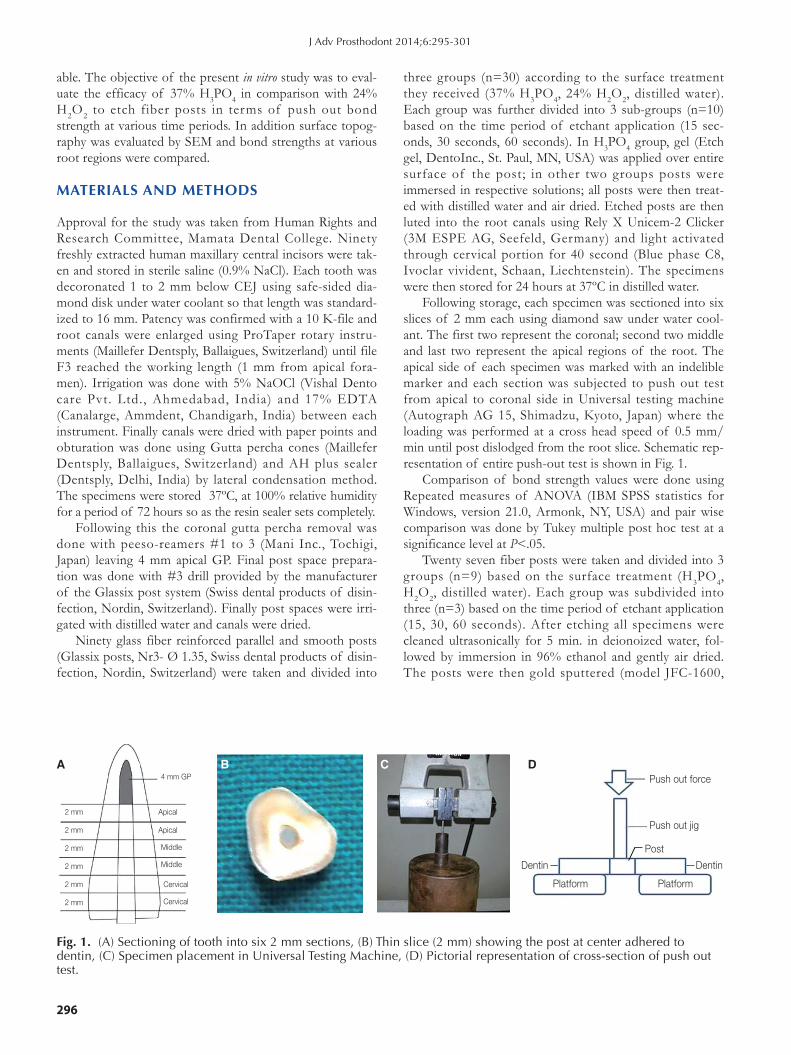
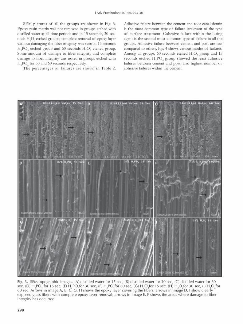
SEM pictures of all the groups are shown in Fig. 3. Epoxy resin matrix was not removed in groups etched with distilled water at all time periods and in 15 seconds, 30 sec-onds H2O2 etched groups; complete removal of epoxy layer without damaging the fiber integrity was seen in 15 seconds H3PO4 etched group and 60 seconds H2O2 etched group. Some amount of damage to fiber integrity and complete damage to fiber integrity was noted in groups etched with H3PO4 for 30 and 60 seconds respectively.
The percentages of failures are shown in Table 2.
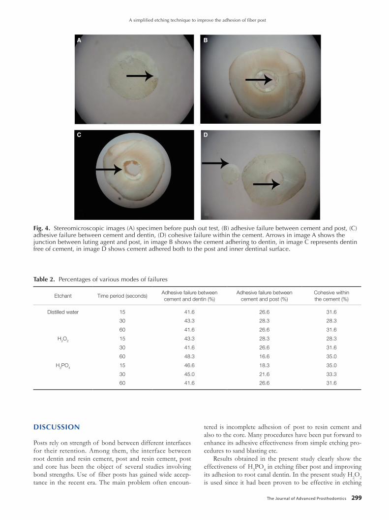
Adhesive failure between the cement and root canal dentin is the most common type of failure irrelevant to the type of surface treatment. Cohesive failure within the luting agent is the second most common type of failure in all the groups. Adhesive failure between cement and post are less compared to others. Fig. 4 shows various modes of failures. Among all groups, 60 seconds etched H2O2 group and 15 seconds etched H3PO4 group showed the least adhesive failures between cement and post, also highest number of cohesive failures within the cement.
fig. 3. SEM topographic images. (A) distilled water for 15 sec, (B) distilled water for 30 sec, (C) distilled water for 60 sec, (D) H3PO4 for 15 sec, (E) H3PO4for 30 sec, (F) H3PO4for 60 sec, (G) H2O2for 15 sec, (H) H2O2for 30 sec, (I) H2O2for 60 sec. Arrows in image A, B, C, G, H shows the epoxy layer covering the fibers; arrows in image D, I show clearly exposed glass fibers with complete epoxy layer removal; arrows in image E, F shows the areas where damage to fiber integrity has occurred.
A
D
G
B
E
H
C
F
I
J Adv Prosthodont 2014;6:295-301
The Journal of Advanced Prosthodontics 299
DISCUSSION
Posts rely on strength of bond between different interfaces for their retention. Among them, the interface between root dentin and resin cement, post and resin cement, post and core has been the object of several studies involving bond strengths. Use of fiber posts has gained wide accep-tance in the recent era. The main problem often encoun-
tered is incomplete adhesion of post to resin cement and also to the core. Many procedures have been put forward to enhance its adhesive effectiveness from simple etching pro-cedures to sand blasting etc.
Results obtained in the present study clearly show the effectiveness of H3PO4 in etching fiber post and improving its adhesion to root canal dentin. In the present study H2O2 is used since it had been proven to be effective in etching
Table 2. Percentages of various modes of failures
Etchant Time period (seconds)Adhesive failure between cement and dentin (%)
Adhesive failure between cement and post (%)
Cohesive within the cement (%)
Distilled water 15 41.6 26.6 31.6
30 43.3 28.3 28.3
60 41.6 26.6 31.6
H2O2 15 43.3 28.3 28.3
30 41.6 26.6 31.6
60 48.3 16.6 35.0
H3PO4 15 46.6 18.3 35.0
30 45.0 21.6 33.3
60 41.6 26.6 31.6
fig. 4. Stereomicroscopic images (A) specimen before push out test, (B) adhesive failure between cement and post, (C) adhesive failure between cement and dentin, (D) cohesive failure within the cement. Arrows in image A shows the junction between luting agent and post, in image B shows the cement adhering to dentin, in image C represents dentin free of cement, in image D shows cement adhered both to the post and inner dentinal surface.
A B
C D
A simplified etching technique to improve the adhesion of fiber post

300
fiber post19 and improving the bond strength; H3PO4 is attempted to use in the present study since it is the com-monly used chair side etchant.
Fiber posts are covered by epoxy resin, which has a high degree of conversion and few reactive sites to chemically bond to the resin cement.13 So to improve the bond between post and adhesive resin, removal of epoxy layer is essential without disturbing the fiber integrity. Unlike the use of corrosive forms of industrial epoxy resin etching techniques such as sodium ethoxide or potassium perman-ganate: H2O2 and H3PO4 etching provides clinically reliable methods to enhance micromechanical retention. Care should be taken not to etch for a longer time period since it affects the fiber integrity similarly to that of HF etching which had been proven to be an aggressive etching proce-dure.17 Dissolution of epoxy resin probably relies on an electrophilic attack of the H2O2 to the cured secondary amine. Thus the spaces created between the fibers provide conditions for the micro-mechanical interlocking of the resin with the post. Etching with H2O2 is a proved subject but studies are still under progress evaluating the efficacy of various concentrations and time periods in altering its etching capability.20-22
SEM images clearly depict the removal of epoxy layer with intact glass fibers in H2O2 and H3PO4 groups at 1 min and 15 seconds time intervals respectively. So the exposed glass fibers aid in improving the bond strength both by micro-mechanical interlocking and by chemical means due interaction between methacrylate group of luting agent and glass fibers.
Push out test was done since it produces shear stress at the interface between post and cement which mimics the stresses under clinical situation.23
The use of adhesive resins have gained popularity from the past few years with their advantage of minimizing the number of steps required and ease of use, so we have selected a self adhesive resin cement. The adhesive proper-ties are claimed to be based upon acidic monomers that demineralize and infiltrate the tooth substrate, and create micromechanical retention and chemical adhesion to hydroxyapatite by forming resin tags.
Our results are similar and support the study by Menezes et al.19 who proved H2O2 etching for one minute improves the bond strength of fiber post. Our results also showed highest bond strength values in the coronal fol-lowed by middle then apical root regions which is in accor-dance with previous studies.23-27 The reason thought for this might be the decrease in the tubule density from coronal to apical region as explained previously by Ferrari et al.28 Most likely explanation for this higher resistance to dislodgement of post in coronal region and decreased resistance in apical region could be due to decreased effectiveness of curing light to penetrate from coronal region to the apex. Also the reduction in the bond strength may be related to the diffi-culties of moisture control in apical third of post space which may resist complete infiltration of the resin cement.
The type of failure modes reveal the least adhesive fail-
ures between the cement and post in 15 seconds H3PO4 group and 60 second H2O2 group indicating the possible improvement in bonding between post and cement after etching. Also due to increased adhesion of cement to the post there might be internal stress created within the cement at the time of loading causing increased number of cohesive failures within the cement in those groups.
CONCLUSION
Within the limitations of the present in vitro study it can be concluded that etching of fiber post is essential to improve adhesion. 37% H3PO4 etching for 15 second is a better and comfortable alternative to other methods in improving the adhesion of fiber post to root canal dentin.
REfERENCES
1. Sorrentino R, Salameh Z, Zarone F, Tay FR, Ferrari M. Effect of post-retained composite restoration of MOD preparations on the fracture resistance of endodontically treated teeth. J Adhes Dent 2007;9:49-56.
2. Salameh Z, Sorrentino R, Papacchini F, Ounsi HF, Tashkandi E, Goracci C, Ferrari M. Fracture resistance and failure pat-terns of endodontically treated mandibular molars restored using resin composite with or without translucent glass fiber posts. J Endod 2006;32:752-5.
3. Schwartz RS, Robbins JW. Post placement and restoration of endodontically treated teeth: a literature review. J Endod 2004;30:289-301.
4. Cagidiaco MC, García-Godoy F, Vichi A, Grandini S, Goracci C, Ferrari M. Placement of fiber prefabricated or custom made posts affects the 3-year survival of endodonti-cally treated premolars. Am J Dent 2008;21:179-84.
5. Ferrari M, Cagidiaco MC, Grandini S, De Sanctis M, Goracci C. Post placement affects survival of endodontically treated premolars. J Dent Res 2007;86:729-34.
6. Monticelli F, Grandini S, Goracci C, Ferrari M. Clinical be-havior of translucent-fiber posts: a 2-year prospective study. Int J Prosthodont 2003;16:593-6.
7. Schmitter M, Rammelsberg P, Gabbert O, Ohlmann B. Influence of clinical baseline findings on the survival of 2 post systems: a randomized clinical trial. Int J Prosthodont 2007;20:173-8.
8. Tay FR, Pashley DH. Monoblocks in root canals: a hypotheti-cal or a tangible goal. J Endod 2007;33:391-8.
9. Goracci C, Raffaelli O, Monticelli F, Balleri B, Bertelli E, Ferrari M. The adhesion between prefabricated FRC posts and composite resin cores: microtensile bond strength with and without post-silanization. Dent Mater 2005;21:437-44.
10. Mazzitelli C, Ferrari M, Toledano M, Osorio E, Monticelli F, Osorio R. Surface roughness analysis of fiber post condition-ing processes. J Dent Res 2008;87:186-90.
11. Valandro LF, Yoshiga S, de Melo RM, Galhano GA, Mallmann A, Marinho CP, Bottino MA. Microtensile bond strength between a quartz fiber post and a resin cement: effect of post surface conditioning. J Adhes Dent 2006;8:105-11.
J Adv Prosthodont 2014;6:295-301
The Journal of Advanced Prosthodontics 301
12. Monticelli F, Toledano M, Tay FR, Cury AH, Goracci C, Ferrari M. Post-surface conditioning improves interfacial ad-hesion in post/core restorations. Dent Mater 2006;22:602-9.
13. Lassila LV, Tanner J, Le Bell AM, Narva K, Vallittu PK. Flexural properties of fiber reinforced root canal posts. Dent Mater 2004;20:29-36.
14. Sahafi A, Peutzfeld A, Asmussen E, Gotfredsen K. Effect of surface treatment of prefabricated posts on bonding of resin cement. Oper Dent 2004;29:60-8.
15. Sahafi A, Peutzfeldt A, Asmussen E, Gotfredsen K. Bond strength of resin cement to dentin and to surface-treated posts of titanium alloy, glass fiber, and zirconia. J Adhes Dent 2003;5:153-62.
16. Monticelli F, Osorio R, Sadek FT, Radovic I, Toledano M, Ferrari M. Surface treatments for improving bond strength to prefabricated fiber posts: a literature review. Oper Dent 2008; 33:346-55.
17. Monticelli F, Toledano M, Tay FR, Sadek FT, Goracci C, Ferrari M. A simple etching technique for improving the re-tention of fiber posts to resin composites. J Endod 2006;32: 44-7.
18. Vano M, Goracci C, Monticelli F, Tognini F, Gabriele M, Tay FR, Ferrari M. The adhesion between fibre posts and com-posite resin cores: the evaluation of microtensile bond strength following various surface chemical treatments to posts. Int Endod J 2006;39:31-9.
19. de Sousa Menezes M, Queiroz EC, Soares PV, Faria-e-Silva AL, Soares CJ, Martins LR. Fiber post etching with hydrogen peroxide: effect of concentration and application time. J Endod 2011;37:398-402.
20. Mosharraf R, Ranjbarian P. Effects of post surface condi-tioning before silanization on bond strength between fiber post and resin cement. J Adv Prosthodont 2013;5:126-32.
21. Cai H, Chen L, Xiong Y. Effects of surface treatment on the fracture resistance of teeth restored with fiber posts and core system. Hua Xi Kou Qiang Yi Xue Za Zhi 2012;30:371-3, 379.
22. Menezes MS, Faria-e-Silva AL, Silva FP, Reis GR, Soares CJ, Stape TH, Martins LR. Etching a fiber post surface with high-concentration bleaching agents. Oper Dent 2014;39: E16-21.
23. Van Meerbeek B, De Munck J, Yoshida Y, Inoue S, Vargas M, Vijay P, Van Landuyt K, Lambrechts P, Vanherle G. Buonocore memorial lecture. Adhesion to enamel and den-tin: current status and future challenges. Oper Dent 2003;28: 215-35.
24. Ngoh EC, Pashley DH, Loushine RJ, Weller RN, Kimbrough WF. Effects of eugenol on resin bond strengths to root canal dentin. J Endod 2001;27:411-4.
25. Bouillaguet S, Troesch S, Wataha JC, Krejci I, Meyer JM, Pashley DH. Microtensile bond strength between adhesive cements and root canal dentin. Dent Mater 2003;19:199-205.
26. Perdigão J, Geraldeli S, Lee IK. Push-out bond strengths of tooth-colored posts bonded with different adhesive systems. Am J Dent 2004;17:422-6.
27. Mallmann A, Jacques LB, Valandro LF, Mathias P, Muench A. Microtensile bond strength of light- and self-cured adhesive
systems to intraradicular dentin using a translucent fiber post. Oper Dent 2005;30:500-6.
28. Ferrari M, Mannocci F, Vichi A, Cagidiaco MC, Mjör IA. Bonding to root canal: structural characteristics of the sub-strate. Am J Dent 2000;13:255-60.
A simplified etching technique to improve the adhesion of fiber post