Embed Size (px)
Citation preview
A specific requirement for PDGF-C in palate formationand PDGFR-a signalingHao Ding1, Xiaoli Wu1, Hans Bostrom2, Injune Kim3, Nicole Wong4, Bonny Tsoi4, Meredith O’Rourke4,Gou Young Koh3, Philippe Soriano5, Christer Betsholtz2, Thomas C Hart6, Mary L Marazita7, L L Field8,Patrick P L Tam4 & Andras Nagy1,9
PDGF-C is a member of the platelet-derived growth factor(PDGF) family, which signals through PDGF receptor (PDGFR)aa and ab dimers1,2. Here we show that Pdgfc�/� mice die inthe perinatal period owing to feeding and respiratorydifficulties associated with a complete cleft of the secondarypalate. This phenotype was less severe than that of Pdgfra�/�
embryos. Pdgfc�/� Pdgfa�/� embryos developed a cleft face,subepidermal blistering, deficiency of renal cortexmesenchyme, spina bifida and skeletal and vascular defects.Complete loss of function of both ligands, therefore,phenocopied the loss of PDGFR-a function, suggesting thatboth PDGF-A and PDGF-C signal through PDGFR-a to regulatethe development of craniofacial structures, the neural tube andmesodermal organs. Our results also show that PDGF-Csignaling is a new pathway in palatogenesis, different from, andindependent of, those previously implicated.
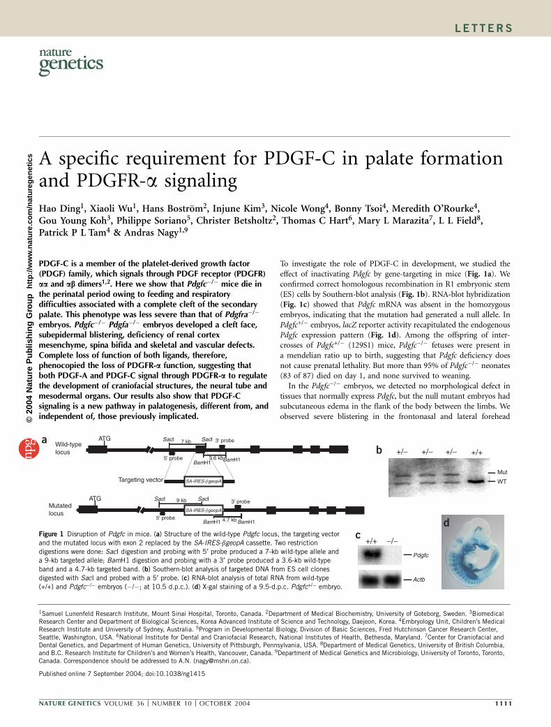
To investigate the role of PDGF-C in development, we studied theeffect of inactivating Pdgfc by gene-targeting in mice (Fig. 1a). Weconfirmed correct homologous recombination in R1 embryonic stem(ES) cells by Southern-blot analysis (Fig. 1b). RNA-blot hybridization(Fig. 1c) showed that Pdgfc mRNA was absent in the homozygousembryos, indicating that the mutation had generated a null allele. InPdgfc+/� embryos, lacZ reporter activity recapitulated the endogenousPdgfc expression pattern (Fig. 1d). Among the offspring of inter-crosses of Pdgfc+/� (129S1) mice, Pdgfc�/� fetuses were present ina mendelian ratio up to birth, suggesting that Pdgfc deficiency doesnot cause prenatal lethality. But more than 95% of Pdgfc�/� neonates(83 of 87) died on day 1, and none survived to weaning.
In the Pdgfc�/� embryos, we detected no morphological defect intissues that normally express Pdgfc, but the null mutant embryos hadsubcutaneous edema in the flank of the body between the limbs. Weobserved severe blistering in the frontonasal and lateral forehead
ATG SacI 7 kb
3.6 kb5' probe
3' probe
ATG SacI
5' probeBamH1
3' probe
BamH1 4.7 kb
SacI9 kb
SA-IRES-�geopA
SA-IRES-�geopA
Wild-typelocus
Targeting vector
Mutatedlocus
WT
Mut
Pdgfc
+/+
SacI
BamH1BamH1
+/− +/− +/− +/+
Actb
−/−
ab
cd
Figure 1 Disruption of Pdgfc in mice. (a) Structure of the wild-type Pdgfc locus, the targeting vector
and the mutated locus with exon 2 replaced by the SA-IRES-bgeopA cassette. Two restriction
digestions were done: SacI digestion and probing with 5¢ probe produced a 7-kb wild-type allele anda 9-kb targeted allele; BamH1 digestion and probing with a 3¢ probe produced a 3.6-kb wild-type
band and a 4.7-kb targeted band. (b) Southern-blot analysis of targeted DNA from ES cell clones
digested with SacI and probed with a 5¢ probe. (c) RNA-blot analysis of total RNA from wild-type
(+/+) and Pdgfc�/� embryos (�/�; at 10.5 d.p.c.). (d) X-gal staining of a 9.5-d.p.c. Pdgfc+/� embryo.
Published online 7 September 2004; doi:10.1038/ng1415
1Samuel Lunenfeld Research Institute, Mount Sinai Hospital, Toronto, Canada. 2Department of Medical Biochemistry, University of Goteborg, Sweden. 3BiomedicalResearch Center and Department of Biological Sciences, Korea Advanced Institute of Science and Technology, Daejeon, Korea. 4Embryology Unit, Children’s MedicalResearch Institute and University of Sydney, Australia. 5Program in Developmental Biology, Division of Basic Sciences, Fred Hutchinson Cancer Research Center,Seattle, Washington, USA. 6National Institute for Dental and Craniofacial Research, National Institutes of Health, Bethesda, Maryland. 7Center for Craniofacial andDental Genetics, and Department of Human Genetics, University of Pittsburgh, Pennsylvania, USA. 8Department of Medical Genetics, University of British Columbia,and B.C. Research Institute for Children’s and Women’s Health, Vancouver, Canada. 9Department of Medical Genetics and Microbiology, University of Toronto, Toronto,Canada. Correspondence should be addressed to A.N. ([email protected]).
NATURE GENETICS VOLUME 36 [ NUMBER 10 [ OCTOBER 2004 1111
LET TERS©
2004
Nat
ure
Pub
lishi
ng G
roup
ht
tp://
ww
w.n
atur
e.co
m/n
atur
egen
etic
s
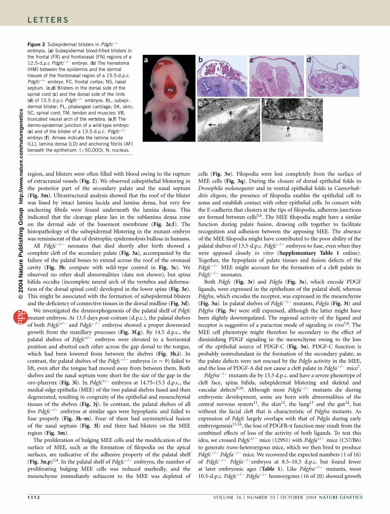
region, and blisters were often filled with blood owing to the ruptureof extracranial vessels (Fig. 2). We observed subepithelial blistering inthe posterior part of the secondary palate and the nasal septum(Fig. 3m). Ultrastructural analysis showed that the roof of the blisterwas lined by intact lamina lucida and lamina densa, but very fewanchoring fibrils were found underneath the lamina densa. Thisindicated that the cleavage plane lies in the sublamina densa zoneon the dermal side of the basement membrane (Fig. 2e,f). Thehistopathology of the subepidermal blistering in the mutant embryowas reminiscent of that of dystrophic epidermolysis bullosa in humans.
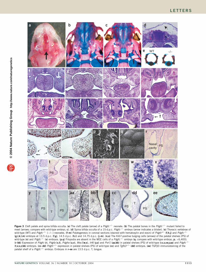
All Pdgfc�/� neonates that died shortly after birth showed acomplete cleft of the secondary palate (Fig. 3a), accompanied by thefailure of the palatal bones to extend across the roof of the oronasalcavity (Fig. 3b; compare with wild-type control in Fig. 3c). Weobserved no other skull abnormalities (data not shown), but spinabifida occulta (incomplete neural arch of the vertebra and deforma-tion of the dorsal spinal cord) developed in the lower spine (Fig. 3e).This might be associated with the formation of subepidermal blistersand the deficiency of connective tissues in the dorsal midline (Fig. 3d).
We investigated the dysmorphogenesis of the palatal shelf of Pdgfcmutant embryos. At 13.5 days post-coitum (d.p.c.), the palatal shelvesof both Pdgfc+/� and Pdgfc�/� embryos showed a proper downwardgrowth from the maxillary processes (Fig. 3f,g). By 14.5 d.p.c., thepalatal shelves of Pdgfc+/� embryos were elevated to a horizontalposition and abutted each other across the gap dorsal to the tongue,which had been lowered from between the shelves (Fig. 3h,i). Incontrast, the palatal shelves of the Pdgfc�/� embryos (n ¼ 9) failed tolift, even after the tongue had moved away from between them. Bothshelves and the nasal septum were short for the size of the gap in theoro-pharynx (Fig. 3i). In Pdgfc+/� embryos at 14.75–15.5 d.p.c., themedial-edge epithelia (MEE) of the two palatal shelves fused and thendegenerated, resulting in congruity of the epithelial and mesenchymaltissues of the shelves (Fig. 3j). In contrast, the palatal shelves of allfive Pdgfc�/� embryos at similar ages were hypoplastic and failed tofuse properly (Fig. 3k–m). Four of them had asymmetrical fusionof the nasal septum (Fig. 3l) and three had blisters on the MEEregion (Fig. 3m).
The proliferation of bulging MEE cells and the modification of thesurface of MEE, such as the formation of filopodia on the apicalsurfaces, are indicative of the adhesive property of the palatal shelf(Fig. 3n,p)3,4. In the palatal shelf of Pdgfc�/� embryos, the number ofproliferating bulging MEE cells was reduced markedly, and themesenchyme immediately subjacent to the MEE was depleted of
cells (Fig. 3o). Filopodia were lost completely from the surface ofMEE cells (Fig. 3q). During the closure of dorsal epithelial folds inDrosophila melanogaster and in ventral epithelial folds in Caenorhab-ditis elegans, the presence of filopodia enables the epithelial cell tosense and establish contact with other epithelial cells. In concert withthe E-cadherin that clusters at the tips of filopodia, adherens junctionsare formed between cells5,6. The MEE filopodia might have a similarfunction during palate fusion, drawing cells together to facilitaterecognition and adhesion between the apposing MEE. The absenceof the MEE filopodia might have contributed to the poor ability of thepalatal shelves of 13.5-d.p.c. Pdgfc�/� embryos to fuse, even when theywere apposed closely in vitro (Supplementary Table 1 online).Together, the hypoplasia of palate tissues and fusion defects of thePdgfc�/� MEE might account for the formation of a cleft palate inPdgfc�/� neonates.
Both Pdgfc (Fig. 3r) and Pdgfa (Fig. 3s), which encode PDGFligands, were expressed in the epithelium of the palatal shelf, whereasPdgfra, which encodes the receptor, was expressed in the mesenchyme(Fig. 3u). In palatal shelves of Pdgfc�/� mutants, Pdgfa (Fig. 3t) andPdgfra (Fig. 3v) were still expressed, although the latter might havebeen slightly downregulated. The regional activity of the ligand andreceptor is suggestive of a paracrine mode of signaling in vivo7,8. TheMEE cell phenotype might therefore be secondary to the effect ofdiminishing PDGF signaling in the mesenchyme owing to the lossof the epithelial source of PDGF-C (Fig. 3o). PDGF-C function isprobably nonredundant in the formation of the secondary palate, asthe palate defects were not rescued by the Pdgfa activity in the MEE,and the loss of PDGF-A did not cause a cleft palate in Pdgfa�/� mice7.Pdgfra�/� mutants die by 15.5 d.p.c. and have a severe phenotype of
cleft face, spina bifida, subepidermal blistering and skeletal andvascular defects9,10. Although most Pdgfa�/� mutants die duringembryonic development, some are born with abnormalities of thecentral nervous system11, the skin12, the lung13 and the gut14, butwithout the facial cleft that is characteristic of Pdgfra mutants. Asexpression of Pdgfc largely overlaps with that of Pdgfa during earlyembryogenesis15,16, the loss of PDGFR-a function may result from thecombined effects of loss of the activity of both ligands. To test thisidea, we crossed Pdgfc+/� mice (129S1) with Pdgfa+/� mice (C57/B6)to generate trans-heterozygous mice, which we then bred to producePdgfc�/� Pdgfa�/� mice. We recovered the expected numbers (1 of 16)of Pdgfc�/� Pdgfa�/�embryos at 8.5–10.5 d.p.c. but found fewerat later embryonic ages (Table 1). Like Pdgfra�/� mutants, most10.5-d.p.c. Pdgfc�/� Pdgfa�/� homozygotes (16 of 20) showed growth
a b c d
e f
AFLD
LL
LD
N
LL
HMFR
FN
SK
SC
VBTM
PL
BL
FCNS
Figure 2 Subepidermal blisters in Pdgfc�/�
embryos. (a) Subepidermal blood-filled blisters in
the frontal (FR) and frontonasal (FN) regions of a
12.5-d.p.c Pdgfc�/� embryo. (b) The hematoma
(HM) between the epidermis and the dermal
tissues of the frontonasal region of a 15.5-d.p.c.
Pdgfc�/� embryo. FC, frontal cortex; NS, nasal
septum. (c,d) Blisters in the dorsal side of the
spinal cord (c) and the dorsal side of the limb
(d) of 15.5 d.p.c Pdgfc�/� embryos. BL, subepi-
dermal blister; PL, phalangeal cartilage; SK, skin;
SC, spinal cord; TM, tendon and muscles; VB,
truncated neural arch of the vertebra. (e,f) The
dermo-epidermal junction of a wild-type embryo
(e) and of the blister of a 13.5-d.p.c. Pdgfc�/�
embryo (f). Arrows indicate the lamina lucida
(LL), lamina densa (LD) and anchoring fibrils (AF)
beneath the epithelium. (�50,000). N, nucleus.
1112 VOLUME 36 [ NUMBER 10 [ OCTOBER 2004 NATURE GENETICS
LET TERS©
2004
Nat
ure
Pub
lishi
ng G
roup
ht
tp://
ww
w.n
atur
e.co
m/n
atur
egen
etic
s
i
WT
a b c d
e
PSNasalcavity
f g
j l m
PS
PS
PS PS PS
PS
n o p q r
k
s t u v w x
PSPS
T PST PS PS PS
TTT PST
PS PS
y z aa bb cc dd ee
−/−
h
Figure 3 Cleft palate and spina bifida occulta. (a) The cleft palate (arrow) of a Pdgfc�/� neonate. (b) The palatal bones in the Pdgfc�/� mutant failed to
meet (arrows; compare with wild-type embryo, c). (d) Spina bifida occulta of a 15-d.p.c. Pdgfc�/� embryo (arrow indicates a blister). (e) Thoracic vertebrae ofwild-type (WT) and Pdgfc�/� (�/�) neonates. (f–m) Palatogenesis in coronal sections (stained with hematoxylin and eosin) of Pdgfc+/� (f,h,j) and Pdgfc�/�
(g,i,k,l,m) embryos at 13.5 d.p.c. (f,g), 14.5 d.p.c. (h,i) and 14.75 d.p.c. (j–m). (n,o) The Ki67-positive bulging cells (arrows) of the palatal shelves (PS) of
wild-type (n) and Pdgfc�/� (o) embryos. (p,q) Filopodia are absent in the MEE cells of a Pdgfc�/� embryo (q; compare with wild-type embryo, p; �6,400).
(r–bb) Expression of Pdgfc (r), Pdgfa (s,t), Pdgfra (u,v), Msx1(w,x), Irf6 (y,z) and Pvrl1 (aa,bb) in palatal shelves (PS) of wild-type (r,s,u,w,y,aa) and Pdgfc�/�
(t,v,x,z,bb) embryos. (cc–dd) Pdgfc�/� expression in palatal shelves (PS) of wild-type (cc) and Tgfb3�/� (dd) embryos. (ee) TGFb3 immunostaining of the
palatal shelf of a Pdgfc�/� embryo. Embryos in r–ee are 13.5 d.p.c. T, tongue.
NATURE GENETICS VOLUME 36 [ NUMBER 10 [ OCTOBER 2004 1113
LET TERS©
2004
Nat
ure
Pub
lishi
ng G
roup
ht
tp://
ww
w.n
atur
e.co
m/n
atur
egen
etic
s
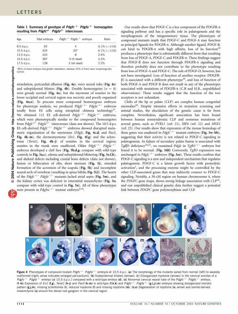
retardation, pericardial effusion (Fig. 4a), wavy neural tube (Fig. 4e)and subepidermal blisters (Fig. 4b). Double homozygotes (n ¼ 4)were grossly normal (Fig. 4a), but the myotome of somites in thelower occipital and cervical region was necrotic and poorly organized(Fig. 4n,o). To procure more compound homozygous embryosfor phenotype analysis, we produced Pdgfc�/� Pdgfa�/� embryostotally from ES cells using tetraploid chimera technology17.We obtained 112 ES cell–derived Pdgfc�/� Pdgfa�/� embryos,which were phenotypically similar to the compound homozygotesfrom Pdgfc+/� Pdgfa+/� intercrosses (data not shown). The 10.5-d.p.cES cell–derived Pdgfc�/� Pdgfa�/� embryos showed disrupted meta-meric organization of the myotomes (Pdgfc, Fig. 4c,d; and Pax3,Fig. 4k–m), the dermomyotome (En1, Fig. 4f,g) and the sclero-tome (Twist1, Fig. 4h–j) of somites in the cervical region;somites in the trunk were unaffected. Older Pdgfc�/� Pdgfa�/�
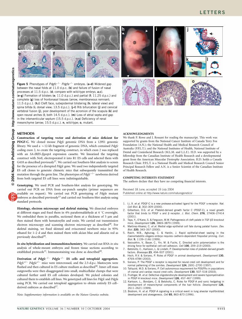
embryos developed a cleft face (Fig. 5b,d,g; compare with wild-typecontrols in Fig. 5a,c), edema and subepidermal blistering (Fig. 5e,f,h),and skeletal defects including cranial bone defects (data not shown),fusion or bifurcation of ribs, short sternum (Fig. 5j), retardedformation of the acromion of the scapula (Fig. 5k) and incompleteneural arch of vertebrae (resulting in spina bifida; Fig. 5i,l). The heartsof the Pdgfc�/� Pdgfa�/� mutants lacked atrial septa (Fig. 5m), andthe kidney cortex was deficient in interstitial mesenchyme (Fig. 5o;compare with wild-type control in Fig. 5n). All of these phenotypeswere present in Pdgfra�/� mutant embryos9,10.
Our results show that PDGF-C is a key component of the PDGFR-asignaling pathway and has a specific role in palatogenesis and themorphogenesis of the integumentary tissue. The phenotypes ofcompound mutants imply that PDGF-C and PDGF-A may functionas principal ligands for PDGFR-a. Although another ligand, PDGF-B,can bind to PDGFR-a with high affinity, loss of its function18
produces a phenotype that is substantially different from that resultingfrom loss of PDGF-A, PDGF-C and PDGFR-a. These findings suggestthat PDGF-B does not function through PDGFR-a signaling andtherefore probably does not contribute to the phenotype resultingfrom loss of PDGF-A and PDGF-C. The role of PDGF-D, however, hasnot been investigated. Loss of function of another receptor (PDGFR-b) is associated with a different phenotype19, and loss of function ofboth PDGF-A and PDGF-B does not result in any of the phenotypesassociated with mutations of PDGFR-a (C.B and H.B., unpublishedobservations). These results suggest that the function of the tworeceptors is not redundant.
Clefts of the lip or palate (CLP) are complex human congenitalanomalies20. Despite intensive efforts in mutation screening andanimal studies, the elucidation of the genetic cause is far fromcomplete. Nevertheless, significant association has been foundbetween human nonsyndromic CLP and nonsense mutations ofseveral genes, such as PVRL1 (ref. 21), IRF6 (ref. 22) and MSX1(ref. 23). Our results show that expression of the mouse homologs ofthese genes was unaltered in Pdgfc�/� mutant embryos (Fig. 3w–bb),suggesting that their activity is not related to PDGF-C signaling inpalatogenesis. As failure of secondary palate fusion is associated withTgfb3 deficiency24,25, we examined Pdgfc in Tgfb3�/� embryos butfound it to be normal. (Fig. 3dd) Conversely, Tgfb3 expression wasunchanged in Pdgfc�/� embryos (Fig. 3ee). These results confirm thatPDGF-C signaling is a new and independent mechanism that regulatespalatogenesis. PDGF-C is a latent growth factor with proteolyticactivation1, and the processing enzyme might be controlled by theother CLP-associated genes that may indirectly connect to PDGF-Csignaling. Notably, a 30-cM region on human chromosome 4, wherethe PDGFC gene maps, shows strong linkage association with CLP26,and our unpublished clinical genetic data further suggest a potentiallink between PDGFC gene polymorphism and CLP.
Table 1 Summary of genotype of Pdgfc�/� Pdgfa�/� homozygotes
resulting from Pdgfc+/� Pdgfa+/� intercrosses
Age Total embryos Pdgfc�/� Pdgfa�/� embryos Ratio
8.5 d.p.c. 65 4 6.1% (B1/16)
10.5 d.p.c. 324 20* 6.1% (B1/16)
13.5 d.p.c. 333 8 2.4%
16.5 d.p.c. 387 9 (5 dead) 2.3%
17.5 d.p.c. 128 3 (all dead) 2.3%
*80% of these embryos had growth retardation, whereas 20% of them were morphologicallynormal.
a b c d e f g
h j k l m n oi
Figure 4 Phenotypes of compound mutant Pdgfc�/� Pdgfa�/� embryos at 10.5 d.p.c. (a) The morphology of the mutants varied from normal (left) to severelymalformed (right; arrow indicates enlarged pericardium). (b) Subepidermal blisters (arrows). (c) Disorganized myotome (arrows) in the cervical somites of a
Pdgfc�/� Pdgfa�/� embryo (at 10.5 d.p.c.) compared with a wild-type embryo (d). (e) Abnormal cervical neural tube of the Pdgfc�/� Pdgfa�/� embryo.
(f–m) Expression of En1 (f,g), Twist1 (h–j) and Pax3 (k–m) in wild-type (f,h,k) and Pdgfc�/� Pdgfa�/� (g,i,j,l,m) embryos showing disorganized meristic
pattern (g,j,m), missing sclerotomes (i), reduced myotome (l) and missing myotome (m). (n,o) Degeneration of myotome (n; arrow) and somite-derived
mesenchyme (o) around the dorsal root ganglion in the cervical region.
1114 VOLUME 36 [ NUMBER 10 [ OCTOBER 2004 NATURE GENETICS
LET TERS©
2004
Nat
ure
Pub
lishi
ng G
roup
ht
tp://
ww
w.n
atur
e.co
m/n
atur
egen
etic
s
METHODSConstruction of targeting vector and derivation of mice deficient for
PDGF-C. We cloned mouse Pdgfc genomic DNA from a 129S1 genomic
library. We used a B12-kb fragment of genomic DNA, which contained Pdgfc
coding exon 2, to create the targeting construct, in which exon 2 was replaced
with an SA-IRES-bgeopA expression cassette. We linearized the targeting
construct with NotI, electroporated it into R1 ES cells and selected them with
G418 as described previously27. We carried out Southern-blot analysis to screen
for the presence of a disrupted Pdgfc gene. We used two independently targeted
ES cell clones to generate chimeric mice that subsequently transmitted the
mutation through the germ line. The phenotypes of Pdgfc�/� newborns derived
from both targeted ES cell lines were indistinguishable.
Genotyping. We used PCR and Southern-blot analysis for genotyping. We
carried out PCR on DNA from ear-punch samples (primer sequences are
available on request). We carried out PCR genotyping of Pdgfa mutant
embryos as described previously13 and carried out Southern-blot analysis using
standard protocols.
Histology, electron microscopy and skeletal staining. We dissected embryos
at different stages and fixed them in 4% paraformaldehyde at 4 1C overnight.
We embedded them in paraffin, sectioned them at a thickness of 5 mm and
then stained them with hematoxylin and eosin. We carried out transmission
electron microscopy and scanning electron microscopy as described28. For
skeletal staining, we fixed skinned and eviscerated newborn mice in 95%
ethanol for 1–2 d and then stained them with alcian blue and alizarin red as
previously described29.
In situ hybridization and immunohistochemistry. We carried out RNA in situ
analysis of whole-mount embryos and frozen tissue sections according to
established protocols15. Immunostaining was done as described28.
Derivation of Pdgfc�/� Pdgfa�/� ES cells and tetraploid aggregation.
Pdgfc+/� Pdgfa+/� mice were intercrossed, and the 2.5-d.p.c. blastocysts were
flushed and then cultured in ES culture medium as described30. Inner cell mass
outgrowths were then disaggregated into small, multicellular clumps that were
cultured further until ES cell colonies developed. We picked colonies and
cultured them to establish cell lines. We genotyped cell lines for Pdgfc and Pdgfa
using PCR. We carried out tetraploid aggregation to obtain entirely ES cell–
derived embryos as described17.
Note: Supplementary information is available on the Nature Genetics website.
ACKNOWLEDGMENTSWe thank P. Rowe and J. Rossant for reading the manuscript. This work wassupported by grants from the National Cancer Institute of Canada Terry FoxFoundation (A.N.); the National Health and Medical Research Council ofAustralia (P.P.L.T.); and the National Institutes of Health, National Institute ofDental and Craniofacial Research (M.L.M. and L.L.F.). H.D. was supported by afellowship from the Canadian Institute of Health Research and a developmentalgrant from the American Muscular Dystrophy Association. H.D. holds a CanadaResearch Chair. P.P.L.T. is a National Health and Medical Research Council SeniorPrincipal Research Fellow and A.N. is a Senior Scientist of the Canadian Instituteof Health Research.
COMPETING INTERESTS STATEMENTThe authors declare that they have no competing financial interests.
Received 18 June; accepted 19 July 2004
Published online at http://www.nature.com/naturegenetics/
1. Li, X. et al. PDGF-C is a new protease-activated ligand for the PDGF a-receptor. Nat.Cell Biol. 2, 302–309 (2000).
2. Gilbertson, D.G. et al. Platelet-derived growth factor C (PDGF-C), a novel growthfactor that binds to PDGF a and b receptor. J. Biol. Chem. 276, 27406–27414(2001).
3. Taya, Y., O’Kane, S. & Ferguson, M.W. Pathogenesis of cleft palate in TGF-b3 knockoutmice. Development 126, 3869–3879 (1999).
4. Martinez-Alvarez, C. et al. Medial edge epithelial cell fate during palatal fusion. Dev.Biol. 220, 343–357 (2000).
5. Raich, W.B., Agbunag, C. & Hardin, J. Rapid epithelial-sheet sealing in theCaenorhabditis elegans embryo requires cadherin-dependent filopodial priming. Curr.Biol. 9, 1139–1146 (1999).
6. Vasioukhin, V., Bauer, C., Yin, M. & Fuchs, E. Directed actin polymerization is thedriving force for epithelial cell-cell adhesion. Cell 100, 209–219 (2000).
7. Betsholtz, C., Karlsson, L. & Lindahl, P. Developmental roles of platelet-derived growthfactors. Bioessays 23, 494–507 (2001).
8. Hoch, R.V. & Soriano, P. Roles of PDGF in animal development. Development 130,4769–4784 (2003).
9. Soriano, P. The PDGF a receptor is required for neural crest cell development and fornormal patterning of the somites. Development 124, 2691–2700 (1997).
10. Tallquist, M.D. & Soriano, P. Cell autonomous requirement for PDGFRa in populationsof cranial and cardiac neural crest cells. Development 130, 507–518 (2003).
11. Fruttiger, M. et al. Defective oligodendrocyte development and severe hypomyelinationin PDGF-A knockout mice. Development 126, 457–467 (1999).
12. Karlsson, L., Bondjers, C. & Betsholtz, C. Roles for PDGF-A and sonic hedgehog indevelopment of mesenchymal components of the hair follicle. Development 126,2611–2621 (1999).
13. Bostrom, H. et al. PDGF-A signaling is a critical event in lung alveolar myofibroblastdevelopment and alveogenesis. Cell 85, 863–873 (1996).
a b c d
e f g
h i
k
j
l
n omFigure 5 Phenotypes of Pdgfc�/� Pdgfa�/� embryos. (a–d) Widened gap
between the nasal folds at 11.0 d.p.c. (b) and failure of fusion of nasal
processes at 11.5 d.p.c. (d; compare with wild-type embryo, a,c).
(e–g) Formation of blisters (e; 11.0 d.p.c.) and partial (f; 11.25 d.p.c.) and
complete (g) loss of frontonasal tissues (arrow, membraneous remnant;
11.5 d.p.c.). (h,i) Cleft face, subepidermal blistering (h; lateral view) and
spina bifida (i; dorsal view; 13.5 d.p.c.). (j–l) Rib bifurcation (j) and cervical
vertebral fusion (j), poor development of the acromion of the scapula (k) andopen neural arches (l; both 14.5 d.p.c.). (m) Loss of atrial septa and gap
in the interventicular septum (15.5 d.p.c.). (n,o) Deficiency of renal
mesenchyme (arrow; 15.5 d.p.c.). n, wild-type; o, mutant.
NATURE GENETICS VOLUME 36 [ NUMBER 10 [ OCTOBER 2004 1115
LET TERS©
2004
Nat
ure
Pub
lishi
ng G
roup
ht
tp://
ww
w.n
atur
e.co
m/n
atur
egen
etic
s
14. Karlsson, L., Lindahl, P., Heath, J.K. & Betsholtz, C. Abnormal gastrointestinaldevelopment in PDGF-A and PDGFR-a deficient mice implicates a novel mesenchymalstructure with putative instructive properties in villus morphogenesis. Development127, 3457–3466 (2000).
15. Ding, H. et al. The mouse Pdgfc gene: dynamic expression in embryonic tissues duringorganogenesis. Mech. Dev. 96, 209–213 (2000).
16. Aase, K., Abramsson, A., Karlsson, L., Betsholtz, C. & Eriksson, U. Expressionanalysis of PDGF-C in adult and developing mouse tissues. Mech. Dev. 110, 187–191 (2002).
17. Nagy, A., Rossant, J., Nagy, R., Abramow-Newerly, W. & Roder, J.C. Derivation ofcompletely cell culture-derived mice from early-passage embryonic stem cells. Proc.Natl. Acad. Sci. USA 90, 8424–8428 (1993).
18. Leveen, P. et al. Mice deficient for PDGF B show renal, cardiovascular, and hemato-logical abnormalities. Genes Dev. 8, 1875–1887 (1994).
19. Soriano, P. Abnormal kidney development and hematological disorders in PDGFb-receptor mutant mice. Genes Dev. 8, 1888–1896 (1994).
20. Murray, J.C. Gene/environment causes of cleft lip and/or palate. Clin. Genet. 61,248–256 (2002).
21. Suzuki, K. et al. Mutations of PVRL1, encoding a cell-cell adhesion molecule/herpesvirus receptor, in cleft lip/palate-ectodermal dysplasia. Nat. Genet. 25,427–430 (2000).
22. Kondo, S. et al. Mutations in IRF6 cause Van der Woude and popliteal pterygiumsyndromes. Nat. Genet. 32, 285–289 (2002).
23. van den Boogaard, M.J., Dorland, M., Beemer, F.A. & van Amstel, H.K. MSX1 mutationis associated with orofacial clefting and tooth agenesis in humans. Nat. Genet. 24,342–343 (2000).
24. Proetzel, G. et al. Transforming growth factor-b 3 is required for secondary palatefusion. Nat. Genet. 11, 409–414 (1995).
25. Kaartinen, V. et al. Abnormal lung development and cleft palate in mice lacking TGF-b3 indicates defects of epithelial-mesenchymal interaction. Nat. Genet. 11, 415–421(1995).
26. Marazita, M.L. et al. Genome scan for loci involved in cleft lip with or withoutcleft palate, in Chinese multiplex families. Am. J. Hum. Genet. 71, 349–364 (2002).
27. Gertsenstein, M., Lobe, C. & Nagy, A. ES cell-mediated conditional transgenesis.Methods Mol. Biol. 185, 285–307 (2002).
28. Ding, H. et al. Astrocyte-specific expression of activated p21-ras results in malignantastrocytoma formation in a transgenic mouse model of human gliomas. Cancer Res.61, 3826–3836 (2001).
29. Nagy, A. et al. Dissecting the role of N-myc in development using a single targetingvector to generate a series of alleles. Curr. Biol. 8, 661–664 (1998).
30. Schoonjans, L. et al. Improved generation of germline-competent embryonic stem celllines from inbred mouse strains. Stem Cells 21, 90–97 (2003).
1116 VOLUME 36 [ NUMBER 10 [ OCTOBER 2004 NATURE GENETICS
LET TERS©
2004
Nat
ure
Pub
lishi
ng G
roup
ht
tp://
ww
w.n
atur
e.co
m/n
atur
egen
etic
s