Embed Size (px)
Citation preview
A STUDY OF APONEUROTIC PTOSIS
AND ITS MANAGEMENT
M.S. DEGREE EXAMINATION
BRANCH III – OPHTHALMOLOGY
Madras Medical College, Chennai
Dissertation Submitted to
THE TAMILNADU Dr. M.G.R. MEDICAL UNIVERSITY CHENNAI.
MARCH – 2007
brought to you by COREView metadata, citation and similar papers at core.ac.uk
provided by ePrints@TNMGRM (Tamil Nadu Dr. M.G.R. Medical University)
CERTIFICATE
This is to certify that Dr. B.Kalaiselvi, MS. Post Graduate Student in
Ophthalmology, Regional Institute of Ophthalmology, Government Ophthalmic
Hospital, Madras Medical College, carried out this dissertation titled A Study of
Aponeurotic ptosis and its management by herself under my guidance and direct
supervision during the period of May 2004 to March 2007. This dissertation is
submitted to Tamil Nadu Dr. M.G.R. Medical University, Chennai in partial
fulfillment of the award of MS Degree (Ophthalmology).
Prof. Dr. M. Radha Krishnan, MS, DO., Prof. Dr.V. Velayutham, MS, DO.,
Chief Orbit and Oculoplasty, Director and Superintendent
Regional Institute of Ophthalmology, Regional Institute of Ophthalmology,
Government Ophthalmic Hospital, Government Ophthalmic Hospital,
Egmore, Chennai. Egmore, Chennai.
Prof. Dr. Kalavathy Ponniraivan, BSc,MD.,
Dean
Madras Medical College,
Government General Hospital,
Chennai.
Date :
Place :

ACKNOWLEDGEMENT
I express my sincere thanks and gratitude to Prof. Dr.
Kalavathy Ponniraivan, BSc, MD, Dean, Madras Medical College for
permitting me to utilise the clinical materials of this hospital.
I am very grateful to Prof. Dr. V. Velayutham, MS, DO, Director,
RIOGOH, Chennai for his valuable advice and guidance in conducting
this study and throughout my post graduate course.
I am very grateful to my Prof. Dr. M. Radhakrishnan, MS, DO,
my unit chief for his valuable guidance in conducting this study and
guiding me throughout my post graduate course.
I am thankful to my Assistant Professors Dr. Malarvizhi,
MS,DO., Dr. Ravindran, MS, for their assistance and help.
I thank all my professors, Assistant professors and colleagues
who have helped me in bringing out this study.
Finally I am thankful to all my patients for their kind cooperation
for completion of this study.
CONTENTS
Section I Page No.
1 Introduction
2. Anatomy of eyelid
3. Physiology of eyelid
4. Classification of Blepharoptosis
5. Literature of Ptosis
6. Evaluation of Ptosis
7. Evolution of Management of Ptosis
8. Management of Ptosis
9. Complication of Blepharoptosis surgery
Section II
1 Aim of the study
2. Materials and Methods
3. Analysis and discussion
4. Conclusion
Section III
1. Bibliography
2. Proforma
3. Master Chart
INTRODUCTION
Blepharoptosis denotes droopiness of the eyelid often the term
is abbreviated as ptosis, which typically denote droopiness of upper
eye lid. A spectrum of conditions affect the upper eyelid height, but the
most common in acquired is aponeurotic Ptosis. This study out lines
aponeurotic ptosis incidence and its management at RIOGOH.
As ptosis leads to an unacceptable cosmetic appearance and
defective vision, the fight against ptosis has been long one, various
surgical techniques for the correction of ptosis are refined and
improved in this modern era of oculoplasty.
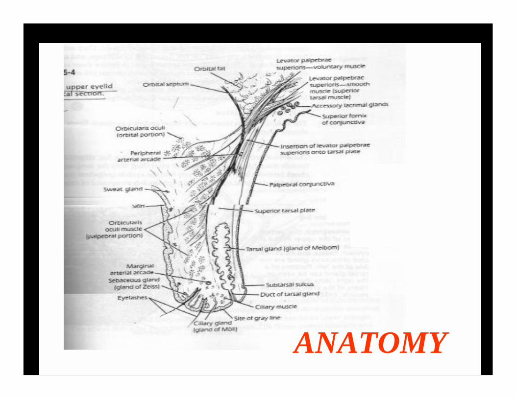
ANATOMY OF EYELID
The upper eyelid is unique in that many important structures in
several lamellae are contained within a thickness of approximately
2mm.
At a level 3mm above the eyelid margin these lamellae are
defined from anterior to posterior as
1. Skin
2. Subcutaneuous tissue
3. Pretarsal orbicularis muscle
4. Post orbicular areolar tissue
5. Tarsal plate
6. Conjunctiva
At a level 15mm above the eyelid margin the lamellae are as follows
1. Skin
2. Subcutaneous tissue
3. Pretarsal orbicularis muscle
4. Post orbicular areolar tissue
5. Orbital septum
6. Preaponeurotic orbital pad of fat
7. Levator aponeurosis
8. Muller’s muscle
9. Conjunctiva
Skin
The eyelid skin is thinnest of the body and unique in having no
subcutaneous fat layer, in both upper and lower eyelid. The pretarsal
tissues are firmly attached to the underlying tissues, the preseptal
tissues are loosely attached.
The upper eyelid crease approximates the attachment of levator
aponeurosis to pretarsal orbicularis bundles and skin. This site is at
the superior border of tarsus.
The Orbicularis muscle
Main protractor of the eyelid, contraction of this muscle, narrows
the palpebral fissure and it is innerrvated by cranial nerve VIl.
The orbicularis muscle is divided into pretarsal, preseptal and
orbital parts. The pretarsal and preseptal parts are involved in
involuntary eyelid movements (blink), orbital portion is involved in
forced eyelid closure.
The pretarsal part of the upper and lower eyelid orbicularis arise
from deep origins at the posterior lacrimal crest and superficial origins
at the anterior limb of medial canthal tendon. The deep head (Hornes
tensor tarsi) encircle both canaliculi to facilitate tear drainage. The
pretarsal orbicularis fuse in the lateral canthal area to become lateral
canthal tendon.
The preseptal part arises from the lacrimal sac and posterior
lacrimal crest and medial canthal tendon and laterally from the lateral
palpebral raphae.
The orbital part arises from the anterior limb of medial canthal
tendon and surrounding periosteum and course over the zygoma
covering the elevator muscle.
Orbital septum
It is a multilayered fibrous tissue arising from the periosteum
over the superior and inferior orbital rim. In the upper eyelid the orbital
septum fuses with the levator aponeurosis 2.5mm above the superior
tarsal border. In the lower eyelid the orbital septum fuses in the
capsulopalpebral fascia. Medially the orbital septum pass behind the
lacrimal sac,blending with the fibres of the posterior crus of the
medial canthal tendon to insert into the posterior lacrimal crest
Preaponeurotic Orbital Fat
Orbital septum serves as a barrier between the orbit and eyelid
to limit the spread of infection.
The orbital fat lies posterior to the orbital septum and anterior to
levator aponeurosis. The central orbital fat is important land mark in
eyelid surgery and eyelid laceration repair.
Levator Palpebrae Superioris Muscle
Levator muscle takes origins from the apex of the orbit from the
periorbita of the lesser wing of the sphenoid just above the Annulus of
Zinn, muscular portion – 40mm long, the aponeurosis is 14 – 20mm
length. The superior transverse ligament (Whitnalls ligament) is a
condensation of levator muscle fibres is the area of transition from
levator muscle to levator aponeurosis. The ligament acts as a fulcrum
for the levator transferring its force from an anterior – posterior to a
superior inferior direction. Medially Whitnalls attaches to connective
tissue around the trochlea. Laterally it forms a septa through the
stroma of the lacrimal gland and attaches to the lateral orbital wall.
The levator aponeurosis continues towards the tarsus and
divides into an anterior and posterior portion. It fans out to occupy the
entire width of eyelid (30 – 35mm). It fuses with the distal fibres of the
orbital septum about 3 – 4mm above the tarsal plate and send fibrous
slips forward which interdigitate between fibres of the orbicularis to
insert into inter muscular septum and the over lying skin
The posterior portion of the aponeurosis inserts onto the anterior
surface of the lower half of tarsus. Most firmly attached 2 – 3mm above
the eyelid margin. Lateral horn divides the lacrimal gland into orbital
and palpebral lobes, medial horn attaches to the medial canthal
tendon and to the posterior lacrimal crest. The levator muscle is
innerrvated by the superior division of the oculomotor Nerve.
Frontalis Muscle
Quadrilateral shaped muscle, arises by a convex upper border
from epicranial aponeurosis mid way between the coronal suture and
the orbital margin. It is inserted into the skin of the eyebrows mingling
with the fibres of the orbicularis and the corrugator. Above there is a
triangular interval between the two frontal muscles. Below, the medial
fibres are joined and intermingle with the procerus. It is supplied by
seventh cranial nerve. It elevates the eyebrows above the line of
vision.
Muller’s Muscle
Originates from the under surface of the levator aponeurosis at
the level of Whitnalls ligament 12 – 14mm above the tarsal margin. It
extends inferiorly to insert along the upper eyelid superior tarsal
margin. It provides 2mm elevation of the upper eyelid. This muscle is
firmly attached to the conjunctiva posteriorly. It is supplied by the
sympathetic nerve.
Procerus and Corrugator Muscle
They are the protractor of the eyelid. The corrugator muscle
draws the head of the eye brows to the nose and is responsible for
vertical furrows on the bridge of the nose. Contraction of the procerus
muscle depresses the head of the eyebrow resulting in horizontal
furrows in the skin overlying the bridge of the nose.
Tarsus
Dense plate of connective tissue serves as the skeleton of
the eyelid. The upper eyelid tarsal plate measure 10 – 12mm vertically,
the lower eyelid measure -4mm. The Tarsal plate have rigid
attachments to the periosteum. Tarsal plate is 1mm thick in the upper
eyelid, the marginal arterial arcade lies 2mm superior to the margin
near the follicle of the cilia. The lower eyelid has only one arterial
arcade.
Conjunctiva
Composed of non-keratinizing squamous epithelium. This forms
the posterior layer of the eyelid and contains the mucus secreting
goblet cells and the accessary glands of Krause and wolfring.
Vascular Supply
Arterial Supply
The arterial supply comes from two main sources
1) Internal carotid artery by way of ophthalmic artery and its
branches.
2) External carotid artery by way of arteries of the face (angular &
temporal)
Venousdrainage
Divided into pretarsal and posttarsal. The pretarsal tissues drain
into angular vein medially, and into the superficial temporal vein
laterally.
Post tarsal drain into the orbital veins and deeper branches of
the anterior facial vein and pterygoid plexus
Lymphatic drainage
Lymphatic vessel serving the medial portion of the eyelid drain
into the submandibular lymph node. The lateral portion of the eyelid
drain into the superficial preauricular node and then into the deeper
cervical node.
Nerve supply
Sensory nerve supply to the eyelid is provided by branches of I
& II division of V cranial nerve. Supraorbital nerve innervates the
forehead and lateral periocular region. Maxillary nerve innervates the
lower eyelid and the cheek
Motor supply is provided by the oculomotor nerve, Facial nerve
and sympathetic nerve.
PHYSIOLOGY OF EYELID
Along with the orbital rim, eyebrows and peri orbital soft tissues,
the eyelid serve to protect the anterior surfaces of the eye.
Eyelid motility
The upper and lower eyelid form a complex system of
movements, which are two opposing motor systems, one for opening
and one for closing the palpebral fissures. These systems involve both
voluntary and involuntary control, via the oculomotor and facial nerve
as well as the sympathetic nervous system.
Opening movements
The upper eyelid
The action of elevation of the upper eyelid is accomplished
primarily via the voluntary skeletal muscle the levator palpebrae which
is innervated by the superior branch of the oculomotor nerve. Muller’s
muscle which is innervated by sympathetic Nerve aid the levator in
maintaining the eyelid position.
The levator muscles of both upper eyelid are bilaterally and
equally innervated. Electromyography study shows complete absence
of orbicular activity in different position of gaze. Frontalis also partly
contributes to lid elevation which raises the eye brows above the line
of vision in extreme upgazes.
Lower eyelid
The lower eyelid retractors are capsulo palpebral fascia and
inferior palpebral muscles.
Supranuclear control
Corticobulbar and extra pyramidal system both contribute to the
levator nucleus and levator tonus is related to the level of arousal.
Levator action is linked to and parallel to that of the superior rectus
muscles in all position of gaze. The exception is during forced lid
closure, where the levator is inhibited while the superior rectus is
activated. (Bell's phenomenon)
Eyelid closure
Voluntary and involuntary closure of the eyelid is produced by
the action of the orbicularis muscle. The functional component of
orbicularis, the pretarsal, preseptal and orbital division has specific
function attributed to it.
Blinking
A blink is a brief closing of the palpebral apertures and refers to
a bilateral action. The blink fibres of the pretarsal orbicularis are the
principal units involved in this contractions. The mixed blink and
volitional fibres of the preseptal orbicularis are involved in a blinking to
a lesser extent.
Blinking may be either periodic, voluntary or reflex winking is a
unilateral form of lid closure, in which the orbicularis and levator
contract simultaneously.
Bell’s phenomenon
The eyes turn upward and slightly outwards the lid are closed so
that the cornea is removed from the region of the palpebral aperture,
occurs bilaterally on closure of the eyes, the excursion of the globes
being equal on both sides.
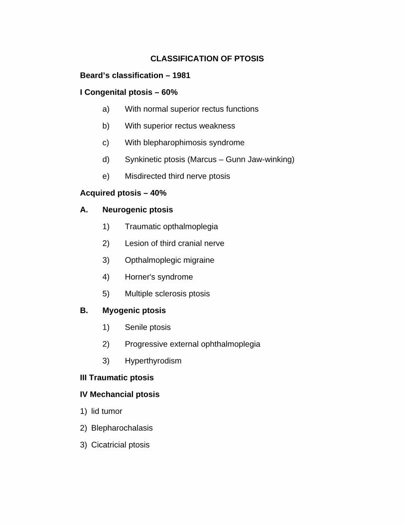
CLASSIFICATION OF PTOSIS
Beard’s classification – 1981
I Congenital ptosis – 60%
a) With normal superior rectus functions
b) With superior rectus weakness
c) With blepharophimosis syndrome
d) Synkinetic ptosis (Marcus – Gunn Jaw-winking)
e) Misdirected third nerve ptosis
Acquired ptosis – 40%
A. Neurogenic ptosis
1) Traumatic opthalmoplegia
2) Lesion of third cranial nerve
3) Opthalmoplegic migraine
4) Horner's syndrome
5) Multiple sclerosis ptosis
B. Myogenic ptosis
1) Senile ptosis
2) Progressive external ophthalmoplegia
3) Hyperthyrodism
III Traumatic ptosis
IV Mechancial ptosis
1) lid tumor
2) Blepharochalasis
3) Cicatricial ptosis
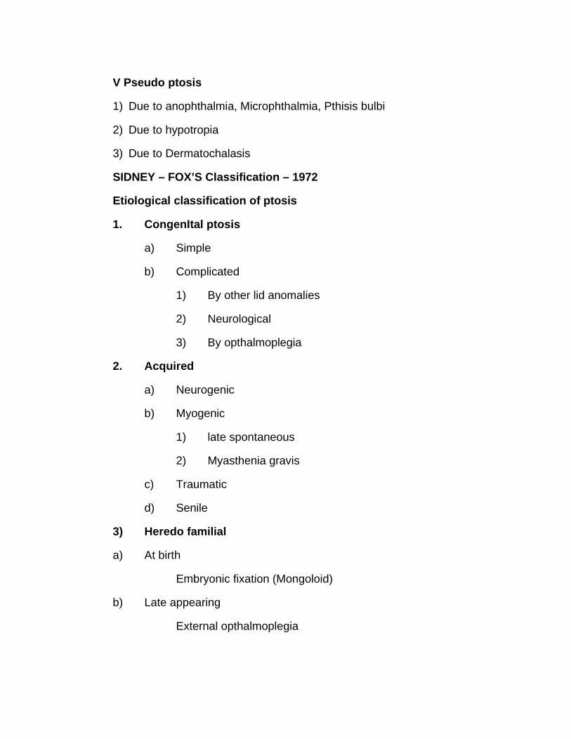
V Pseudo ptosis
1) Due to anophthalmia, Microphthalmia, Pthisis bulbi
2) Due to hypotropia
3) Due to Dermatochalasis
SIDNEY – FOX’S Classification – 1972
Etiological classification of ptosis
1. CongenItal ptosis
a) Simple
b) Complicated
1) By other lid anomalies
2) Neurological
3) By opthalmoplegia
2. Acquired
a) Neurogenic
b) Myogenic
1) late spontaneous
2) Myasthenia gravis
c) Traumatic
d) Senile
3) Heredo familial
a) At birth
Embryonic fixation (Mongoloid)
b) Late appearing
External opthalmoplegia
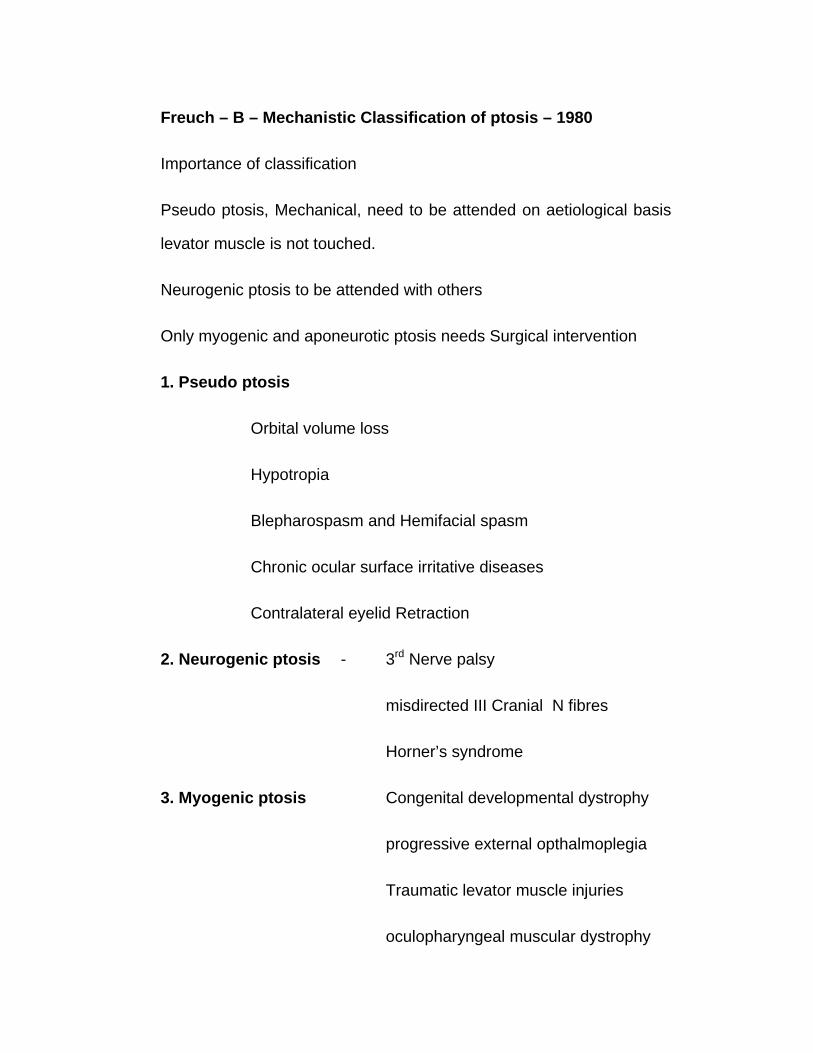
Freuch – B – Mechanistic Classification of ptosis – 1980
Importance of classification
Pseudo ptosis, Mechanical, need to be attended on aetiological basis
levator muscle is not touched.
Neurogenic ptosis to be attended with others
Only myogenic and aponeurotic ptosis needs Surgical intervention
1. Pseudo ptosis
Orbital volume loss
Hypotropia
Blepharospasm and Hemifacial spasm
Chronic ocular surface irritative diseases
Contralateral eyelid Retraction
2. Neurogenic ptosis - 3rd Nerve palsy
misdirected III Cranial N fibres
Horner’s syndrome
3. Myogenic ptosis Congenital developmental dystrophy
progressive external opthalmoplegia
Traumatic levator muscle injuries
oculopharyngeal muscular dystrophy
Myotonic dystrophy
Myasthenia gravis
Toxic myopathy
Late – acquired hereditary ptosis
Non – hereditary acquired myopathy
4. Aponeurotic ptosis
Aponeurotic dehiscence or disinsertion
Senile aponeurotic redundancy
5. Mechanical ptosis
Eyelid or orbital tumor
Eyelid oedema / infection, haematoma
Dermatochalasis
Brow ptosis
Upper eyelid skin disease
LITERATURE OF PTOSIS
Myogenic Ptosis:-
Congenital : - results from dysgenesis of the levator muscle.
Instead of usual muscle fibres, fibrous or adipose tissue is present in
the muscle belly. Congenital myogenic ptosis with an associated poor
bell’s phenomenon or vertical strabismus may indicate concomitant
maldevelopment of the superior rectus(double elevator palsy) or
monocular elevation deficiency.
Acquired myogenic ptosis is uncommon and result from
localized or dffuse muscular disease such as myotonic dystrophy,
chronic progressive external ophthalmoplegia, myasthenia gravis.
Aponeurotic ptosis:-
The levator aponeurosis transmits levator force to the eyelid.
Thus any disruption in its anatomy or function can lead to ptosis.
Congenital aponeurotic ptosis is caused by failure of the aponeurosis
to insert in its normal position on the anterior surface of the tarsus
characterised by good upper eyelid excursion and high upper eyelid
crease. It is a rare case of congenital ptosis and is associated with
birth trauma.
Acquired aponeurotic ptosis is the most common form of all form
of ptosis caused by stretching, dehiscence or disinsertion of the levator
aponeurosis from its normal position. Common causes are involutional

attenuation or repetitive traction of the eyelid. Aponeurotic ptosis may
also be caused or exacerbated by intraocular surgery or eyelid surgery
through multiple mechanism.
Eyelid with aponeurotic ptosis characteristically have a high or
absent upper eyelid crease secondary to upward displacement or loss
of the insertion of levator fibres into the skin.
The levator muscle itself being healthy, the levator function in
aponeurotic ptosis is usually normal (approximately 15mm)
Comparison between congenital and acquired aponeurotic ptosis.
Congenital Myogenic Ptosis
Aponeurotic ptosis
1 Palpebral fissure height mild to severe ptosis mild to severe ptosis
2 Upper eyelid crease weak or absent crease Higher than normal crease
3. Levator function reduced Near normal
4. ondown gaze eyelid lag Eyelid drop
Neurogenic ptosis:-
Congenital neurogenic ptosis is caused by innervational defects
that occur during embryonic development congenital oculomotor nerve
palsy manifest as Blepharoptosis associated with defective Extraocular
movements
Congenital Horner’s syndrome may case mild ptosis associated
with miosis, anhidrosis and decreased pigmentation of the iris and
areola on the involved side.
Congenital myogenic ptosis may also be synkinetic. Marcus
Gunn jaw winking syndrome is the most common form. In this the
unilaterally ptotic eyelid elevates with Jaw movement. This is thought
to be caused by abberant connection between the motor division of
cranial V and levator muscle.
Acquired myogenic ptosis is caused by acquired oculomotor
nerve palsy.
Acquired Horner’s syndrome lead to mild ptosis (2mm)
Mechanical ptosis
The condition in which a swelling in the upper eyelid pulls down
the lid. It can be due to congenital abnormality such as plexiform
neurofibroma or by an acquired neoplasm.
Traumatic ptosis
Blunt or sharp trauma to the levator aponeurosis or the levator
muscle may also cause ptosis. Orbital and neurosurgical procedures
also lead to traumatic ptosis.
EVALUATION OF PTOSIS
History
The patent’s history usually distinguishes congenital from
acquired ptosis. Ptosis in other family members should alert the
observer the possibility of a familiar disorder.
Rapidity of onset should be questioned in all patients, gradual
onset is most typical in acquired cases, but any association with
diplopia or other neurologic symptoms requires further investigation.
A history of normal lid height in the morning progressing to
moderate ptosis by evening, may indicate myasthenia gravis. History
of previous surgery is important.
The patient should be questioned about previous episodes of
eyelid oedema as with allergic angio neurotic oedema which can result
in aponeurotic defect.
OBSERVATION
While taking the history the surgeon should observe the patients
eyes and face. It should be noted whether the ptosis is unilateral or
bilateral and whether there is any associated neuromuscular disorder
such as facial muscle weakness or blepharospasm. The presence of
concurrent anatomical deformities such as brow ptosis,
dermatochalasis, Blepharophimosis should be recorded.
A head turn or tilt should lead to a careful evaluation of ocular
motility to rule out the presence of associated strabismus. The ptotic
eyelid is then more carefully observed, the degree to which the eyelid
margin overlaps the pupil and the general contour of the eyelid is
noted. The presence of any eyelid scar, mass lesion is noted.
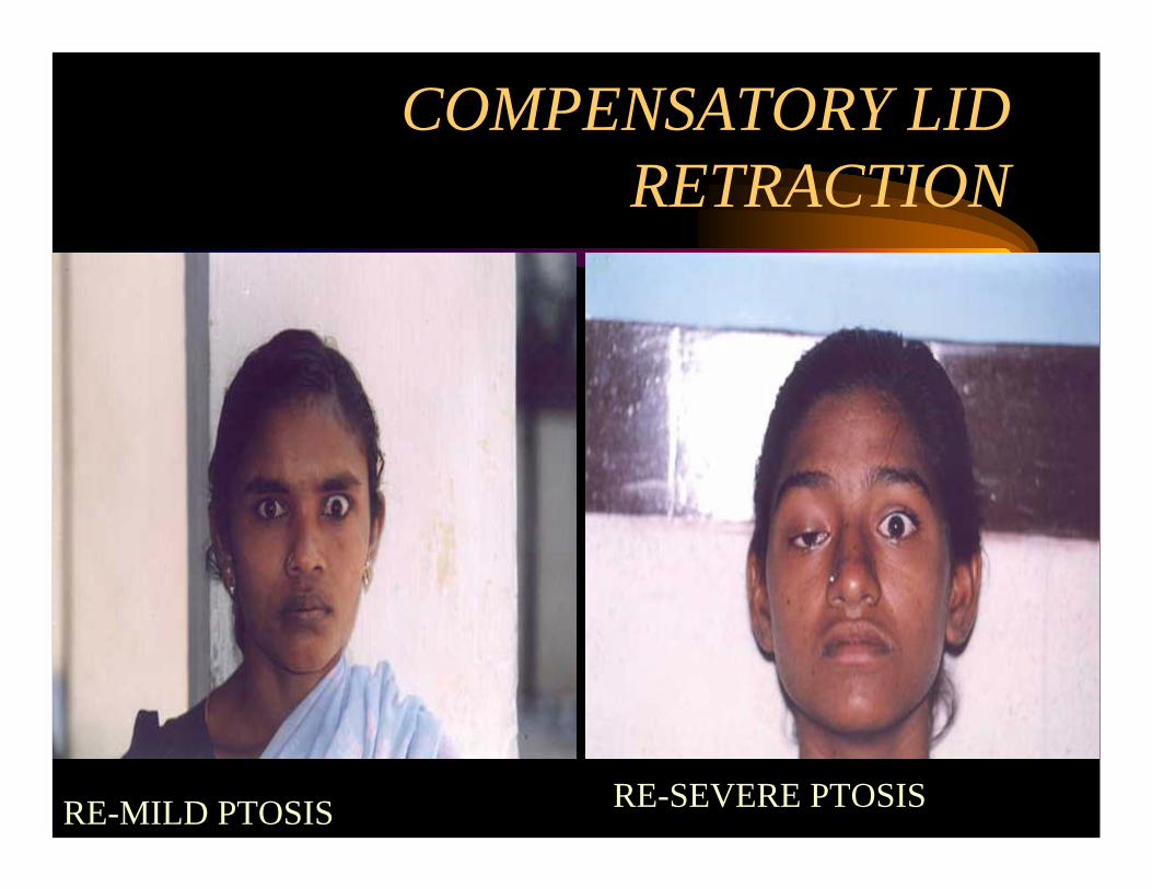
Retraction of the lower eyelid and / or the contralateral upper eyelid
should raise suspicion of early thyroid opthalomopathy.
The lack of spontaneous eyelid movement may suggest
progressive external ophthalmoplegia. The presence of anisocoria
should raise the possibility of Horners syndrome, third nerve Injury or
ocular trauma.

PHYSICAL EXAMINATION
1. Visual acuity.
The ocular examinations should include a record of best
corrected visual acuity to evaluate the presence of amblyopia on the
ptotic eye.
2. Vertical interpalpebral fissure height
This is measured at the widest point between the lower eyelid
and the upper eyelid with the patent fixing on a distant object in
primary gaze.
3. Margin reflex distance (MRD)
This is the distance from the upper eyelid margin to the corneal
light reflex in primary position, the single most effective measurement
in describing the amount of ptosis. MRD2 is the distance from the
corneal light reflex to the lower eyelid margin, sum of MRD1 & MRD2
should equal the vertical interpalpebral fissure height.
Amount of ptosis may be classified
1. Mild ptosis - 2mm or less
2. Moderate ptosis - 3 mm
3. Severe ptosis - 4mm or more
4. Levator function:-
Estimation of levator function is the single most important aspect
of ptosis evaluation for surgical planning.
Berkes method
Measures the excursion of the upperlid from extreme downgaze
to extreme up gaze, with action of frontalis muscle blocked. The
readings are recorded in millimeter. The measurement needs to be
accurate.
The levator function is classified as
8mm or more - good
5-7 mm - Fair
4mm - Poor
Margin Crease distance :
The distance from the upper eyelid crease to the eyelid margin
is measured. The upper eyelid crease is often shallow or absent in
congenital ptosis. The crease is usually elevated in patients with
involutional ptosis.
Pharmacologial test
Helpful in confirming the clinical diagnosis
Edrophonium or Tensilon Test
Tensilon test is used to diagnose myasthenia Gravis.
Edrophonium chloride is an ultra short acting acetyl cholinergic agent.
10mg of edrophonium is prepared in a tuberculin syringe. The needle
is left in situ after 2mg is injected intravenously over a 15-30 second
period. If no untoward effects are noted within one minute, the
remaining 8mg is slowly injected. If there is no improvement of ptosis
in 1-5 minutes the patients probably does not have myasthenia gravis.
Adverse reaction
Pallor, lacrimation, salivation, flushing, abdominal cramping,
bradycardia, an injection of 0.5mg of atropine is give intravenously.
Icepack test
Helps in the diagnosis of myasthenia gravis. The icepack test is
a simple procedure with a few potential side effect. An icepack is
applied to the patients eyelid for about 2 minutes. If MG is present the
ptosis often improves because of the enhancement of neuromuscular
transmission that occurs with inhibition of acetyl cholinesterase.
Phenylephrine test :
Phenylephrine 10% drops used in diagnosis of ptosis due to
Horner’s syndrome.
Laboratory Test
Myasthenia Gravis serum assay of acetyl choline receptor
antibody.
CSF analysis – multiple sclerosis.
Chronic progressive external ophthalmoloplegia. –
Electromyography, Electroretinography, Mitochondrial assay.
Thyroid eye disease – T3,T4 and TSH
Imaging techniques
May be required in
1. Neurological deficit and multiple sclerosis.
2. Horner’s syndrome
3. Thyroid related ophthalmopathy
4. Myasthenia gravis
EVOLUTION OF MANAGEMENT OF PTOSIS
Numerous surgical procedures have been advanced for the
correction of ptosis over the past one hundred and fifty years. Many
represent minor variations of more standard techniques.
1. Non surgical methods or palliative measures
1. Lid Crutches
Gold Heieshen (1890) Kaufmann (1893), Meyer (1893) Dodge
(1935).
2. Contact lens with a shelf
Dudragne (1946), Watillon and pivont (1957), Cochet (1967) and
Barridson (1970).
3. Use of magnetic force
Conway 1973, suggested elevation of the lid by magnetic force
Surgical methods
1. Skin resection
Crescents of skin of the upper lid were removed by the Arabian
surgeons. Von Graefe tried excision of skin and the underlying
muscular tissue.
2. Tarsus Resection
Carried out in Trachoma IV stage with mechanical ptosis was
the begining of this type of ptosis surgery. As recently as 1961 a type
of tarsectomy for lesser of ptosis was reported by fasanella and servat.
3. Full thickness lid Resection
1996. Restivo, Mandridi and Valvo, 1975, Mecord. 1975,
Mustarde did a split level lid resection
levator Muscle Utilisation
Bowman is credited with first levator muscle resection procedure
(1857) via conjunctival approach upper border of tarsus excised with ¾
of levator aponeurosis and muscle.
1883, Everbusch, via the anterior approach, 1896, wolft, 1923
Blascovics via the conjunctival approach. Modification by Agatson
1924, Berke 1952, Jone 1964, Mustarde 1988, Shortened the levator
by tucking it.
5. Resection of Mullers muscle and Conjunctiva
Putterman and urist 1975, treated small degree of ptosis by
resection of mullers muscle and its overlying conjunctiva.
llif 1976 modified fasanella – servat procedure.
Suspension from Brow
Brow suspension by skin flaps
1866, Pavas, Tanscy 1895, Miachek 1915
Brow suspension by orbicularis oculi
Reese 1924
Brow suspension by Tendon
In some parts of England, Plantar tendons of the fifth toe is
used Palmaris longus tendon is also used.
Brow Suspension by Collagen Strips
Illiff, 1963, used scleral slips.
Brow Suspension by fascia lata strips
Wright 1922, Lexer 1923, Rosenberg – 1990, Fox 1966,
Mandeland and Crawford 1972, Craw ford 1966.
Brow Suspension by Non-Absorbable Sutures
1n 1880, Drarasard used non-absorbable sutures. Hess 1893
and Koster 1899 developed procedures Tillet and Tillet 1966 used a
silicone rubber sling. Rama and peduzzi 1973, used 1 mm diameter
silicone rod. Downes and collin, 1989, have described successful
results in 17 ptotic lids corrected with mersilene slings.

MANAGEMENT OF PTOSIS
Non-Surgical
1. Ocular irritation / myasthenia Graves,
2. Eyelid movement obstruction / Orbital disorders
3. Crutch Glasses – CPEO
STANDARD SURGICAL PROCEDURES
Anaesthesia
Local anaesthesia is preferable to general anaesthesia if the
patient will tolerate it, since the voluntary movement of the levator
muscle aids in the identification of lid structures and a better operative
assessment of lid level is possible.
Surgical Procedures
1. Myogenic ptosis 1- 2 mm ptosis with 8-15 mm action –
minimal fasanella servat
2. Moderate to severe 3 – 8 mm action – levator resection to
be done.
3. Severe ptosis with 0-2 mm frontalis sling surgery.
Aponeurotic advancement surgery
Indication:-
Mild to moderate ptosis with very good levator action (5-15mm)
Good down gaze and high tarsal fold.
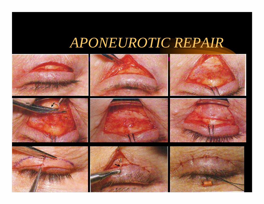
Procedure:-
Under local anaesthesia.
The skin incision marked along the eyelid crease is made
symmetric to the contralateral crease. If bilateral – crease should be
made 8-10 mm from the central lid margin.
The marking is extended nasally to the area just above the
upper punctum to avoid the formation of undesirable webbing or
redundant skin in the medial canthal area.
The incision is made along the skin line with a 15 Bard Parker
blade. Incision should be deep enough to spread the skin and visualise
the subcutaneous tissue. Dissection of an inferior skin flap in the
central portion of the eyelid exposes the anterior surface the tarsus.
Excess bleeding can occur due to damage to the underlying peripheral
vascular arcade.
The edge of the dehiscent levator aponeurosis is seen as a
white line of tissue superior to the upper border of tarsus between the
orbital fat and mullers muscle. The entire orbital septum is incised
horizontally.
Once the levator complex has been exposed the eyelid is lifted
off the globe. A double armed suture is passed through the central
tarsus with partial thickness bites 1 – 2 mm below the superior tarsal
border. A central 5mm broad based bite through the tarsus is
recommended for better eyelid contour.
Both arm of suture passed through levator aponeurosis in a
mattress fashion. The suture is tied temporarily to allow for adjustment.
The patient opens both eyes and the surgeon assess the eyelid height
and contour. An intra-operative over correction of 1mm should be
aimed since the lid tends to droop in the post operative period. The
suture is tied with a permanent square knot and the lid height is
evaluated before skin closure.
Skin is closed by a running sub cuticular 6’0’ prolene suture for a
seam less skin closure and painless removal.
Lubricants and antibiotic ointment is used post operatively.
Post – Operative care
With levator resection and sling procedures. Lagophthalmos is
expected, the lower lid is pulled up to cover the cornea with a modified
frost suture immediately after surgery. Antibiotic ointment and patch for
1 day.
After one week sutures can be removed.

COMPLICATION OF SURGERY
Under correction
Most frequent complication. This is not a complication but a fault
of judgement and some times of techniques.
Over correction
Usually rare
Lid lag, lagophthalmos & Keratitis
Lid lag is seen to some degree after most ptosis surgery
Entropion and ectropion:
Loss of lashes – rare
Lid crease, lid fold abnormalities
A lid crease that is too high or too low results from a skin incision
that has been too high or too low if the skin approach has been used.
Conjunctival prolapse
Prolapse of conjunctiva from the upper fornix is rare. It will
usually retract spontaneously within a week or two.
Post operative haemorrhage
Rare : A few moment of pressure over the lid followed by a
pressure dressing for a day or two may be enough
Infection
Infection is rare, when infection does occur it can be
successfully treated with antibiotics except in brow suspension with a
non – absorbable suture when infection recurs when the antibiotic is
discontinued. Removal of the suture and resurgery is the procedure of
choice.
AIM OF THE STUDY
Aponeurotic Ptosis the most commonest of acquired ptosis, its
evaluation and management are analysed in this study.
MATERIAL AND METHODS
It is felt in the recent past, oculo plastic surgery particularly of lid
and adnexa have been generally advancing. This may be due to the
improved technology in the field of the plastic surgery and also the
oculoplastic surgery has also become a subspeciality in the field of the
ophthalmology.
The study was conducted at Regional Institute of
Ophthalmology, Government Ophthalmic Hospital for a period of 24
months from August 2004 to August 2006.
Cases were from Chennai, referred from different parts of the
state to this tertiary care hospital.
Incidence of ptosis in the Regional Institute during the study
period was 109 cases of which congenital ptosis – 60 cases
Aponeurotic Ptosis – 26 cases, Myogenic ptosis – 11 cases and post
traumatic ptosis – 12 cases.
Only 26 cases of aponeurotic ptosis was taken up for study.
Inclusion Criteria
All patients with aponeurotic ptosis, with good levator function
were taken up for this study.
Exclusion Criteria
• Congenital ptosis
• Myogenic ptosis
• Post traumatic ptosis
• Post inflammatory ptosis
• Mechanical ptosis and other causes.
A detailed history was taken in each case. Examination of the
patient consisted of a detailed ocular examination, complete
examination of the ptosis.
Ptosis was examined thoroughly as given on the proforma with
special emphasis on the degree of ptosis, the amount of levator
functions, Bells’ phenomenon, orbicularis muscle Power, corneal
sensation and staining. Adequate and appropriate investigations were
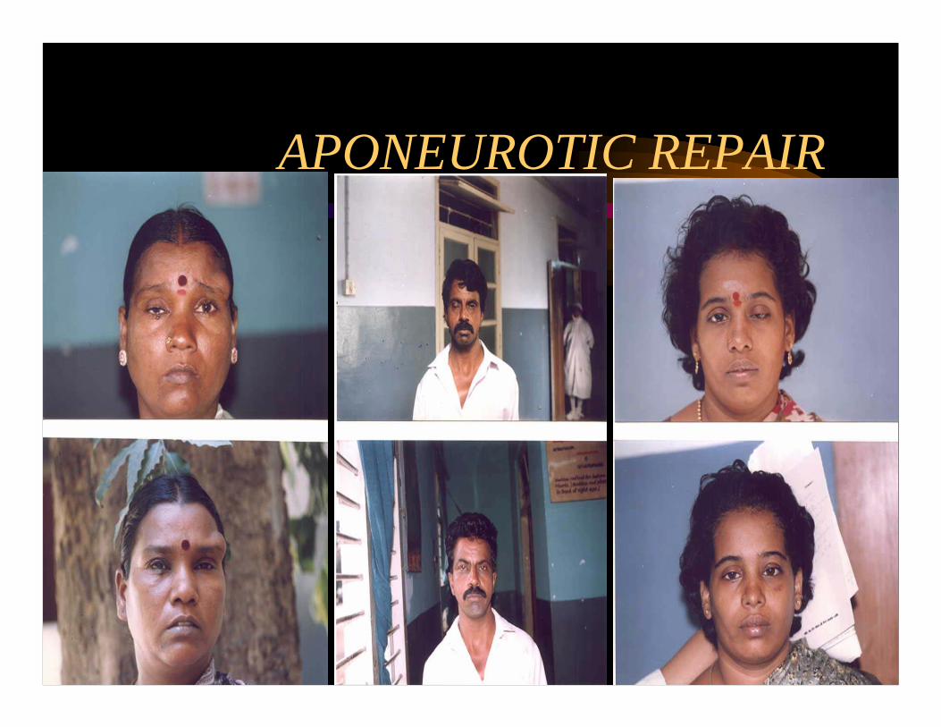
also done before taking up the patient for surgery. Photographic
documentation was done as a part of pre-operative as well post-
operative evaluation.
Out of 26 patients, 7 patients had bilateral ptosis and so the
number of ptosis evaluated was 33. Based on the amount of levator
function and degree of ptosis, surgical procedures were planned and
performed.
For all mild, moderate and severe ptosis with good levator
function. Levator advancement surgery through the transcutaneous
approach (external) was performed.
Post – operative evaluation was done on the first and fifth post –
operative day, monthly follow up was done for about 2 months.
Patients were evaluated 6 months after surgery to determine the
success of surgery as well as to find out the incidence of recurrence.

ANALYSIS AND DISCUSSION
The prospective study on evaluation and surgical management
of Aponeurotic Ptosis was conducted at Regional Institute of
Ophthalmology Government Ophthalmic hospital, Chennai. The study
was conducted between August 2004 and August 2006.
All cases of aponeurotic ptosis with good levator function – 26
patients were evaluated and were operated and taken up for this
study. The various findings in this study were analysed and details are
as follows:
Incidence
Incidence of ptosis in Regional Institute of Ophthalmology during
the study period was 109 of which congenital ptosis – 60 cases(55%),
aponeurotic ptosis – 26 (24%) cases, Myogenic ptosis – 11 (10%)
cases and post traumatic ptosis – 12 (11%).
26 cases of aponeurotic ptosis were taken up for the study.
Table – 1
Incidence of Ptosis
Sl.No. Congenital ptosis
aponeurotic ptosis
Myogenic ptosis
Post traumatic
ptosis
Total
1. 60
(55%)
26
(24%)
11
(10%)
12
(11%)
109

60
26
11 12
0
10
20
30
40
50
60
Congenital ptosis aponeurotic ptosis Myogenic ptosis Post traumatic ptosis
IncIdence of PtosIs

Gender distribution
Out of 26 patients taken up in this study 16 male patients
(61.5%) and 10 female patients (38.5%)
Table – 2
Sl. No. Male Female Total
1. 16
(61.5%)
10
(38.5%)
26
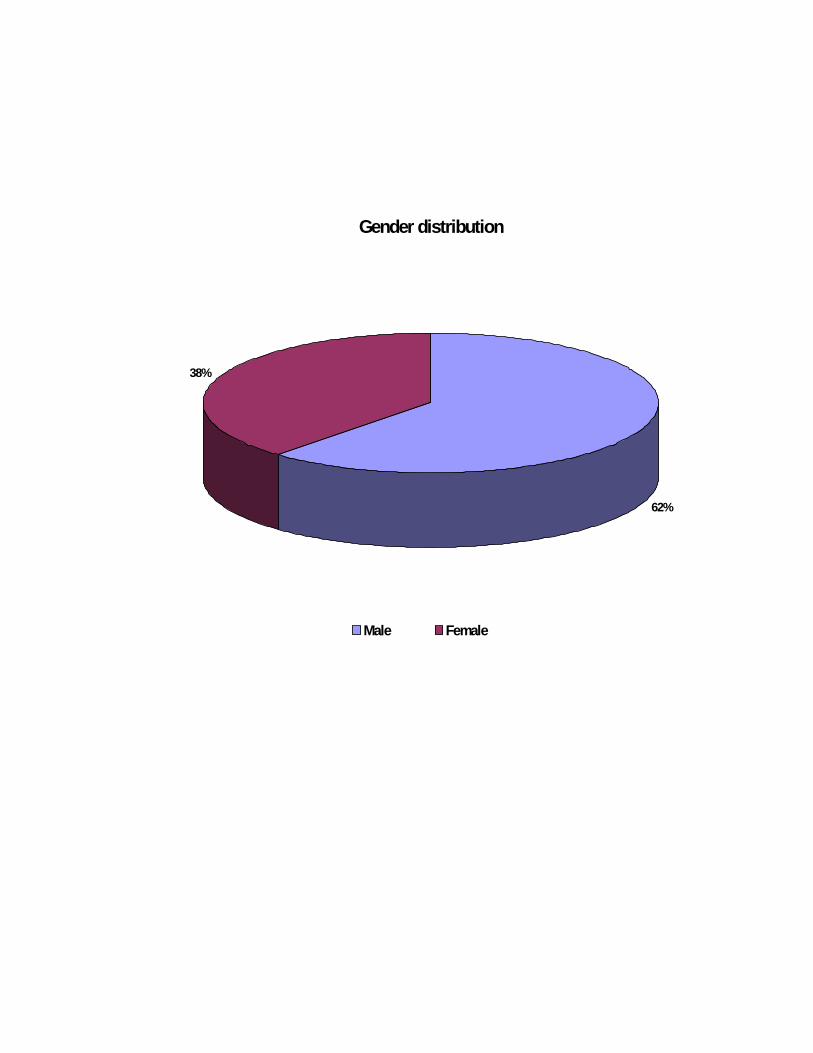
Gender distribution
62%
38%
Male Female
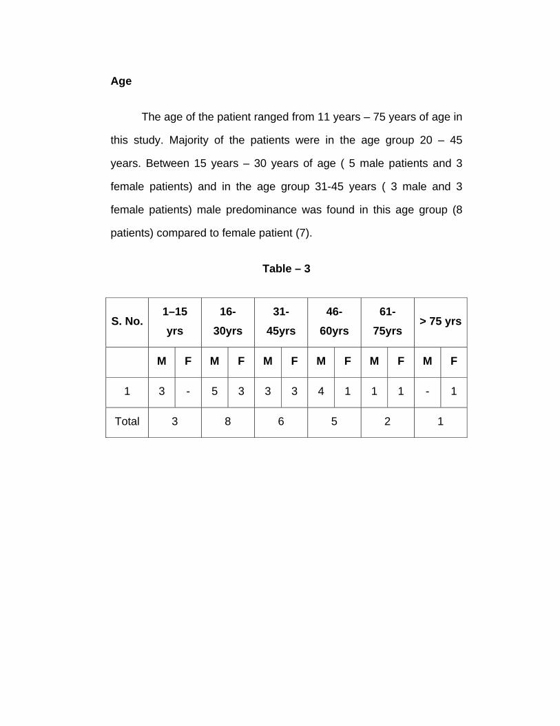
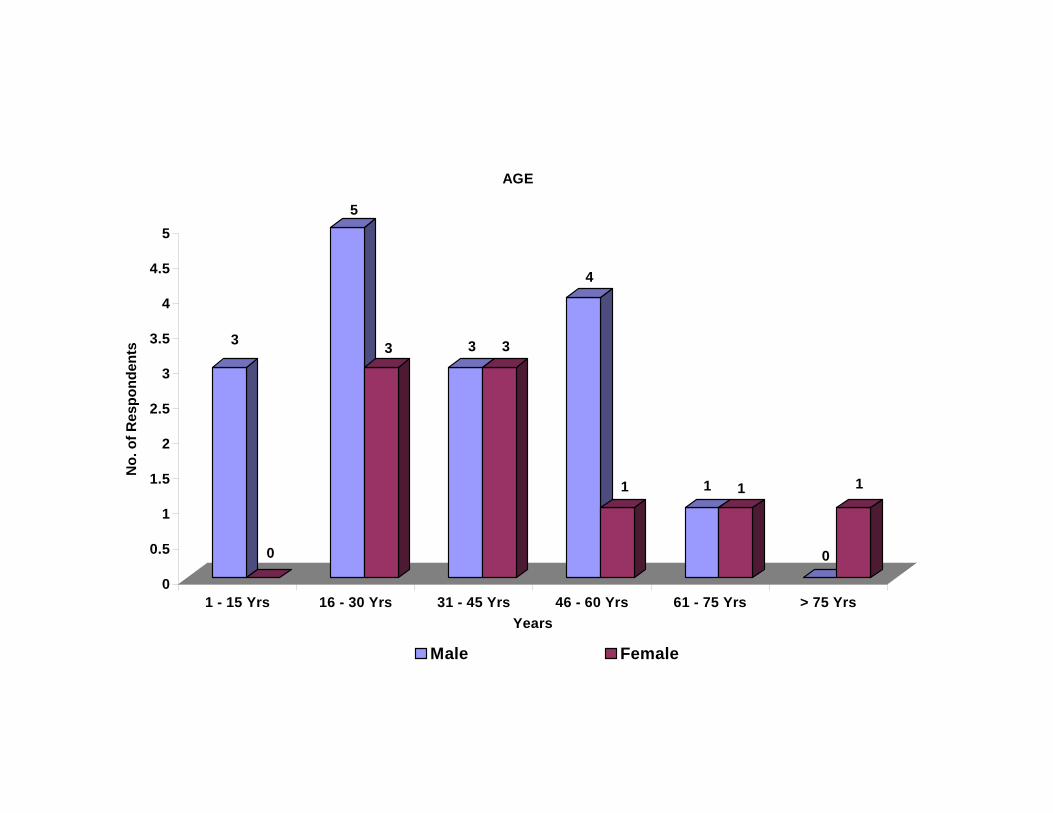
Age
The age of the patient ranged from 11 years – 75 years of age in
this study. Majority of the patients were in the age group 20 – 45
years. Between 15 years – 30 years of age ( 5 male patients and 3
female patients) and in the age group 31-45 years ( 3 male and 3
female patients) male predominance was found in this age group (8
patients) compared to female patient (7).
Table – 3
S. No. 1–15 yrs
16-30yrs
31-45yrs
46-60yrs
61-75yrs
> 75 yrs
M F M F M F M F M F M F
1 3 - 5 3 3 3 4 1 1 1 - 1
Total 3 8 6 5 2 1
3
0
5
3 3 3
4
1 1 1
0
1
0
0.5
1
1.5
2
2.5
3
3.5
4
4.5
5
No.
of R
espo
nden
ts
1 - 15 Yrs 16 - 30 Yrs 31 - 45 Yrs 46 - 60 Yrs 61 - 75 Yrs > 75 YrsYears
AGE
Male Female
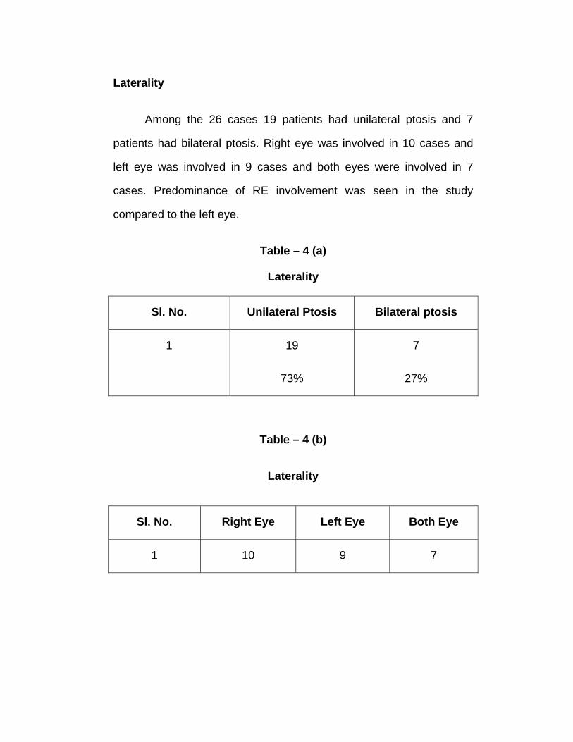
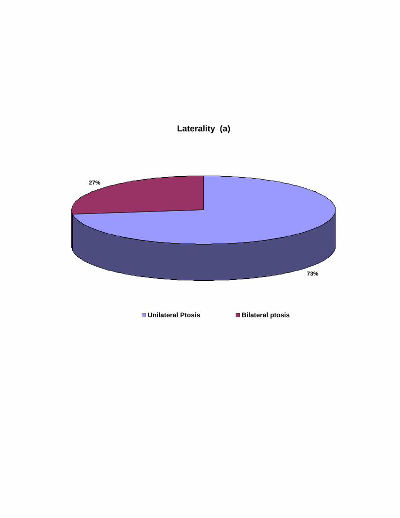
Laterality
Among the 26 cases 19 patients had unilateral ptosis and 7
patients had bilateral ptosis. Right eye was involved in 10 cases and
left eye was involved in 9 cases and both eyes were involved in 7
cases. Predominance of RE involvement was seen in the study
compared to the left eye.
Table – 4 (a)
Laterality
Sl. No. Unilateral Ptosis Bilateral ptosis
1 19
73%
7
27%
Table – 4 (b)
Laterality
Sl. No. Right Eye Left Eye Both Eye
1 10 9 7
Laterality (a)
73%
27%
Unilateral Ptosis Bilateral ptosis
Degree of Ptosis
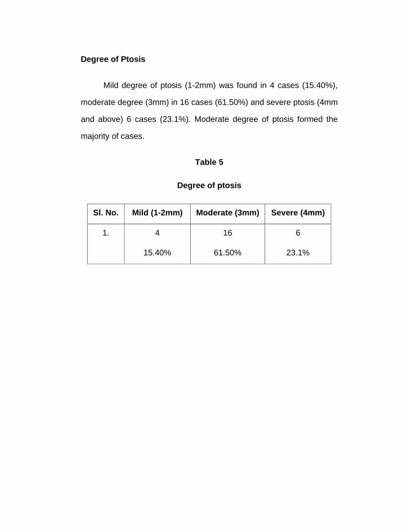
Mild degree of ptosis (1-2mm) was found in 4 cases (15.40%),
moderate degree (3mm) in 16 cases (61.50%) and severe ptosis (4mm
and above) 6 cases (23.1%). Moderate degree of ptosis formed the
majority of cases.
Table 5
Degree of ptosis
Sl. No. Mild (1-2mm) Moderate (3mm) Severe (4mm)
1. 4
15.40%
16
61.50%
6
23.1%
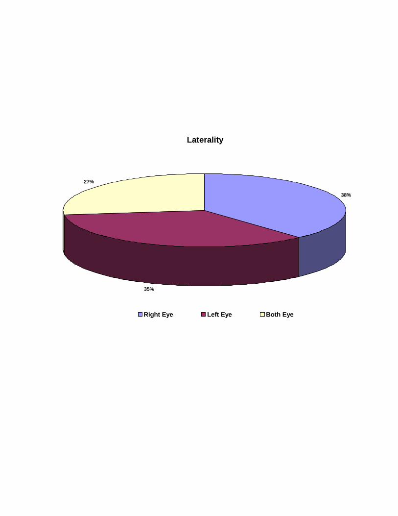
Laterality
38%
35%
27%
Right Eye Left Eye Both Eye
Management of Ptosis
Of 33 ptosis, of 26 patients 30 ptosis were corrected surgically, 3
cases of bilateral ptosis did not have the other eye operated. Of this 26
patients, 3 patients were operated under General anesthesia and all
the other cases were operated under local anesthesia 4 mild ptosis
cases, 16 moderate ptosis cases and 6 severe ptosis cases were
corrected surgically.
16 cases were male and 10 were female majority belonging to
the age group 20-45 year of age group. Most probably due to the
awareness and cosmetic consciousness in this age group and also
lack of interest for cosmetic correction in older age group.
All the patients were operated using the technique of levator
aponeurotic advancement through transcutaneous approach.
Most of the case were operated under local anesthesia. It gives
the greatest advantage to the surgeon, because on the table the eyelid
skin incision which form future skin crease can be marked by
comparing it with the contra lateral eyelid and also the final suture
adjustment can also be made by asking the patient to open both eyes
and the surgeon can assess the eyelid height and contour. (Anderson
Rl et al. in his study showed that local anesthesia during aponeurotic
advancement surgery is advantageous)
Technique of levator aponeurotic advancement performed in this
study was transcutaneous approach.
Pre-operative and on the table in all cases of unilateral ptosis
the possibility of post operative ptosis in the contra lateral eyelid was
assessed by lifting the ptotic eyelid to the desired level with one finger
and observing any changes on the contra lateral eyelid height.
Attention was given to the eyelid crease and fold, especially
when considering a blepharoplasty combined with ptosis surgery.
Accuracy of ptosis repair by levator advancement depends on
the intra-operative co-operation of the patient in opening the eyes to
verify the appropriate eyelid position.
The incision was marked along the eyelid crease symmetric to
the contra lateral crease. The marking is extended nasally to the area
just above the upper punctum to avoid redundant skin in the medial
canthal area.
The incision is made with a 15 Bard parker blade. Incision is
deep enough to spread the skin and visualize the subcutaneous
tissue. Dissection of an inferior skin flap in the central portion of the
eyelid exposes the anterior surface the tarsus.
The edge of the dehiscent levator aponeurosis is seen as a
white line of tissue superior to the upper border of the tarsus between
the orbital fat and mullers muscle. The entire septum is incised
horizontally.
Once the levator has been exposed a double armed suture is
passed through the central tarsus with partial thickness bites 1-2mm
below the superior tarsal border. A central 5mm broad based bite is
passed through the tarsus for better eyelid contour.
In 20 cases of our study three suture technique was used for
levator advancement (Ibrar Hussain et al 3 suture technique gave
good cosmetic results and also can be used in congenital ptosis with
good levator function.)
6 cases of aponeurotic ptosis was performed using a single
broad based mattress suture. This eliminates the need for medial and
lateral suture. Making the procedure easier to perform and results in
better contour of eyelid.
(liu-D et al – used single suture technique for levator
advancement, the procedure is simple and effective and versatile used
in the correction of involutional, post traumatic ptosis).
Most of the cases had a thinned out aponeurosis (Hosal Banu
M.M.D et.al. measure thickness of levator aponeurosis using UBM and
found that in, aponeurotic ptosis most common pathology is thinned
out aponeurosis.).
Both arm of suture passed through the levator aponeurosis. The
suture is tied temporarily to allow for adjustment. The patient is asked
to open both eyes and the surgeon assess the eyelid height and
contour and the permanent suture knot is applied.
Skin closed with a sub cuticular suture for a seamless skin
closure and painless removal.
All the cases, mild, moderate and severe degree of ptosis were
operated through anterior transcutaneous approach in this study
(Anderson Rl et al, shovlin Jp et al. caraway Jh et al in their study have
shown that anterior transcutaneous approach gave good results for all
degree of ptosis)
Advantages of this technique being
1. Easier and better exposure of the tissues
2. Lid need not be averted
3. More access to levator aponeurosis
4. Conjunctiva need not be cut, hence cornea is not injured.
5. All tissues are freely visible and aponeurosis can be identified
easily.
6. Lid fold, excess skin can be excised and forming a future lid
crease is easier.
7. Blepharoplasty can be combined with levator advancement
procedures.
One case was performed with combined procedure of leavtor
advancement and blepharoplasty. Combined procedures can be done
using a transcutaneous approach levator advancement. (Wilkins RB et
al. did combined procedure levator advancement and blepharoplasty
with good results cosmetically and functionally).
Complications
One patient had a conjunctival prolapse which did not get
corrected within a week so it was corrected surgically by excising the
prolapsed conjunctiva and suturing it.
One patient developed dermatochalasis after surgery which was
more on looking up which was corrected by excising the excess skin.
Two patient had mild lid peaking.
CONCLUSION
1. Aponeurotic ptosis is the commonest among the acquired
ptosis.
2. Majority of patients belong to the age group of 20-45 yrs of
age.
3. Aponeurotic ptosis surgery – levator advancement,
transcutaneous approach gives good results functionally and
cosmetically.
4. In this procedure excess skin can be excised and forming a
future lid crease is easier.
5. Levator advancement can be combined with Blepharoplasty.
6. This procedure can be performed not only by the plastic
surgeon but also by the General ophthalmologist.
BIBLIOGRAPHY
Text books
1. Albert Hornblass.. carl.N. Honig – Oculoplastic and orbital
Reconstructive surgery vol. I, 1988 william & wilkins.
2. Bosniak – Principle & Practice ophthalmic plastic and
Reconstructive Surgery 1996 – Vol.- I.
3. Brhan G. Brazzo – Complications in ophthalmic plastic surgery
2003. Springer.
4. Byron C. Smith and Frank A. Ness – Practical Techniques in
ophthalmic plastic surgery, 1981, C.V. Mosby Company.
5. Clinton D.MC Cord Jr. and Myron tanenbaum Oculoplastic
Surgery second edition, 1987 Raven Press.
6. Collin JRO A. Manual of systematic eyelid surgery, Second
edition, 1989, Churchill livingstone.
7. Crowell Beard – Ptosis, third edition, 1987 C.V. mosby and
company.
8. Duke elder S. system of ophthalmology, Vol XII Part I st. Louis,
CV. Mosby, 1974.
9. Frederic A. Jakobiec, Jesse sigelman – Advanced techniques in
ocular surgery WB saunders, 1984.
10. Geoffrey J. Gladstone – oculoplastic surgery atlas 2002 –
Springer.
10. Iliff CE, Iliff WJ. NT –Oculoplastic surgery WB saunders – 1979.
11. Mark R. leine – Manual of Oculoplastic Surgery
12. Mustarde S.C. – Repair and Reconstruction in the orbital region,
third edition 1991. Churchil Livining Stone.
13. Paymen, Sanders and Goldberg – Principles and practice of
ophthalmology, Vol. III First Edition 1987, W.B. Saunders and
Company.
14. Ralph. E. Wesley Techniques in Ophthalmic plastic surgery,
1986 John Wiley and Sons.
15. Sidney A Fox – Surgery of Ptosis, 1980 William and Wilkins
16. Thomas D. Duane – Clinical Ophthalmology vol. 5 1986 Harper
and Row publishers.
17. Yonoff – Orbit and Oculoplasty.
Journals
1. Anderson RL, Dixon RS – Levator aponeurotic advancement
procedure of Choice for a acquired ptosis. Arch oph. 1979 June
97/6 (1123 – 8)
2. Barbara J. Rutledge Ptosis surgery in cosmetic Blepharoplasty;
Cosmetic surgery Nov-Dec. 2004.
3. Bartley R. Freuh M.D. et al., New involutional ptosis correction
Trans. am. oph. society 2004 Dec., 199 – 208.
4. Ben-simon GJ External levator advancement Vs Mullers
conjunctival resection for correction of involutional ptosis Am.J.
Oph. 2005 Sep. 40 (3) 426-32.
5. Carraway JH Levator advancement tuck for eyelid ptosis plastic
reconstructive surgery 1986 March 77 (3)
6. Erb MH, Kersten RL, Effect of unilateral ptosis repair on
contralateral eyelid position oph. plastic reconstructive surgery
2004 Nov. 20 (6)
7. Epstein G Putterman AM Acquired blepharoptosis secondary to
contact lens wear AJO 91:634-638, 1987.
8. Harris WA – Levator tuck simplified procedure Ann. Opn. 1975
(Jun) 7(6) 873 – 8.
9. Hosal Banu M.MD – UBM Levator aponeurosis in congenital
and acquired ptosis art. oph. & plastic Recons surgery July
2004.
10. Ibrar Hussain – Cosmetic outcome of 3 suture technique of
aponeurotic ptosis repair Dept. of oph. Khyber treatment
hospital.
11. Jone LT – Cure of ptosis –aponeurotic repair Arch. Oph. 1975
Aug. 629 – 34.
12. Kersten RL – Acquired Ptosis in young and middle age
individuals oph. 1999 June 924-8.
13. Liu –D Ptosis repair by single suture technique oph. 1993 sep.
100, 1278-9
14. Martin DA – Congenital aponeurotic ptosis Aust. NZ J. Oph.
1988 Nov. 16 291-4.
15. Older JJ Levator aponeurosis tuck treatment for ptosis oph.
surgery – 1978 Aug. 9 (4) 102 – 10.
16. Shovlin JP Aponeurotic approach for the amount of ptosis (Int.
oph. clinic 1997 Summer 37(3) 133 – 50)
17. Steven C. Dressner M.D. Blepharoplasty cosmetic surgery.
18. Wilkins RD – Surgical repair of levator dehiscence
simultaneously with a blepharoplasty Plastic reconstructive surg.
1982. Oct. (431 – 4)
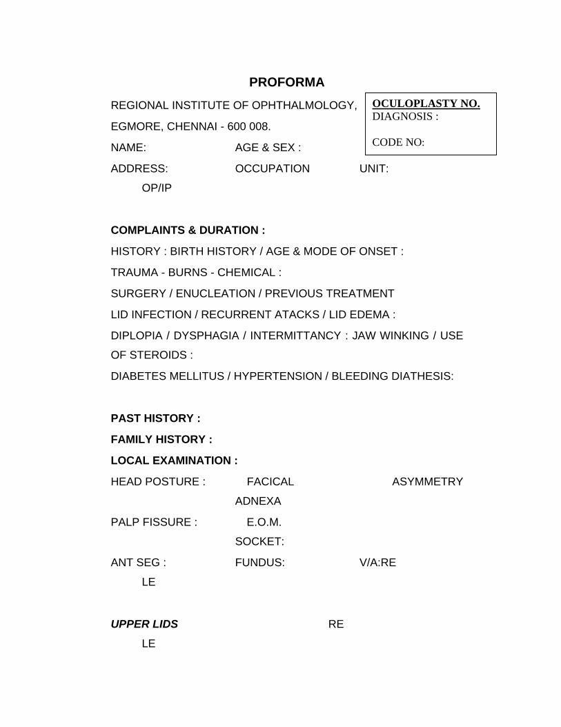
PROFORMA
REGIONAL INSTITUTE OF OPHTHALMOLOGY,
EGMORE, CHENNAI - 600 008.
NAME: AGE & SEX :
ADDRESS: OCCUPATION UNIT:
OP/IP
COMPLAINTS & DURATION :
HISTORY : BIRTH HISTORY / AGE & MODE OF ONSET :
TRAUMA - BURNS - CHEMICAL :
SURGERY / ENUCLEATION / PREVIOUS TREATMENT
LID INFECTION / RECURRENT ATACKS / LID EDEMA :
DIPLOPIA / DYSPHAGIA / INTERMITTANCY : JAW WINKING / USE
OF STEROIDS :
DIABETES MELLITUS / HYPERTENSION / BLEEDING DIATHESIS:
PAST HISTORY :
FAMILY HISTORY :
LOCAL EXAMINATION :
HEAD POSTURE : FACICAL ASYMMETRY
ADNEXA
PALP FISSURE : E.O.M.
SOCKET:
ANT SEG : FUNDUS: V/A:RE
LE
UPPER LIDS RE
LE
OCULOPLASTY NO. DIAGNOSIS : CODE NO:
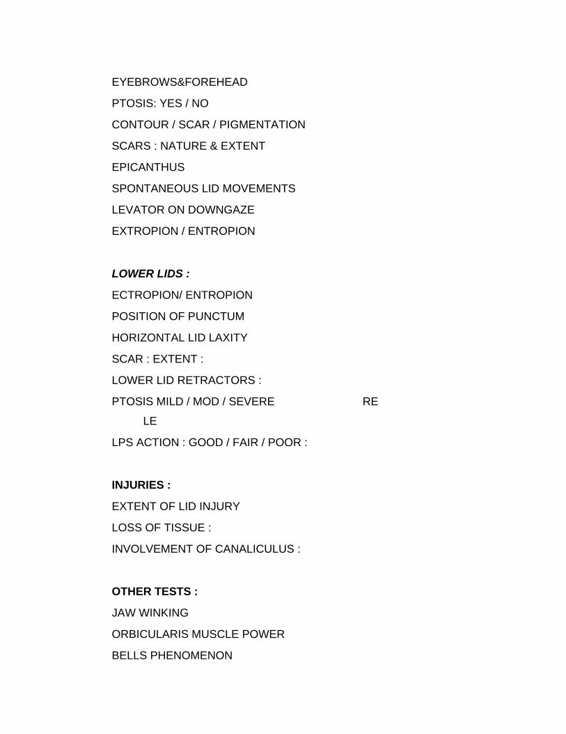
EYEBROWS&FOREHEAD
PTOSIS: YES / NO
CONTOUR / SCAR / PIGMENTATION
SCARS : NATURE & EXTENT
EPICANTHUS
SPONTANEOUS LID MOVEMENTS
LEVATOR ON DOWNGAZE
EXTROPION / ENTROPION
LOWER LIDS :
ECTROPION/ ENTROPION
POSITION OF PUNCTUM
HORIZONTAL LID LAXITY
SCAR : EXTENT :
LOWER LID RETRACTORS :
PTOSIS MILD / MOD / SEVERE RE
LE
LPS ACTION : GOOD / FAIR / POOR :
INJURIES :
EXTENT OF LID INJURY
LOSS OF TISSUE :
INVOLVEMENT OF CANALICULUS :
OTHER TESTS :
JAW WINKING
ORBICULARIS MUSCLE POWER
BELLS PHENOMENON
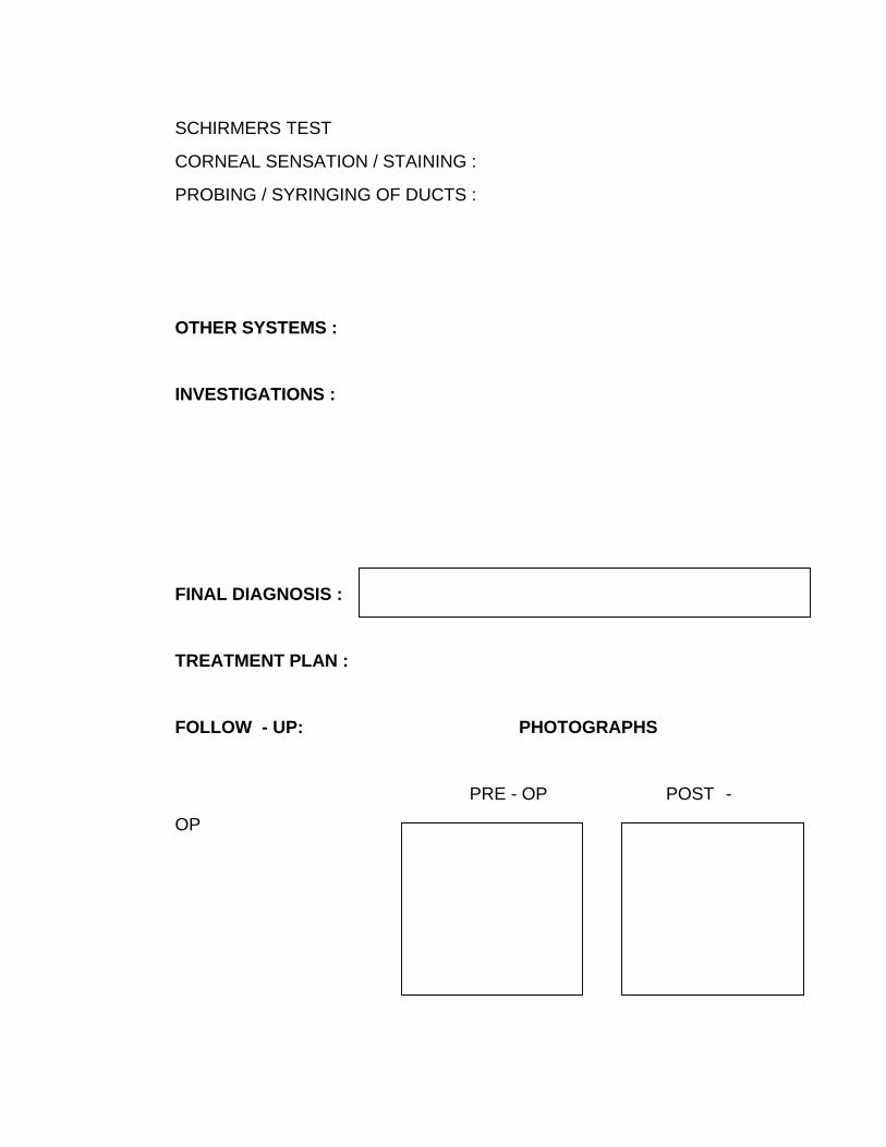
SCHIRMERS TEST
CORNEAL SENSATION / STAINING :
PROBING / SYRINGING OF DUCTS :
OTHER SYSTEMS :
INVESTIGATIONS :
FINAL DIAGNOSIS :
TREATMENT PLAN :
FOLLOW - UP: PHOTOGRAPHS
PRE - OP POST -
OP
MASTER CHART
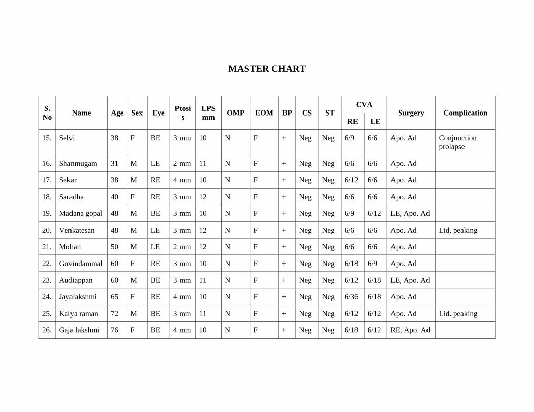
CVA S. No Name Age Sex Eye Ptosis
LPS
mmOMP EOM BP CS ST
RE LE Surgery Complication
1 Dhinesh 11 M RE 4 mm 10 N F + Neg Neg 6/9 6/6 Apo. Ad
2. Jai Kumar 11 M RE 4 mm 10 N F + Neg Neg 6/9 6/6 RE, Apo, Ad with Blepharoplasty
Dermato chalasis
3. Sarath kumar 13 M LE 3 mm 11 N F + Neg Neg 6/6 6/6 Apo. Ad
4. Subash chandra Bose
21 M LE 3 mm 11 N F + Neg Neg 6/6 6/6 Apo. Ad
5. Solochana 23 F LE 3 mm 10 N F + Neg Neg 6/6 6/6 Apo. Ad
6. Radha 20 F LE 2 mm 11 N F + Neg Neg 6/6 6/6 Apo. Ad
7. Muniappan 24 M LE 3 mm 11 N F + Neg Neg 6/6 6/9 Apo. Ad
8. Selvi 25 F BE 3 mm 10 N F + Neg Neg 6/9 6/12 Apo. Ad
9. Suresh 28 M LE 3 mm 11 N F + Neg Neg 6/9 6/6 Apo. Ad
10. Sundar 21 M RE 3 mm 12 N F + Neg Neg 6/9 6/6 Apo. Ad
11. Jayanthi 27 F RE 4 mm 10 N F + Neg Neg 6/9 6/6 Apo. Ad
12. Jarina 33 F RE 3 mm 11 N F + Neg Neg 6/6 6/9 Apo. Ad
13. Mohan 30 M BE 3 mm 10 N F + Neg Neg 6/9 6/6 Apo. Ad
14. Senthil kumar 32 M RE 2 mm 11 N F + Neg Neg 6/6 6/9 Apo. Ad
MASTER CHART
CVA S. No Name Age Sex Eye Ptosi
s LPS mm OMP EOM BP CS ST
RE LE Surgery Complication
15. Selvi 38 F BE 3 mm 10 N F + Neg Neg 6/9 6/6 Apo. Ad Conjunction prolapse
16. Shanmugam 31 M LE 2 mm 11 N F + Neg Neg 6/6 6/6 Apo. Ad
17. Sekar 38 M RE 4 mm 10 N F + Neg Neg 6/12 6/6 Apo. Ad
18. Saradha 40 F RE 3 mm 12 N F + Neg Neg 6/6 6/6 Apo. Ad
19. Madana gopal 48 M BE 3 mm 10 N F + Neg Neg 6/9 6/12 LE, Apo. Ad
20. Venkatesan 48 M LE 3 mm 12 N F + Neg Neg 6/6 6/6 Apo. Ad Lid. peaking
21. Mohan 50 M LE 2 mm 12 N F + Neg Neg 6/6 6/6 Apo. Ad
22. Govindammal 60 F RE 3 mm 10 N F + Neg Neg 6/18 6/9 Apo. Ad
23. Audiappan 60 M BE 3 mm 11 N F + Neg Neg 6/12 6/18 LE, Apo. Ad
24. Jayalakshmi 65 F RE 4 mm 10 N F + Neg Neg 6/36 6/18 Apo. Ad
25. Kalya raman 72 M BE 3 mm 11 N F + Neg Neg 6/12 6/12 Apo. Ad Lid. peaking
26. Gaja lakshmi 76 F BE 4 mm 10 N F + Neg Neg 6/18 6/12 RE, Apo. Ad
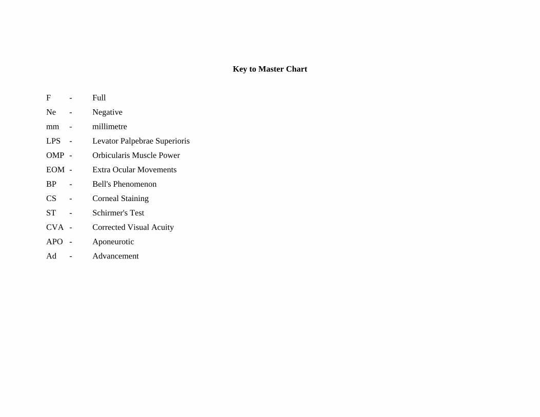
Key to Master Chart
F - Full
Ne - Negative
mm - millimetre
LPS - Levator Palpebrae Superioris
OMP - Orbicularis Muscle Power
EOM - Extra Ocular Movements
BP - Bell's Phenomenon
CS - Corneal Staining
ST - Schirmer's Test
CVA - Corrected Visual Acuity
APO - Aponeurotic
Ad - Advancement
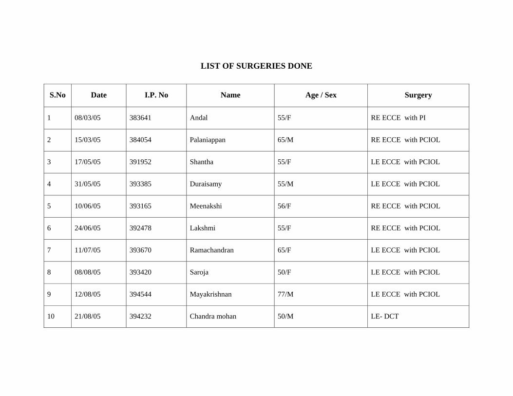
LIST OF SURGERIES DONE
S.No Date I.P. No Name Age / Sex Surgery
1 08/03/05 383641 Andal 55/F RE ECCE with PI
2 15/03/05 384054 Palaniappan 65/M RE ECCE with PCIOL
3 17/05/05 391952 Shantha 55/F LE ECCE with PCIOL
4 31/05/05 393385 Duraisamy 55/M LE ECCE with PCIOL
5 10/06/05 393165 Meenakshi 56/F RE ECCE with PCIOL
6 24/06/05 392478 Lakshmi 55/F RE ECCE with PCIOL
7 11/07/05 393670 Ramachandran 65/F LE ECCE with PCIOL
8 08/08/05 393420 Saroja 50/F LE ECCE with PCIOL
9 12/08/05 394544 Mayakrishnan 77/M LE ECCE with PCIOL
10 21/08/05 394232 Chandra mohan 50/M LE- DCT
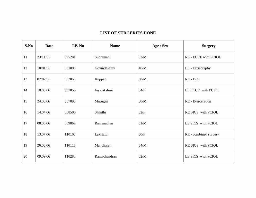
LIST OF SURGERIES DONE
S.No Date I.P. No Name Age / Sex Surgery
11 23/11/05 395281 Subramani 52/M RE - ECCE with PCIOL
12 10/01/06 001098 Govindasamy 40/M LE - Tarssoraphy
13 07/02/06 002853 Kuppan 50/M RE - DCT
14 10.03.06 007856 Jayalakshmi 54/F LE ECCE with PCIOL
15 24.03.06 007890 Murugan 50/M RE - Evisceration
16 14.04.06 008506 Shanthi 52/F RE SICS with PCIOL
17 08.06.06 009869 Ramanathan 51/M LE SICS with PCIOL
18 13.07.06 110102 Lakshmi 60/F RE - combined surgery
19 26.08.06 110116 Manoharan 54/M RE SICS with PCIOL
20 09.09.06 110283 Ramachandran 52/M LE SICS with PCIOL
ANATOMY
COMPENSATORY LID RETRACTION
RE-MILD PTOSIS RE-SEVERE PTOSIS

MEASURING PTOSIS
PALP- APERTURE MRD LEVAT-ACTION
APONEUROTIC REPAIR
APONEUROTIC REPAIR