Embed Size (px)
Citation preview
DOI: 10.21276/sjams
Available online at http://saspublisher.com/sjams/ 3349
Scholars Journal of Applied Medical Sciences (SJAMS) ISSN 2320-6691 (Online)
Sch. J. App. Med. Sci., 2017; 5(8E):3349-3365 ISSN 2347-954X (Print) ©Scholars Academic and Scientific Publisher
(An International Publisher for Academic and Scientific Resources)
www.saspublisher.com
A Study of Outcome of Foramen Magnum Decompression and Duraplasty for
Chiari Malformations Dr. Sreedharala Srinivasa Satynarayana
1, Dr. Palukuri Lakshmi
2*, Dr. Sandeep Raja Pittala
3, Dr. Uday Goutam
Nookathota3, Dr. Alluri Neeraja
3, Dr. Kotha Arjun Reddy
3, G. Prakash Rao
4
1M.S., M.Ch., I/c Prof. of NeuroSurgery, Gandhi Medical College, Telangana State, India
2Assoc Prof. of Plastic Surgery, Osmania Medical College, Hyderabad, Telangana State, India
3Postgraduates in Department of Neurosurgery Department of Neurosurgery, Gandhi Medical College, Gandhi Hospital,
Secunderabad, Telangana 4Professor of Neurosurgery, Gandhi Medical College, Secunderabad
*Corresponding author Dr. Palukuri Lakshmi
Email: [email protected]
Abstract: Chiari malformations appears to be complex in its presentation, cause and natural history. The true incidence
is not known. Around 3% in children and 1% in adults demonstrate radiological evidence, and all cases need not be
symptomatic. There are various surgical options, expansion of foramen magnum has been widely accepted. There are
controversies in current surgical strategies. We evaluated the efficacy of foramen magnum decompression and duraplasty
alone without any additional procedure in 30 patients. The outcome was assessed by using Chicago Chiari Outcome
Scale (CCOS). We observed that, the foramen magnum decompression and duraplasty in cases of chiari malformations
with Syrinx is associated with good clinical and radiological outcome. In conclusion, foramen magnum decompression
and duraplasty is alone enough, even for the management of Syrinx in cases of chiari malformation with syrinx.
Keywords: Chiari malformations, Chicago Chiari Outcome Scale (CCOS), foramen magnum
INTRODUCTION Chiari malformations were described in detail
by Dr. Hans Chiari, and defined by him as tonsillar
ectopia located below the level of foramen magnum.
The malformations appears to be complex in its
presentation, cause, and natural history. These are now
being identified increasingly as a result of advances in
MR imaging. The true incidence of these malformations
is not known. Large institutional studies have shown
that around 3% of children and 1% of adults
demonstrate radiographic evidence of Chiari
malformation and all the cases need not be symptomatic
.The disease has considerable morbidity in terms of
affecting the quality of life.
There are various surgical treatment options
for Chiari malformations with and without
syringomyelia. The surgical options include, foramen
magnum decompression with or without duraplasty and
cerebellar tonsillectomy. For syrinx, syringo-
subarachnoid shunt placement is tried along with
Foramen magnum decompression. The optimal
approach is still unclear as there are controversies in
current surgical strategies. However, the expansion of
foramen magnum has been widely accepted as a single
surgical procedure of choice.
The shape of cerebellar tonsils and medulla
undergoes a gradual recovery to normalcy after surgery
as seen in imaging studies.
The present study is aimed at evaluating the
efficacy of Foramen Magnum Decompression and
Duraplasty alone without any additional procedure in
Chiari malformation and its effect on the symptoms and
imageology, particularly on the associated syrinx.
PATIENTS AND METHODS
This study is a retrospective and prospective
study consisting of 30 patients who presented to our
Original Research Article
Sreedharala Srinivasa Satynarayana et al., Sch. J. App. Med. Sci., Aug 2017; 5(8E):3349-3365
Available online at http://saspublisher.com/sjams/ 3350
institute between July 2011 to December 2016. All
patients who are diagnosed to have Chiari
malformations and who underwent Foramen Magnum
Decompression and duraplasty were included in this
study.
The surgery was done in prone position, a
standard suboccipital craniectomy of 3- to 4-cm
diameter with C1 laminectomy was done, dura was
opened in Y shaped manner, any arachnoid adhesions
present were released. Duraplasty was done with
autologous fascia lata, closure was done in layers and
muscle layer was closed tightly to prevent CSF leaks.
The preoperative symptoms were documented
and thorough clinical examination was done along with
preoperative MRI. Following surgery the postoperative
symptoms were evaluated and compared with
preoperative symptoms. Follow up MRI was done
within 6 months of surgery and syrinx status was
compared with preoperative syrinx. Functional outcome
was assessed at the end of 6 months /1 year /2 year and
3 year.
The cases with severe motor symptoms in
form of wasting of muscles were excluded from this
study as these cases needed syringothecal shunt
placement along with Foramen Magnum
Decompression. Outcomes were assessed using
Chicago Chiari Outcome Scale (CCOS) which uses 4
postoperative outcome categories - pain symptoms,
non- pain symptoms, functionality and complications.
Informed consent was taken from all the
patients and institutional ethical committee approval
was obtained for the study. Statistical analysis of data
was done using appropriate tools.
RESULTS
Age
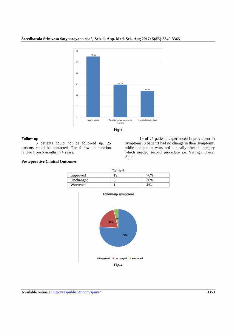
The mean age of patients in the present study
is 27.53 years (SD 14.85). Five patients were below 15
years of age and three patients were above 50 years of
age.
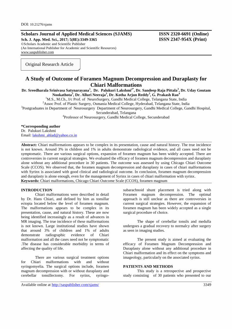
Sex
22 (73.3%) of the patients were male and
8(26.7%) were females.
Fig-1:
Duration of symptoms
The mean duration of symptoms noted in this
study is 14.77 months (SD 10.32).
The following are the presenting symptoms:
(Table-1)
Sreedharala Srinivasa Satynarayana et al., Sch. J. App. Med. Sci., Aug 2017; 5(8E):3349-3365
Available online at http://saspublisher.com/sjams/ 3351
Table 1:
Symptom Number of Patients %
Headache 9 30%
Neck Pain 23 76.7%
Upper Extremity Pain 9 30%
Lower Extremity Pain 1 3.3%
Tingling Numbness UE 22 73.3%
Tingling Numbness LE 3 10%
Weakness of UE 13 43.3%
Weakness of LE 4 13.3%
Blurring of Vision 4 13.3%
Double Vision 1 3.3%
Head Tilt 2 6.7%
Difficulty in Swallowing 1 3.3%
Decreased Sensations UE 9 30%
Decreased Sensations LE 2 6.7%
Decreased Sensations Trunk 2 6.7%
Others 2 6.7%
The following are signs present on examination:
(Table-2)
Table 2:
Signs Number of Patients %
Sensory Deficits 21 70%
Balance Impairment 7 23.3%
Upper Extremity Weakness 16 53.3%
Nystagmus 1 3.3%
Diminished Gag Reflex 1 3.3%
Lower Extremity Weakness 7 23.3%
Gait Impairment 12 40%
Scoliosis 2 6.7%
Increased Lower Extremity Tone 12 40%
Up Going Plantars 6 20%
Others 3 10%
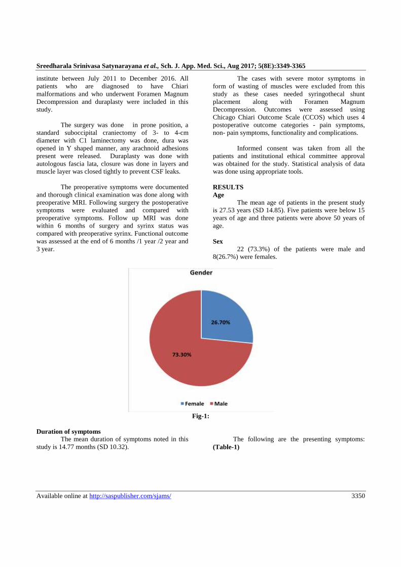
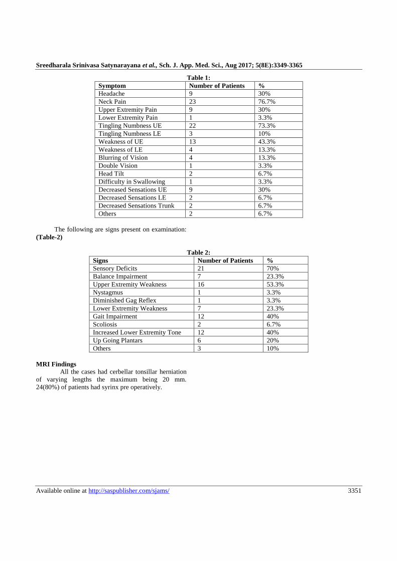
MRI Findings All the cases had cerbellar tonsillar herniation
of varying lengths the maximum being 20 mm.
24(80%) of patients had syrinx pre operatively.
Sreedharala Srinivasa Satynarayana et al., Sch. J. App. Med. Sci., Aug 2017; 5(8E):3349-3365
Available online at http://saspublisher.com/sjams/ 3352
Fig-2:
Table 3
Location of Syrinx Number of Patients %
Cervical 2 8.3%
Cervical/ Dorsal 18 75%
Cervical /Dorsal /Lumbar 4 16.6%
Associated Conditions
5 (16.6%) patients had Hydrocephalus which
needed placement of VP Shunt. Scoliosis was present in
2 (6.6%) patients. Meningomyelocele were present in
2(6.6%) patients which were operated earlier.
Surgical Procedure All the 30 patients underwent Foramen
Magnum Decompression with Duraplasty using fascia
lata graft. One patient with worsening symptoms
needed an additional procedure i.e. Syringo Thecal
shunt. One patient needed Tracheostomy for breathing
difficulty postoperatively and one patient needed re
exploration for persistent CSF leak from the wound.
Complications
The following complications were noted:
(Table-4)
Table 4
Nil 24 80 %
Persistent CSF Leak 1 3.3%
Meningitis 1 3.3%
Pseudomeningocele 1 3.3%
Breathing Difficulty 1 3.3%
Superficial Wound infection 2 6.7%
Hospital Stay
The mean Hospital stay was 11.97 days (SD
5.29) (Table-5)
Table 5
Mean SD N
Age in years 27.53 14.85 30
Duration of symptoms in months 14.77 10.32 30
Hospital stay in days 11.97 5.29 30
Sreedharala Srinivasa Satynarayana et al., Sch. J. App. Med. Sci., Aug 2017; 5(8E):3349-3365
Available online at http://saspublisher.com/sjams/ 3353
Fig-3
Follow up
5 patients could not be followed up. 25
patients could be contacted. The follow up duration
ranged from 6 months to 4 years.
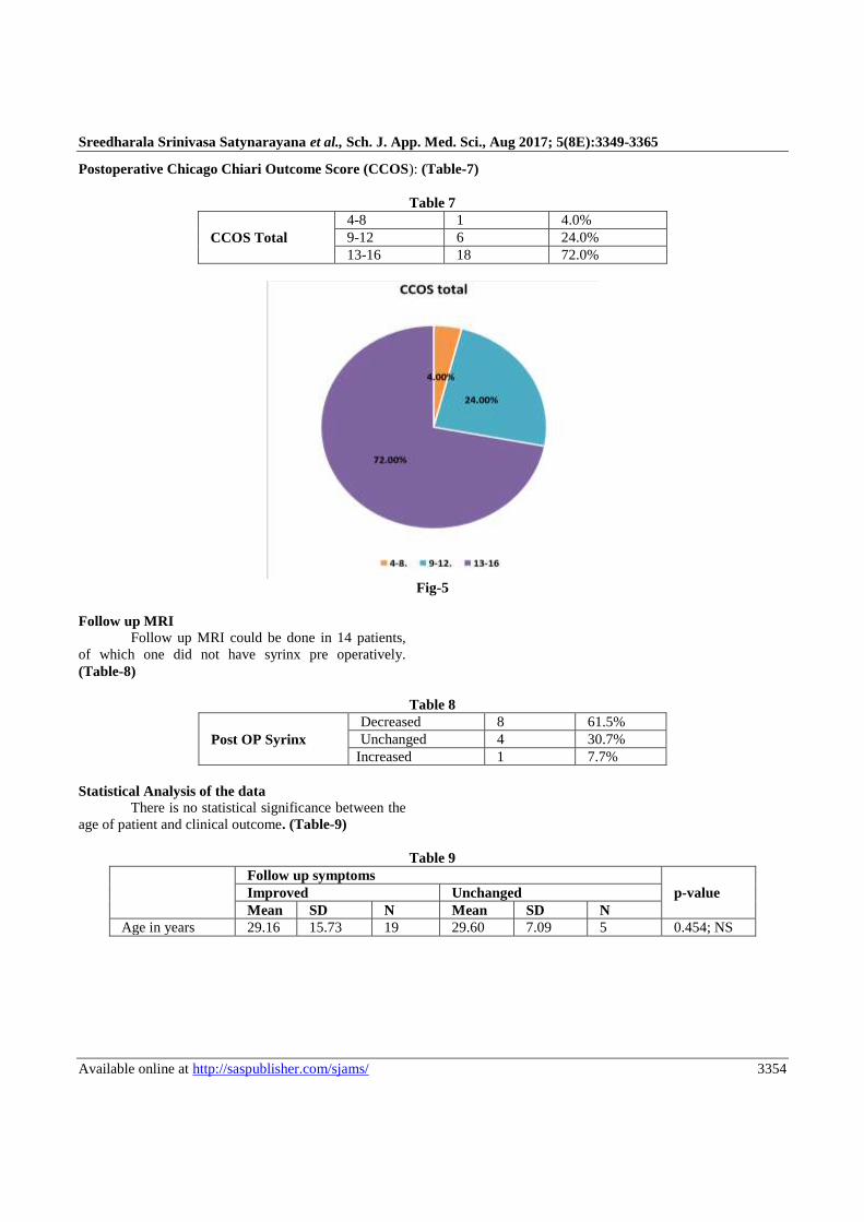
Postoperative Clinical Outcomes
19 of 25 patients experienced improvement in
symptoms, 5 patients had no change in their symptoms,
while one patient worsened clinically after the surgery
which needed second procedure i.e. Syringo Thecal
Shunt.
Table 6
Improved 19 76%
Unchanged 5 20%
Worsened 1 4%
Fig-4
Sreedharala Srinivasa Satynarayana et al., Sch. J. App. Med. Sci., Aug 2017; 5(8E):3349-3365
Available online at http://saspublisher.com/sjams/ 3354
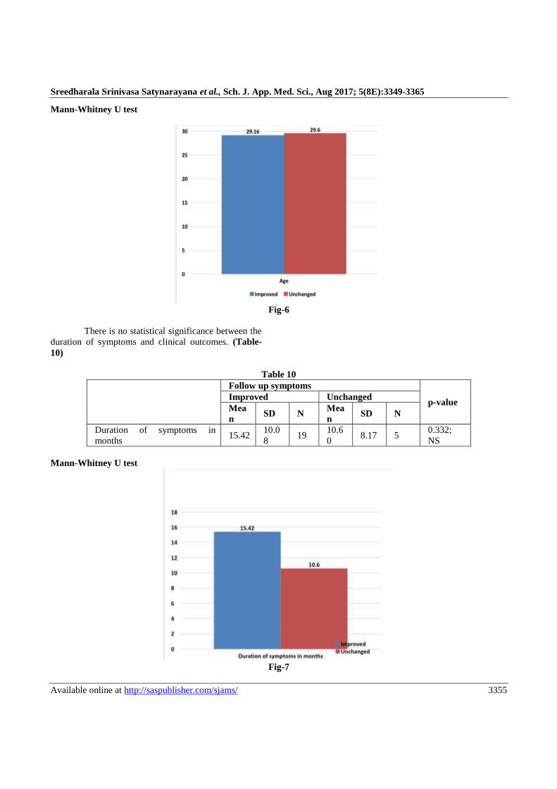
Postoperative Chicago Chiari Outcome Score (CCOS): (Table-7)
Table 7
CCOS Total
4-8 1 4.0%
9-12 6 24.0%
13-16 18 72.0%
Fig-5
Follow up MRI
Follow up MRI could be done in 14 patients,
of which one did not have syrinx pre operatively.
(Table-8)
Table 8
Post OP Syrinx
Decreased 8 61.5%
Unchanged 4 30.7%
Increased 1 7.7%
Statistical Analysis of the data
There is no statistical significance between the
age of patient and clinical outcome. (Table-9)
Table 9
Follow up symptoms
p-value Improved Unchanged
Mean SD N Mean SD N
Age in years 29.16 15.73 19 29.60 7.09 5 0.454; NS
Sreedharala Srinivasa Satynarayana et al., Sch. J. App. Med. Sci., Aug 2017; 5(8E):3349-3365
Available online at http://saspublisher.com/sjams/ 3355
Mann-Whitney U test
Fig-6
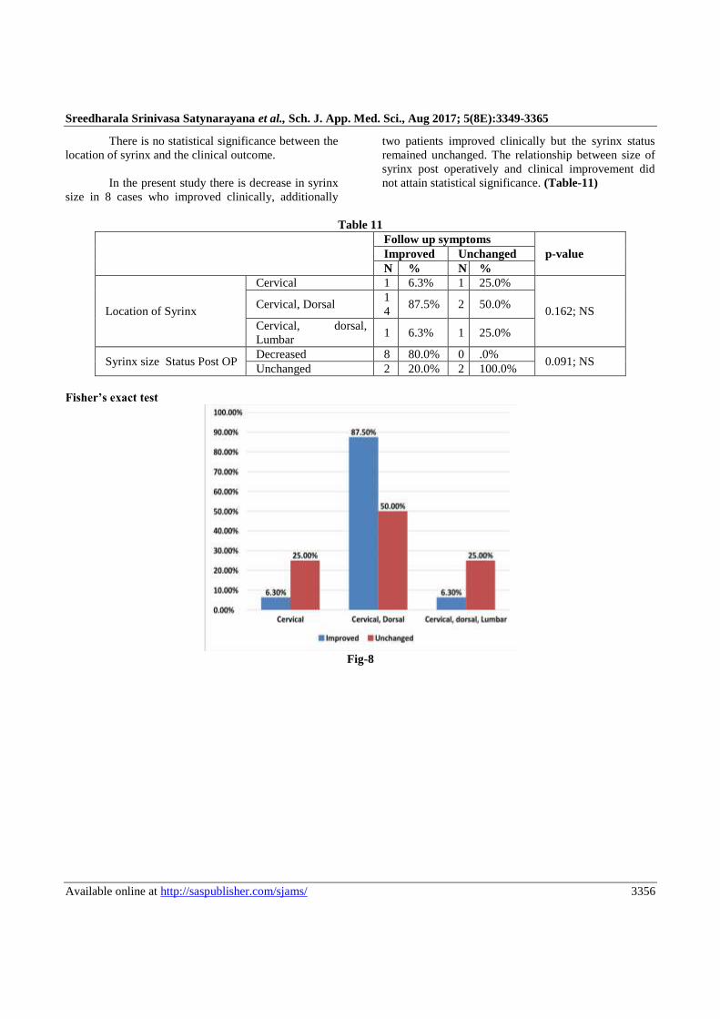
There is no statistical significance between the
duration of symptoms and clinical outcomes. (Table-
10)
Table 10
Follow up symptoms
p-value Improved Unchanged
Mea
n SD N
Mea
n SD N
Duration of symptoms in
months 15.42
10.0
8 19
10.6
0 8.17 5
0.332;
NS
Mann-Whitney U test
Fig-7
Sreedharala Srinivasa Satynarayana et al., Sch. J. App. Med. Sci., Aug 2017; 5(8E):3349-3365
Available online at http://saspublisher.com/sjams/ 3356
There is no statistical significance between the
location of syrinx and the clinical outcome.
In the present study there is decrease in syrinx
size in 8 cases who improved clinically, additionally
two patients improved clinically but the syrinx status
remained unchanged. The relationship between size of
syrinx post operatively and clinical improvement did
not attain statistical significance. (Table-11)
Table 11
Follow up symptoms
p-value Improved Unchanged
N % N %
Location of Syrinx
Cervical 1 6.3% 1 25.0%
0.162; NS Cervical, Dorsal
1
4 87.5% 2 50.0%
Cervical, dorsal,
Lumbar 1 6.3% 1 25.0%
Syrinx size Status Post OP Decreased 8 80.0% 0 .0%
0.091; NS Unchanged 2 20.0% 2 100.0%
Fisher’s exact test
Fig-8
Sreedharala Srinivasa Satynarayana et al., Sch. J. App. Med. Sci., Aug 2017; 5(8E):3349-3365
Available online at http://saspublisher.com/sjams/ 3357
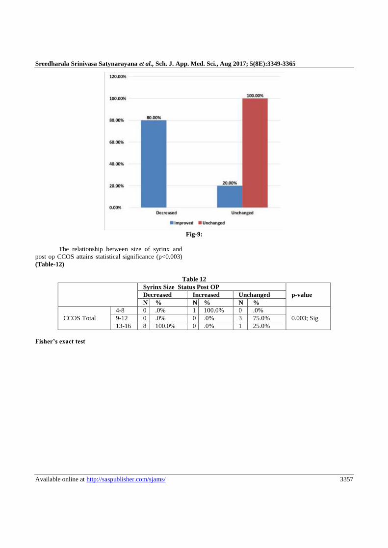
Fig-9:
The relationship between size of syrinx and
post op CCOS attains statistical significance (p<0.003)
(Table-12)
Table 12
Syrinx Size Status Post OP
p-value Decreased Increased Unchanged
N % N % N %
CCOS Total
4-8 0 .0% 1 100.0% 0 .0%
0.003; Sig 9-12 0 .0% 0 .0% 3 75.0%
13-16 8 100.0% 0 .0% 1 25.0%
Fisher’s exact test
Sreedharala Srinivasa Satynarayana et al., Sch. J. App. Med. Sci., Aug 2017; 5(8E):3349-3365
Available online at http://saspublisher.com/sjams/ 3358
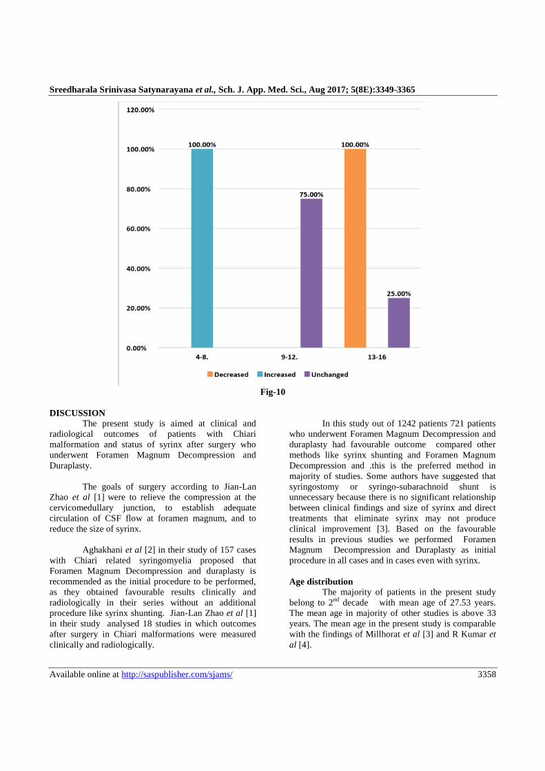
Fig-10
DISCUSSION
The present study is aimed at clinical and
radiological outcomes of patients with Chiari
malformation and status of syrinx after surgery who
underwent Foramen Magnum Decompression and
Duraplasty.
The goals of surgery according to Jian-Lan
Zhao et al [1] were to relieve the compression at the
cervicomedullary junction, to establish adequate
circulation of CSF flow at foramen magnum, and to
reduce the size of syrinx.
Aghakhani et al [2] in their study of 157 cases
with Chiari related syringomyelia proposed that
Foramen Magnum Decompression and duraplasty is
recommended as the initial procedure to be performed,
as they obtained favourable results clinically and
radiologically in their series without an additional
procedure like syrinx shunting. Jian-Lan Zhao et al [1]
in their study analysed 18 studies in which outcomes
after surgery in Chiari malformations were measured
clinically and radiologically.
In this study out of 1242 patients 721 patients
who underwent Foramen Magnum Decompression and
duraplasty had favourable outcome compared other
methods like syrinx shunting and Foramen Magnum
Decompression and .this is the preferred method in
majority of studies. Some authors have suggested that
syringostomy or syringo-subarachnoid shunt is
unnecessary because there is no significant relationship
between clinical findings and size of syrinx and direct
treatments that eliminate syrinx may not produce
clinical improvement [3]. Based on the favourable
results in previous studies we performed Foramen
Magnum Decompression and Duraplasty as initial
procedure in all cases and in cases even with syrinx.
Age distribution
The majority of patients in the present study
belong to 2nd
decade with mean age of 27.53 years.
The mean age in majority of other studies is above 33
years. The mean age in the present study is comparable
with the findings of Millhorat et al [3] and R Kumar et
al [4].
Sreedharala Srinivasa Satynarayana et al., Sch. J. App. Med. Sci., Aug 2017; 5(8E):3349-3365
Available online at http://saspublisher.com/sjams/ 3359
Table 13:
Study Mean age in years
D Mueller and J J Oro [6]
40 (SD 11.3)
Scott Parker et al [7]
39(SD 13)
Xiafeng Deng et al [8]
33.1 (SD 11.2)
Aghakhani et al [2]
30.9( SD 14.9)
JorgKlekamp [9]
40(SD 16)
V N Vakharia et al [10]
33.4(SD 17.2)
R Kumar et al [4]
26.14 (SD 12.24)
Millhorat et al [3]
24.9 (SD 15.8)
Leonard Aliaga et al [11]
20.0 (SD 16.7)
Present study 27.53 (SD 14.85)
Sex distribution
There is a clear female preponderance in all
the studies .In the present study there were 22 males
and 8 females and only in the study by R Kumar et al
[4] there is male preponderance.
Table 14:
Study M : F
D Mueller and J J Oro [6]
82:104
Scott Parker et al [7]
14: 36
Xiafeng Deng et al [8]
52:100
Aghakhani et al [2]
74:83
JorgKlekamp [9]
118:241
V N Vakharia et al [10]
21:40
Jian- Lan Zhao et al [1]
470:713
R Kumar et al [4]
48:39
Present study 22:8
Duration of symptoms
The mean duration of symptoms in our study is
1.23 years which is shortest among all the previous
studies. In the study by Aghakhani et al [2] the mean
duration was 8.22 years the longest amongst all studies.
Table 15:
Study Years No. of Patients
Aghakhani et al [2]
8.22 157
D Mueller and J J Oro [6]
6.3 265
Xiafeng Deng et al [8]
5.5 152
Z Q Zhang et al [5]
4.51 316
N M F El Ghandour [12]
1.96 46
Present study 1.23 30
Presenting signs and symptoms
The most common presenting symptom in the
present study is neck pain (76%) which is present in 48
% of the cases in the study by R G Ellenbogen et al [13]
and in 78% of patients in the study by D Muller and JJ
Oro [6]. In this study the neck pain is described as
stiffness, dull or aching or stabbing, range of motion
was also affected .Similar findings were noted in the
present study.
Weakness of extremity was present in 76.7%
of the patients which was seen in 65% of patients in the
study by D Muller and JJ Oro [6]. Headache was seen
in 30% of the patients in the present study compared to
66% in study by C Hayhurst et al [14] and 97% by D
Muller and JJ [6]. Scoliosis was seen in two (6.7%) of
the patients in contrast to 9% in study by C Hayhurst et
al [14].
Sreedharala Srinivasa Satynarayana et al., Sch. J. App. Med. Sci., Aug 2017; 5(8E):3349-3365
Available online at http://saspublisher.com/sjams/ 3360
Dysphagia and lower cranial nerve palsy was
found in one patient. An uncommon presenting feature
in this study is head tilt in two(6.7%) children which
was found in one child in a study by Sunil V Furtado et
al [15]. Hydrocephalus was present in 5 patients
needing Ventriculo peritoneal shunting Two children
had meningomyelocele which was repaired earlier.
Table 16:
Study/No of patients Symptoms/Signs Percentage
R G Ellenbogen et al [13]
(n=29)
Headache
Neck Pain
Sensory Loss
Ataxia
Scoliosis
100%
48%
45%
6%
1%
R Kumar et al [4]
(n=87)
Neck pain
Weakness
Spasticity
Headache
Dysphagia
50.6%
39.1%
25%
21.8%
12.6%
C Hayhurst et al [14]
(n=96)
Headache
Weakness
Altered Sensation
Ataxia
Scoliosis
66%
35%
15%
14%
9%
D Muller and JJ Oro
(n=112) [6]
Headache
Neck pain
Vision changes
Extremity weakness
Extremity numbness
97%
78%
71%
65%
65%
Aghaknani et al [2]
(n=152)
Neck pain
Sensory
disturbances
Motor weakness
Ataxia
Scoliosis
Dysphagia
Diplopia
38.2%
22.92%
17.2%
8.91%
5.73%
1.91%
0.64%
Xiaofeng Deng et al [8]
(n=152)
Motor dysfunction
Numbness
Neck pain
Dysphagia
Ataxia
Diplopia
25.6%
28.2%
8.5%
6.5%
3.94%
1.9%
Present study
(n=30)
Neck pain
Extremity weakness
Sensory deficits
Extremity pain
Headache
Visual disturbances
Scoliosis
Head tilt
Dysphagia
76.7%
76.7%
70%
33.3%
30%
16.7%
6.7%
6.7%
3.3%
Sreedharala Srinivasa Satynarayana et al., Sch. J. App. Med. Sci., Aug 2017; 5(8E):3349-3365
Available online at http://saspublisher.com/sjams/ 3361
Common Presenting symptoms and signs.
Syrinx in MRI
Pre operative MRI in all cases had tonsillar
descent the maximal descent was observed to be 20
mm. In the present study 80%(24) of cases had
syringomyelia.
Table 17:
Tisell M et al [16]
50 %
C Hayhurst et al [14]
55.4 %
N M F El Ghandour [12]
69.5%
Richard Ellenbogen [13]
76%
Present study 80%
Material used for duraplasty
In this study fascia lata was used for duraplasty
and except in one cases with postoperative CSF leak no
complication was reported.
Historically a variety of dural substitutes were
used like cadaver pericardium, autologous pericranium.
autologous fascia lata and synthetic dural substitute like
e PTFE (expanded polytetrafluoroethylene). Abla AA
et al [17] in their study of dural grafts in Chiari
decompression surgery found that none among
autologous or nonautologous grafts were superior, they
however recommended that autologous pericranium to
be used as it is non-immunogenic, inexpensive and
capable of creating a watertight closure with the dura.
In contrast. JorgKlekamp et al [9] say that
autologous material use was associated with higher
rates of postoperative deterioration compared with
alloplastic materials. This may be attributed to
formation of adhesions underneath the autologous
material compromising CSF flow
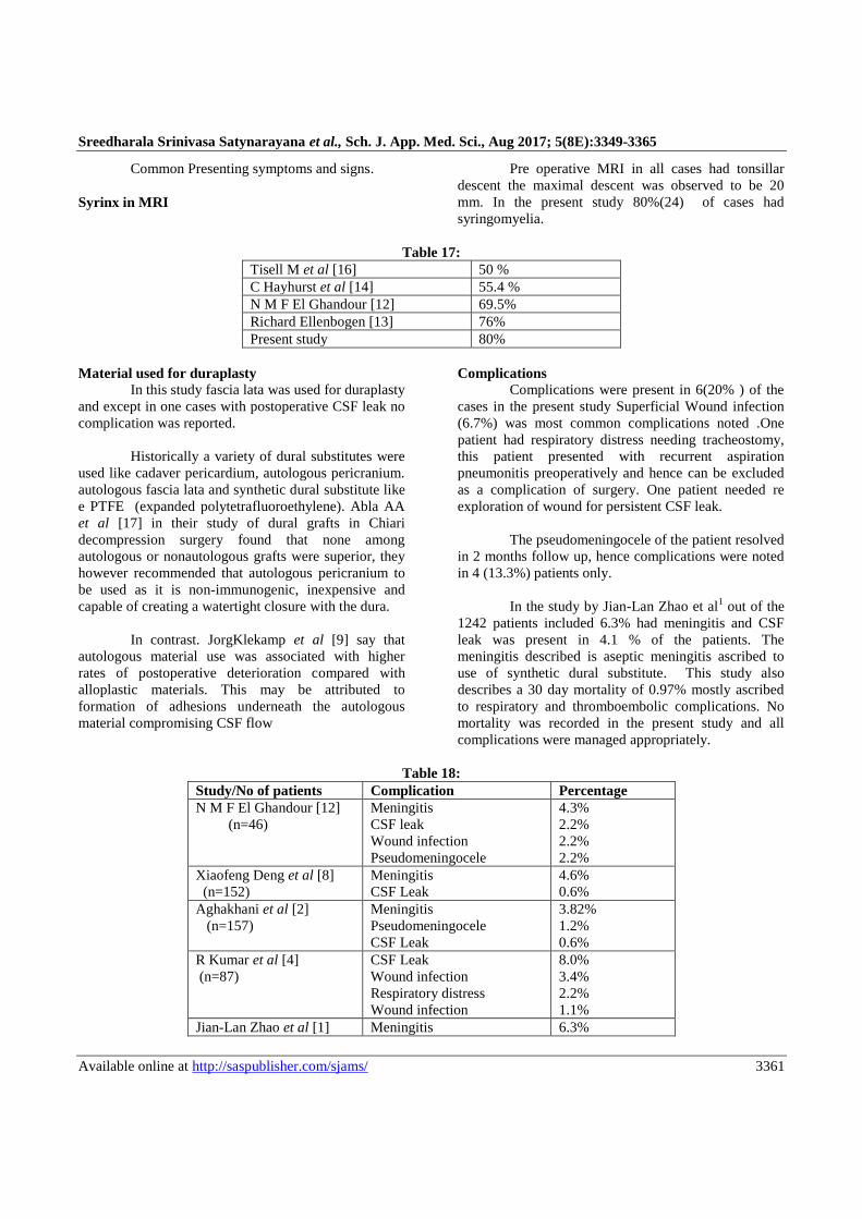
Complications
Complications were present in 6(20% ) of the
cases in the present study Superficial Wound infection
(6.7%) was most common complications noted .One
patient had respiratory distress needing tracheostomy,
this patient presented with recurrent aspiration
pneumonitis preoperatively and hence can be excluded
as a complication of surgery. One patient needed re
exploration of wound for persistent CSF leak.
The pseudomeningocele of the patient resolved
in 2 months follow up, hence complications were noted
in 4 (13.3%) patients only.
In the study by Jian-Lan Zhao et al1 out of the
1242 patients included 6.3% had meningitis and CSF
leak was present in 4.1 % of the patients. The
meningitis described is aseptic meningitis ascribed to
use of synthetic dural substitute. This study also
describes a 30 day mortality of 0.97% mostly ascribed
to respiratory and thromboembolic complications. No
mortality was recorded in the present study and all
complications were managed appropriately.
Table 18:
Study/No of patients Complication Percentage
N M F El Ghandour [12]
(n=46)
Meningitis
CSF leak
Wound infection
Pseudomeningocele
4.3%
2.2%
2.2%
2.2%
Xiaofeng Deng et al [8]
(n=152)
Meningitis
CSF Leak
4.6%
0.6%
Aghakhani et al [2]
(n=157)
Meningitis
Pseudomeningocele
CSF Leak
3.82%
1.2%
0.6%
R Kumar et al [4]
(n=87)
CSF Leak
Wound infection
Respiratory distress
Wound infection
8.0%
3.4%
2.2%
1.1%
Jian-Lan Zhao et al [1] Meningitis 6.3%
Sreedharala Srinivasa Satynarayana et al., Sch. J. App. Med. Sci., Aug 2017; 5(8E):3349-3365
Available online at http://saspublisher.com/sjams/ 3362
(n=1242)
CSF Leak
Wound infection
Swallowing difficulty
Respiratory distress
Pseudomeningocele
4.1%
1.4%
0.9%
0.6%
0.5%
Present study
(n=30)
Superficial wound infection
Persistent CSF Leak
Meningitis
Pseudomeningocele
Respiratory distress
6.7%
3.3%
3.3%
3.3%
3.3%
Post operative status
Out of 25 patients who could be followed up
postoperatively at least 6 months of surgery to 4 years
of surgery 19 (76%) showed improvement in their
preoperative symptoms This is comparable to the
studies of J Lan Zhao et al [1], Z Q Zhang et al [5], J
Klekamp [9], C Hayhurst et al [14].
In the present study the symptoms were static
in 5(20%) patients and in one patient there was clinical
worsening. These percentages are comparable with
previous studies.
Table 19:
J Lan Zhao
et al [1]
(n=1242)
ZQ Zhang
et al [5]
(n=218)
J Klekamp
[9]
(n=371)
R Kumar
et al [4]
(n=87)
C Hayhurst
et al [14]
(n=96)
Aghakhani
et al [2]
(n=157)
Xiaofeng
Deng et al
[8]
(n=152)
Present
study
(n=25)
Improved 79.95% 80.4% 73.6% 45.9% 78% 63.06% 82.9% 76%
Unchanged 14.09% 15.6% 21.0% 43.7% 20% 30.58% 13.8% 20%
Worsened 6.09% 3.9% 5.5% 10.3% 2% 5.73% 3.3% 4%
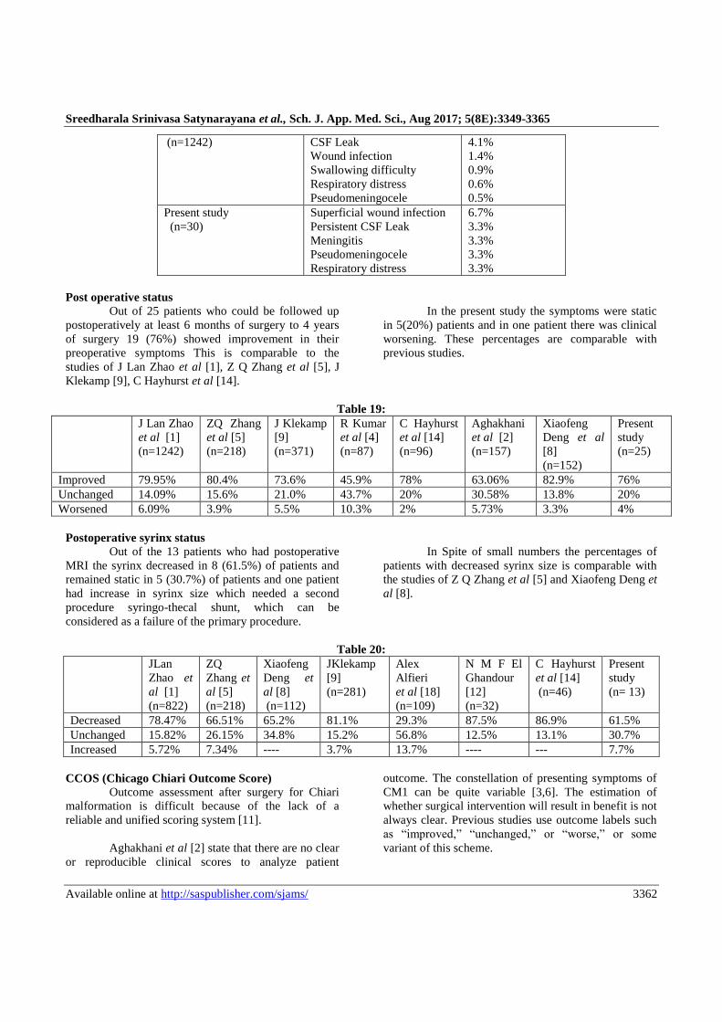
Postoperative syrinx status
Out of the 13 patients who had postoperative
MRI the syrinx decreased in 8 (61.5%) of patients and
remained static in 5 (30.7%) of patients and one patient
had increase in syrinx size which needed a second
procedure syringo-thecal shunt, which can be
considered as a failure of the primary procedure.
In Spite of small numbers the percentages of
patients with decreased syrinx size is comparable with
the studies of Z Q Zhang et al [5] and Xiaofeng Deng et
al [8].
Table 20:
JLan
Zhao et
al [1]
(n=822)
ZQ
Zhang et
al [5]
(n=218)
Xiaofeng
Deng et
al [8]
(n=112)
JKlekamp
[9]
(n=281)
Alex
Alfieri
et al [18]
(n=109)
N M F El
Ghandour
[12]
(n=32)
C Hayhurst
et al [14]
(n=46)
Present
study
(n= 13)
Decreased 78.47% 66.51% 65.2% 81.1% 29.3% 87.5% 86.9% 61.5%
Unchanged 15.82% 26.15% 34.8% 15.2% 56.8% 12.5% 13.1% 30.7%
Increased 5.72% 7.34% ---- 3.7% 13.7% ---- --- 7.7%
CCOS (Chicago Chiari Outcome Score)
Outcome assessment after surgery for Chiari
malformation is difficult because of the lack of a
reliable and unified scoring system [11].
Aghakhani et al [2] state that there are no clear
or reproducible clinical scores to analyze patient
outcome. The constellation of presenting symptoms of
CM1 can be quite variable [3,6]. The estimation of
whether surgical intervention will result in benefit is not
always clear. Previous studies use outcome labels such
as “improved,” “unchanged,” or “worse,” or some
variant of this scheme.
Sreedharala Srinivasa Satynarayana et al., Sch. J. App. Med. Sci., Aug 2017; 5(8E):3349-3365
Available online at http://saspublisher.com/sjams/ 3363
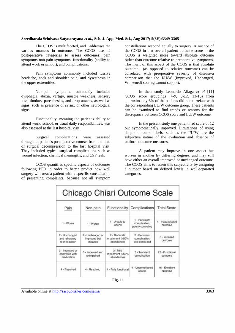
The CCOS is multifaceted, and addresses the
various nuances in outcome. The CCOS uses 4
postoperative categories to assess outcomes: pain
symptoms non-pain symptoms, functionality (ability to
attend work or school), and complications.
Pain symptoms commonly included tussive
headache, neck and shoulder pain, and dysesthesia in
the upper extremities.
Non-pain symptoms commonly included
dysphagia, ataxia, vertigo, muscle weakness, sensory
loss, tinnitus, paresthesias, and drop attacks, as well as
signs, such as presence of syrinx or other neurological
signs.
Functionality, meaning the patient's ability to
attend work, school, or usual daily responsibilities, was
also assessed at the last hospital visit.
Surgical complications were assessed
throughout patient's postoperative course, from the time
of surgical decompression to the last hospital visit.
They included typical surgical complications such as
wound infection, chemical meningitis, and CSF leak.
CCOS quantifies specific aspects of outcomes
following PFD in order to better predict how well
surgery will treat a patient with a specific constellation
of presenting complaints, because not all symptom
constellations respond equally to surgery. A nuance of
the CCOS in that overall patient outcome score in the
CCOS is weighted more toward absolute outcome
rather than outcome relative to preoperative symptoms.
The merit of this aspect of the CCOS is that absolute
outcome (as opposed to relative outcome) can be
correlated with preoperative severity of disease-a
comparison that the I/U/W (Improved, Unchanged,
Worsened) scoring cannot support.
In their study Leonardo Aliaga et al [11]
CCOS score groupings (4-9, 8-12, 13-16) from
approximately 8% of the patients did not correlate with
the corresponding I/U/W outcome group. These patients
can be examined to find trends or reasons for the
discrepancy between CCOS score and I/U/W outcome.
In the present study one patient had score of 12
but symptomatically improved. Limitations of using
simple outcome labels, such as the I/U/W, are the
subjective nature of the evaluation and absence of
uniform outcome measures.
A patient may improve in one aspect but
worsen in another by differing degrees, and may still
have either an overall improved or unchanged outcome.
The CCOS aims to lessen this subjectivity by assigning
a number based on defined levels in well-separated
categories.
Fig-11
Sreedharala Srinivasa Satynarayana et al., Sch. J. App. Med. Sci., Aug 2017; 5(8E):3349-3365
Available online at http://saspublisher.com/sjams/ 3364
Table 21:
CCOS Total
4-8 1 4.0%
9-12 6 24.0%
13-16 18 72.0%
Table 22:
Improved 19 76%
Unchanged 5 20%
Worsened 1 4%
One patient in the improved category had
CCOS score 12 and hence discrepancy seen in CCOS
scores versus I/U/W classification
Analysis of data
In the present study there is no statistical
significance between improvement and age of patient
(p=0.454).
In the study by Alex Alfieri et al [18] they
found that there is statistical significance between age
and clinical improvement, and according to this study
older age at surgery negatively influenced the outcome.
Similarly, Aghakhani et al2 in their study found that
there is a higher probability of improvement or
stabilization in young patients.
There is no statistical significance between
duration of symptoms and clinical improvement
(p=0.332) Alex Alfieri et al [18] found that longer
duration of symptoms negatively influence the
outcome.
The relationship between size of syrinx post
operatively and clinical improvement did not attain
statistical significance.(p=0.091)In the present study
there is decrease in syrinx size in 8 cases who improved
clinically ,additionally two patients improved clinically
but the syrinx status remained unchanged, N M F El
Ghandour [12] in their study state that clinical
improvement occurs earlier than syrinx size in
postoperative MR imaging.
There is no correlation between specific syrinx
size and clinical condition, this is reflected in two
patients in the present study who improved clinically
but the syrinx remained unchanged in MR imaging.
According to Xiaofeng Deng et al [8] patients
with motor dysfunction revealed a lower recovery rate
than the others. In the present study out of 5 patients
whose symptoms remain unchanged 3 had motor
dysfunction and one patient who worsened also had
motor dysfunction.
An important finding in this study is that the
relationship between size of syrinx and post op CCOS
attains statistical significance (p<0.003).
Table 23:
Syrinx Size Status Post OP
p-value Decreased Increased Unchanged
N % N % N %
CCOS Total
4-8 0 .0% 1 100.0% 0 .0%
0.003; Sig 9-12 0 .0% 0 .0% 3 75.0%
13-16 8 100.0% 0 .0% 1 25.0%
CONCLUSION
Chiari malformations with syrinx are a
neurosurgical challenge, with complex pathophysiology
and with varied surgical options. Foramen Magnum
Decompression and Duraplasty is a safe and effective
surgical option, it provides significant and sustained
improvement in symptoms by establishing CSF flow at
foramen magnum and reducing the size of syrinx
radiologically.
Foramen Magnum Decompression and
Duraplasty in cases of Chiari malformations with syrinx
is associated with good clinical and radiological
outcomes. Clinical improvement occurs earlier than
Sreedharala Srinivasa Satynarayana et al., Sch. J. App. Med. Sci., Aug 2017; 5(8E):3349-3365
Available online at http://saspublisher.com/sjams/ 3365
radiological improvement and patients with motor
dysfunction have lower recovery rates of existing
neurological status as compared with other patients.
The improved clinical outcomes are sustained over a
period of time as measured by CCOS (Chicago Chiari
Outcome Score).
Foramen Magnum Decompression and
Duraplasty significantly reduces the need for second
surgery in cases with syrinx. In the present study only
one patient (3.3%) needed syringo-thecal shunt after
Foramen Magnum Decompression.
Hence in conclusion, in cases of Chiari
malformation with syrinx, Foramen Magnum
Decompression and Duraplasty is alone enough, even
for the management of syrinx and only small percentage
of cases need a second procedure (syringo-thecal
shunt).
Sponsors & Financial support – NIL
Conflicts of interest – NIL
REFERENCES 1. Zhao JL, Li MH, Wang CL, Meng W. A
Systematic Review of Chiari I Malformation :
Techniques and Outcome s World Neurosurg. 2016
Apr;88:7-14.
2. Aghakhani N, Parker F, David P, Morar S. Long
term analysis of 157 surgically treated cases.
Neurosurgery. 2009; 64:308-315.
3. Milhorat T, Chou M, Trinidad E. CIMs redefined:
clinical and radiographic findings for 364
symptomatic patients. Neurosurgery.
1999;44:1005–17.
4. Kumar R, Kalra SK, Vaid VK, Mahapatra AK.
Chiari 1 malformation : surgical experience over a
decade of management Br J Neurosurg. 2008
Jun;22(3):409-14.
5. Zhang ZQ, Chen YQ, Chen YA, Wu X, Wang YB,
Li XG. Chiari I malformation associated with
syringomyelia: a retrospective study of 316
surgically treated patientsSpinal Cord. 2008; 46:
358–36.
6. Mueller DM, Oro’ JJ. Prospective analysis of
presenting symptoms among 265 patients with
radiographic evidence of Chiari malformation type
I with or without syringomyelia. J Am Acad Nurse
Pract. 2004;16:134–8.
7. Parker SL, Godil SS, Zuckerman SL, Mendenhall
SK, Wells JA, Shau DN. Comprehensive
assessment of 1-year outcomes and determination
of minimum clinically important difference in pain,
disability, and quality of life after suboccipital
decompression for Chiari malformation I in adults.
Neurosurgery. 2013;73:569–581.
8. Deng X, Yang C, Gan J, Wu L, Yang T, Yang J,
Xu Y. Long-term outcomes after small-bone-
window posterior fossa decompression and
duraplasty in adults with Chiari malformation type
I. World neurosurgery. 2015 Oct 31;84(4):998-
1004.
9. Klekamp J. Surgical Treatment of Chiari I
Malformation— Analysis of Intraoperative
Findings, Complications, and Outcome for 371
Foramen Magnum Decompressionsn
Neurosurgery. 2012; 7(1):365–380.
10. Vakharia VN, Guilfoyle MR, Laing RJ.
Prospective study of outcome of foramen magnum
decompressions in patients with syrinx and non-
syrinx associated Chiari malformations Br J
Neurosurg. 2012 Feb;26(1):7-11.
11. Aliaga L, Hekman KE, Yassari R, Straus D, Luther
G, Chen J, Sampat A, Frim D. A novel scoring
system for assessing Chiari malformation type I
treatment outcomes. Neurosurgery. 2011 Aug
12;70(3):656-65.
12. Nasser M, El-Ghandour F. Long-term outcome of
surgical management of adult Chiari I
malformation Neurosurg Rev. 2012; 35:537–547.
13. Ellenbogen RG, Armonda RA, Shaw DWW, Winn
HR. Toward a rational treatment of Chiari I
malformation and
syringomyelia Neurosurg Focus. 2000; 8 (3): 6.
14. Hayhurst C, Richards O, Zaki H, Findlay G, Pigott
TJ. Hindbrain decompression for Chiari–
syringomyelia complex: an outcome analysis
comparing surgical techniques. British journal of
neurosurgery. 2008 Jan 1;22(1):86-91.
15. Furtado SV, Thakar S, Hegde AS. Correlation of
functional outcome and natural history with
clinicoradiological factors in surgically managed
pediatric Chiari I malformation. Neurosurgery.
2011 Feb 1;68(2):319-28.
16. Tisell M, Wallskog J, Linde M. Long-term
outcome after surgery for Chiari I malformation.
ActaNeurolScand 2009: 120: 295–299.
17. Abla AA, Link T, Fusco D, Wilson DA, Sonntag
VK. Comparison of dural grafts in Chiari
decompression surgery: review of the literature. J
Craniovertebr Junction Spine. 2010;1(1):29-37.
18. Alfieri A, Pinna G. Long-term results after
posterior fossa decompression in syringomyelia
with adult Chiari Type I malformation. Journal of
Neurosurgery: Spine. 2012 Nov;17(5):381-7.