Embed Size (px)
Citation preview
R
As
DSa
b
c
h
•••••
a
ARRAA
KEFSTHAP
1
p
oI
h0
Behavioural Brain Research 273 (2014) 144–154
Contents lists available at ScienceDirect
Behavioural Brain Research
jou rn al hom epage: www.elsev ier .com/ locate /bbr
esearch report
study on fear memory retrieval and REM sleep in maternaleparation and isolation stressed rats
ayalan Sampatha, K.R. Sabithaa, Preethi Hegdea, H.R. Jayakrishnana, Bindu M. Kuttya,umantra Chattarji b, Govindan Rangarajanc, T.R. Laxmia,∗
National Institute of Mental Health and Neurosciences (NIMHANS), Bangalore 560029, IndiaNational Center for Biological Sciences (NCBS), GKVK Campus, Bangalore 560065, IndiaIndian Institute of Science (IISc), Bangalore 560012, India
i g h l i g h t s
Maternal separation stress was done during stress hyporesponsive period.We examined changes in sleep and fear memory.Increase in the fear memory was negatively correlated with home cage exploration.Resistance to the immediate fear extinction and increased REM sleep was observed.Altered neuronal activities in hippocampus–amygdala–cortical loop during REM sleep.
r t i c l e i n f o
rticle history:eceived 5 June 2014eceived in revised form 18 July 2014ccepted 21 July 2014vailable online 30 July 2014
eywords:arly maternal separation stressear memoryleepheta–gamma oscillationsippocampusmygdalarefrontal cortex
a b s t r a c t
As rapid brain development occurs during the neonatal period, environmental manipulation during thisperiod may have a significant impact on sleep and memory functions. Moreover, rapid eye movement(REM) sleep plays an important role in integrating new information with the previously stored emotionalexperience. Hence, the impact of early maternal separation and isolation stress (MS) during the stresshyporesponsive period (SHRP) on fear memory retention and sleep in rats were studied. The neonatalrats were subjected to maternal separation and isolation stress during postnatal days 5–7 (6 h daily/3 d).Polysomnographic recordings and differential fear conditioning was carried out in two different sets ofrats aged 2 months. The neuronal replay during REM sleep was analyzed using different parameters.MS rats showed increased time in REM stage and total sleep period also increased. MS rats showed feargeneralization with increased fear memory retention than normal control (NC). The detailed analysis ofthe local field potentials across different time periods of REM sleep showed increased theta oscillationsin the hippocampus, amygdala and cortical circuits. Our findings suggest that stress during SHRP
has sensitized the hippocampus–amygdala–cortical loops which could be due to increased release ofcorticosterone that generally occurs during REM sleep. These rats when subjected to fear conditioningexhibit increased fear memory and increased fear generalization. The development of helplessness,anxiety and sleep changes in human patients, thus, could be related to the reduced thermal, tactile andsocial stimulation during SHRP on brain plasticity and fear memory functions.. Introduction
The early postnatal period, specifically, stress-hypo responsiveeriod (SHRP) is a critical phase for normal brain development,
∗ Corresponding author at: Department of Neurophysiology, National Institutef Mental Health and Neurosciences (NIMHANS), Hosur Road, Bangalore 560 029,ndia. Tel.: +91 80 26995178; fax: +91 80 26562121/26564830.
E-mail addresses: [email protected], [email protected] (T.R. Laxmi).
ttp://dx.doi.org/10.1016/j.bbr.2014.07.034166-4328/© 2014 Elsevier B.V. All rights reserved.
© 2014 Elsevier B.V. All rights reserved.
characterized by an enormous capacity of structural and functionalreorganization of neural circuitry. Bowlby’s [1] discovery of attach-ment theory incited studies on maternal separation stress affectingneuroendocrine and behavioral alterations in different develop-mental period [2]. In particular, the stress produced by repeatedseparation from the mother during early postnatal life maternal
separation stress (MS) can lead to juvenile and adult mood disor-ders and reduced cognitive abilities [3].Exposure to early maternal separation stress often causes patho-logical changes that principally affect three functionally well
Brain
cmscadsf((
Rh[hbrds
iRgatedpp
2
2
AMmae(w(ad
sgo2
efihvTscaimmtc
D. Sampath et al. / Behavioural
onnected brain regions – amygdala [4], hippocampus [5] andedial prefrontal cortex (mPFC) [6]. Interestingly, the effects of
tress on these regions are not the same – while the infralimbicortex (ILC) showed higher synaptic densities to early maternal sep-ration stress, the anterior cingulate cortex showed a reduced spineensity [7]. The present study aimed to understand if early maternaleparation together with isolation stress during SHRP affects dif-erentially on the hippocampus (CA1 subregion) (CA1), amygdalaLateral nucleus of amygdala) (LA) and medial prefrontal cortexinfralimbic cortex) (ILC) in adult rats.
In our previous study, we reported that increased duration ofEM sleep was associated with increased synchronized amygdalo-ippocampal theta activities in adult rats exposed to chronic stress8]. These changes may be related to the profound alteration of theormonal stress axis and to neurotrophic factor expression in therain [9] and imbalances in neurotransmitter release in these brainegions [10]. Thus MS-induced brain pathology could result in theevelopment of helplessness, anxiety and sleep changes that reflectymptoms in human patients.
Whether hippocampus and amygdala processes differentiallynfluence the explicit and implicit memory during non-REM andEM sleep still remains controversial [11]. Previous studies haveiven substantial evidence to suggest that early life stress cause
delayed effect on emotional processing in rats [12]. To addresshis issue, rats subjected to early maternal separation stress werexposed to differential fear conditioning, and the impact of stressuring SHRP of rats on the synchronous firing between hippocam-us, amygdala and medial prefrontal cortex during REM sleep wasrobed.
. Materials and methods
.1.1. Rats
Pregnant Wistar rats were bred and raised in the Centralnimal Research Facility (CARF) of the National Institute ofental Health and Neurosciences (NIMHANS). These rats wereaintained in polypropylene cages, 1 week before gestation
nd were provided with ad libitum food and water. It wasnsured that rats were maintained on a regular light–dark cycle12 h:12 h) and was properly ventilated. Experimental protocolsere approved by the Institutional Animal Ethics Committee
IAEC) and maximum care was taken to minimize the painnd discomfort to the experimental animals during the proce-ures.
The pregnant rats were first randomly assigned to maternaleparation stress (MS) and normal control groups (NC). Polysomno-raphic recordings and differential fear conditioning was carriedut in two different sets of male rats aged 2 months weighing50–300 g.
MS procedure was carried out under temperature controllednvironment different from the normal rearing environment. Atrst, dam was separated from the pups and placed in a differentome cage. Immediately after separation, rat pups were indi-idually placed in a different home cage having six partitions.hus, there were two types of stress: (1) maternal separationtress, (2) isolation stress for the pups. Maternal separation wasarried out during stress hyporesponsive period (SHRP) suchs postnatal days 5–7 (P5–P7) (MS) and was carried out dur-ng 9 AM to 3 PM for 6 h daily. The room temperature was
aintained at 20–22 ◦C Rats during the MS procedure (7). Onlyale offspring were used in the study. The rats left undis-
urbed during pre-weaning period were considered as normalontrols.
Research 273 (2014) 144–154 145
2.1.2. Effect of MS stress on the spontaneous behavior of the rat inthe familiar home cage
Before subjecting them to differential fear conditioning, ratsfrom control (n = 10) and MS groups (n = 18) were tested for theirspontaneous behaviors in their familiar home cage for a periodof 15 min during the dark phase of the cycle (between 7 PM and10 PM). The spontaneous locomotor behaviors and quiet behaviorof the rat were video-taped and stored for offline analysis. The aver-age time spent on each behavior was classified into exploratorylocomotion (exploratory walking and rearing) and quiet behav-ior (immobility and grooming) during 15 min recording period foreach rat which was converted into percentages. The mean valuesbetween the NC and MS were subjected to statistical analysis.
2.1.3. Effect of MS on differential fear conditioning
The differential fear conditioning was carried out to evaluatethe effect of early maternal separation stress on the associative fearmemory and generalization effect in a fear-conditioning chamber(Coulbourn Instruments Inc., USA). The fear conditioning was car-ried out in context A (foreground context) consisted of aluminumand Plexiglas walls and a grid floor spaced at 1.8 cm. In addition,video camera was mounted on the ceiling and tone generator onthe sidewalls. The fear retention test was in context B (backgroundcontext) which was similar to that of context A but was placed in adifferent room consisting of ventilation fans, lights-on and withoutwhite noise.
3. Fear conditioning procedure
3.1. Habituation
The rats were first habituated to the conditioning chambers ofboth contexts for 5 min each. During the habituation period, ratswere presented with six auditory tones (2.5 kHz, 70 dB intensity)for 10 s (CS−). The inter tone interval was for 20 s. The rats werepresented with two habituation protocols first in context A andafter 24 h into the context B. The house light remained switched onthroughout the protocol.
3.2. Training
After 24 h of habituation (day 3), fear conditioning was car-ried out in context A (Fig. 4(a)). Each rat was placed into thefear-conditioning chamber (context A) and allowed to explorefor 120 s without presentation of any conditioned stimulus (CS).Immediately after 120 s, each rat was presented with three con-ditioned stimulus (CS+) (10 kHz auditory tone, 10 s, 90 dB) andco-terminated with unconditioned stimulus (US) (0.5 mA scram-bled foot shock for a period of 1 s). After the last presentation ofCS–US pairings, rat was allowed to stay in the chamber for another120 s before returning it to their home cage. The conditioning pro-tocol was repeated on the subsequent day with the same CS–USstimulus pairings for the sufficient consolidation of fear memory.
3.3. Testing
Testing for the retention of fear memory was carried out after24 h of fear conditioning. The retention test was carried out in thecontext B. The retention protocol comprised of presentation of four
CS− tones followed by four CS+ tones without co-terminating withelectric footshock stimulus (US). The rats were allowed to remainin the chamber for 120 s after the termination of the last CS+. Theentire chamber was wiped with cotton and alcohol to prevent the
1 Brain
pibs
3
ttasitDl+lWcer(tefitcgtihbtta
4
thmsfsacpwaottwo
5
aiEimc
46 D. Sampath et al. / Behavioural
resentation of odor cues to the next experimental rat. The behav-or of the rat was video-recorded for the offline analysis of freezingehavior to cues and context. The time spent in freezing was con-idered as an index of fear memory.
.3.1. Surgery for sleep–wake recordingRats from both MS (n = 8) and NC (n = 6) were anesthetized with
he combination of ketamine (80 mg/kg body weight, intraperi-oneal injections) and xylazine (10 mg/kg body weight, IP). Inddition, xylocaine (2%) was injected subcutaneously before theurgery. These rats were stereotaxically (Stoelting Co., USA)mplanted with insulated nichrome wires of 250 �m diame-er in CA1 (antero-posterior: −3.3 mm; medio-lateral: 1.5 mm;orso-ventral:2.6 mm), LA (antero-posterior: −3.3 mm; medio-
ateral: 5.2 mm; Dorso-ventral:8.0 mm) and ILC (antero-posterior,3.0 mm; medio-lateral, 0.5 mm; dorso-ventral, 5.0 mm), on theeft hemisphere for measuring the LFP during sleep. Paxinos and
atson [13] rat brain atlas was used to locate all the stereotaxicoordinates in relation to Bregma and Lambda. An external screwlectrode was implanted above the cerebellum subdurally as aeference electrode. In addition, electrodes for ElectrooculogramEOG) and electromyogram (EMG) were implanted in external can-hus of the eye and posterior nuchal muscle, respectively. Thisntire electrode ensemble was soldered into a 10-pin socket andxed on the skull using dental acrylic powder. Healex was appliedo all the wound edges and the rat finally placed back into its homeage for 5 days to recover from the surgical effects. Following sur-ical recovery, polysomnographic recordings were carried out inhe rat home cage continuously for 6 h. Once the behavioral exper-ments were completed, the rats were deeply anesthetized withalothane and transcardially perfused with 10% formaldehyde. Therains were processed for Nissl staining for the histological verifica-ion of electrode placement. The rats with confirmed electrodes inhe target were only used for the further evaluation of sleep–wakerchitecture in both control and MS groups.
. Procedure for sleep recording
After the complete recovery from surgery, the rats were takeno the recording room and left in its home cage for an hour forabituation. Once the rats were familiar with the new environ-ent, they were connected to the swivel commutator first and
ubsequently basal LFP recordings were carried out continuouslyor 6 h during light phase of the cycle (12.00–6.00 PM) [14]. The LFPignals were amplified (10×), band-pass filtered (0.3 Hz–0.3 KHz)nd sampled at the rate of 1000 Hz, digitized by a CED A/Donverter (CED 1401A/D converter, Cambridge, UK). During thiseriod, the spontaneous behavior of the rat was monitored andas recorded online with the spike 2 software. The rats had free
ccess to food and water during the entire recording session. Inrder to observe the prolonged effect of early maternal separa-ion and isolation stress on the sleep and associated changes inhe amygdalo–hippocampal–cortical network activities, these ratsere taken to the recording room and LFP recording was carried
ut on day 70.
. Recording and quantification of the sleep architecture
The 6 ho sleep–wakefulness cycles were split into 15 s epochsnd visually scored on the basis of LFP, EMG and EOG, by twondependent judges who were blind to the experimental condition.
ach epoch was classified into a particular stage based on predom-nant sleep stage in a 15 s epoch that belonged [14]. The scoringethod described here in detail, is primarily based on five-stagelassification of sleep–wake for rats as proposed earlier [15–18].
Research 273 (2014) 144–154
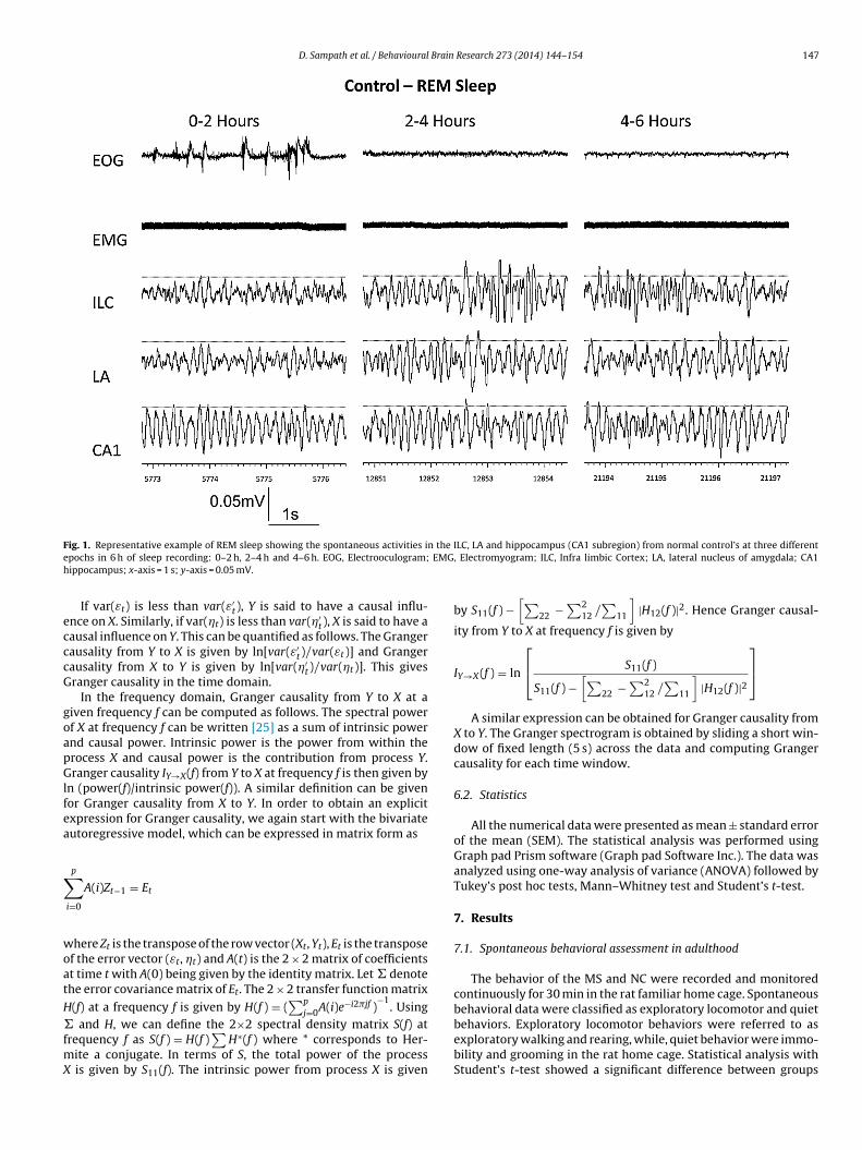
The wakeful period was classified into active wakefulness (W1)based on the presence of Type 1 theta rhythm in CA1, high fre-quency LFP in ILC, tonic EMG level and phasic EOG movements; andquiet wakefulness (W2) showing reduced tonic EMG, occasionalEOG movements, Type 2 theta rhythm in CA1 and desynchronizedILC LFP. The sleep period was classified into light slow wave sleep(SWS) (S1) characterized by the presence of high amplitude and lowfrequency LFP activity in CA1 and ILC, reduced EMG activity in theabsence of locomotor activity and reduced EOG movements; deepSWS (S2) showing synchronized low frequency (delta range) andhigh amplitude–low frequency LFP activity in CA1 and ILC, EMGhypotonia and no EOG movements. Figs. 1 and 2 are the repre-sentative examples from NC and MS rats showing the presence ofsynchronized neural activities in CA1 and LA, EOG with rapid burstsof eye movements and EMG showing muscle atonia during differ-ent timings of rapid eye movement (REM) sleep. REM onset latencywas calculated based on the appearance of the first episode of REMsleep from the beginning of sleep recording.
The percentage distribution of sleep stages was calculated fromthe total recording time that includes, SWS percentage (TSWS/totalrecording time); REM percentage (total REM sleep/total recordingtime); wake time percentage (total waking time/total recordingtime).
6. Quantification of the changes with local field potentials(LFPs)
The analysis starts with the LFPs recorded (CED analog-to-digital (A/D) converter (Cambridge, UK)) at 1 kHz sampling ratefrom the three sites. For the quantification, the longest first,middle and the last episode of REM was selected from whicha 15 s duration was considered for further analysis. Mathemati-cally, we model these LFPs as a multivariate autoregressive (AR)process of order p [19]. The optimal order p is obtained byusing the Akaike Information Criterion [20]. The AR model isfitted to the data using the Levinson–Wiggins–Robinson algo-rithm [20]. Once the AR model is obtained, the 3 × 3 spectraldensity matrix for the process can be obtained directly fromthe model [21]. This matrix is a function of the frequency. Thethree diagonal elements of the spectral density matrix give thepower spectrum of the three LFPs as a function of the fre-quency.
Using power spectral data for REM sleep, two major types ofanalysis were performed: (1) power of the theta frequency rangemeasured from hippocampal, amygdalar and cortical LFP duringREM sleep and (2) coherence level between brain regions.
6.1. Granger causality analysis
Granger causality analysis [21] is a leading statistical techniqueused to quantify causal influences between two time series. This hasbeen used extensively to analyze both LFP [20,22,23] and spike train[24] data. Mathematically, given two time series (say Xt and Yt),they can be jointly modeled as a realization of a bivariate stationaryautoregressive process of order p [19]:
Xt = a1Xt−1 + a2Xt−2 + · · · + apXt−p + b1Yt−1 + b2Yt−2 + · · · + bpYt−p + εt
Xt = c1Xt−1 + c2Xt−2 + · · · + cpXt−p + d1Yt−1 + d2Yt−2 + · · · + dpYt−p + �t
Where t denotes the discrete time, εt and �t are the residualerrors and the optimal model order p is determined using AkaikeInformation Criterion [20]. Alternatively, Xt and Yt can be individ-
ually modeled asXt = ˛1Xt−1 + ˛2Xt−2 + · · · + ˛pXt−p + ε′t
Yt = ˇ1Yt−1 + ˇ2Yt−2 + · · · + ˇpYt−p + �′t
D. Sampath et al. / Behavioural Brain Research 273 (2014) 144–154 147
F n the Ie EMGh
ecccG
goapGlfea
∑
woat
H�fmX
ig. 1. Representative example of REM sleep showing the spontaneous activities ipochs in 6 h of sleep recording: 0–2 h, 2–4 h and 4–6 h. EOG, Electrooculogram;ippocampus; x-axis = 1 s; y-axis = 0.05 mV.
If var(εt) is less than var(ε′t), Y is said to have a causal influ-
nce on X. Similarly, if var(�t) is less than var(�′t), X is said to have a
ausal influence on Y. This can be quantified as follows. The Grangerausality from Y to X is given by ln[var(ε′
t)/var(εt)] and Grangerausality from X to Y is given by ln[var(�′
t)/var(�t)]. This givesranger causality in the time domain.
In the frequency domain, Granger causality from Y to X at aiven frequency f can be computed as follows. The spectral powerf X at frequency f can be written [25] as a sum of intrinsic powernd causal power. Intrinsic power is the power from within therocess X and causal power is the contribution from process Y.ranger causality IY→X(f) from Y to X at frequency f is then given by
n (power(f)/intrinsic power(f)). A similar definition can be givenor Granger causality from X to Y. In order to obtain an explicitxpression for Granger causality, we again start with the bivariateutoregressive model, which can be expressed in matrix form as
p
i=0
A(i)Zt−1 = Et
here Zt is the transpose of the row vector (Xt, Yt), Et is the transposef the error vector (εt, �t) and A(t) is the 2 × 2 matrix of coefficientst time t with A(0) being given by the identity matrix. Let � denotehe error covariance matrix of Et. The 2 × 2 transfer function matrix
(f) at a frequency f is given by H(f ) = (∑p
j=0A(i)e−i2�jf )−1
. Using
and H, we can define the 2×2 spectral density matrix S(f) atrequency f as S(f ) = H(f )∑
H∗(f ) where * corresponds to Her-ite a conjugate. In terms of S, the total power of the process
is given by S11(f). The intrinsic power from process X is given
LC, LA and hippocampus (CA1 subregion) from normal control’s at three different, Electromyogram; ILC, Infra limbic Cortex; LA, lateral nucleus of amygdala; CA1
by S11(f ) −[∑
22 −∑2
12 /∑
11
]|H12(f )|2. Hence Granger causal-
ity from Y to X at frequency f is given by
IY→X (f ) = ln
⎡⎣ S11(f )
S11(f ) −[∑
22 − ∑212 /
∑11
]|H12(f )|2
⎤⎦
A similar expression can be obtained for Granger causality fromX to Y. The Granger spectrogram is obtained by sliding a short win-dow of fixed length (5 s) across the data and computing Grangercausality for each time window.
6.2. Statistics
All the numerical data were presented as mean ± standard errorof the mean (SEM). The statistical analysis was performed usingGraph pad Prism software (Graph pad Software Inc.). The data wasanalyzed using one-way analysis of variance (ANOVA) followed byTukey’s post hoc tests, Mann–Whitney test and Student’s t-test.
7. Results
7.1. Spontaneous behavioral assessment in adulthood
The behavior of the MS and NC were recorded and monitoredcontinuously for 30 min in the rat familiar home cage. Spontaneousbehavioral data were classified as exploratory locomotor and quiet
behaviors. Exploratory locomotor behaviors were referred to asexploratory walking and rearing, while, quiet behavior were immo-bility and grooming in the rat home cage. Statistical analysis withStudent’s t-test showed a significant difference between groups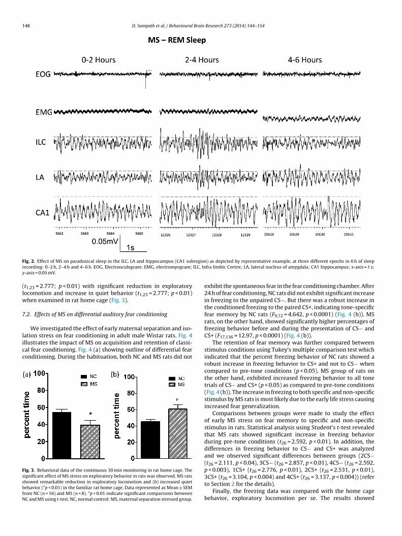
148 D. Sampath et al. / Behavioural Brain Research 273 (2014) 144–154
F regionr ILC, Iny
(lw
7
licc
FssbfN
ig. 2. Effect of MS on paradoxical sleep in the ILC, LA and hippocampus (CA1 subecording: 0–2 h, 2–4 h and 4–6 h. EOG, Electrooculogram; EMG, electromyogram;
-axis = 0.05 mV.
t1,23 = 2.777; p < 0.01) with significant reduction in exploratoryocomotion and increase in quiet behavior (t1,23 = 2.777; p < 0.01)
hen examined in rat home cage (Fig. 3).
.2. Effects of MS on differential auditory fear conditioning
We investigated the effect of early maternal separation and iso-
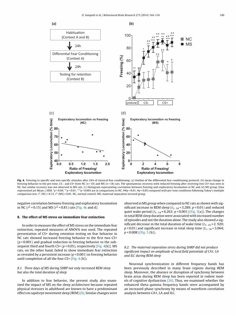
ation stress on fear conditioning in adult male Wistar rats. Fig. 4llustrates the impact of MS on acquisition and retention of classi-al fear conditioning. Fig. 4 (a) showing outline of differential fearonditioning. During the habituation, both NC and MS rats did notig. 3. Behavioral data of the continuous 30 min monitoring in rat home cage. Theignificant effect of MS stress on exploratory behavior in rats was observed. MS ratshowed remarkable reduction in exploratory locomotion and (b) increased quietehavior (*p < 0.01) in the familiar rat home cage. Data represented as Mean ± SEMrom NC (n = 16) and MS (n = 8). *p < 0.01 indicate significant comparisons betweenC and MS using t-test. NC, normal control; MS, maternal separation stressed group.
) as depicted by representative example, at three different epochs in 6 h of sleepfra limbic Cortex; LA, lateral nucleus of amygdala; CA1 hippocampus; x-axis = 1 s;
exhibit the spontaneous fear in the fear conditioning chamber. After24 h of fear conditioning, NC rats did not exhibit significant increasein freezing to the unpaired CS−. But there was a robust increase inthe conditioned freezing to the paired CS+, indicating tone-specificfear memory by NC rats (F9,72 = 4.642, p < 0.0001) (Fig. 4 (b)). MSrats, on the other hand, showed significantly higher percentages offreezing behavior before and during the presentation of CS− andCS+ (F17,136 = 12.97, p < 0.0001) (Fig. 4 (b)).
The retention of fear memory was further compared betweenstimulus conditions using Tukey’s multiple comparison test whichindicated that the percent freezing behavior of NC rats showed arobust increase in freezing behavior to CS+ and not to CS− whencompared to pre-tone conditions (p < 0.05). MS group of rats onthe other hand, exhibited increased freezing behavior to all tonetrials of CS− and CS+ (p < 0.05) as compared to pre-tone conditions(Fig. 4 (b)). The increase in freezing to both specific and non-specificstimulus by MS rats is most likely due to the early life stress causingincreased fear generalization.
Comparisons between groups were made to study the effectof early MS stress on fear memory to specific and non-specificstimulus in rats. Statistical analysis using Student’s t-test revealedthat MS rats showed significant increase in freezing behaviorduring pre-tone conditions (t26 = 2.592, p < 0.01). In addition, thedifferences in freezing behavior to CS− and CS+ was analyzedand we observed significant differences between groups (2CS−(t26 = 2.111, p < 0.04), 3CS− (t26 = 2.857, p < 0.01), 4CS− (t26 = 2.592,p < 0.003), 1CS+ (t26 = 2.776, p < 0.01), 2CS+ (t26 = 2.531, p < 0.01),
3CS+ (t26 = 3.104, p < 0.004) and 4CS+ (t26 = 3.137, p < 0.004)) (referto Section 2 for the details).Finally, the freezing data was compared with the home cagebehavior, exploratory locomotion per se. The results showed
D. Sampath et al. / Behavioural Brain Research 273 (2014) 144–154 149
Habitua�on (Co ntext A an d B)
Differen�al Fear Condi�oning (Context A)
Tes�ng for reten�on (Context B)
24h
24h
(a) (b)
0
20
40
60
80
100NCMS
pretone CS- CS+
***
***** ** ***
***
**
# #
# #
# #
# ## # # #
# ## #
##
Free
zing
(%)
(c) (d)Exploratory l ocomotion vs free zing
(NC)
0.0 0.5 1.0 1.5 2.040
50
60
70
80
Ratio of Free zing /Exploratory locomotion
Expl
orat
ory
Loco
mot
ion
(% ti
me) Exploratory l ocomotion vs free zing
(MS)
0 2 4 6 80
20
40
60
80
Ratio of Free zing /Exploratory locomotion
Expl
orat
ory
Loco
mot
ion
(% ti
me)
Fig. 4. Freezing to specific and non-specific stimulus after 24 h of classical fear conditioning. (a) Outline of the differential fear conditioning protocol; (b) mean change infreezing behavior to the pre-tone, CS− and CS+ from NC (n = 10) and MS (n = 18) rats. The spontaneous recovery with reduced freezing after receiving two CS+ was seen inN correlr NC; #c aratio
ni
8
epN(srau
8b
ipe
C, but similar recovery was not observed in MS rats. (c) Histogram representing
epresented are Mean ± SEM. *p < 0.04; **p < 0.01; ***p < 0.003 are in comparisons toomparison test. r2 (NC) = 0.15, r2 (MS) = 0.81. NC, normal control; MS, maternal sep
egative correlation between freezing and exploratory locomotionn NC (r2 = 0.15) and MS (r2 = 0.81) rats (Fig. 4c and d).
. The effect of MS stress on immediate fear extinction
In order to measure the effect of MS stress on the immediate fearxtinction, repeated measures of ANOVA was used. The repeatedresentation of CS+ during retention testing on fear behavior inC rats showed increased freezing behavior to the first two CS+
p < 0.001) and gradual reduction in freezing behavior to the sub-equent third and fourth CS+ (p < 0.05), respectively (Fig. 4(b)). MSats, on the other hand, failed to show immediate fear extinctions revealed by a persistent increase (p < 0.001) in freezing behaviorntil completion of all the four CS+ (Fig. 4 (b)).
.1. Three days of MS during SHRP not only increased REM sleeput also the total duration of sleep
In addition to fear behavior, the present study also exam-ned the impact of MS on the sleep architecture because repeatedhysical stressors in adulthood are known to have a predominantffect on rapid eye movement sleep (REM) [8]. Similar changes were
ation between freezing and exploratory locomotion in NC and (d) MS group. Data#p < 0.01; #p < 0.05 compared with pre-tone conditions following Tukey’s multiplen stressed group.
observed in MS group when compared to NC rats as shown with sig-nificant increase in REM sleep (t1, 14 = 3.289; p < 0.01) and reducedquiet wake period (t1, 14 = 6.263; p < 0.001) (Fig. 5(a)). The changesin total REM sleep duration were associated with increased numberof episodes and not the duration alone. The study also showed a sig-nificant decrease in the total duration of wake time (t1, 14 = 2. 920;p < 0.01) and significant increase in total sleep time (t1, 14 = 3.064;p < 0.008) (Fig. 5 (b)).
8.2. The maternal separation stress during SHRP did not producesignificant impact on amplitude of local field potentials of CA1, LAand ILC during REM sleep
Neuronal synchronization in different frequency bands hasbeen previously described in many brain regions during REMsleep. Moreover, the absence or disruption of synchrony betweenbrain areas during REM sleep has been reported in rodent mod-
els of cognitive dysfunction [26]. Thus, we examined whether theenhanced theta–gamma frequency bands were accompanied byan increased phase synchrony by means of waveform correlationanalysis between CA1, LA and ILC.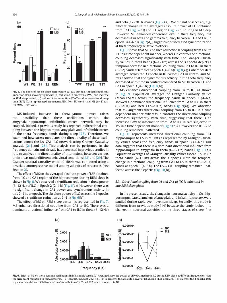
150 D. Sampath et al. / Behavioural Brain
(a) (b)
0
20
40
60
** *
NCMS
W1 W2 S1 S2 REM
Perc
ent t
ime
0
20
40
60
80
TWT TSWS TST
*
**NCMS
Perc
ent t
ime
Fig. 5. The effect of MS on sleep architecture. (a) MS during SHRP had significantimpact on sleep showing significant (a) reduction in quiet wake (W2) and increaseit*
tacpielafrbGbS
fs(nts
Md
Ftr
n REM sleep period; (b) reduced total wake time (TWT) and increased total sleepime (TST). Data represented are mean ± SEM from NC (n = 6) and MS (n = 8) rats*p < 0.001; *p < 0.01.
MS-induced increase in theta–gamma power raiseshe possibility that these oscillations within themygdala–hippocampal–infralimbic cortex network may beoupled. Indeed, a previous study has reported bidirectional cou-ling between the hippocampus, amygdala and infralimbic cortex
n the theta frequency bands during sleep [27]. Therefore, wexamined how stress modulates the directionality of these oscil-ations across the LA–CA1–ILC network using Granger-Causalitynalysis [21] and [25]. This analysis can be performed in therequency domain and already has been used in previous studies inats to analyze the directionality of interactions between variousrain areas under different behavioral conditions [28] and [29]. Theranger spectral causality within 0–50 Hz was computed using aivariate autoregressive model among all pairs of structures (seeection 2).
The effect of MS on the averaged absolute power of LFP obtainedrom ILC and CA1 region of the hippocampus during REM sleep ishown in Fig. 6. We observed a significant reduction in theta power6–12 Hz) of ILC in Epoch 2 (2–4 h) (Fig. 6(a)). However, there waso significant change in CA1 power and synchronous activity inhis 2–4 hour epoch. The absolute power of ILC across the 3 epochs
howed a significant reduction at 2–4 h (Fig. 6(b)).The effect of MS on REM sleep pattern is represented in Fig. 7.S enhances directional coupling from CA1 to ILC. There was a
ominant directional influence from CA1 to ILC in theta (6–12 Hz)
(a)
0-4 4-8 6-12 12-20 20-400.000 0
0.000 0
0.000 1
0.0001NCMS
Abs
olut
e Po
wer
(dB
)
Frequency (Hz)
**
ig. 6. Effect of MS on theta–gamma oscillations in infralimbic cortex. (a) Averaged absolhe significant reduction in theta power (6–12 Hz) of ILC in Epoch 2 (2–4 h). (b) Represenepresented as Mean ± SEM from NC (n = 5) and MS (n = 7), **p < 0.007 when compared to
Research 273 (2014) 144–154
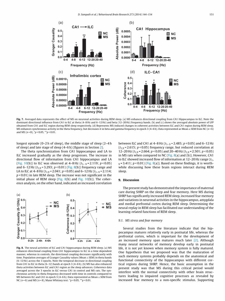
and beta (12–20 Hz) bands (Fig. 7 (a)). We did not observe any sig-nificant change in the averaged absolute power of LFP obtainedfrom CA1 (Fig. 7(b)) and ILC region (Fig. 7 (c)) during REM sleep.However, MS enhanced coherence level in theta frequency, butdecreases it in beta and gamma frequency between ILC and CA1 inepoch 3 (4–6 h) (Fig. 7(d)), suggestive of increased synchronizationat theta frequency relative to others.
Fig. 8 shows that MS enhances directional coupling from CA1 toILC in a time dependent manner, whereas in control the directionalcoupling decreases significantly with time. The Granger Causal-ity values in theta bands (6–12 Hz) across the 3 epochs depicts atemporal decrease in directional coupling from CA1 to ILC in theta(6–12) bands at late sleep epoch 3 (4–6 h) (Fig. 8(a)). Coherence dataaveraged across the 3 epochs in ILC verses CA1 in control and MSrats showed that the synchronous activity in the theta frequencydecreased with time in controls compared to MS between ILC andCA1 in epoch 3 (4–6 h) (Fig. 8(b)).
MS enhances directional coupling from LA to ILC as shownin Fig. 9. Population averages of Granger Causality values(Mean ± SEM) across the frequency bands in epoch 3 (4–6 h)showed a dominant directional influence from LA to ILC in theta(6–12 Hz) and beta (12–20 Hz) bands (Fig. 9(a)). We observedthat MS augments directional coupling from LA to ILC in a timedependent manner, whereas in control’s the directional couplingdecreases significantly with time, suggesting that there is anincreased flow of information from LA to ILC in rats subjected toMS in a time dependent manner (Fig. 9(b)). However the ILC → LAcoupling remained unaffected.
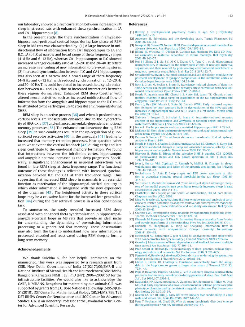
Fig. 10 represents increased directional coupling from CA1hippocampus to LA in MS rats as represented by Granger Causal-ity values across the frequency bands in epoch 3 (4–6 h). Ourdata suggests that there is a dominant directional influence fromhippocampus to amygdala in theta (6–12 Hz) bands (Fig. 10(a)).Population averages of Granger Causality values (Mean ± SEM) intheta bands (6–12 Hz) across the 3 epochs. Note the temporalchange in directional coupling from CA1 to LA in theta (6–12 Hz)bands at epoch 3 (4–6 h). The LA → CA1 coupling remained unaf-fected across the 3 epochs (Fig. 10(b)).
8.3. Directional coupling from LA and CA1 to ILC is enhanced inlate REM sleep phase
In the present study, the changes in neuronal activity in CA1 hip-
pocampus, Lateral nucleus of amygdala and infralimbic cortex werestudied during rapid eye movement sleep. Secondly, this study isdifferent from previous study (14) because the study looked intochanges in neuronal activities during three stages of sleep–first(b)
0-2h 2-4h 4-6h0.0000 0
0.00002
0.00004
0.0000 6
0.00008
0.0001 0NCMS**
Abs
olut
e po
wer
(dB
)
ute power of LFP obtained from ILC during REM sleep at different frequencies. Notets the absolute power of ILC during REM sleep at 6–12 Hz across the 3 epochs. Data
NC.
D. Sampath et al. / Behavioural Brain Research 273 (2014) 144–154 151
(a) (b)
(d)(c)
0-4 4-8 6-12 12-20 20-400.000 0
0.000 1
0.000 2
0.000 3
0.000 4
0.000 5
Abs
olut
e Po
wer
(dB
)
Frequ ency (Hz)
NCMS
CA1 Hippoca mpu s
0-4 4-8 6-12 12-20 20-400.0
0.1
0.2
0.3
0.4
0.5NCMS
Frequency (Hz)
**
**G
rang
er C
ausa
lity
*
CA1 I LC
0-4 4-8 6-12 12-20 20-400.0000 0
0.0000 2
0.0000 4
0.0000 6
0.0000 8
0.0001 0 NCMS
Abs
olut
e Po
wer
(dB
)
Frequ ency (Hz)
Infrali mbic cortexILC - CA1
0-4 4-8 6-12 12-20 20-400.0
0.1
0.2
0.3
0.4
0.5NCMS
Frequ ency (Hz)
*
* *
*
Coh
eren
ceFig. 7. Averaged data represents the effect of MS on neuronal activities during REM sleep. (a) MS enhances directional coupling from CA1 Hippocampus to ILC. Note thedominant directional influence from CA1 to ILC at theta (4–8 Hz and 6–12 Hz) and beta (12–20 Hz) frequency bands. (b) and (c) shows the averaged absolute power of LFPobtained from CA1 and ILC region during REM sleep respectively. (d) Represents MS-induced changes in coherent activities between ILC and CA1 region during REM sleep.M d gama
lo
Id(aLpie
(
Femt(ftacMN
S enhances synchronous activity in the theta frequency, but decreases it in beta annd MS (n = 8), *p < 0.05, **p < 0.01.
ongest episode (0–2 h of sleep), the middle stage of sleep (2–4 hf sleep) and late stage of sleep (4–6 h) (figures in Section 2).
The theta synchronization from CA1 hippocampus and LA toLC increased gradually as the sleep progresses. The increase inirectional flow of information from CA1 hippocampus and LAFig. 10(b)) to ILC was observed at 4–8 Hz (t1, 9 = 2.119; p < 0.05)nd 6–12 Hz (t1,9 = 3.293; p < 0.01) (Fig. 8(b)) frequency range andA to ILC at 4–8 Hz (t = 2.041; p < 0.05) and 6–12 Hz (t , = 2.114;
1,9 1 9< 0.01) in late REM sleep. The increase was not significant in thenitial phase of REM sleep (Fig. 8(b) and Fig. 10(b)). The coher-nce analysis, on the other hand, indicated an increased correlation
a) (b)
0-2h 2-4h 4-6h0.0
0.1
0.2
0.3
0.4
0.5NCMS**
Gra
nger
Cau
salit
y (6
-12H
z)
0-2h 2-4h 4-6h0.0
0.1
0.2
0.3
0.4
0.5NCMS
*
Coh
eren
ce (6
-12H
z)
ig. 8. The neural activities of ILC and CA1 hippocampus during REM sleep. (a) MSnhances directional coupling from CA1 hippocampus to ILC in a time dependentanner, whereas in controls, the directional coupling decreases significantly with
ime. Population averages of Granger Causality values (Mean ± SEM) in theta bands6–12 Hz) across the 3 epochs. Note the temporal decrease in directional couplingrom CA1 to ILC in theta (6–12) bands at epoch 3 (4–6 h). (b) MS has also enhancedheta activities between ILC and CA1 region as the sleep advances. Coherence dataveraged across the 3 epochs in ILC versus CA1 in control and MS rats. The syn-hronous activity in theta frequency decreased with time in controls compared toS between ILC and CA1 in epoch 3 (4–6 h). Data represented as Mean ± SEM fromC (n = 6) and MS (n = 8), Mann Whitney test: *p < 0.05, **p < 0.01.
ma frequency in epoch 3 (4–6 h). Data represented as Mean ± SEM from NC (n = 6)
between ILC and CA1 at 4–8 Hz (t1, 9 = 2.485; p < 0.05) and 6–12 Hz(t1,9 = 2.615; p < 0.05) frequency range, but reduced correlation at12–20 Hz (t1,9 = 2.664; p < 0.05) and 20–40 Hz (t1,9 = 2.501; p < 0.05)in MS rats when compared to NC (Fig. 8(a) and (b)). However, CA1to ILC showed increased flow of information at 12–20 Hz range (t1,9 = 3.411; p < 0.01) (Fig. 8(a)). Based on these findings, it is worth-while discussing how these brain regions interact during REMsleep.
9. Discussion
The present study has demonstrated the importance of maternalcare during SHRP on the sleep and fear memory. Here MS duringSHRP has significantly increased REM sleep, increased fear memoryand variations in neuronal activities in the hippocampus, amygdalaand medial prefrontal cortex during REM sleep. Determining theneural replay in REM sleep has facilitated our understanding of thelearning-related functions of REM sleep.
9.1. MS stress and fear memory
Several studies from the literature indicate that the hip-pocampus matures relatively early in postnatal life, whereas theprefrontal cortex, which is important for the development ofan increased memory span matures much later [2]. Althoughmany neural networks of memory develop early in postnatallife, it is not yet known when memory system is fully matured.One hypothesis that we proposed was that the maturation ofsuch memory systems probably depends on the anatomical andfunctional connectivity of the hippocampus with different cor-tical regions during SHRP. Hence, the basic assumption in the
present study was that MS during this critical period wouldinterfere with the normal connectivity with other brain struc-tures leading to impaired cognitive processes as revealed byincreased fear memory to a non-specific stimulus. Supporting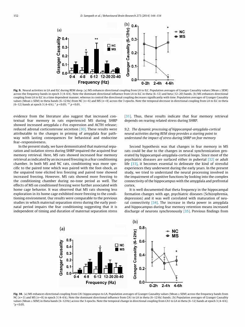
152 D. Sampath et al. / Behavioural Brain Research 273 (2014) 144–154
Fig. 9. Neural activities in LA and ILC during REM sleep. (a) MS enhances directional coupling from LA to ILC. Population averages of Granger Causality values (Mean ± SEM)across the frequency bands in epoch 3 (4–6 h). Note the dominant directional influence from LA to ILC in theta (6–12) and beta (12–20) bands. (b) MS enhances directionalcoupling from LA to ILC in a time dependent manner, whereas in control the directional coupling decreases significantly with time. Population averages of Granger Causalityv the 3(
etsrawf
rmrcctitehetsni
FNv*
alues (Mean ± SEM) in theta bands (6–12 Hz) from NC (n = 6) and MS (n = 8) across6–12) bands at epoch 3 (4–6 h); * p < 0.05; ** p < 0.01.
vidence from the literature also suggest that increased con-extual fear memory in rats experienced MS during SHRPhowed increased amygdala c-Fos expression and ACTH release;educed adrenal corticosterone secretion [30]. These results werettributable to the changes in priming of amygdala fear path-ay with lasting consequences for behavioral and endocrine
ear–responsiveness.In the present study, we have demonstrated that maternal sepa-
ation and isolation stress during SHRP impaired the acquired fearemory retrieval. Here, MS rats showed increased fear memory
etrieval as indicated by an increased freezing in a fear conditioninghamber. In both MS and NC rats, conditioning was more spe-ific to the paired tone which was paired with the foot-shock, ashe unpaired tone elicited less freezing and paired tone showedncreased freezing. However, MS rats showed more freezing tohe conditioning chamber during no-tone period as well. Theffects of MS on conditioned freezing were further associated withome cage behavior. It was observed that MS rats showing lessxploration in its home cage exhibited more freezing to the condi-
ioning environment. Our results were comparable to the previoustudies in which maternal separation stress during the early post-atal period impairs the fear conditioning suggesting that it isndependent of timing and duration of maternal separation stress
ig. 10. (a) MS enhances directional coupling from CA1 hippocampus to LA. Population avC (n = 3) and MS (n = 8) in epoch 3 (4–6 h). Note the dominant directional influence fromalues (Mean ± SEM) in theta bands (6–12 Hz) across the 3 epochs. Note the temporal chap < 0.05.
epochs. Note the temporal decrease in directional coupling from LA to ILC in theta
[31]. Thus, these results indicate that fear memory retrievaldepends on rearing related stress during SHRP.
9.2. The dynamic processing of hippocampal-amygdala-corticalneural activities during REM sleep provides a starting point tounderstand the impact of stress during SHRP on fear memory
Second hypothesis was that changes in fear memory in MSrats could be due to the changes in neural synchronization gen-erated by hippocampal-amygdala-cortical loops. Since most of thepsychiatric diseases are surfaced either in pubertal [32] or adultlife [33], it becomes essential to delineate the kind of stressfulexperiences they underwent during the early years. In the presentstudy, we tried to understand the neural processing involved inthe impairment of cognitive functions by looking into the complexconnectivity of the hippocampus with the amygdala and prefrontalcortex.
It is well documented that theta frequency in the hippocampalformation changes with age, psychiatric diseases (Schizophrenia,
depression) and it was well correlated with maturation of neu-ral connectivity [34]. The increase in theta power in amygdalaand hippocampus during fear memory retention means increaseddischarge of neurons synchronously [35]. Previous findings fromerages of Granger Causality values (Mean ± SEM) across the frequency bands from CA1 to LA in theta (6–12 Hz) bands; (b) Population averages of Granger Causality
nge in directional coupling from CA1 to LA in theta (6–12) bands at epoch 3 (4–6 h);
Brain
osa
hsdI(ia(w(attaibS
cimsciastaifonsfwoltt
aafpmpl
A
mCNBiCs5DSt
[
[[
[
[
[
[
[
[
[
[
[
[
[
[
[
[
[
[
[
[
D. Sampath et al. / Behavioural
ur laboratory showed a direct correlation between increased REMleep in stressed rats with enhanced theta synchronization in LAnd CA1 hippocampus [8].
In the present study, the theta synchronization in amygdalo-ippocampal-prefrontal cortical loops during late phase of REMleep in MS rats was characterized by: (1) A large increase in uni-irectional flow of information from CA1 hippocampus to LA and
LC, LA to ILC at narrow and broad range of theta frequency ranges4–8 Hz and 6–12 Hz), whereas CA1 hippocampus to ILC showedncreased Granger causality ratio at 12–20 Hz and 20–40 Hz reflectn increase in encoding of new information to pre-existing stores.2) Increased synchronization between ILC and CA1 hippocampusas also seen at a narrow and a broad range of theta frequency
4–8 Hz and 6–12 Hz) with reduced synchronization at 12–20 Hznd 20–40 Hz. This could be related to increased theta synchroniza-ion between ILC and CA1, due to increased interactions betweenhese regions during sleep. Enhanced REM sleep together withltered neural activities, such as increased unidirectional flow ofnformation from the amygdala and hippocampus to the ILC coulde attributed to the early exposure to stressful environments duringHRP.
REM sleep is an active process [36] and when it predominates,ortisol levels are consistently enhanced due to the hyperactiv-ty of HPA-axis [37] and enhanced amygdala-dependent emotional
emory processes [38]. The enhanced corticosterone during REMleep [39] in such conditions results in the up-regulation of gluco-orticoid receptor activation [40] in the amygdala [41] leading toncreased fear memory retention processes. Next question raisess to what extent the cortisol feedback [42] during early and lateleep contribute to the emotional memory formation. We foundhat interactions between the infralimbic cortex, hippocampusnd amygdala neurons increased as the sleep progresses. Specif-cally, a significant enhancement in neuronal interactions wasound in late REM sleep at theta frequency range. The functionalutcome of these findings is reflected with increased synchro-ization between ILC and CA1 at theta frequency range. Thusuggesting that increased REM sleep in mammals is an essentialunction as reactivation of the hippocampal-cortical circuitry inhich older information is integrated with the new experience
f the organism [43]. The heightened activity in these corticaloops may be a causative factor for the increased fear generaliza-ion [44] during the fear retrieval process in a fear conditioningask.
To summarize, the study revealed increased REM sleepssociated with enhanced theta synchronization in hippocampal-mygdala-cortical loops in MS rats that provide an ideal nicheor understanding the dynamicity of pre-existing informationrocessing to a generalized fear memory. These observationsay also form the basis to understand how new information is
rocessed, encoded and reactivated during REM sleep to form aong term memory.
cknowledgements
We thank Sulekha S. for her helpful comments on theanuscript. This work was supported by a research grant from
SIR, New Delhi, Government of India 27(0217)/09/EMR-II andational Institute of Mental Health and Neurosciences (NIMHANS),angalore, Karnataka NIMH: ES: PhD (NP): 2006–2009: SD for the
nfrastructure facilities. We would also like to acknowledge theARF, NIMHANS, Bengaluru for maintaining our animals.G.R. wasupported by grants from J.C. Bose National Fellowship (SR/S2/JCB-
1/2010), DST Center for Mathematical Biology (SR/S4/MS:799/12),ST IRHPA Center for Neuroscience and UGC Center for Advancedtudies. G.R. is an Honorary Professor at the Jawaharlal Nehru Cen-er for Advanced Scientific Research.[
[
Research 273 (2014) 144–154 153
References
[1] Bowlby J. Developmental psychiatry comes of age. Am J Psychiatry1988;145:1–10.
[2] Andersen SL. Stimulants and the developing brain. Trends Pharmacol Sci2005;26:237–43.
[3] Newport DJ, Stowe ZN, Nemeroff CB. Parental depression: animal models of anadverse life event. Am J Psychiatry 2002;159:1265–83.
[4] Rilling JK, Winslow JT, O’Brien D, Gutman DA, Hoffman JM, Kilts CD. Neu-ral correlates of maternal separation in rhesus monkeys. Biol Psychiatry2001;49:146–57.
[5] Hui J-J, Zhang Z-J, Liu S-S, Xi G-J, Zhang X-R, Teng G-J, et al. Hippocampalneurochemistry is involved in the behavioural effects of neonatal maternalseparation and their reversal by post-weaning environmental enrichment: amagnetic resonance study. Behav Brain Res 2011;217:122–7.
[6] Ovtscharoff W, Braun K. Maternal separation and social isolation modulate thepostnatal development of synaptic composition in the infralimbic cortex ofOctodon degus. Neuroscience 2001;104:33–40.
[7] Bock J, Gruss M, Becker S, Braun K. Experience-induced changes of dendriticspine densities in the prefrontal and sensory cortex: correlation with develop-mental time windows. Cereb Cortex 2005;15:802–8.
[8] Hegde P, Jayakrishnan HR, Chattarji S, Kutty BM, Laxmi TR. Chronic stress-induced changes in REM sleep on (oscillations in the rat hippocampus andamygdala. Brain Res 2011;1382:155–64.
[9] Faure J, Uys JDK, Marais L, Stein DJ, Daniels WMU. Early maternal separa-tion followed by later stressors leads to dysregulation of the HPA-axis andincreases in hippocampal NGF and NT-3 levels in a rat model. Metab BrainDis 2006;21:181–8.
10] Ziabreva I, Poeggel G, Schnabel R, Braun K. Separation-induced receptorchanges in the hippocampus and amygdala of Octodon degus: influence ofmaternal vocalizations. J Neurosci 2003;23:5329–36.
11] Nadel L. The hippocampus and space revisited. Hippocampus 1991;1:221–9.12] McEwen BS. Physiology and neurobiology of stress and adaptation: central role
of the brain. Physiol Rev 2007;87:873–904.13] Paxinos G, Watson C. The rat brain stereotaxic coordinates. 2nd ed. Sydney:
Academic Press; 1986.14] Hegde P, Singh K, Chaplot S, Shankaranarayana Rao BS, Chattarji S, Kutty BM,
et al. Stress-induced changes in sleep and associated neuronal activity in rathippocampus and amygdala. Neuroscience 2008;153:20–30.
15] Bjorvatn B, Ursin R. Effects of the selective 5-HT1B agonist, CGS 12066B,on sleep/waking stages and EEG power spectrum in rats. J Sleep Res1994;3:97–105.
16] John J, Kumar VM, Gopinath G, Ramesh V, Mallick H. Changes in sleep-wakefulness after kainic acid lesion of the preoptic area in rats. Jpn J Physiol1994;44:231–42.
17] Neckelmann D, Ursin R. Sleep stages and EEG power spectrum in rela-tion to acoustical stimulus arousal threshold in the rat. Sleep 1993;16:467–77.
18] Vetrivelan R, Mallick HN, Kumar VM. Tonic activity of alpha1 adrenergic recep-tors of the medial preoptic area contributes towards increased sleep in rats.Neuroscience 2006;139:1141–51.
19] Chatfield C. The analysis of time series: an introduction. 6th ed. Boca Raton:Chapman and Hall/CRC; 2003.
20] Ding M, Bressler SL, Yang W, Liang H. Short-window spectral analysis of corti-cal event-related potentials by adaptive multivariate autoregressive modeling:data preprocessing, model validation, and variability assessment. Biol Cybern2000;83:35–45.
21] Granger CWJ. Investigating causal relations by econometric models and cross-spectral methods. Econometrica 1969;37:424–38.
22] Dhamala M, Rangarajan G, Ding M. Estimating Granger causality from Fourierand wavelet transforms of time series data. Phys Rev Lett 2008;100:018701.
23] Dhamala M, Rangarajan G, Ding M. Analyzing information flow inbrain networks with nonparametric Granger causality. Neuroimage2008;41:354–62.
24] Nedungadi AG, Rangarajan G, Jain N, Ding M. Analyzing multiple spike trainswith nonparametric Granger causality. J Comput Neurosci 2009;27:55–64.
25] Geweke J. Measurement of linear dependence and feedback between multipletime series. J Am Stat Assoc 1982;77:304–13.
26] Pace-Schott EF, Hobson JA. The neurobiology of sleep: genetics, cellular physi-ology and subcortical networks. Nat Rev Neurosci 2002;3:591–605.
27] Pignatelli M, Beyeler A, Leinekugel X. Neural circuits underlying the generationof theta oscillations. J Physiol Paris 2012;106:81–92.
28] Ghosh S, Laxmi TR, Chattarji S. Functional connectivity from the amyg-dala to the hippocampus grows stronger after stress. J Neurosci 2013;33:7234–44.
29] Popa D, Duvarci S, Popescu AT, Léna C, Paré D. Coherent amygdalocortical thetapromotes fear memory consolidation during paradoxical sleep. Proc Natl AcadSci U S A 2010;107:6516–9.
30] Daskalakis NP, Diamantopoulou A, Claessens SEF, Remmers E, Tjälve M, OitzlMS, et al. Early experience of a novel-environment in isolation primes a fearfulphenotype characterized by persistent amygdala activation. Psychoneuroen-docrinology 2014;39:39–57.
31] Kosten TA, Lee HJ, Kim JJ. Early life stress impairs fear conditioning in adultmale and female rats. Brain Res 2006;1087:142–50.
32] Paus T, Keshavan M, Giedd JN. Why do many psychiatric disorders emergeduring adolescence? Nat Rev Neurosci 2008;9:947–57.

1 Brain
[
[
[
[
[
[
[
[
[
[
[43] Buzsáki G. Memory consolidation during sleep: a neurophysiological perspec-
54 D. Sampath et al. / Behavioural
33] Paradis AD, Reinherz HZ, Giaconia RM, Fitzmaurice G. Major depression in thetransition to adulthood: the impact of active and past depression on youngadult functioning. J Nerv Ment Dis 2006;194:318–23.
34] Benes FM, Berretta S. Amygdalo-entorhinal inputs to the hippocampalformation in relation to schizophrenia. Ann N Y Acad Sci 2000;911:293–304.
35] Seidenbecher T, Laxmi TR, Stork O, Pape H-C. Amygdalar and hippocam-pal theta rhythm synchronization during fear memory retrieval. Science2003;301:846–50.
36] Wagner U, Born J. Memory consolidation during sleep: interactive effects ofsleep stages and HPA regulation. Stress 2008;11:28–41.
37] O’Brien JT, Ames D, Schweitzer I, Mastwyk M, Colman P. Enhanced adrenal
sensitivity to adrenocorticotrophic hormone (ACTH) is evidence of HPA axishyperactivity in Alzheimer’s disease. Psychol Med 1996;26:7–14.38] Wagner U, Degirmenci M, Drosopoulos S, Perras B, Born J. Effects of cortisol sup-pression on sleep-associated consolidation of neutral and emotional memory.Biol Psychiatry 2005;58:885–93.
[
Research 273 (2014) 144–154
39] Aisa B, Tordera R, Lasheras B, Del Río J, Ramírez MJ. Cognitive impairmentassociated to HPA axis hyperactivity after maternal separation in rats. Psy-choneuroendocrinology 2007;32:256–66.
40] Plihal W, Born J. Memory consolidation in human sleep depends on inhibitionof glucocorticoid release. Neuroreport 1999;10:2741–7.
41] Schulkin J, Gold PW, McEwen BS. Induction of corticotropin-releasing hor-mone gene expression by glucocorticoids: implication for understanding thestates of fear and anxiety and allostatic load. Psychoneuroendocrinology1998;23:219–43.
42] Hasselmo M. Neuromodulation: acetylcholine and memory consolidation.Trends Cogn Sci 1999;3:351–9.
tive. J Sleep Res 1998;7(Suppl. 1):17–23.44] Likhtik E, Stujenske JM, Topiwala MA, Harris AZ, Gordon JA. Prefrontal entrain-
ment of amygdala activity signals safety in learned fear and innate anxiety. NatNeurosci 2014;17(1):106–13.