Embed Size (px)
Citation preview
IOSR Journal Of Pharmacywww.iosrphr.org
(e)-ISSN: 2250-3013, (p)-ISSN: 2319-4219
Volume 7, Issue 10 Version. 1 (October 2017), PP. 46-61
46
A Study on the applications of Silver nanoparticles synthesized
usingaqueous extract and purified secondary metabolites of
seaweed Hypneacervicornis
Leela.K1 and Anchana Devi.C
2*
1, 2*PG & Research Department of Biotechnology, Women’s Christian college, College road, Chennai - 600006,
Tamilnadu, India
*Corresponding author: Anchana Devi. C, PG & Research Department of Biotechnology, Women’s Christian
college, College road, Chennai - 600006, Tamilnadu, India
Abstract:Seaweeds are commonly referred to as macroalgae and are plant like organisms that live attached to
rock or any other hard substrata. They are one among the commercially important sources which possess the
ability to synthesize numerous secondary metabolites that have wide applications in the field of medicine as
anti-inflammatory or anticancer agents. They are also widely utilized in the production of polysaccharides such
as carrageenan, fucoidan etc., and have both therapeutic as well as industrial applications. The Present study
was carried out on the synthesis, characterization and applications of silver nanoparticle using the aqueous
extract and the individual purified compounds of Seaweed Hypneacervicornis. The Silver nanoparticles were
synthesized and characterized by means of UV-Visible spectroscopy, Fourier transform infrared spectroscopy
and Scanning electron microscopy. The applications of these nanoparticles were determined by studying their
antimicrobial, antioxidant and antidiabetic activity. Not much work has been reported before on the
nanoparticle synthesis using the individual secondary compounds from seaweed Hypneacervicornis so this
study was one such attempt to identify the stabilizing and reducing ability of the secondary compounds in the
synthesis of silver nanoparticle that could prove to be useful to the mankind for different applications. Keywords:Hypneacervicornis, Silver nanoparticle, Secondary compounds, UV-Visible spectroscopy, Fourier
transform infrared spectroscopy, Scanning electron microscope, Applications.
----------------------------------------------------------------------------------------------------------------------------- ----------
Date of Submission: 17-10-2017 Date of acceptance: 27-10-2017
----------------------------------------------------------------------------------------------------------------------------- ----------
I. INTRODUCTION Seaweeds are marine non flowering plantsfound to grow abundantly in shallow waters of seas and
estuaries.They are considered to be potentially beneficial sources of highly bioactive secondary metabolites that
could be used widely for different applications. The compounds extracted from seaweeds are found to possess
antimicrobial, antioxidant, antitumor, larvicidal and antiviral properties [1]. Compounds such as laminaran,
porphyran, ulvanderived from seaweeds are found to reduce cholesterol and high blood pressure levels so they
are used as drugs in pharmaceutical industries in the form of nutraceuticals[2].
Hypneacervicornis is a red macroalgae that belongs to the family Hypneaceae and also referred to as
hooked red weed. It remains attached to the upper branches of other reef algae in the subtidal region. These are
generally yellowish green in colour but appear to be red in shaded conditions[3].They produce numerous
secondary compounds that play an important role in food and pharmaceutical industries. They are the chief
source in production of polysaccharides such as carrageenan which is used as a binding and smoothening agent
in ice creams, tooth paste and also acts as a principal component in bacterial culture media.
Nanotechnology is an emerging field of scientific study that incorporates biotechnology at a nanoscale.
Nanoparticle is a microscopic particle with dimension less than 100nm and can be of different types based on
the metal used such as silver, gold, copper, zinc etc. Among them silver nanoparticles are mostly preferred due
to their specialized magnetic, electrical and optical properties. Silver nanoparticles are widely utilized for
different applications such as in textile industries, in cosmetics, in water treatment and as antimicrobial drugs
etc. where they are found to possess effective antimicrobial activity[4](Fig 1).They are also been extensively
studied in different fields such as materials science, electronics and in medicine [5]. Silver nanoparticles can be
produced by different methods but biological synthesis is widely preferred due to low toxicity, eco-friendly and
low cost.

A study on applications of silver nanoparticles synthesized usingaqueous extract and purified ..
47
Figure 1: SEM Image of Silver nanoparticles
Secondary metabolites are organic compounds widely synthesized by various natural sources such as
bacteria, fungi, algae, plants and animals and are not involved in the growth and development of an organism.
These are generally produced during a transition state from growth to stationary phase and are widely utilized as
colorant, aromatic agents and as medicinal drugs. In the present study Silver nanoparticles were synthesized
using two different secondary metabolites they are Glycosides and Alkaloids. Glycosides are organic
compounds in which a sugar molecule is bound to a functional group through a glycosidic bond while alkaloids
are chemical compoundsthat contain basic nitrogen atoms both the compounds play a vital role and possess
wide pharmaceutical and industrial applications.
Thus the present study was aimed at determining the antimicrobial, antioxidant and antidiabetic
applications of Silver nanoparticles synthesized using the aqueous extract as well as the purified glycoside and
alkaloid compounds of SeaweedHypneacervicornis.
II. MATERIALS AND METHODS 2.1 Sample Preparation:
The Seaweed Hypneacervicornis samples were collected manually from Pazhaverkadu brackish water
area at Thiruvallur district, Chennai and were identified based on their morphological characteristics [6]. The
samples were cleaned, washed under tap water and distilled water to remove the dirt and were dried for about 3
days and powdered. The powdered samples were stored in clean bottles for further analysis.
2.2 Extraction of Samples:
2.2.1 Methanolic Extraction: The crude extract from the lichen sample was obtained by means of cold
extraction method using methanol as solvent. About 50g of the powdered lichen sample was added to 500ml of
methanol in a conical flask, covered with aluminium foil and kept on a rotary shaker for 3 days at room
temperature. The solution was filtered with the help of Whatman No.1 filter paper and the filtrate obtained was
evaporated. The dried extracts were then stored for further experiments [7]. The yield of respective crude extract
was calculated as:
Percentage yield (%) = (dry weight of extract/dry weight of samples) × 100.
2.2.2 Aqueous Extraction: For aqueous extraction about 50g of the powdered lichen sample was added to
500ml of double distilled water separately in a conical flask and was kept in water bath at 65ºc for about 1 hour
to enhance complete extraction. The aqueous solution thus obtained was filtered with the help of a Whatman no
1 filter paper and the filtrates thus obtained were collected in a beaker and was utilized for further analysis.
2.3 Identification, Isolation and Purification of Secondary metabolites:
2.3.1 Thin Layer Chromatography:
Thin layer chromatography (TLC) is a chromatographic technique used for separating different
components from a mixture. It is generally carried out on a thin sheet of plastic, glass or an aluminium foil
coated with an absorbent material. The adsorbent could be cellulose, silica or aluminium oxide (alumina). The
layer of adsorbent material is called as stationary phase while the solvent or mixture of solvents acts as a mobile
phase via capillary action. TLC plate is cut and using a pencil line is drawn about 1cm from one end of the edge.
The samples were spotted on to the plate using a capillary tube for each spot. The spots should be air dried. The
TLC chamber consists of a glass jar with a lid and the solvent mixture is added to chamber. The Mobile phase
used for isolation of glycosides are Toluene: methanol: glacial acetic acid: water = 7:4:3:1 and for alkaloids
are Butanol: acetic acid: water = 4:1:3. The TLC plates were placed carefully in the TLC chamber. The
solvent mixture should be below the spot and the chamber is kept closed. As the solvent moves up due to
capillary movement, the compounds present in the extract are separated. The TLC plates are then removed from
the chamber and kept for drying. The dried plates are observed for spots by using a suitable spraying reagent
[8].For glycosides Iodine chamber is used while for alkaloids Dragendroff’s reagent is used for identification [9,
A study on applications of silver nanoparticles synthesized usingaqueous extract and purified ..
48
10]. The formation of brown band indicates the presence of glycosides and the formation of reddish orange band
indicates the presence of alkaloids. The Rf value is calculated using the formula:
Rf= Distance travelled by the solute / Distance travelled by the solvent
2.3.2 Column Chromatography:
Silica gel (100 - 200 mesh) was chosen as the stationary phase. The column was packed with silica gel
using methanol once packed the crude residue from methanol extract was transferred onto the bed of silica gel.
The column was run by using mobile phase (Toluene: methanol: glacial acetic acid: water in the ratio of
35:20:15:10) for glycosides and (Butanol: acetic acid: water in the ratio of 40:10:30) for alkaloids. The
fractions were collected at an interval of 5ml each and are monitored by means of thin layer chromatography.
The fractions obtained were stored and utilized for the identification of individual compounds present in the
sample by means of Gas chromatography – Mass spectrometry (GC-MS) [11].
2.3.3 Confirmatory test: [12]
1. Glycosides: Keller –Killani Test: To 1ml of sample add 1ml of glacial acetic acid and 1ml of concentrated
Sulphuric acid. Appearance of reddish brown colour at the junction of 2 layers indicates the presence of
glycosides.
2. Alkaloids: Dragendroff’s test: To 2ml of sample 2-3 drops of Dragendroff’s reagent is added. Appearance
of orange red coloured complex indicates the presence of alkaloids
2.4 Identification of Secondary metabolites:
2.4.1 Gas chromatography – Mass spectrometry (GC-MS): It is an analytical method used for identifying different substances within a test sample. It also helps in
identifying trace elements in a sample. The Clarus 680 GC was used for the analysis and a fused silica column
was employed and packed with Elite-5MS (5% biphenyl 95% dimethylpolysiloxane, 30 m × 0.25 mm ID ×
250μm df) and the components were separated using Helium as carrier gas at a constant flow of 1 ml/min. The
injector temperature was set at 260°C during the chromatographic run. The 1μL of extract sample was injected
into the instrument and the oven temperature was as maintained as follows: 60 °C (2 min); followed by 300 °C
at the rate of 10 °C min−1; and 300 °C, where it was held for 6 min. The mass detector conditions were:
transfer line temperature 240 °C; ion source temperature 240 °C; and ionization mode electron impact at 70 eV,
a scan time 0.2 sec and scan interval of 0.1 sec. The fragments were obtained from 40 to 600 Da. The spectrums
of the components were compared with the database of spectrum of known components stored in the GC-MS
NIST (2008) library [13].
2.5 Synthesis of Silver nanoparticles: [14]
The Silver nanoparticles were synthesized using the aqueous extracts and the purified glycoside and alkaloid
compounds obtained through column chromatography from Seaweed Hypneacervicornissample respectively.
2.5.1 Using the Aqueous extract:
About 30ml of 1mM silver nitrate was prepared and taken in a beaker. The beaker containing
magnets were placed on the magnetic stirrer with a rotation of 5,000 rpm at 60ºc. 10ml of Seaweed
(Hypneacervicornis) aqueous extracts were taken in a conical flask and was added drop wise into the beaker
containing silver nitrate. The colour change from colourless to dark brown indicates the formation of silver
nanoparticles. The reduction of the Ag+ ions was measured by the UV-Vis spectrum.
2.5.2 Using the Purified fractions:
About 30ml of 1mM silver nitrate was prepared and taken in a beaker. The beaker containing
magnets were placed on the magnetic stirrer with a rotation of 5,000 rpm at 60ºc. 10ml of the purified glycoside
and alkaloid fractions obtained through column chromatography were taken in a conical flask and was added
drop wise into the beaker containing silver nitrate. The colour change from colourless to brown indicates the
formation of silver nanoparticles. The reduction of the Ag+ ions was measured by the UV-Vis spectrum.
2.6 Characterization of Silver nanoparticles:
2.6.1 Ultraviolet - Visible Spectroscopy: Instrument model: UV-Visible Spectrophotometer - UV-1800.
UV-Vis spectral analysis was done by using UV-Visible spectrophotometer. Ultraviolet-visible spectroscopy is
a type of absorption spectroscopy in which light of ultraviolet region (400-500nm) is absorbed by the molecule.
Absorption of the ultra violet radiations results in the excitation of the electrons from the ground state to the
higher energy state. The energy of UV radiation absorbed is equal to the energy difference between the ground
A study on applications of silver nanoparticles synthesized usingaqueous extract and purified ..
49
state and the higher energy state. The absorbance was recorded at different nanometers from 400-500nm and the
Optical density (OD) was calculated [15].
2.6.2 Fourier Transform Infrared Spectroscopy (FTIR): Fourier transform infrared spectroscopy is a technique which is used to obtain an infrared spectrum of
absorption or emission of a solid, liquid, gas.
Instrument Model: Shimadzu IRTracer-100
KBr Pellet Method: To prepare a liquid sample to IR analysis, firstly place a drop of the liquid on the face of a
highly polished salt plate (such as NaCl, AgCl or KBr), then place a second plate on top of the first plate so as to
spread the liquid in a thin layer between the plates, and clamp the plates together. Finally wipe off the liquid out
of the edge of plate. Mount the sandwich plate onto the sample holder. After finishing the experiment, clean the
plates with isopropanol and returned to the desiccators [16].
2.6.3 Scanning Electron Microscopy (SEM): The Synthesized nanoparticles were centrifuged at 5,500 rpm for 15 minutes. The supernatant was discarded and
the pellet was dissolved in double distilled water and centrifugation was repeated twice to remove the
impurities. The pellet obtained finally was taken in petriplates and kept in hot air oven at 70ºc for about 30
minutes for drying. After it has completely dried the nanoparticles were scrapped off, stored in eppendorf and
utilized for SEM analysis[17].
2.7 Applications:
2.7.1 Antimicrobial activity:
The antibacterial activity of Seaweed(Hypneacervicornis) crude methanolic extract, Silver
nanoparticle from aqueous extracts, Silver nanoparticle from glycoside and alkaloid fractions were screened
against both Gram positive and Gram negative bacteria such as Staphylococcus aureus, Streptococcus spp,
Escherichia coli, Klebsiellapneumoniae, Salmonella sppand Pseudomonas aeruginosawhile the antifungal
activity was carried out against AspergillussppandCandida albicans.
Agar well diffusion method:
The stock cultures were maintained at 4°C on the nutrient agar slant slopes. Nutrient broth was
prepared for about 50ml and a loop-full of stock cultures were transferred to 50ml of nutrient broth and were
incubated at 37°C for 24 hours. The strains were inoculated in nutrient broth for 24 hours. Potato dextrose broth
was prepared for the growth of fungal strains and incubated for 48 hours. About 250ml of Muller Hinton agar
medium (MHA) was prepared for antibacterial activity while Potato Dextrose agar medium (PDA) was prepared
for antifungal activity and poured into the petriplates and was allowed to solidify. Once solidified the bacterial
cultures and the fungal cultures were swabbed onto the agar medium using a sterile cotton swab .The wells were
punctured using a sterile cork borer. Different concentrations of samples (20, 40, 60, 80mg/ml) were dispensed
into each well using a micropipette. The petriplates were then incubated at 37°C for 24 hours for bacteria and
37°C for 3-4 days for fungi and observed for the zone of inhibition. The diameter of the zone of inhibition was
measured in mm [18].
2.7.2 Antioxidant activity:
Hydrogen peroxide scavenging assay:
Solution of Hydrogen peroxide (40mM) was prepared in phosphate buffer (pH 7.4).1ml of the test sample was
added to 3ml of Hydrogen peroxide solution and was incubated at room temperature for 10 minutes and the
absorbance was determined at 230nm in a spectrophotometer. Ascorbic acid was used as the standard.
Phosphate buffer without hydrogen peroxide served as blank [19]. It can be calculated using the formula:
% scavenged (H2O2) = (A of Control – A of test / A of Control) × 100
2.7.3 Antidiabetic activity:
The antidiabetic activity of Seaweed (Hypneacervicornis) crude methanolic extract, Silver nanoparticle from
aqueous extracts, Silver nanoparticle from glycoside and alkaloid fractions were determined by means of Alpha
amylase inhibition assay.
Alpha amylase inhibition assay: About 1ml of the sample was added to 1ml starch solution and was incubated
for 10 minutes at room temperature.0.5ml of the prepared enzyme solution was added to the mixture and was
incubated at 25ºc for about 10 minutes. The reaction was then terminated by the addition of 1ml of colorimetric
reagent and was kept in water bath for 5 minutes and cooled to room temperature. This was further diluted by
adding 10ml of distilled water and the absorbance of the mixture was measured at 540nm in colorimeter.
Sample without extract served as blank [20].The % inhibition was calculated using the formula:
A study on applications of silver nanoparticles synthesized usingaqueous extract and purified ..
50
% inhibition = (A of Control – A of test / A of Control) × 100
III. RESULTS & DISCUSSION
3.1 Extraction of Sample:
The Seaweed (Hypneacervicornis) sample was extracted by means of cold extraction method using methanol
and water (aqueous). The Percentage yield of Seaweed crude extract was found to be 3.16 % and it is the total
yieldobtained for about 50 grams of the dry weight of the sample(Table 1).The Percentage yield was calculated
according to the paper [21].
S.NO SAMPLE DRY WEIGHT
OF SAMPLE (g)
DRY WEIGHT OF
EXTRACT (g)
YIELD
(%)
1
Seaweed
50 Grams
1.58 Grams
3.16 %
Table 1:Estimation of Percentage yield of Seaweed sample
Methanol was highly preferred for extraction purposes because it has a polarity index of 5.1 and was found to be
capable of dissolving polar compounds and the solvent was also reported to have been used by other authors for
their extraction purposes[22]. Water has a polarity index of about 10.2 and is also used for extraction in order to
compare its ability with that of methanol in extracting the compounds. A Study has reported the extraction of
Seaweed Sargassumwightii using 4 different solvents such as hexane, diethyl ether, acetone and methanol
wherein the percentage yield was found out to be maximum in acetone extract (0.969%) and minimum in
hexane extract (0.225%) respectively [23].
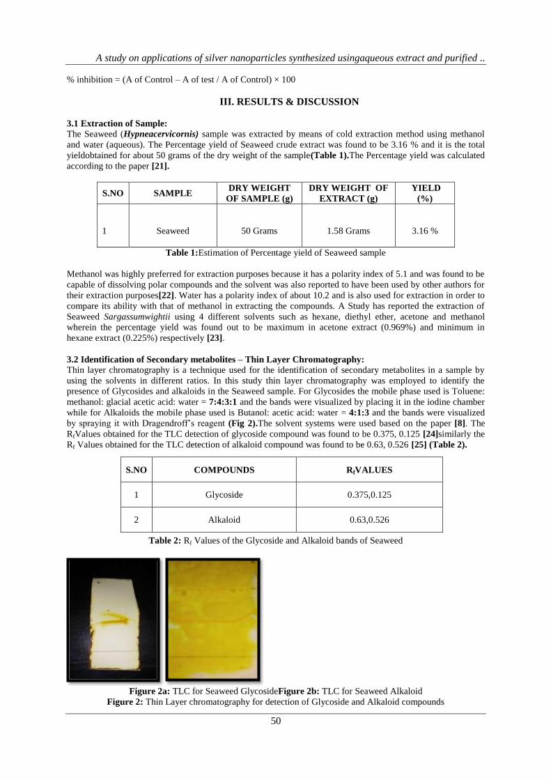
3.2 Identification of Secondary metabolites – Thin Layer Chromatography:
Thin layer chromatography is a technique used for the identification of secondary metabolites in a sample by
using the solvents in different ratios. In this study thin layer chromatography was employed to identify the
presence of Glycosides and alkaloids in the Seaweed sample. For Glycosides the mobile phase used is Toluene:
methanol: glacial acetic acid: water = 7:4:3:1 and the bands were visualized by placing it in the iodine chamber
while for Alkaloids the mobile phase used is Butanol: acetic acid: water = 4:1:3 and the bands were visualized
by spraying it with Dragendroff’s reagent (Fig 2).The solvent systems were used based on the paper [8]. The
RfValues obtained for the TLC detection of glycoside compound was found to be 0.375, 0.125 [24]similarly the
Rf Values obtained for the TLC detection of alkaloid compound was found to be 0.63, 0.526 [25] (Table 2).
Table 2: Rf Values of the Glycoside and Alkaloid bands of Seaweed
Figure 2a: TLC for Seaweed GlycosideFigure 2b: TLC for Seaweed Alkaloid
Figure 2: Thin Layer chromatography for detection of Glycoside and Alkaloid compounds
S.NO COMPOUNDS RfVALUES
1 Glycoside 0.375,0.125
2 Alkaloid 0.63,0.526
A study on applications of silver nanoparticles synthesized usingaqueous extract and purified ..
51
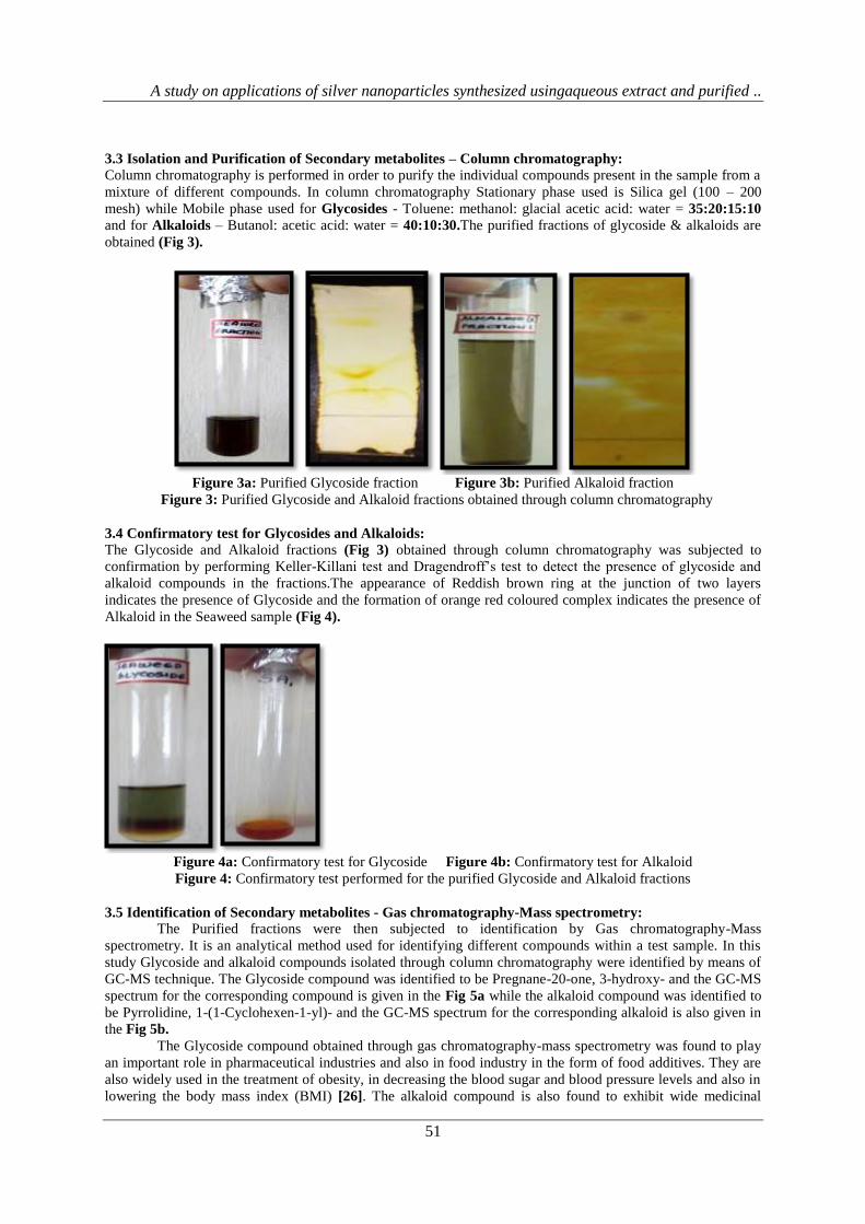
3.3 Isolation and Purification of Secondary metabolites – Column chromatography: Column chromatography is performed in order to purify the individual compounds present in the sample from a
mixture of different compounds. In column chromatography Stationary phase used is Silica gel (100 – 200
mesh) while Mobile phase used for Glycosides - Toluene: methanol: glacial acetic acid: water = 35:20:15:10
and for Alkaloids – Butanol: acetic acid: water = 40:10:30.The purified fractions of glycoside & alkaloids are
obtained (Fig 3).
Figure 3a: Purified Glycoside fraction Figure 3b: Purified Alkaloid fraction
Figure 3: Purified Glycoside and Alkaloid fractions obtained through column chromatography
3.4 Confirmatory test for Glycosides and Alkaloids:
The Glycoside and Alkaloid fractions (Fig 3) obtained through column chromatography was subjected to
confirmation by performing Keller-Killani test and Dragendroff’s test to detect the presence of glycoside and
alkaloid compounds in the fractions.The appearance of Reddish brown ring at the junction of two layers
indicates the presence of Glycoside and the formation of orange red coloured complex indicates the presence of
Alkaloid in the Seaweed sample (Fig 4).
Figure 4a: Confirmatory test for Glycoside Figure 4b: Confirmatory test for Alkaloid
Figure 4: Confirmatory test performed for the purified Glycoside and Alkaloid fractions
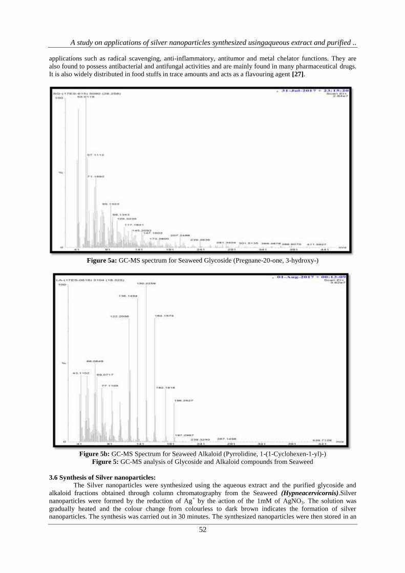
3.5 Identification of Secondary metabolites - Gas chromatography-Mass spectrometry:
The Purified fractions were then subjected to identification by Gas chromatography-Mass
spectrometry. It is an analytical method used for identifying different compounds within a test sample. In this
study Glycoside and alkaloid compounds isolated through column chromatography were identified by means of
GC-MS technique. The Glycoside compound was identified to be Pregnane-20-one, 3-hydroxy- and the GC-MS
spectrum for the corresponding compound is given in the Fig 5a while the alkaloid compound was identified to
be Pyrrolidine, 1-(1-Cyclohexen-1-yl)- and the GC-MS spectrum for the corresponding alkaloid is also given in
the Fig 5b.
The Glycoside compound obtained through gas chromatography-mass spectrometry was found to play
an important role in pharmaceutical industries and also in food industry in the form of food additives. They are
also widely used in the treatment of obesity, in decreasing the blood sugar and blood pressure levels and also in
lowering the body mass index (BMI) [26]. The alkaloid compound is also found to exhibit wide medicinal
A study on applications of silver nanoparticles synthesized usingaqueous extract and purified ..
52
applications such as radical scavenging, anti-inflammatory, antitumor and metal chelator functions. They are
also found to possess antibacterial and antifungal activities and are mainly found in many pharmaceutical drugs.
It is also widely distributed in food stuffs in trace amounts and acts as a flavouring agent [27].
Figure 5a: GC-MS spectrum for Seaweed Glycoside (Pregnane-20-one, 3-hydroxy-)
Figure 5b: GC-MS Spectrum for Seaweed Alkaloid (Pyrrolidine, 1-(1-Cyclohexen-1-yl)-)
Figure 5: GC-MS analysis of Glycoside and Alkaloid compounds from Seaweed
3.6 Synthesis of Silver nanoparticles:
The Silver nanoparticles were synthesized using the aqueous extract and the purified glycoside and
alkaloid fractions obtained through column chromatography from the Seaweed (Hypneacervicornis).Silver
nanoparticles were formed by the reduction of Ag+ by the action of the 1mM of AgNO3. The solution was
gradually heated and the colour change from colourless to dark brown indicates the formation of silver
nanoparticles. The synthesis was carried out in 30 minutes. The synthesized nanoparticles were then stored in an

A study on applications of silver nanoparticles synthesized usingaqueous extract and purified ..
53
air tight bottle at room temperature for further characterization studies(Fig 6).A study has reported the synthesis
of silver nanoparticle from the aqueous extract of seaweed Sargassumcinereum and the nanoparticle formation
was confirmed based on the colour change from yellowish green to orange[28].
Figure 6: Synthesis of silver nanoparticle using the aqueous extract and purified fractions
3.7 Characterization of Silver nanoparticles:
The Silver nanoparticles synthesized using the aqueous extract and purified fractions were further characterized
by means of UV-Visible spectroscopy, Fourier transform infrared spectroscopy (FTIR) and Scanning electron
microscope (SEM).
3.7.1 UV-Visible Spectrophotometer analysis: The Silver nanoparticles synthesized using the aqueous extracts and purified fractions were characterized by
means of UV-Visible spectrophotometer. The wavelength was selected from 400 to 500nm and the absorbance
was recorded. The maximum absorption peak was obtained at 430nm for Seaweed aqueous extracts while
showed peak at 440 nm for nanoparticle synthesized using Seaweed purified glycoside and alkaloid fractions
(Fig 7).A study has reported the characterization of silver nanoparticles from Murrayakoenigii and Zea mays by
UV-Visible spectroscopy wherein the absorption peak was found to be obtained at 420 - 440nm and thus
confirmed the formation of nanoparticle [29].
Figure 7a: UV-Visible spectra showing absorbance for Silver nanoparticle synthesized using Seaweed aqueous
extract
0.1050.167
0.286
0.335 0.311
0.284
0.273
0.135
0.102
0.064
0.035
0
0.1
0.2
0.3
0.4
AB
SOR
BA
NC
E (O
.D)
WAVELENGTH (nm)
SEAWEED AQUEOUS NANOPARTICLE
SEAWEED AQUEOUS NANOPARTICLE
0.123
0.1740.198
0.2950.345
0.3180.268
0.259
0.101
0.0340.021
0
0.1
0.2
0.3
0.4
AB
SOR
BA
NC
E (O
.D)
WAVELENGTH (nm)
SEAWEED GLYCOSIDE NANOPARTICLE
SEAWEED GLYCOSIDE NANOPARTICLE

A study on applications of silver nanoparticles synthesized usingaqueous extract and purified ..
54
Figure 7b: UV-Visible spectra showing absorbance for Silver nanoparticle synthesized using purified glycoside
fraction of Seaweed
Figure 7c: UV-Visible spectra showing absorbance for Silver nanoparticle synthesized using purified alkaloid
fraction of Seaweed
Figure 7: UV-Visible spectroscopic analysis of Silver nanoparticles from Seaweed
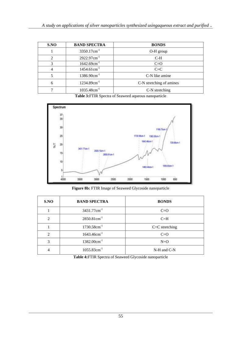
3.7.2 Fourier Transform Infrared Spectroscopy (FTIR):
Fourier transform infrared spectroscopy is a technique which is used to obtain an infrared spectrum of
absorption or emission of a solid, liquid, gas. The FTIR results are interpreted according to a paper [16]. The
FTIR spectrum for the silver nanoparticles synthesized using the aqueous extract was analysed(Fig 8a) and
absorption bands were observed at 3350.17cm-1
which corresponds to O-H group. The reduction of Ag+ to Ag
0
was due to C=O stretch. The peak at 1234.89 cm-1
,1386.90cm-1
corresponds with C-N stretching vibrations of
aromatic and aliphatic amines, peak at 2922.97 cm-1
, indicates presence of C-H stretching of aromatic
compounds and 1642.69cm-1
indicated presence of C=O group (Table 3).The FTIR spectrum for the silver
nanoparticles synthesized using the purified glycoside fraction was analysed(Fig 8b) and absorption bands were
observed at 3431.77cm-1
which corresponds to C=O group. The peak at 1055.83cm-1
corresponds with C-N
stretching vibrations of aromatic and aliphatic amines and the peak at 2850.81 cm-1
, indicates presence of C-H
stretching of aromatic compounds , peak at 1643.46cm-1
indicated presence of C=O group and peak at
1730.58cm-1
indicates presence of C=C stretching (Table 4). The FTIR spectrum for the silver nanoparticles
synthesized using the purified alkaloid fraction was analysed(Fig 8c) and absorption bands were observed at
3421.67cm-1
and 2919.56cm-1
which corresponds to O-H and C-H group. The peak at 1733.77 cm-1
corresponds
with C-C stretching, peak at 2850.54cm-1
, indicates presence of C-H stretching of aromatic compounds and
1634.00cm-1
indicated presence of C=O group (Table 5).
Figure 8a: FTIR Image of Seaweed aqueous nanoparticle
0.116
0.154
0.188
0.320.332 0.245
0.2170.164
0.1030.094
0.0430
0.1
0.2
0.3
0.4
AB
SOR
BA
NC
E (O
.D)
WAVELENGTH (nm)
SEAWEED ALKALOID NANOPARTICLE
SEAWEED ALKALOID NANOPARTICLE
A study on applications of silver nanoparticles synthesized usingaqueous extract and purified ..
55
S.NO BAND SPECTRA BONDS
1 3350.17cm-1
O-H group
2 2922.97cm-1
C-H
3 1642.69cm-1
C=O
4 1454.61cm-1
C=C
5 1386.90cm-1
C-N like amine
6 1234.89cm-1
C-N stretching of amines
7 1035.48cm-1
C-N stretching
Table 3:FTIR Spectra of Seaweed aqueous nanoparticle
Figure 8b: FTIR Image of Seaweed Glycoside nanoparticle
Table 4:FTIR Spectra of Seaweed Glycoside nanoparticle
S.NO BAND SPECTRA BONDS
1 3431.77cm-1
C=O
2 2850.81cm-1
C=H
1 1730.58cm-1
C=C stretching
2 1643.46cm-1
C=O
3 1382.00cm-1
N=O
4 1055.83cm-1
N-H and C-N
A study on applications of silver nanoparticles synthesized usingaqueous extract and purified ..
56
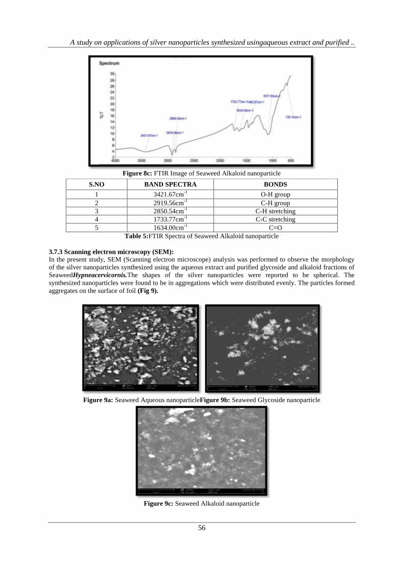
Figure 8c: FTIR Image of Seaweed Alkaloid nanoparticle
Table 5:FTIR Spectra of Seaweed Alkaloid nanoparticle
3.7.3 Scanning electron microscopy (SEM):
In the present study, SEM (Scanning electron microscope) analysis was performed to observe the morphology
of the silver nanoparticles synthesized using the aqueous extract and purified glycoside and alkaloid fractions of
SeaweedHypneacervicornis.The shapes of the silver nanoparticles were reported to be spherical. The
synthesized nanoparticles were found to be in aggregations which were distributed evenly. The particles formed
aggregates on the surface of foil (Fig 9).
Figure 9a: Seaweed Aqueous nanoparticleFigure 9b: Seaweed Glycoside nanoparticle
Figure 9c: Seaweed Alkaloid nanoparticle
S.NO BAND SPECTRA BONDS
1 3421.67cm-1
O-H group
2 2919.56cm-1
C-H group
3 2850.54cm-1
C-H stretching
4 1733.77cm-1
C-C stretching
5 1634.00cm-1
C=O
A study on applications of silver nanoparticles synthesized usingaqueous extract and purified ..
57
Figure 9: SEM images of Silver nanoparticle synthesized using aqueous extract and purified fractions of
Seaweed Hypneacervicornis
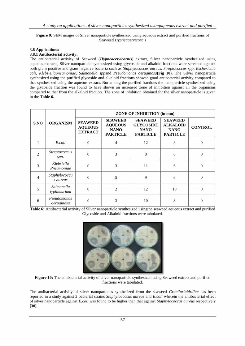
3.8 Applications:
3.8.1 Antibacterial activity:
The antibacterial activity of Seaweed (Hypneacervicornis) extract, Silver nanoparticle synthesized using
aqueous extracts, Silver nanoparticle synthesized using glycoside and alkaloid fractions were screened against
both gram positive and gram negative bacteria such as Staphylococcus aureus, Streptococcus spp, Escherichia
coli, Klebsiellapneumoniae, Salmonella sppand Pseudomonas aeruginosa(Fig 10). The Silver nanoparticle
synthesized using the purified glycoside and alkaloid fractions showed good antibacterial activity compared to
that synthesized using the aqueous extract. But among the purified fractions the nanoparticle synthesized using
the glycoside fraction was found to have shown an increased zone of inhibition against all the organisms
compared to that from the alkaloid fraction. The zone of inhibition obtained for the silver nanoparticle is given
in the Table 6.
Table 6: Antibacterial activity of Silver nanoparticle synthesized usingthe seaweed aqueous extract and purified
Glycoside and Alkaloid fractions were tabulated.
Figure 10: The antibacterial activity of silver nanoparticle synthesized using Seaweed extract and purified
fractions were tabulated.
The antibacterial activity of silver nanoparticles synthesized from the seaweed Gracilariabirdiae has been
reported in a study against 2 bacterial strains Staphylococcus aureus and E.coli wherein the antibacterial effect
of silver nanoparticle against E.coli was found to be higher than that against Staphylococcus aureus respectively
[30].
S.NO ORGANISM
ZONE OF INHIBITION (in mm)
SEAWEED
AQUEOUS
EXTRACT
SEAWEED
AQUEOUS
NANO
PARTICLE
SEAWEED
GLYCOSIDE
NANO
PARTICLE
SEAWEED
ALKALOID
NANO
PARTICLE
CONTROL
1 E.coli 0 4 12 8 0
2 Streptococcus
spp. 0 3 8 6 0
3 Klebsiella
Pneumoniae 0 3 11 6 0
4 Staphylococcu
s aureus 0 5 9 6 0
5 Salmonella
typhimurium 0 2 12 10 0
6 Pseudomonas
aeruginosa 0 3 10 8 0
A study on applications of silver nanoparticles synthesized usingaqueous extract and purified ..
58
3.8.2 Antifungal activity:
The antifungal activity of Seaweed (Hypneacervicornis) extract, Silver nanoparticle synthesized using aqueous
extracts, Silver nanoparticle synthesized using glycoside and alkaloid fractions were screened against two fungal
organisms namely Aspergillusniger and Candida albicans.In case of antifungal activity the nanoparticle
synthesized using the aqueous extract showed an increased zone of inhibition against both the fungal organisms
when compared to that of the purified fractions. The zone of inhibition obtained for the silver nanoparticle is
given in the Table 7.
Table 7: Antifungal activity ofSilver nanoparticle synthesized usingthe aqueous extract and purified Glycoside
and Alkaloid fractions of Seaweed were tabulated.
A study by has reported the antifungal activity of the silver nanoparticles synthesized using the brown seaweed
Padinaboergeseniiagainst 3 fungal pathogens namely Aspergillusfumigatus, Aspergillusnidulansand Candida
albicans respectively wherein the maximum inhibition was found against Aspergillusfumigatus (21.2mm)
followed by Aspergillusnidulans (17.3mm) and moderately against Candida albicans (17mm) [31].
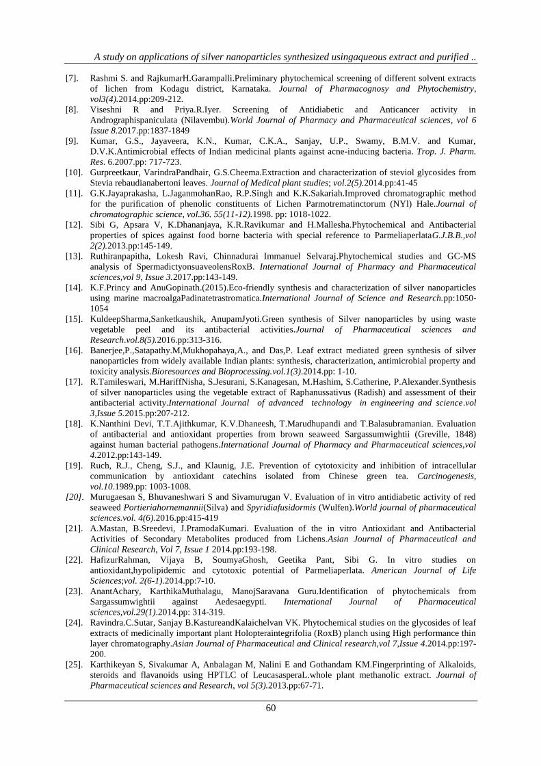
3.9 Antioxidant activity:
3.9.1 Hydrogen peroxide scavenging assay:
Hydrogen peroxide is a weak oxidizing agent. It is capable of penetrating within the cell and thereby reacts with
Fe2+
or Cu2+
ion and gets converted into hydroxyl radical that could cause harmful effects leading to cell
damage. So it is important to prevent the formation of hydroxyl radical by preventing the accumulation of
hydrogen peroxide within the cell. This assay is based on the principle of decrease in absorbance of hydrogen
peroxide upon oxidation of hydrogen peroxide. The ability of the extracts to scavenge the hydrogen peroxide is
determined [32]. The Hydrogen peroxide scavenging assay for the Silver nanoparticle synthesized using
aqueous extract and purified glycoside and alkaloid fractions of Seaweed Hypneacervicorniswere determined. In
this assay the nanoparticle synthesized using the purified fractions and the nanoparticle from aqueous extract
(19.95%) exhibited good scavenging percentage but among both the purified fractions the nanoparticle
synthesized using glycoside fraction (32.48%) was found to show better scavenging percentage compared to that
of the alkaloid fraction (18.36%) respectively (Fig 11).
Figure 11: Hydrogen peroxide scavenging assay for Silver nanoparticle synthesized using seaweed aqueous
extract and purified glycoside and alkaloid fractions.
The Hydrogen peroxide scavenging activity of the silver nanoparticles synthesized from the methanolic extract
of brown seaweed Himanthaliaelongata have been reported in a study wherein the nanoparticles were found to
have exhibited inhibition percentage of 39.1% to 77.4% at different concentrations from 200 to 800 ppm [33].
3.10 Antidiabetic activity: The antidiabetic activity of the silver nanoparticle synthesized using the aqueous
extract and the purified Glycoside as well as Alkaloid fractions of Seaweed Hypneacervicornis were determined
0
0.5
1
SEAWEED AQUEOUS
NANOPARTICLE
SEAWEED GLYCOSIDE
NANOPARTICLE
SEAWEED ALKALOID
NANOPARTICLE
0.942 0.942 0.9420.754
0.6360.769
19.95%32.48%
18.36%
% S
CA
VEN
GIN
G
SAMPLE
HYDROGEN PEROXIDE SCAVENGING ASSAY
CONTROL O.D
SAMPLE O.D
SCAVENGING %
S.NO ORGANISM
ZONE OF INHIBITION (in mm)
SEAWEED
AQUEOUS
NANOPARTICLE
SEAWEED
GLYCOSIDE
NANOPARTICLE
SEAWEED
ALKALOID
NANOPARTICLE
1 Aspergillusniger 13 6 2
2 Candida albicans 11 3 2

A study on applications of silver nanoparticles synthesized usingaqueous extract and purified ..
59
by means of alpha amylase inhibition assay. It is based on the principle of starch hydrolysis in the presence of α-
amylase enzyme. This process is quantified using iodine which reacts with starch giving blue colour. The
reduction in the intensity of blue colour indicates the breakdown of starch by the enzyme into monosaccharides.
If the extracts show increased activity then the intensity of blue colour will be more or higher thus the intensity
of blue colour developed is directly proportional to the alpha amylase inhibitory activity [34]. The Silver
nanoparticle synthesized using the aqueous extract (27.77%) as well as the purified fractions showed good
inhibition percentage but on a comparative basis the nanoparticle synthesized using the purified fractions
showed good inhibition percentage than that from the aqueous extract. Among the 2 purified fractions the
nanoparticle synthesized using the glycoside fraction (55.55%) showed an increased inhibition % than that of
the alkaloid fraction (31.48%) respectively (Fig 12).The antidiabetic activity of silver nanoparticles synthesized
using the seaweed Halymeniapophyroideshas been studied at different concentrations as 0.2, 0.4, 0.6, 0.8 and
1ml respectively wherein the inhibition percentage was found to have shown an increase in dose dependent
manner as 20% inhibition at a concentration of 0.2ml, 47% inhibition at 0.4ml, 61% inhibition at 0.6ml and 83%
inhibition at 0.8ml [35].
Figure 12: Alpha amylase inhibition assay for Silver nanoparticle synthesized using seaweed aqueous extract
and purified glycoside and alkaloid fractions.
IV. CONCLUSION The Current study was carried out in order to determine the therapeutic potential of Silver nanoparticles
synthesized using the aqueous extract and purified glycoside and alkaloid fractions of Seaweed
Hypneacervicornis.Nanotechnology remains as the major field of interest due to its application in various fields
and among the metal nanoparticles silver nanoparticle remains as the most promising source due to its wide
importance in various disciplines. Silver nanoparticles exhibit various medicinal as well as industrial
applications. Many reports have been proposed on the synthesis of silver nanoparticles using the aqueous extract
of natural sources such as plants, fungi, algae etc. Since the compounds are derived from natural sources they
have been found to possess numerous pharmacological properties thereby do not cause any health impacts or
don’t pose any threat to the environment. Not much work has been carried out on the nanoparticle synthesis
using a specific secondary compound so this study was an attempt to synthesize silver nanoparticles using the
purified secondary metabolites from SeaweedHypneacervicornisso that the combined efficacy of these
nanoparticles with the secondary compounds could prove to be beneficial to the mankind in different aspects.
REFERENCES
[1]. Johnson Marimuthu, PetchiammalEssakimuthu, JanakiramanNarayanan,BabuAnantham,Renisheya Joy
Jeba malar Tharmaraj,SivaramanArumugam. Phytochemical characterization of brown seaweed
Sargassumwightii.Asian Pacific Journal of Tropical diseases. 2012. pp: 109-113.
[2]. Kiriyama S, Okazaki Y, Yoshida A.Hypocholesterolemic effect of polysaccharides and polysaccharide
foodstuffs in cholesterol fed rats.J. Nutr.1968. pp:382–388.
[3]. Littler DS, Littler MM, Bucher KE, Norris JN.(Ed.3).Marine Plants of the Caribbean, a Field Guide from
Florida to Brazil. Washington, DC: Smithsonian Institution Press.1989.
[4]. Rai M., Yadav A. and Gade A. Biotech. Adv., vol.27.2009.pp: 76-83.
[5]. Ahamed M, Alsalhi.M. S, Siddiqui.M. K. J. Clinicachimicaacta. 2010. pp: 1841-1848.
[6]. Abbott.I.A.New species and notes on marine algae from Hawaii. Pacific science.vol.50.1996.pp: 142-
156.
0
0.2
0.4
0.6
SEAWEED AQUEOUS
NANOPARTICLE
SEAWEED GLYCOSIDE
NANOPARTICLE
SEAWEED ALKALOID
NANOPARTICLE
0.39
0.24
0.37
0.54 0.54 0.54
27.77%
55.55%
31.48%
% IN
HIB
ITIO
N
SAMPLES
ALPHA AMYLASE INHIBITION ASSAY
O.D AT 540nm
CONTROL (α-amylase)
% INHIBITION
A study on applications of silver nanoparticles synthesized usingaqueous extract and purified ..
60
[7]. Rashmi S. and RajkumarH.Garampalli.Preliminary phytochemical screening of different solvent extracts
of lichen from Kodagu district, Karnataka. Journal of Pharmacognosy and Phytochemistry,
vol3(4).2014.pp:209-212.
[8]. Viseshni R and Priya.R.Iyer. Screening of Antidiabetic and Anticancer activity in
Andrographispaniculata (Nilavembu).World Journal of Pharmacy and Pharmaceutical sciences, vol 6
Issue 8.2017.pp:1837-1849
[9]. Kumar, G.S., Jayaveera, K.N., Kumar, C.K.A., Sanjay, U.P., Swamy, B.M.V. and Kumar,
D.V.K.Antimicrobial effects of Indian medicinal plants against acne-inducing bacteria. Trop. J. Pharm.
Res. 6.2007.pp: 717-723.
[10]. Gurpreetkaur, VarindraPandhair, G.S.Cheema.Extraction and characterization of steviol glycosides from
Stevia rebaudianabertoni leaves. Journal of Medical plant studies; vol.2(5).2014.pp:41-45
[11]. G.K.Jayaprakasha, L.JaganmohanRao, R.P.Singh and K.K.Sakariah.Improved chromatographic method
for the purification of phenolic constituents of Lichen Parmotrematinctorum (NYl) Hale.Journal of
chromatographic science, vol.36. 55(11-12).1998. pp: 1018-1022.
[12]. Sibi G, Apsara V, K.Dhananjaya, K.R.Ravikumar and H.Mallesha.Phytochemical and Antibacterial
properties of spices against food borne bacteria with special reference to ParmeliaperlataG.J.B.B.,vol
2(2).2013.pp:145-149.
[13]. Ruthiranpapitha, Lokesh Ravi, Chinnadurai Immanuel Selvaraj.Phytochemical studies and GC-MS
analysis of SpermadictyonsuaveolensRoxB. International Journal of Pharmacy and Pharmaceutical
sciences,vol 9, Issue 3.2017.pp:143-149.
[14]. K.F.Princy and AnuGopinath.(2015).Eco-friendly synthesis and characterization of silver nanoparticles
using marine macroalgaPadinatetrastromatica.International Journal of Science and Research.pp:1050-
1054
[15]. KuldeepSharma,Sanketkaushik, AnupamJyoti.Green synthesis of Silver nanoparticles by using waste
vegetable peel and its antibacterial activities.Journal of Pharmaceutical sciences and
Research.vol.8(5).2016.pp:313-316.
[16]. Banerjee,P.,Satapathy.M,Mukhopahaya,A., and Das,P. Leaf extract mediated green synthesis of silver
nanoparticles from widely available Indian plants: synthesis, characterization, antimicrobial property and
toxicity analysis.Bioresources and Bioprocessing.vol.1(3).2014.pp: 1-10.
[17]. R.Tamileswari, M.HariffNisha, S.Jesurani, S.Kanagesan, M.Hashim, S.Catherine, P.Alexander.Synthesis
of silver nanoparticles using the vegetable extract of Raphanussativus (Radish) and assessment of their
antibacterial activity.International Journal of advanced technology in engineering and science.vol
3,Issue 5.2015.pp:207-212.
[18]. K.Nanthini Devi, T.T.Ajithkumar, K.V.Dhaneesh, T.Marudhupandi and T.Balasubramanian. Evaluation
of antibacterial and antioxidant properties from brown seaweed Sargassumwightii (Greville, 1848)
against human bacterial pathogens.International Journal of Pharmacy and Pharmaceutical sciences,vol
4.2012.pp:143-149.
[19]. Ruch, R.J., Cheng, S.J., and Klaunig, J.E. Prevention of cytotoxicity and inhibition of intracellular
communication by antioxidant catechins isolated from Chinese green tea. Carcinogenesis,
vol.10.1989.pp: 1003-1008.
[20]. Murugaesan S, Bhuvaneshwari S and Sivamurugan V. Evaluation of in vitro antidiabetic activity of red
seaweed Portieriahornemannii(Silva) and Spyridiafusidormis (Wulfen).World journal of pharmaceutical
sciences.vol. 4(6).2016.pp:415-419
[21]. A.Mastan, B.Sreedevi, J.PramodaKumari. Evaluation of the in vitro Antioxidant and Antibacterial
Activities of Secondary Metabolites produced from Lichens.Asian Journal of Pharmaceutical and
Clinical Research, Vol 7, Issue 1 2014.pp:193-198.
[22]. HafizurRahman, Vijaya B, SoumyaGhosh, Geetika Pant, Sibi G. In vitro studies on
antioxidant,hypolipidemic and cytotoxic potential of Parmeliaperlata. American Journal of Life
Sciences;vol. 2(6-1).2014.pp:7-10.
[23]. AnantAchary, KarthikaMuthalagu, ManojSaravana Guru.Identification of phytochemicals from
Sargassumwightii against Aedesaegypti. International Journal of Pharmaceutical
sciences,vol.29(1).2014.pp: 314-319.
[24]. Ravindra.C.Sutar, Sanjay B.KastureandKalaichelvan VK. Phytochemical studies on the glycosides of leaf
extracts of medicinally important plant Holopteraintegrifolia (RoxB) planch using High performance thin
layer chromatography.Asian Journal of Pharmaceutical and Clinical research,vol 7,Issue 4.2014.pp:197-
200.
[25]. Karthikeyan S, Sivakumar A, Anbalagan M, Nalini E and Gothandam KM.Fingerprinting of Alkaloids,
steroids and flavanoids using HPTLC of LeucasasperaL.whole plant methanolic extract. Journal of
Pharmaceutical sciences and Research, vol 5(3).2013.pp:67-71.
A study on applications of silver nanoparticles synthesized usingaqueous extract and purified ..
61
[26]. GarciaV.P, Bermeio J, Rubio S, Quintana J, Estevez F. Pregnane Steroidal glycosides and their cytostatic
activites. Glycobiology, vol. 21(5).2011.pp:619-624.
[27]. ChinmayBhat.Synthetic studies of alkaloids containing Pyrrolidine and Piperidine structural motifs,
Chemistry open, 4.2015. pp: 192-196.
[28]. C. Mohandass, A.S. Vijayaraj, R. Rajasabapathy, S. SatheeshBabu, S.V. Rao, C. Shiva and L. De Mello
.Biosynthesis of silver nanoparticles from marine seaweed Sargassumcinereum and their antibacterial
activity. Indian Journal of Pharmaceutical sciences.2013.pp:606-610.
[29]. Sushmita Deb. Synthesis of silver nanoparticles using Murrayakoenigii (Green curry leaves) and Zea
mays (Baby corn) and its antimicrobial activity against pathogens. International Journal of
Pharmaceutical Technology and Research.2014.pp:91-96.
[30]. Anderson Passos de Aragao, Taiane Maria de Oliveria, Patrick VerasQuelemes, Marcia Lerana Gomes
Perfeito, Maria CarvalhoAraujo, Janaina de Araujo, Sousa Santiago, ViniciusS.Cardoso, Pedro
Quaresma, Jose Roberto de Souza de Almeida Leite, DurcileneAlves da Silva. Green synthesis of silver
nanoparticles using the seaweed Gracilariabirdiae and their antibacterial activity.Arabian Journal of
Chemistry.2016.pp:1-7.
[31]. Dr.P.Senthilkumar, Bhuvaneshwari, Janani, Prakash, Lakshmi Priya and RanjithSanthosh Kumar. Green
synthesis and characterization of silver nanoparticles from aqueous extract of brown seaweed
Padinaboergesenii and its antifungal activity. World Journal of Pharmacy and Pharmaceutical
sciences.vol 4, Issue 10.2015.pp:1858-1870.
[32]. Priyanka B, Anitha K, Shirisha K, Janipasha SK, Dipankar B, Rajesh K..Evaluation of antioxidant
activity of ethanolic root extract of AlbiziaLebbeck (L.) Benth .International Journal of Pharmaceutical
and Applied sciences. Vol. 3(2).2013.pp: 93-101.
[33]. GauravRajauria, AmitJaiswal, NissreenAbu-Ghannam, Shilpi Gupta.Antimicrobial, antioxidant and free
radical scavenging capacity of brown seaweed Himanthaliaelongata from Western coast of
Ireland.Journal of Food Biochemistry.2012.pp:1-14.
[34]. Sheikh, J.H., Iyo, Tsujiyama, M.T., Md.Ashabul I., Rajat, S.B., Hitoshi,A. Total phenolic
content,antioxidative,anti-amylase,antiglucosidase and antihistamine release activities of Bangladeshi
fruits.Food Science and Technology Research.,14.2008.pp:261-268.
[35]. Vishnu Kiran M and Murugesan S.Biogenic silver nanoparticles by Halymeniaporphyroides and its
Invitroantidiabeticefficacy.Journal of Chemical and Pharmaceutical Research.Vol.5(12).2013.pp:1001-
1008.