Embed Size (px)
Citation preview
Muthukumaravel, K. and Rajaraman, P. J. Pure Appl. Zool., 1(2): 122-126, 2013
122
ISSN Print/Online: 2320-9577/2320-9585
INTERNATIONAL JOURNAL OF PURE AND APPLIED ZOOLOGY
Volume 1, Issue 2, June 2013
Available online at: http://www.ijpaz.com
RISHAN PUBLICATIONS
RESEARCH ARTICLE OPEN ACCESS
A STUDY ON THE TOXICITY OF CHROMIUM ON THE HISTOLOGY
OF GILL AND LIVER OF FRESHWATER FISH LABEO ROHITA
1K. MUTHUKUMARAVEL* AND
2P. RAJARAMAN
1PG And Research Department of Zoology, Khadir Mohideen College,
Adirampattinam – 614 701. Tamilnadu, India
2Thiru Vi Ka Govt. Arts College, Tiruvarur-610 003, Tamilnadu, India
*Corresponding Author Email: [email protected], Tel: +91 9791387363
Article History: Received: 06.06.2013, Accepted: 19.06.2013, Published: 26.06.2013
ABSTRACT
The toxic effect of chromium on the histology of gill and liver of Labeo rohita fingerlings was studied. The
fingerlings were exposed for 10, 20 and 30 days in 10% sublethal concentration of 96 h LC50 of chromium (3.5
ppm). The gills exposed to sublethal concentration of chromium showed mild histological alterations during 10
days of exposure. However after 30 days, fusion of gill lamellae, hypertrophy and degeneration of epithelium
were prominent. Liver lesions consisted of vacuolation, degeneration of hepatocytes and disintegration of cell
boundaries of hepatocytes. These changes occurred predominantly in the 30 days exposure.
Keywords: Heavy metal, chromium, fish, histology, gills and liver.
INTRODUCTION
Fish is an excellent source of protein in
human diet. The unique feature which
differentiates fish food other animal protein
sources is the presence of omega-3 fatty acids
such as Linolenic acid, Decosa hexaenoic acid
(DHA) and Eicosa pentaenoic acid (EPA). DHA
promotes learning ability in children and
improved memory in adults. DHA is also
essential for the foetal growth and development.
Omega fatty acid is also good for heart and helps
to control diabetics by improving insulin action
(Lee and Reasner, 2000 and Lemos et al., 2005).
That’s why it must be included in human diet at
least 1.3 kg per week (FAO, 1989). However, the
fish habitates are being contaminated alarmingly
through a number of aquatic pollutants. Among
these pollutants heavy metals are most injurious
to fish. These pollutants have not only depleted
the fish stock but also have threatened the human
health by incorporating into food chain (Pip,
1995). Heavy metal chromium is widely used in
metallurgic, refractory, chemical and tannery
industries. Tanneries are the major industries that
use chromium for the treatment of leather and
nearly 40% of the chromium used is released into
the environment. In the present work, an attempt
was made to evaluate the long term exposure
effect of chromium on the histology of gill and
liver of the freshwater fish Labeo rohita.
MATERIALS AND METHOD
The fish, L. rohita fingerlings (Wt:8 0.5g;
Length 7.1 cm) were collected from the
Katherasan Aqua Farm near Thanjavur, Tamil
Nadu. They were acclimatized for 15 days in
large cement tanks (Temperature 24.2°C, pH 7.2
±0.3 and Dissolved Oxygen 7.9±0.7 mg/l)

Muthukumaravel, K. and Rajaraman, P. J. Pure Appl. Zool., 1(2): 122-126, 2013
123
previously washed with 1% potassium
permanganate. The water as renewed every 24 h.
The LC50 of potassium dichromate for 96h was
found out by using Probit method (Finney,
1971). Stock solution of chromium was prepared
and the toxicity tests were conducted to be 3.5
ppm. For histological studies L. rohita were
reared in sublethal concentration (10% of 96
hours LC50) for a period of 10, 20 and 30 days.
The gill arches and liver were dissected out and
fixed in Bouin’s fluid. The tissues were
embedded in paraffin (58°C) and sectioned at 6µ
thickness. The sections were stained with
haemotoxylin and eosin (Gurr, 1959). The
stained slides were examined for
histopathological changes and were
photomicrographed.
RESULTS AND DISCUSSION
In control fish, the secondary gill lamellae
(SGL) appeared as finger-like structures. The
SGL was thin, slender and attached on either side
of the primary gill lamellae (PGL). The
secondary gill lamellae are highly vascularised
and surrounded by a thin layer of epithelial cells
(Figure 1).
The overall observed results in the present
investigation indicates that marked
histopathological changes have been found in the
gill and liver of fish L. rohita under sublethal
concentrations of chromium in chronic exposure.
Fusion and shortening lamellae, hypertrophy,
degeneration of epithelium and necrosis were
found in the gills of chromium treated L. rohita
(Figures 2 & 3). Higher degree of hypertrophy
and fusion of gill lamellae were prominent in the
gills of fish exposed to 30 days. Hemalatha and
Banerjee (1977) and Gupta and Kumar (2006)
noted similar types of gill lesions in zinc treated
Heteropneustes fossilis and mercury treated
Cirrhinus mrigala respectively. Kaoud and
El-Dahshan (2010) observed severe hyperplasia
in secondary gill lamellae which lead to complete
embedding in adjacent lamellae in copper,
cadmium, lead and mercury treated Oreochromis
niloticus. In the present study, hypertropy and
degeneration of secondary lamellae were
apparent in L. rohita exposed to chromium
(Figure 4). These observations are quite
comparable to pathological lesions induced in
gills by mercuric chloride in Acipenser persicus
fry (Khoshnood et al., 2011 ), by lead and
cadmium treatment in Cyprinus carpio (Patnaik
et al., 2011), Lates calcarifer (Thophon et al.,
2003), Brachydanio rerio and Salmo gairdneri
(Karlson-Norgren et al., 1985). Patel and
Bahadur (2010) also noted severe gill lesions in
copper treated Catla catla. In the present
investigation the gill epithelium of chromium
treated fish was completely desquamated, fuion
and shapeless secondary lamellae and were
broken at several places (Figure 4). Daoust et al.
(1984) also observed similar pathological lesions
in the gill of copper treated rainbow trout, Salmo
gairdneri. Further, Hemalatha and Banerjee
(1997) and Al-Attar (2007) also observed such
gill damages in zinc chloride and nickel treated
Heteropneustes fossilis and Oreochromis
niloticus.
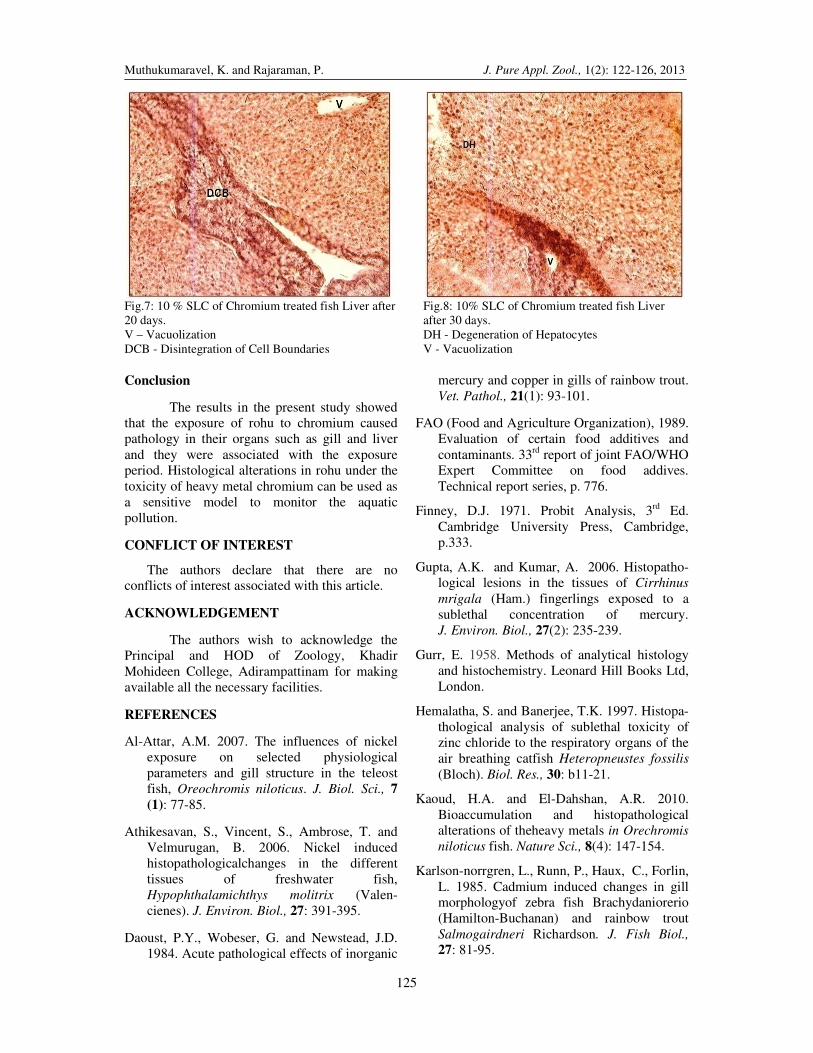
Liver of fish is responsible for digestion,
filtration and storage of glycogen. The liver also
produces many enzymes that stored in the gall
bladder. The liver functions to store food energy.
The normal liver is made up of continuous mass
of hepatocytes with large number of blood
sinusoids (Figure 5). The fish exposed to the
sublethal concentrations of chromium showed
the vacuolation, loose arrangement of hepatic
cells, histolysis and disintegration of cell
boundries (Figures 6-8).The damage as more
severe and progressive after 30 days exposure.
Histological changes in the liver of fishes have
been extensively reported. Athikesavan et al.
(2006) reported the histological lesions in the
liver of Hypophthalamichthys molitrix exposed
to nickel. They observed marked changes like
degeneration of blood vessels hypertrophy,
vacuolisation necrosis and pyknotic nuclei of the
exposed fish. The histological lesions due to
cadmium and zinc poisoning were reported by
Van Dyk et al. (2007) in Oreochromis
mossambicus. Loganathan et al. (2006) and
Radhakrishnan and Hemalatha (2010) have also
observed the histological changes in the liver of
zinc treated L. rohita and cadmium chloride
treated Channa striatus. They observed that the
cytoplasmic vacuolization of hepatocytes,
congestion of blood vessel, leucocytic infiltration
and necrosis. The results of the present
observations in L. rohita exposed to chromium
were in agreement with those of the earlier
workers especially in the vacuolization and
necrosis in hepatic tissue. Intracellular
vacuolation, necrosis and shrinkage of nuclei
were also apparent in the present study in
chromium treated L. rohita. Similar changes are
observed by Loganathan et al. (2006) in zinc
treated L. rohita. The development of necrosis,

Muthukumaravel, K. and Rajaraman, P. J. Pure Appl. Zool., 1(2): 122-126, 2013
124
congestion of hepatic blood vessels and
vacuolization in chromium treated L. rohita were
mainly due to large scale accumulation of these
metals in liver. Liver is the vital organ for
detoxificationof unwanted and toxic substances.
Accumulation and elimination processes of
metals ions in the liver may lead to hepatic
lesions.
Figure 1. Control fish gill.
Figure 2. 10 % SLC of Chromium treated fish gill
10 days.
PGL - Primary Gill Lamellae
SGL - Secondary Gill Lamellae
EGL - Erosion of Secondary Lamellae
FSL - Fusion of Secondary Lamellae
Figure 3. 10 % SLC of Chromium treated fish gill
after 20 days.
Figure 4. 10 % SLC of Chromium treated fish gill
after 30 days.
DE - Degeneration of Epithelium, H – Hypertrophy
FSL - Fusion of Secondary Lamellae
DE - Degeneration of Epithelium, HT-
Hypertrophy,
FSL - Fusion of Secondary Lamellae
Figure 5. Control fish liver.
Figure 6. 10 % SLC of Chromium treated fish
Liver after 10 days.
NH - Normal hepatocytes,
BS - Blood Sinus.
DH - Degeneration of hepatocytes,
V – Vacuolization.
Muthukumaravel, K. and Rajaraman, P. J. Pure Appl. Zool., 1(2): 122-126, 2013
125
Fig.7: 10 % SLC of Chromium treated fish Liver after
20 days.
Fig.8: 10% SLC of Chromium treated fish Liver
after 30 days.
V – Vacuolization
DCB - Disintegration of Cell Boundaries
DH - Degeneration of Hepatocytes
V - Vacuolization
Conclusion
The results in the present study showed
that the exposure of rohu to chromium caused
pathology in their organs such as gill and liver
and they were associated with the exposure
period. Histological alterations in rohu under the
toxicity of heavy metal chromium can be used as
a sensitive model to monitor the aquatic
pollution.
CONFLICT OF INTEREST
The authors declare that there are no
conflicts of interest associated with this article.
ACKNOWLEDGEMENT
The authors wish to acknowledge the
Principal and HOD of Zoology, Khadir
Mohideen College, Adirampattinam for making
available all the necessary facilities.
REFERENCES
Al-Attar, A.M. 2007. The influences of nickel
exposure on selected physiological
parameters and gill structure in the teleost
fish, Oreochromis niloticus. J. Biol. Sci., 7
(1): 77-85.
Athikesavan, S., Vincent, S., Ambrose, T. and
Velmurugan, B. 2006. Nickel induced
histopathologicalchanges in the different
tissues of freshwater fish,
Hypophthalamichthys molitrix (Valen-
cienes). J. Environ. Biol., 27: 391-395.
Daoust, P.Y., Wobeser, G. and Newstead, J.D.
1984. Acute pathological effects of inorganic
mercury and copper in gills of rainbow trout.
Vet. Pathol., 21(1): 93-101.
FAO (Food and Agriculture Organization), 1989.
Evaluation of certain food additives and
contaminants. 33rd
report of joint FAO/WHO
Expert Committee on food addives.
Technical report series, p. 776.
Finney, D.J. 1971. Probit Analysis, 3rd Ed.
Cambridge University Press, Cambridge,
p.333.
Gupta, A.K. and Kumar, A. 2006. Histopatho-
logical lesions in the tissues of Cirrhinus
mrigala (Ham.) fingerlings exposed to a
sublethal concentration of mercury.
J. Environ. Biol., 27(2): 235-239.
Gurr, E. 1958. Methods of analytical histology
and histochemistry. Leonard Hill Books Ltd, London.
Hemalatha, S. and Banerjee, T.K. 1997. Histopa-
thological analysis of sublethal toxicity of
zinc chloride to the respiratory organs of the
air breathing catfish Heteropneustes fossilis
(Bloch). Biol. Res., 30: b11-21.
Kaoud, H.A. and El-Dahshan, A.R. 2010.
Bioaccumulation and histopathological
alterations of theheavy metals in Orechromis
niloticus fish. Nature Sci., 8(4): 147-154.
Karlson-norrgren, L., Runn, P., Haux, C., Forlin,
L. 1985. Cadmium induced changes in gill
morphologyof zebra fish Brachydaniorerio
(Hamilton-Buchanan) and rainbow trout
Salmogairdneri Richardson. J. Fish Biol.,
27: 81-95.

Muthukumaravel, K. and Rajaraman, P. J. Pure Appl. Zool., 1(2): 122-126, 2013
126
Khoshnood Z., Khodabandeh S., Shahryari
Moghaddam M., Mosafer Khorjestan S.
2011. Histopathological and pathomor-
phological effects of mercuric chloride on
the gills of Persian sturgen, Acipenser
persicus, fry. Int. J. Nat. Resour. Mar. Sci.,
1(1): 23-32.
Lee, N A and Reasner C A. 2000. Beneficial
effect of chromium supplementation on
serum triglycerides levels in Cyprinus
carpio. Diab. Care., 17: 1449-1452.
Lemos, N G, Dias A L, Silva S T and Mantovani
M S. 2005. Evaluation of environmental
water using the comet assay in Tilapia
rendalli. Environ. Toxicol. Pharmacol., 9:
197-201.
Loganathan K, Velmurugan B, Howrelia J H,
Selvanayagam M and Patnaik B B. 2006.
Zinc induced histological changes in brain
and liver of Labeo rohita (Ham). J Environ
Biol., 27(1): 107-110.
Patel, J.M. and Bahadur, A. 2010. Histopatho-
logical alterations in Catla catla induced by
chronic exposure of copper ions. J. Cell.
Tissue Res., 10(3): 2365-2370.
Patnaik, B.B., Howrela, H.J., Mathews, T. and
Selvanayagam, M. 2011. Histopathology of
gill, liver, muscle and brain of Cyprinus
carpio communis L exposed to sublethal
concentration of lead and cadmium. African.
J. Biotech., 10(57): 12218-12223.
Pip, E. 1995. Cadmium, lead and copper in
freshwater mussel from Assiniboine river,
Manitoba, Canada. J. Moll. Stud., 61: 295-
302.
Radhakrishnan, M.V and Hemalatha S. 2010.
Sublethal toxicity effects of cadmium
chloride to liverof freshwater fish Channa
striatus (Bloch) Am-Emuras. J. Toxicol. Sci.,
2(1): 54-56.
Thophon S, Kruatrache M, Upatham E.S,
Pokethitiyook P, Sahaphong S. and
Jaritkhuan S. 2003. Histopathological
alterations of white seabass Lates calcarifer
in acute and sub chronic cadmium exposure.
Environ. Poll., 121: 307-320.
Van Dyk. J., Pieterse, G. and Van Vuren,
J. 2007. Histological changes in the liver of
Oreochromis mossambicus (Cichlidae) after
exposure to cadmium and zinc. Ecotoxicol.
Environ. Saf., 66: 432-440.
Cite this article as:
Muthukumaravel, K. and Rajaraman, P. 2013. A study on the toxicity of chromium on the histology of gill and
liver of freshwater fish Labeo rohita. J. Pure Appl. Zool., 1(2): 122-126.