Embed Size (px)
Citation preview
i
A SURVEY OF FACTORS ASSOCIATED WITH
IDIOPATHIC CLUBFOOT RELAPSE AFTER PONSETI
TREATMENT
DR GRACE MUTHONI KINYANJUI,
MBCh.B. (UoN)
H58/68802/2011
Department of Orthopedics
School of Medicine, University of Nairobi
A Thesis submitted in partial fulfillment of the requirements for the Award of
the Degree of Master of Medicine in Orthopedic Surgery of the University of
Nairobi
2017
ii
DECLARATION
STUDENT’S DECLARATION
I hereby declare that this thesis is my original work and has not been presented for a degree at
any other university.
Dr. Grace Muthoni Kinyanjui– Principal Investigator.
Registration Number: H58/68802/2011
MMed. Student, Department Of Orthopedic Surgery
MBCh.B. (UoN).
Sign ...................................................Date....................................................
iii
SUPERVISORS’ DECLARATION
This thesis has been submitted with our approval as university supervisors.
Dr. Edward Gakuya
MBCh.B (NRB), Mmed (NRB)
Consultant Orthopedic Surgeon
Lecturer, Department of Orthopedic Surgery
University of Nairobi
Signed..............................................................Date.......................................
Dr. Vincent Mutiso
MBCh.B (NRB), Mmed (NRB)
Consultant Orthopedic Surgeon
Senior Lecturer, Department of Orthopedic Surgery
University of Nairobi
Signed .................................................................Date.............................................
iv
CERTIFICATE OF AUTHENTICITY
This thesis has been submitted for examination with the approval of the Chairman and the
Orthopedic Department of University of Nairobi.
Prof. John E.O. Atinga
Professor of Orthopedic Surgery,
Chairman,
Department of Orthopedic Surgery,
University Of Nairobi.
Signed:......................................................................Date:................................................................
v
DEDICATION
I dedicate this study to my loving parents Stephen Kinyanjui and Olive Kinyanjui for their
continued support throughout my education to my beautiful triplets Keira, Kqurtney and
Kylie.
vi
ACKNOWLEDGEMENT
I would like to acknowledge the following people:
- My supervisors Dr. Gakuya and Dr. Mutiso for their input throughout the study
- Dr. Mangoli and Kijabe even staff for assisting me in my study logistics.
vii
TABLE OF CONTENTS
STUDENT’S DECLARATION ..................................................................................................... ii
SUPERVISORS’ DECLARATION .............................................................................................. iii
CERTIFICATE OF AUTHENTICITY ......................................................................................... iv
DEDICATION ................................................................................................................................ v
ACKNOWLEDGEMENT ............................................................................................................. vi
TABLE OF FIGURES .................................................................................................................... x
LIST OF ABBREVIATIONS ........................................................................................................ xi
DEFINITIONS .............................................................................................................................. xii
1.2 INTRODUCTION ............................................................................................................... 1
2.1 LITERATURE REVIEW .................................................................................................... 4
2.1.1 INFLUENCE OF BRACING IN RELAPSE RATES .................................................. 4
2.1.2 AGE AT PRESENTATION ......................................................................................... 5
2.1.3 INITIAL SEVERITY OF DEFORMITY ..................................................................... 5
2.1.4 RELATIONSHIP OF GENDER AND RELAPSE ...................................................... 6
2.1.5 ATYPICAL DEFORMITY .......................................................................................... 6
2.1.6 RACE/ETHINICITY .................................................................................................... 7
2.1.7 PARENTAL FACTORS .............................................................................................. 7
2.1.8 PREVIOUS TREATMENT ......................................................................................... 7
2.1.9 NUMBER OF CASTS.................................................................................................. 8
2.2 CONCLUSION .................................................................................................................... 8
3.1 STUDY QUESTION ........................................................................................................... 9
3.2 STUDY JUSTIFICATION .................................................................................................. 9
3.3 STUDY OBJECTIVES ........................................................................................................ 9
viii
3.3.1 MAIN OBJECTIVE ..................................................................................................... 9
3.3.2 SPECIFIC OBJECTIVES............................................................................................. 9
3.4 METHODOLOGY ............................................................................................................ 10
3.4.1 STUDY DESIGN ....................................................................................................... 10
3.4.2 STUDY SETTING ..................................................................................................... 10
3.4.3 STUDY POPULATION ............................................................................................. 10
3.4.4 SAMPLE SIZE ........................................................................................................... 10
3.4.5 INCLUSION CRITERIA ........................................................................................... 12
3.4.6 EXCLUSION CRITERIA .......................................................................................... 12
3.4.7 OUTCOME MONITORING ...................................................................................... 12
3.4.8 DATA COLLECTION, MANAGEMENT AND ANALYSIS .................................. 12
3.5 STUDY LIMITATIONS ................................................................................................... 13
3.6 DISSEMINATION OF FINDINGS .................................................................................. 13
3.7 ETHICAL CONSIDERATIONS ....................................................................................... 13
4.1 RESULTS .......................................................................................................................... 15
4.1.1 DISTRIBUTION BY GENDER ................................................................................ 15
4.1.2 AGE AT INTERVIEW BY STUDY GROUP ........................................................... 16
4.1.3 PARENTAL EDUCATION ....................................................................................... 17
4.1.4 NUMBER OF PARENTS .......................................................................................... 18
4.1.5 AFFECTED LIMB ..................................................................................................... 19
4.1.6 INITIAL PIRANI SCORE- (RIGHT SIDE) .............................................................. 20
4.1.7 INITIAL PIRANI SCORE-(LEFT) ............................................................................ 21
4.1.8 AGE AT FIRST CAST............................................................................................... 22
4.1.9 NUMBER OF CAST CHANGES .............................................................................. 23
ix
4.1.10 PIRANI SCORE AT INTERVIEW-(RIGHT) ........................................................... 24
4.1.11 PIRANI SCORE AT INTERVIEW-(LEFT) .............................................................. 25
5.1 DISCUSSION .................................................................................................................... 26
5.2 LIMITATIONS .................................................................................................................. 28
5.3 CONCLUSIONS................................................................................................................ 28
5.4 RECOMMENDATIONS ................................................................................................... 29
7.1 DATA COLLECTION SHEET ......................................................................................... 34
7.2 CONSENT INFORMATION ............................................................................................ 37
7.2.1 ENGLISH VERSION ................................................................................................. 37
7.2.2 KISWAHILI VERSION ............................................................................................. 40
x
TABLE OF FIGURES
Figure 1.2-1: Talipes Equinovarus .................................................................................................. 1
Figure 1.2-2: Casting in the Ponseti Method .................................................................................. 2
Figure 1.2-3: Dennis Brown Brace ................................................................................................. 2
Graph 4.1-1: Distribution by Gender within the Study Groups .................................................... 15
Table 4.1-1: Distribution by Age within the Study Groups at time of Interview ......................... 16
Graph 4.1-2: Parental Education Level within the Study Groups ................................................ 17
Graph 4.1-3: Number of Parents by Study Group ........................................................................ 18
Graph 4.1-4: Distribution of Affected Limb by Study Group ...................................................... 19
Table 4.1-2: Initial Pirani score for the Right Limb ..................................................................... 20
Table 4.1-3: Initial Pirani score for the Left Limb ........................................................................ 21
Graph 4.1-5: Age at First Cast ...................................................................................................... 22
Table 4.1-4: Number of Cast Changes .......................................................................................... 23
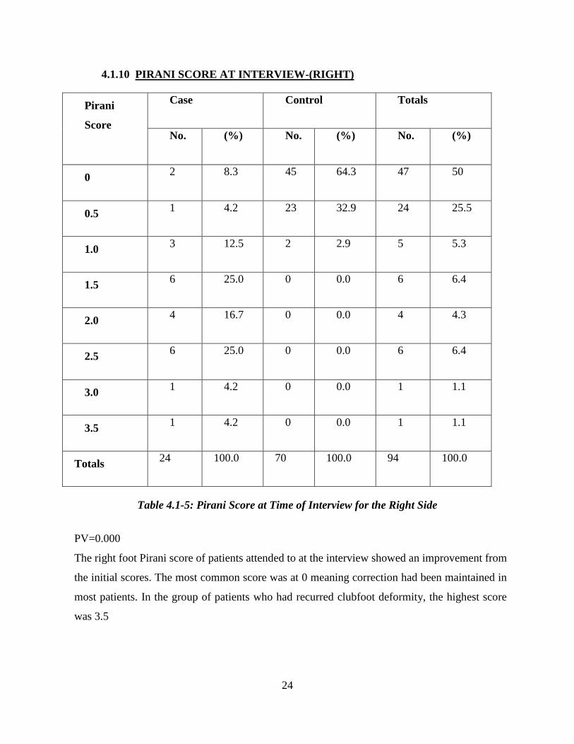
Table 4.1-5: Pirani Score at Time of Interview for the Right Side ............................................... 24
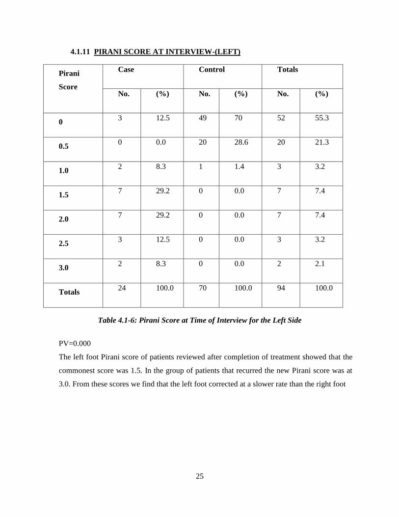
Table 4.1-6: Pirani Score at Time of Interview for the Left Side ................................................. 25
xi
LIST OF ABBREVIATIONS
KNH Kenyatta National Hospital
UON University of Nairobi
FAB Foot abduction brace
POP Plaster of Paris
CTEV Congenital talipes equinovarus
AIC African Inland Church
FHL Flexor Hallucis Longus
FDL Flexor Digitorium Longus
AFO Ankle Foot Orthosis
xii
DEFINITIONS
ADHERENCE: Brace worn 23hours/day in the 1st 3months followed by 12hours night
time wear for 3-4years
NON-ADHERENCE: Failure of following bracing protocol
CORRECTION: Pirani score 0
RELAPSE: Pirani score 1 or more
xiii
ABSTRACT
Worldwide, Congenital Talipes Equinovarus (CTEV) is a common foot deformity encountered in
the pediatric population with an incidence of 1 in every 1000 births. This problem is more
common in low-middle income countries. In Africa, the prevalence of CTEV is 2/1000live births
(Uganda).At the Kenyatta National Hospital (KNH) in Kenya, an average of 260 children are
diagnosed with CTEV annually.
While a lot of effort has been made to treat CTEV, success rates are not always 100% and about
25% of operated clubfeet will develop recurrence or show a marked residual deformity. Between
3% - 5% rates of recurrence of clubfoot after Ponseti treatment have been reported across the
world. Studies have attributed CTEV relapse after Ponseti manipulation to poor adherence to
treatment regime and improper use of foot braces.
At KNH Treatment for CTEV is both operative and non-operative. The gold standard for non-
operative treatment is Ponseti manipulation.
There is need to study relapse after Ponseti manipulation to determine risk factors and identify
corrective measures especially in low resource settings like Kenya.
Objective: To determine the factors associated with clubfoot recurrence after Ponseti treatment.
Design: Case-Control study.
Setting: Foot clinic at KNH and the outpatient clinic at Kijabe AIC Cure Hospital.
Patient and methods: Patients diagnosed with idiopathic CTEV and had used the Foot
Abduction Brace (FAB) for at least one year were recruited. Sample size was 24 cases and 70
controls. Data on socio-demographic characteristics, duration of treatment, compliance in use of
brace, presence of CTEV relapse, type of CTEV relapse and mitigating efforts employed by care
providers to contain the relapse. The following parameters were used to determine the presence
of CTEV relapse; Pirani score, foot bisector, thigh foot angle and foot progression angle.
xiv
Absence of relapse was defined as having a Pirani score of 0 and foot bisector passing through
second toe.
The frequency of flexibility/stiffness of ankle joint, presence of callosities, gait characteristics
(toe walking, side stepping), and parental/guardian satisfaction, was tabulated.
The study was carried out over a six week period through the months of December 2016 and
April 2017.
Relapse factors were compared and analyzed in terms of socio-demographic characteristics,
history of treatment for clubfoot, duration of treatment for clubfoot and the outcome measures.
Data collection through structured questionnaires was analyzed using IBM statistics (SPSS)
version 21. Results are presented using tables, textual write up, charts and graphs.
1
1. CHAPTER ONE
1.2 INTRODUCTION
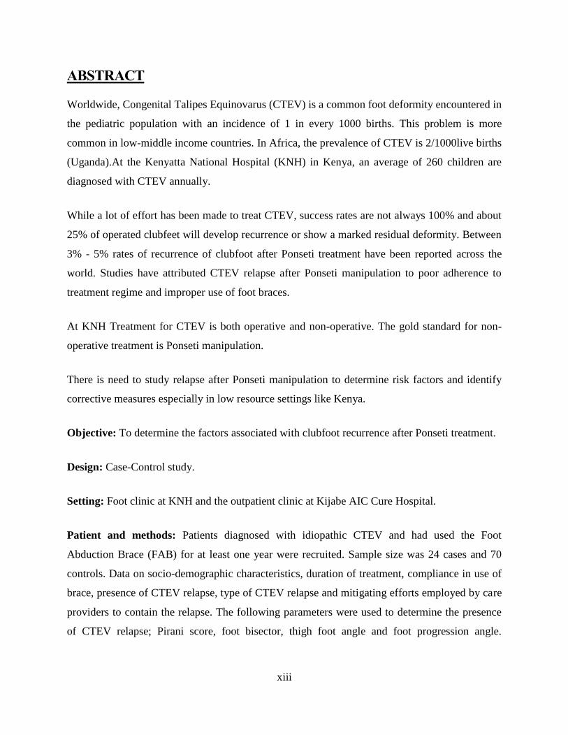
Clubfoot, also referred to as talipes equinovarus, is a complex foot and ankle deformity
involving forefoot adduction and supination, midfoot cavus, hindfoot varus and equinus,
inversion at the subtalar joint, adduction at midtarsal joint and internal tibial torsion (1)
Forefoot adductus is attributed to tight tibialis posterior, midfoot cavus is due to tight FHL,
FDL & intrincic muscles. Hindfoot varus due to tight tibialis posterior, tendoachilles &
hindfoot equinus due to tight tendoachilles (5). Treatment of clubfoot has evolved over the
years. Non operative management includes Ponseti technique, Kite technique and French
technique, the more popularized Ponseti technique is what is used at our local facilities (7).
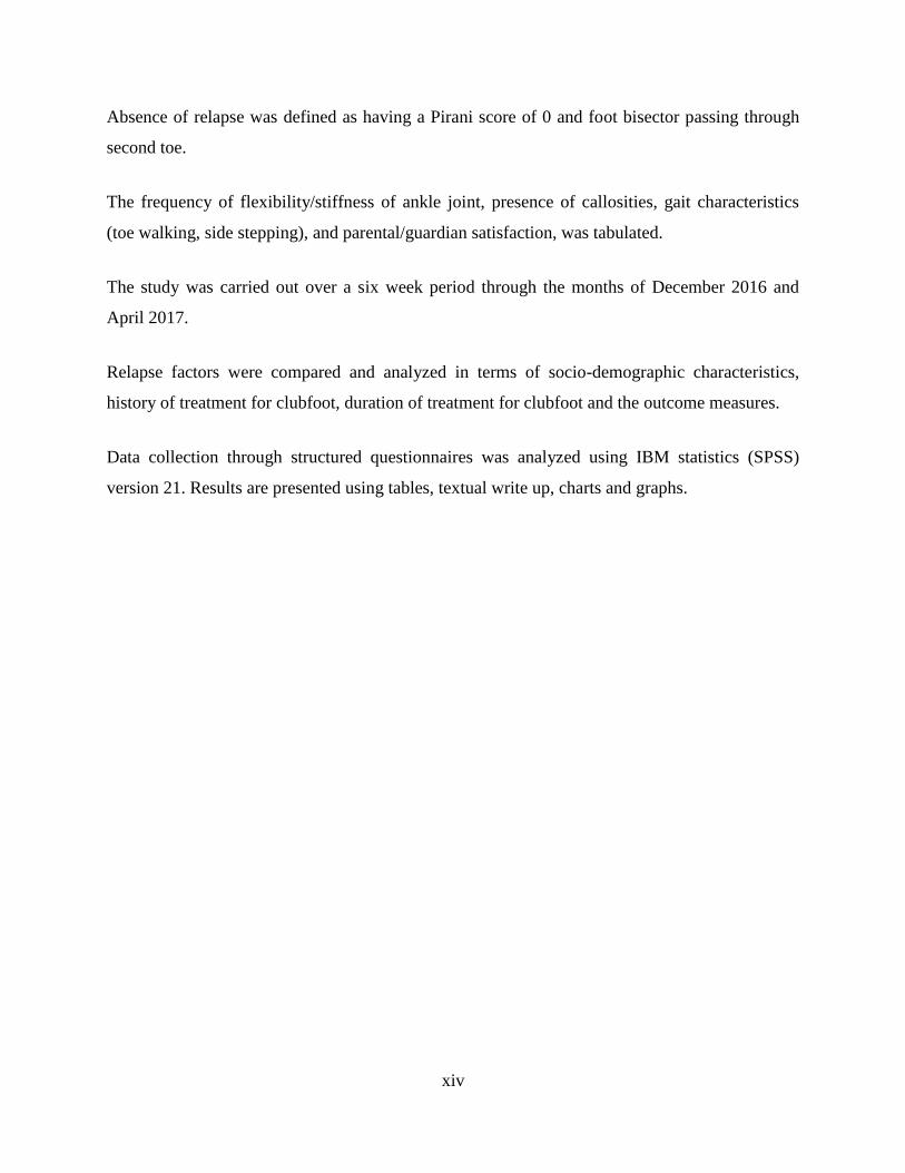
Ponseti involves two phases, phase one involves manipulation of deformed foot through a
weekly series of movements at the subtalar joint, supination of forefoot and dorsiflexion of 1st
ray corrects the cavus, abduction with gentle pressure under 1st ray and fulcrum at the head of
talus corrects adduction and varus, dorsiflexion plus or minus tenotomy corrects equinus,
manipulation takes approximately 5-6 wks, after every manipulation a long leg cast is applied
to maintain the position (2).
Figure 1.2-1: Talipes Equinovarus
2
Figure 1.2-2: Casting in the Ponseti Method
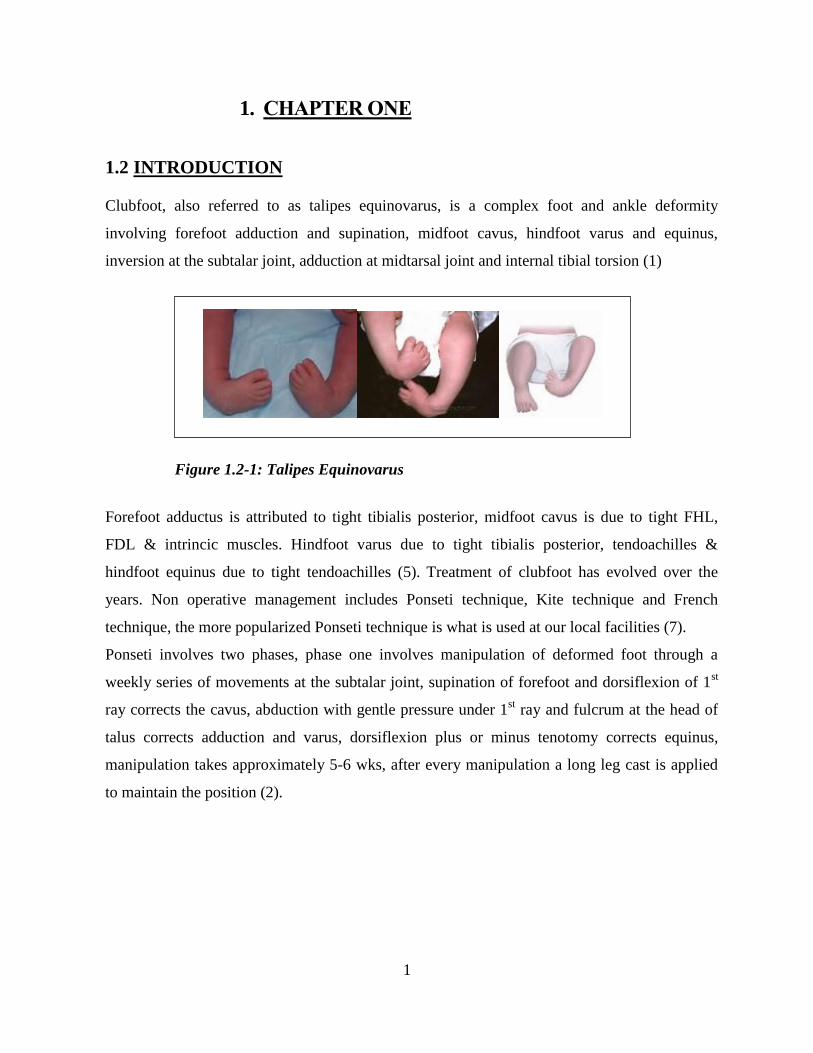
After full correction is achieved, the second phase of treatment includes the use of foot
abduction braces (FAB), worn to maintain correction achieved post manipulation and prevent
relapse (4)
Figure 1.2-3: Dennis Brown Brace
Annually, 150,000 children are born with clubfoot representing 1.2/1000 live births worldwide.
Eighty (80%) of these cases are in developing nations (1,3,4). In Africa and specifically in
Uganda 1-2 cases of clubfoot were reported in every 1000 live births (24).
Ponseti technique has a success rate of 90-98%(1,2,11).A recurrence rate reported by Ponseti 1-
2/10 cases with the gap occurring during the foot abduction brace phase due to noncompliance
to protocol. Incidence of recurrence reduced significantly when foot abduction bracing was
emphasized (1, 2, 10, 16).
3
Clubfoot is a condition that can be disabling to the affected individual. It causes pain, abnormal
foot position and if not corrected gait is altered (4,34). Callosities form on the lateral aspect of
the foot which leads to difficulty in shoe wear (22).
The family and community are affected psychosocially (9,13) . Treatment can be expensive,
time consuming and emotionally draining (16).
Recurrence rates and the factors contributing to relapse are unknown in our population; this
information will help close the gap in the treatment of the deformity.

4
2 CHAPTER TWO
2.1 LITERATURE REVIEW
2.1.1 INFLUENCE OF BRACING IN RELAPSE RATES
Bracing is an integral part of Ponseti treatment. This phase starts after full correction of the
deformity has been achieved. It involves wearing a FAB, which comprises of well fitted, open
toed, high- top straight-lace shoes. These are mounted onto a bar that corresponds to the child´s
shoulder width. Full time wear for 23hours/day in the first 3months followed by part time wear
for 12-14 hours/day for 3-4 years (1, 9, 10, 11, 12).
If this is not adhered to, then recurrences are observed. Ponseti et al described relapse as the
reappearance of any of the components of the initial deformity, which include forefoot
adduction, hindfoot varus or equinus. This definition is similar to the IOWA group. Haft et al
studied 51 children and found a recurrence rate of 41%; Ponseti et al established a recurrence
rate of 1-2/10 case. Studies have attributed recurrence to low educational level of parents, Native
American ethnicity, and annual family income of < US$20,000. The most common factor
however was non-compliance of brace wear in the reviewed papers. Non-compliance rate varies
from 36%-60%(1,3,5,10,11,13,16,25).
A difference in the definitions of compliance and non-compliance was observed in many studies.
Morcuende et al defined non compliance as not using the FAB for 10hours/day, Dobbs et al
defined non compliance as complete discontinuation of brace wear, this was similar to
Abdelgawad et al, Avilucea et al described it as premature discontinuation of bracing, while
Panjavi et al defined it as lack of full time bracing in the 1st 3months or night time bracing for 9
months thereafter. Differences in definitions could contribute to difference in results (3, 5, 7, 25,
26).
Treatment centers have adopted different protocols of bracing. Laaveg et al and Ponseti et al
advised on 22-23hours/day for the 1st 2-3 months followed by night time 10-12hours/day for 2-
4years, Morcuende et al agreed with the 23hours/day but night time preferred 12-14hours/day for
3-4years. Some authors suggested wearing the brace full time for 2-4years then night time
thereafter. These differences may contribute to significant differences in the results, the less the
hours spent in the brace the higher the recurrence noted (1, 4, 7, 11, 13, 27, 28).

5
2.1.2 AGE AT PRESENTATION
Early presentation has several benefits, soft tissues are more supple therefore easier to
manipulate, bones have not ossified making it easier to manipulate and the stretch period is
reduced due to viscoelastic properties and collagen organization (16,18,29).
Ponseti treatment showed a success rate of 80% as per Smoley et al in the initial stage, later in
the 1990ˈs after revisions and more understanding of CTEV the rate improved to 90-98%.
Smoley et al selected children between 1wk-6mnths and had 56% recurrence, Patil et al
compared two groups, 1st group <6mnths had relapse of 7.14%, 2
nd group >6mnths had a
relapse rate of 15.5%. Verma et al selected 1-3yr olds and had a success rate of 89%. Most
studies targeted children who were below 1 year and they reported a success rate of 90-98
%.Morcuende et al had full correction in older children (5, 6, 7, 13, 29, 30, 31). Differences in
ages of children in studies can contribute to significant difference in results, having more
relapses in children above 2 years.
2.1.3 INITIAL SEVERITY OF DEFORMITY
Numerous systems have been proposed to classify severity of deformity. This allows planning
of treatment and predicting out. Catterall/Pirani and Dimeglio/Bensahel are systems where
components of the deformity are assigned numerical scores; higher scores indicate a more
severe deformity. Both systems are reported to have a high interobserver reliability after a short
course of learning (17, 18, 19, 20, 21, 23, 22).
Dyer et al used the Catterall/Pirani system to estimate the number of weekly casts required,
they also used the hindfoot score to predict the need for tenotomy. There was a significant
association between the initial Pirani score and the number of cast changes required to correct
the deformity (21).
Some authors like Morcuende et al, Dobbs et al, Wainwright et al and others showed that there
is no correlation between initial severity and treatment outcomes or risk of relapse. Out of these
studies Wainwright proposed that Dimeglio system was more reliable in categorization of
deformity (3, 7, 23).Comparing children with varying severity of deformity can lead to a
difference in treatment success and the risk of recurrence results. Most studies showed Pirani
scoring being easier to use, it´s reliability has been proved (17,18).
6
2.1.4 RELATIONSHIP OF GENDER AND RELAPSE
Idiopathic clubfoot affects males more often than females with ratios of 2.5:1 and 6:1.1.
clubfoot deformity occurs bilaterally in 50% of the cases. Zionts et al had 240 patients with
idiopathic clubfoot, their Dimeglio scores were 13 for the males and 13.6 for females. There
was no significant difference in severity of deformity due to sex, the p value=0.61. Bilateral
cases showed no increase in severity but had a large range of severity when compared to
unilateral cases. Willis et al and Dobbs et al showed no significant relationship between gender
and risk of recurrence (2, 3, 15, 31, 32, 33).
Kruse et al had results that suggested that female patients are 5.5 times more likely than male
patients to transmit idiopathic clubfoot to their children. A study on need of surgery depending
on genetics was done by Goldstein et al; they stated that female patients were 5.3 times more
likely to need surgery (15, 34).
In these studies the incidence is higher in boys due to an inherent difference in susceptibility to
the deformity. Girls have a higher chance of transmitting the deformity to their children.
2.1.5 ATYPICAL DEFORMITY
Results of treatment depend on the type of clubfoot. Morcuende et al described an atypical
deformity which comprised of small, bean shaped, stiff feet with short big toe and volar crease.
These feet were resistant to manipulation and kept having recurrences. Bensahel et al reviewed
children with idiopathic, neurogenic and malformative clubfoot. Malformative were associated
with other congenital deformities. One surgeon using the same method treated all cases. They
reported 88% success rate in idiopathic feet and 25% success rate in malformative feet (7, 19).
Wudbhav et al suggested using gait analysis preoperatively to pick the subtle deformities not
evident on visual observation. Patients with recurrence were referred to them from different
centers. Gait analysis which was clinical and computerized revealed that 30 out of 35 patients
had additional deformities. Surgical plans of 19 patients had to be changed (63%) of cases.
In clubfoot deformity thorough evaluation is a must; this involves a detailed family history,
careful visual observation and gait analysis. Pretreatment classification helps with planning for
the appropriate management depending on the type of clubfoot (35).
7
2.1.6 RACE/ETHINICITY
Lochmiller et al carried out a study on epidemiology in the USA. Whites 1.2/1000, Hispanics
1.3/1000, African American 1.14/1000. Mkandawire et al showed an incidence of 2/1000 live
births in Malawi. In Uganda, Mathias et al had an incidence of 1.2/1000 births.
Dobbs et al compared whites and nonwhites and the rate of recurrence. Results showed no
significant relationship between race and risk of recurrence. In Newzealand Haft et al showed a
high recurrence (41%) but could not attribute results to high proportion of deformity in the
Polynesian children. The Polynesian patients had a less severe deformity and were less likely to
require surgery than the white patients. Although they did not score the feet of those patients
who chose operative treatment, they had an equal number of Polynesian patients in each group.
Avilucea et al showed an increase in recurrence in Native Americans living in rural areas than
those in urban centers and other ethnicities. They suggested that the rate could be attributed to
problems in communication (3, 16, 24, 25, 33, 36).
2.1.7 PARENTAL FACTORS
Parents play a major role in success of Ponseti treatment. The education level, income and
general attitude to the treatment can contribute to recurrence. Dobbs et al found a significant
relationship between parental education and recurrence rates. Parents with high school level or
below carried a 10 fold increased risk of relapse post Ponseti treatment. Fact. Haft in 2007 also
found no association between any parental factors and recurrence rate (3, 16).
Avilucea had a significant relationship between relapse and 1) unmarried parents 2) parental
education especially high school level or less 3) family income of less than $20,000. All these
factors like parental marital status and parental income, led to parents not embracing the
treatment fully (25).
2.1.8 PREVIOUS TREATMENT
Multiple studies have reported that idiopathic clubfoot deformities, previously treated with non
operative interventions respond well to Ponseti manipulation and casting. Alves et al
manipulated and casted children of 2yrs and below. They reported 93% success rate. Bor et al
re-manipulated infants referred to them, one patient (2.8%) required surgery at the end of
8
treatment. A previous study done at the same institution at a previous date included older
children and reported similar results (7, 37, 38).
No association has been found between previous non-operative treatment and risk of recurrence
after Ponseti treatment for children up to 4 years (25, 36, 47).
2.1.9 NUMBER OF CASTS
Several authors have attempted to link the number of casts required for correction with the risk
of recurrence. Dobbs et al found that the more severe the initial deformity the greater the
number of cast changes. Morcuende et al reported that the number of casts required was not a
long-term prognostic factor for recurrence after treatment.
Others have found a significant difference in the number of casts required for those with
recurrence compared with those who did not (3, 7, 20, 25, 37, 42, 47, 54).
Number of casts depends on technique of casting, stretch period needed and discomfort on the
child.
2.2 CONCLUSION
In the studies reviewed, the main factor associated with clubfoot relapse is non-adherence to
bracing. Low parental education and poor attitude to bracing contributed to non-adherence.
Children selected in the studies were of different ages and included both sexes that could
influence results. Locally no studies are available that evaluate factors associated with
idiopathic clubfoot relapse after Ponseti treatment. Settings in these regions differ from our
setting, which makes it difficult to compare results

9
3 CHAPTER THREE
3.1 STUDY QUESTION
What factors contribute to clubfoot recurrence after Ponseti treatment?
3.2 STUDY JUSTIFICATION
Clubfoot is the commonest deformity affecting the pediatric population. Treatment of choice in
our facilities locally is the Ponseti manipulation for idiopathic type.
Relapsed deformities are either manipulated and casted again or treated surgically. Burden of
retreatment can be reduced if the condition and factors affecting recurrence are better
understood.
A gap exists locally in knowledge and awareness of the condition and the factors that contribute
to recurrence.
This study surveyed the factors that contribute to relapse and permitted development of
strategies in prevention.
3.3 STUDY OBJECTIVES
3.3.1 MAIN OBJECTIVE
To survey factors that contribute to clubfoot relapse after Ponseti treatment
3.3.2 SPECIFIC OBJECTIVES
To determine the influence of brace adherence in recurrence of clubfoot deformity.
To determine parental factors that may contribute to relapse of deformity.
To determine the relationship between gender and the rate of recurrence of clubfoot
10
3.4 METHODOLOGY
3.4.1 STUDY DESIGN
Case control Study.
3.4.2 STUDY SETTING
AIC Cure Kijabe Hospital wards and outpatient clinics, this is a pediatric orthopedic hospital
that deals with childhood deformities both congenital and acquired, KNH pediatric foot clinic.
These two centers have personnel that are trained in the Ponseti manipulation; training begins
with the curriculum including this manipulation, constant workshop training, training in FAB
manufacture and utilization through apprenticeship.
3.4.3 STUDY POPULATION
Children with idiopathic clubfoot deformity who have successfully completed first phase of
treatment (serial casting) and are in the second phase (bracing) for at least 1 year.
CASE DEFINITION
For the purpose of this study, a case is defined as a recurrence of deformity, occurring after
successful correction (Pirani 0).
SELECTION OF CONTROLS
Control group comprised the clubfoot clients who had maintained correction (Pirani 0).
3.4.4 SAMPLE SIZE
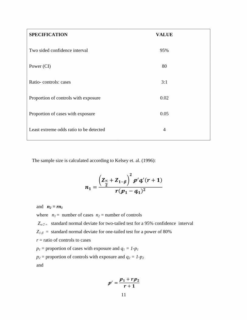
The sample size is calculated using OPENEPI based on Kelsey et al. (1996) with the following
specifications:
11
SPECIFICATION VALUE
Two sided confidence interval 95%
Power (CI) 80
Ratio- controls: cases 3:1
Proportion of controls with exposure 0.02
Proportion of cases with exposure 0.05
Least extreme odds ratio to be detected 4
The sample size is calculated according to Kelsey et. al. (1996):
(
)
( )
( )
and n2 = rn1
where n1 = number of cases n2 = number of controls
Zα/2 = standard normal deviate for two-tailed test for a 95% confidence interval
Z1-β = standard normal deviate for one-tailed test for a power of 80%
r = ratio of controls to cases
p1 = proportion of cases with exposure and q1 = 1-p1
p2 = proportion of controls with exposure and q2 = 1-p2
and

12
and q’ = 1 – p’
This worked out to 24 cases and 70 controls.
3.4.5 INCLUSION CRITERIA
1. Children (5years and below) diagnosed with idiopathic clubfoot deformity and have
completed first phase of treatment.
2. Children (5 years and below) already on foot abduction braces for at least one year
3. Willing and consenting parents to clubfoot clients
3.4.6 EXCLUSION CRITERIA
1. Children (5years and below) with neurogenic or syndromic CTEV
2. Children (5years and below) with Pirani score >0 at the end of manipulation
3. Unwilling and non-consenting parents of clubfoot clients
3.4.7 OUTCOME MONITORING
Assessment of correction maintenance or relapse was undertaken using the Pirani scoring
system, foot bisector and foot progression angle.The Pirani score obtained at the interview was
compared to that documented at the completion of treatment.
Questionnaires provided a subjective assessment of the parent/guardian/caretaker of state of the
patient’s feet, their feelings on the Ponseti method.
Brace wear was evaluated by the state of the laces, leather, sole, callosities and the number of
times the brace had been changed.
3.4.8 DATA COLLECTION, MANAGEMENT AND ANALYSIS
Data was collected using structured questionnaires and entered into a password protected
Microsoft Access Database. The hard copy data forms were stored in a lockable cabinet either
in the statistician’s office or the Principal Investigator’s office. Upon completion of Data entry,
hard copy forms were compared with the entered data to identify errors and corrections made
appropriately.
13
Descriptive statistics of variables included; frequencies and percentages while continuous
variables were summarized using measures of central tendency such as mean, median, mode
and standard deviation.
Associations between two variables were determined using Chi-squared tests and Fisher’s exact
tests for categorical variables and t-tests for continuous variables. Multivariate analyses,
independent factors associated with clubfoot recurrence were identified using binary stepwise
backward logistic regression.
Data was analyzed using IBM Statistics (SPSS) Version 21. Results have been presented using
tables, textual write up, charts and graphs.
3.5 STUDY LIMITATIONS
1. Proof of brace compliance; relied on truthfulness of the parents.
2. Guardians opting out of study; education on purpose of study was done at enrollment.
3. Loss to follow up; some patients receiving treatment were already lost to follow up.
4. Lack of the initial Pirani score.
3.6 DISSEMINATION OF FINDINGS
Copies of the study will be availed to the following places:
The University of Nairobi College of Health Sciences Library.
Library of the Department of Orthopedics, University of Nairobi.
Library of AIC Cure Kijabe Hospital.
Kenyatta National Hospital Research Department.
Published in peer-reviewed journal.
Conference presentation.
3.7 ETHICAL CONSIDERATIONS
Approval for the study was obtained from department of orthopedic surgery, University of
Nairobi, AIC Cure hospital and the KNH ethics and research committee (KNH/ERC) before
commencement.
14
Informed consent was obtained from the patients parents/guardians who had accepted to
participate in the study. For those who did not consent and had relapsed, they were managed as
per the Ponseti relapse protocol but were not included in the study. Their treatment was not
affected by refusal to give consent.
15
4 CHAPTER FOUR
4.1 RESULTS
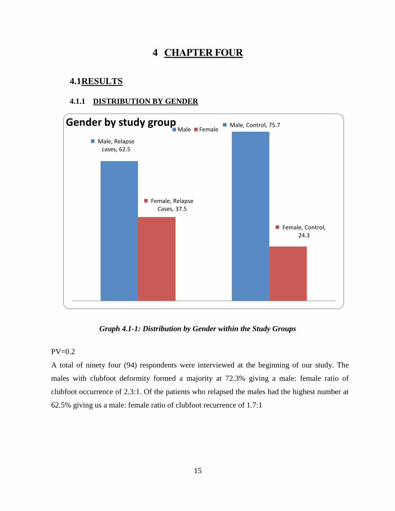
4.1.1 DISTRIBUTION BY GENDER
Graph 4.1-1: Distribution by Gender within the Study Groups
PV=0.2
A total of ninety four (94) respondents were interviewed at the beginning of our study. The
males with clubfoot deformity formed a majority at 72.3% giving a male: female ratio of
clubfoot occurrence of 2.3:1. Of the patients who relapsed the males had the highest number at
62.5% giving us a male: female ratio of clubfoot recurrence of 1.7:1
Male, Relapse cases, 62.5
Male, Control, 75.7
Female, Relapse Cases, 37.5
Female, Control, 24.3
Gender by study group Male Female
16
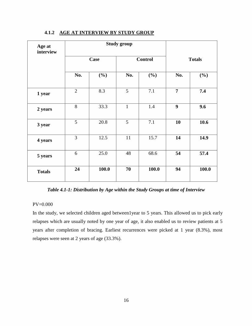
4.1.2 AGE AT INTERVIEW BY STUDY GROUP
Age at
interview
Study group
Case Control Totals
No. (%) No. (%) No. (%)
1 year 2 8.3 5 7.1 7 7.4
2 years 8 33.3 1 1.4 9 9.6
3 year 5 20.8 5 7.1 10 10.6
4 years 3 12.5 11 15.7 14 14.9
5 years 6 25.0 48 68.6 54 57.4
Totals 24 100.0 70 100.0 94 100.0
Table 4.1-1: Distribution by Age within the Study Groups at time of Interview
PV=0.000
In the study, we selected children aged between1year to 5 years. This allowed us to pick early
relapses which are usually noted by one year of age, it also enabled us to review patients at 5
years after completion of bracing. Earliest recurrences were picked at 1 year (8.3%), most
relapses were seen at 2 years of age (33.3%).
17
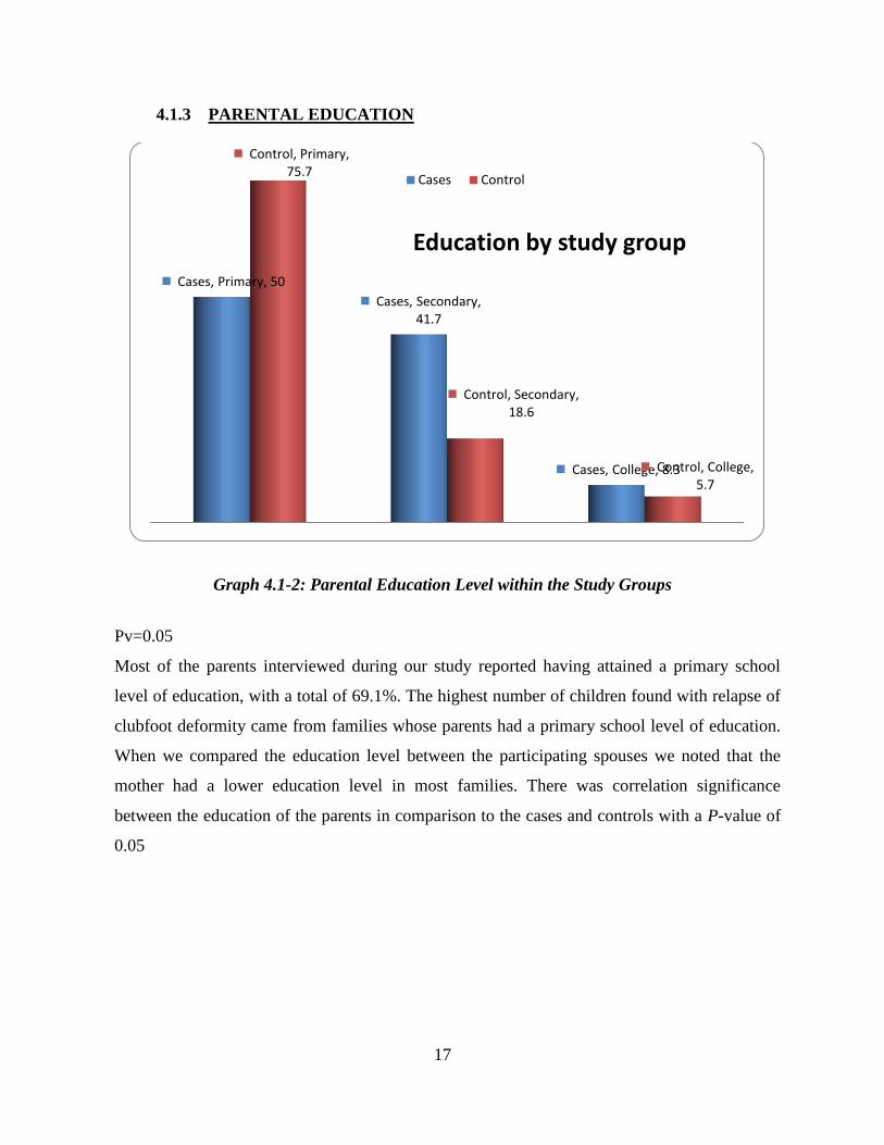
4.1.3 PARENTAL EDUCATION
Graph 4.1-2: Parental Education Level within the Study Groups
Pv=0.05
Most of the parents interviewed during our study reported having attained a primary school
level of education, with a total of 69.1%. The highest number of children found with relapse of
clubfoot deformity came from families whose parents had a primary school level of education.
When we compared the education level between the participating spouses we noted that the
mother had a lower education level in most families. There was correlation significance
between the education of the parents in comparison to the cases and controls with a P-value of
0.05
Cases, Primary, 50
Cases, Secondary, 41.7
Cases, College, 8.3
Control, Primary, 75.7
Control, Secondary, 18.6
Control, College, 5.7
Education by study group
Cases Control
18
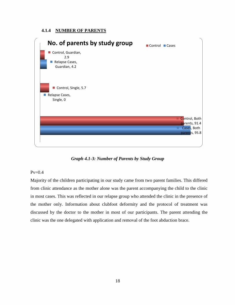
4.1.4 NUMBER OF PARENTS
Graph 4.1-3: Number of Parents by Study Group
Pv=0.4
Majority of the children participating in our study came from two parent families. This differed
from clinic attendance as the mother alone was the parent accompanying the child to the clinic
in most cases. This was reflected in our relapse group who attended the clinic in the presence of
the mother only. Information about clubfoot deformity and the protocol of treatment was
discussed by the doctor to the mother in most of our participants. The parent attending the
clinic was the one delegated with application and removal of the foot abduction brace.
Cases, Both parents, 95.8
Relapse Cases, Single, 0
Relapse Cases, Guardian, 4.2
Control, Both parents, 91.4
Control, Single, 5.7
Control, Guardian, 2.9
No. of parents by study group Control Cases
19
4.1.5 AFFECTED LIMB
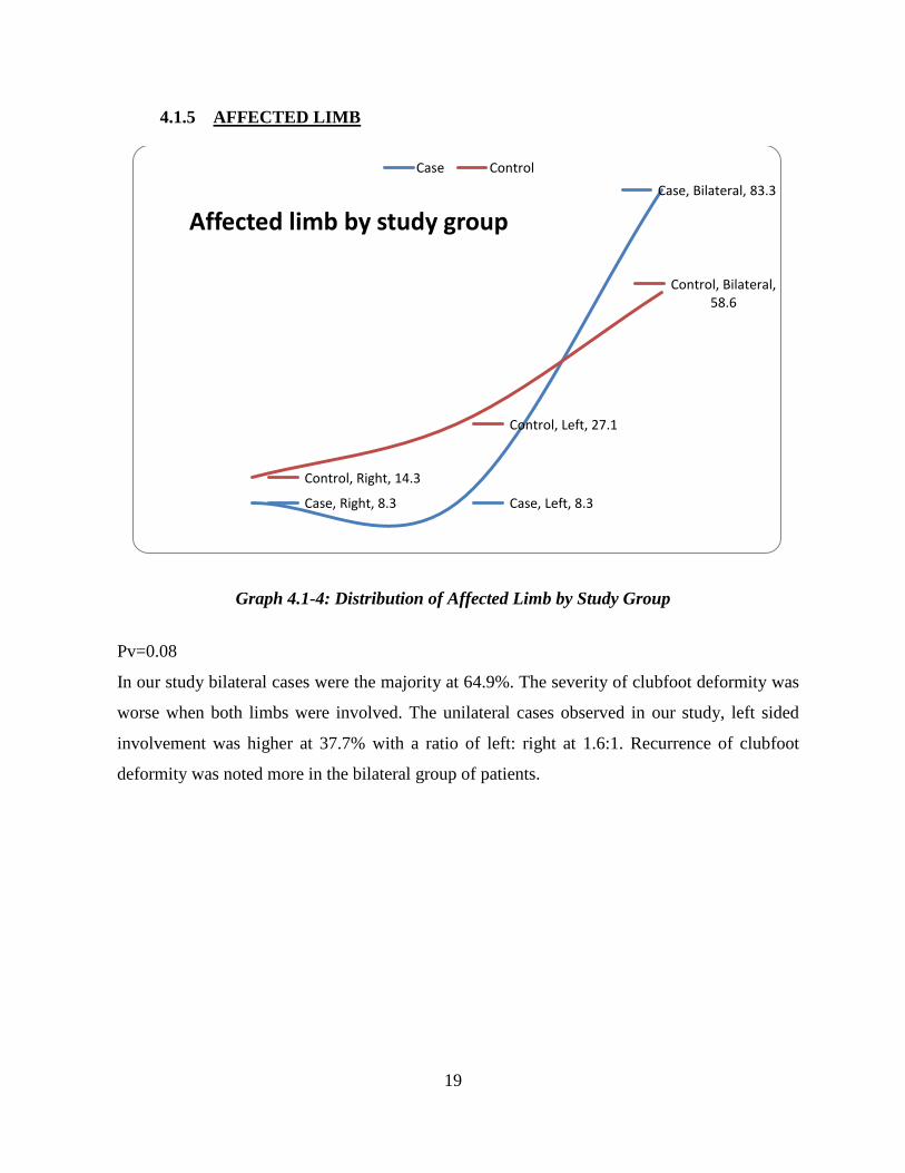
Graph 4.1-4: Distribution of Affected Limb by Study Group
Pv=0.08
In our study bilateral cases were the majority at 64.9%. The severity of clubfoot deformity was
worse when both limbs were involved. The unilateral cases observed in our study, left sided
involvement was higher at 37.7% with a ratio of left: right at 1.6:1. Recurrence of clubfoot
deformity was noted more in the bilateral group of patients.
Case, Right, 8.3 Case, Left, 8.3
Case, Bilateral, 83.3
Control, Right, 14.3
Control, Left, 27.1
Control, Bilateral, 58.6
Affected limb by study group
Case Control

20
4.1.6 INITIAL PIRANI SCORE- (RIGHT SIDE)
Pirani
Score
Case Control Totals
No. (%) No. (%) No. (%)
0.5 0 0.0 2 2.9 2 2.1
1.0 0 0.0 1 1.4 1 1.1
1.5 3 12.5 8 11.4 11 11.7
2.0 2 8.3 7 10.0 9 9.6
2.5 2 8.3 3 4.3 5 5.3
3.0 2 8.3 3 4.3 5 5.3
3.5 2 8.3 3 4.3 5 5.3
4.0 3 12.5 6 8.6 9 9.6
4.5 4 16.7 6 8.6 10 10.6
5.0 1 4.2 3 4.3 4 4.3
5.5 2 8.3 3 4.3 6 6.4
6.0 0 0.0 6 8.6 6 6.4
Totals 24 100.0 70 100.0 94 100.0
Table 4.1-2: Initial Pirani score for the Right Limb
PV=0.5
Pirani scores were taken before treatment of every patient and documented. From our
evaluation most children had a Pirani score of 1.5. The worst Pirani score recorded was 6.0. In
the group of participants who had recurrence of clubfoot deformity they started out with a
Pirani score of 5.5
21
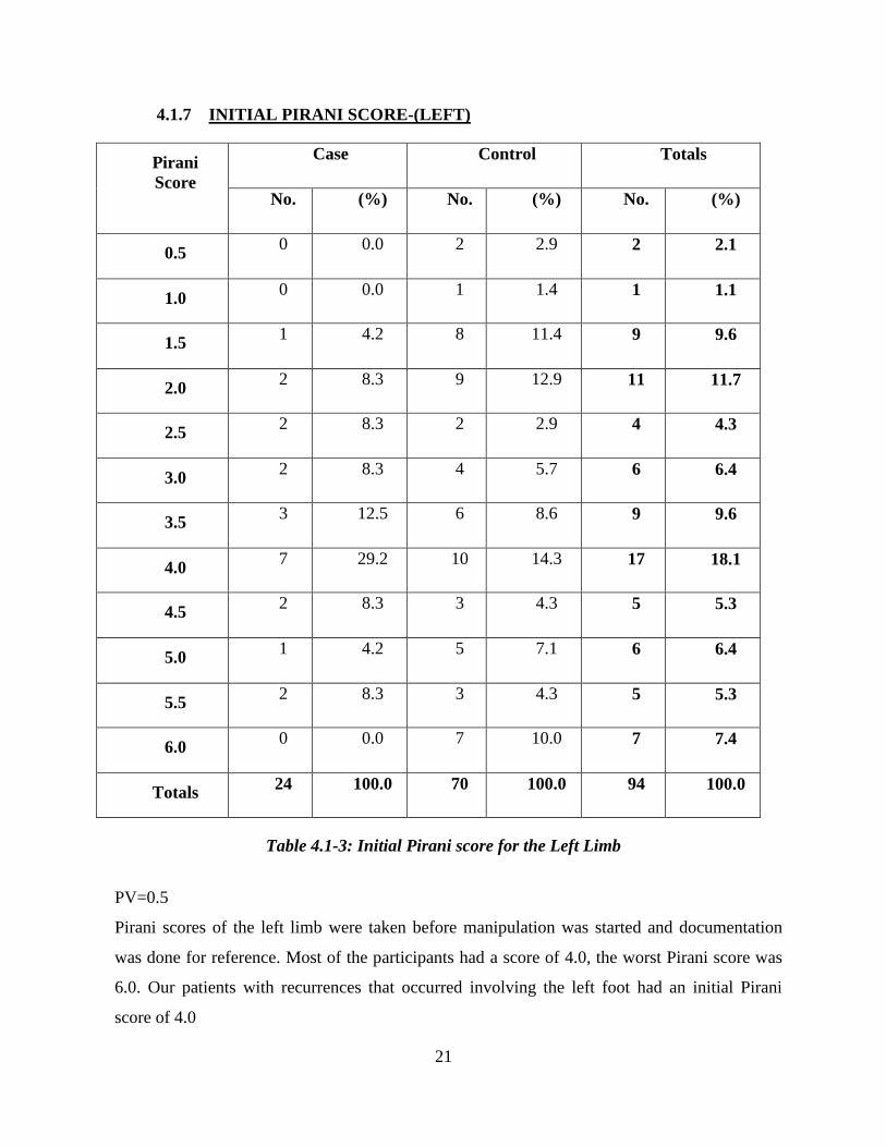
4.1.7 INITIAL PIRANI SCORE-(LEFT)
Pirani
Score
Case Control Totals
No. (%) No. (%) No. (%)
0.5 0 0.0 2 2.9 2 2.1
1.0 0 0.0 1 1.4 1 1.1
1.5 1 4.2 8 11.4 9 9.6
2.0 2 8.3 9 12.9 11 11.7
2.5 2 8.3 2 2.9 4 4.3
3.0 2 8.3 4 5.7 6 6.4
3.5 3 12.5 6 8.6 9 9.6
4.0 7 29.2 10 14.3 17 18.1
4.5 2 8.3 3 4.3 5 5.3
5.0 1 4.2 5 7.1 6 6.4
5.5 2 8.3 3 4.3 5 5.3
6.0 0 0.0 7 10.0 7 7.4
Totals 24 100.0 70 100.0 94 100.0
Table 4.1-3: Initial Pirani score for the Left Limb
PV=0.5
Pirani scores of the left limb were taken before manipulation was started and documentation
was done for reference. Most of the participants had a score of 4.0, the worst Pirani score was
6.0. Our patients with recurrences that occurred involving the left foot had an initial Pirani
score of 4.0
22
4.1.8 AGE AT FIRST CAST
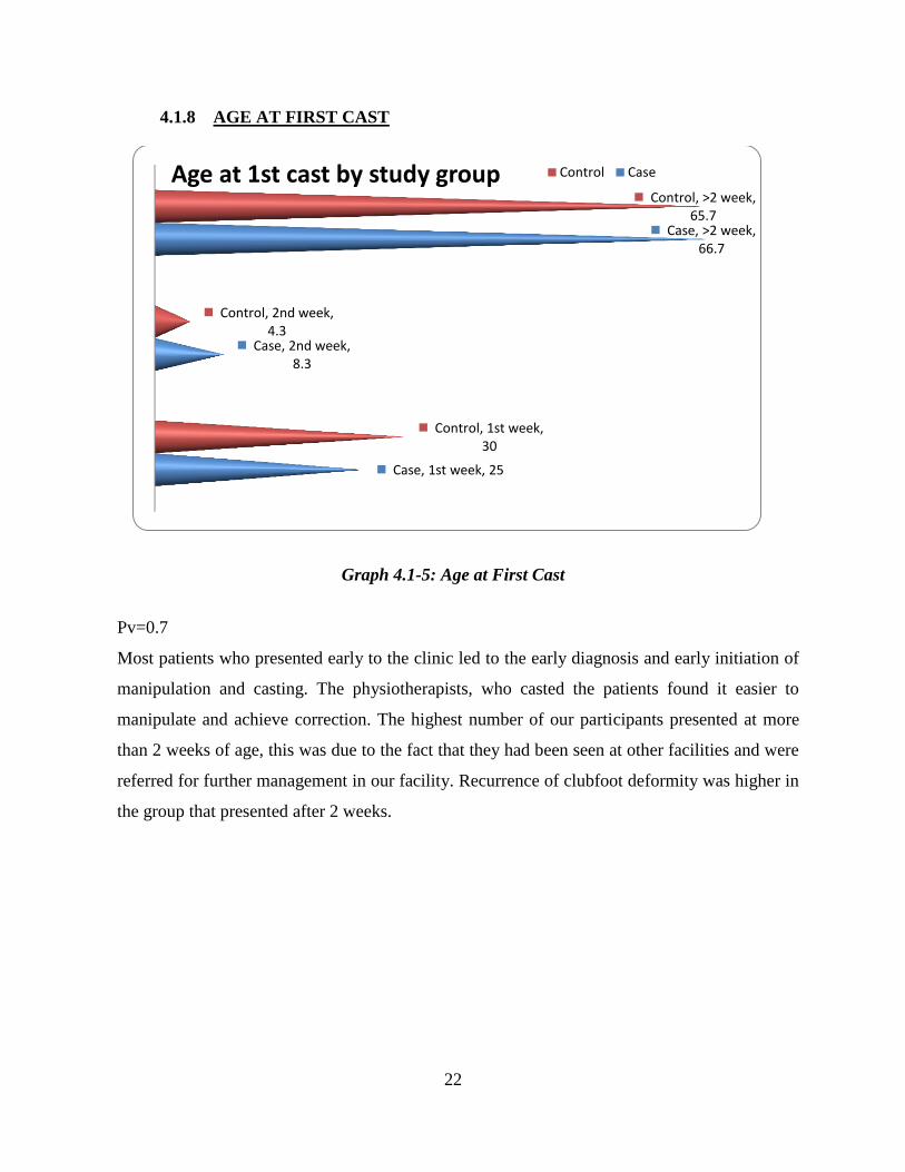
Graph 4.1-5: Age at First Cast
Pv=0.7
Most patients who presented early to the clinic led to the early diagnosis and early initiation of
manipulation and casting. The physiotherapists, who casted the patients found it easier to
manipulate and achieve correction. The highest number of our participants presented at more
than 2 weeks of age, this was due to the fact that they had been seen at other facilities and were
referred for further management in our facility. Recurrence of clubfoot deformity was higher in
the group that presented after 2 weeks.
Case, 1st week, 25
Case, 2nd week, 8.3
Case, >2 week, 66.7
Control, 1st week, 30
Control, 2nd week, 4.3
Control, >2 week, 65.7
Age at 1st cast by study group Control Case
23
4.1.9 NUMBER OF CAST CHANGES
Number of
Cast
Changes
Case Control Totals
No. (%) No. (%) No. (%)
4 4 16.7 13 18.6 17 18.1
5 4 16.7 18 25.7 22 23.4
6 7 29.2 15 21.4 22 23.4
7 1 4.2 11 15.7 12 12.8
8 6 25.0 7 10.0 13 13.8
9 1 4.2 3 4.3 4 4.3
10 0 0.0 1 1.4 1 1.1
12 1 4.2 0 0.0 1 1.1
13 0 0.0 2 2.9 2 2.1
Totals 24 100.0 70 100.0 94 100.0
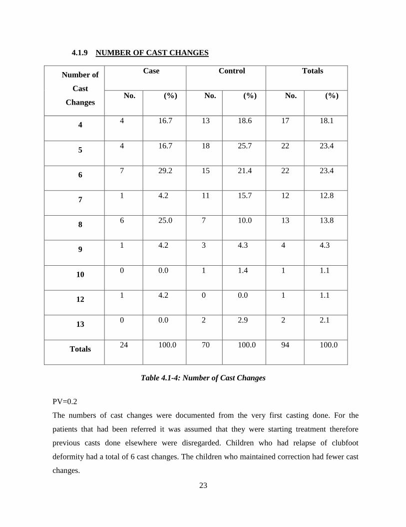
Table 4.1-4: Number of Cast Changes
PV=0.2
The numbers of cast changes were documented from the very first casting done. For the
patients that had been referred it was assumed that they were starting treatment therefore
previous casts done elsewhere were disregarded. Children who had relapse of clubfoot
deformity had a total of 6 cast changes. The children who maintained correction had fewer cast
changes.
24
4.1.10 PIRANI SCORE AT INTERVIEW-(RIGHT)
Pirani
Score
Case Control Totals
No. (%) No. (%) No. (%)
0 2 8.3 45 64.3 47 50
0.5 1 4.2 23 32.9 24 25.5
1.0 3 12.5 2 2.9 5 5.3
1.5 6 25.0 0 0.0 6 6.4
2.0 4 16.7 0 0.0 4 4.3
2.5 6 25.0 0 0.0 6 6.4
3.0 1 4.2 0 0.0 1 1.1
3.5 1 4.2 0 0.0 1 1.1
Totals 24 100.0 70 100.0 94 100.0
Table 4.1-5: Pirani Score at Time of Interview for the Right Side
PV=0.000
The right foot Pirani score of patients attended to at the interview showed an improvement from
the initial scores. The most common score was at 0 meaning correction had been maintained in
most patients. In the group of patients who had recurred clubfoot deformity, the highest score
was 3.5
25
4.1.11 PIRANI SCORE AT INTERVIEW-(LEFT)
Pirani
Score
Case Control Totals
No. (%) No. (%) No. (%)
0 3 12.5 49 70 52 55.3
0.5 0 0.0 20 28.6 20 21.3
1.0 2 8.3 1 1.4 3 3.2
1.5 7 29.2 0 0.0 7 7.4
2.0 7 29.2 0 0.0 7 7.4
2.5 3 12.5 0 0.0 3 3.2
3.0 2 8.3 0 0.0 2 2.1
Totals 24 100.0 70 100.0 94 100.0
Table 4.1-6: Pirani Score at Time of Interview for the Left Side
PV=0.000
The left foot Pirani score of patients reviewed after completion of treatment showed that the
commonest score was 1.5. In the group of patients that recurred the new Pirani score was at
3.0. From these scores we find that the left foot corrected at a slower rate than the right foot
26
5 CHAPTER FIVE
5.1 DISCUSSION
In this study, we set out to find the factors that are associated with idiopathic clubfoot relapse
after Ponseti treatment.
We had a total of 94 children with idiopathic clubfoot deformity, 24 patients had a relapse of
the deformity while 70 maintained the correction (figure 1).
Out of our participants, males had the greatest numbers (figure 2). We had more male patients
showing recurrence. Most studies report a higher prevalence of idiopathic clubfoot deformity in
male patients (1,11,15,32). Our higher number in male recurrence could be attributed to a
higher number of males in the total group, or the lower number of cases compared to controls
(15.32). The study by Goldstein et al showed a higher incidence in boys due to an inherent
difference in susceptibility to clubfoot.
Children who participated in our study were aged between1 year-5years (figure 4). Out of 94
children 54 (57.4%) were aged 5yrs. This ensured that there was adequate follow up post
manipulation before the interview. Most relapses were seen at 2years of age, followed by 5
years of age. Earliest recurrences that were visible to the caretakers/parents occurred after one
year. Our research team picked subtle recurrences during the interview using the Pirani score.
The commonest relapse seen in our study was dynamic forefoot adduction. The relapses were
treated by full time splint application, re-casting. Splint compliance was compromised in both
groups.
Overall, bilateral cases were highest at 64.9%, followed by unilateral left foot. The least group
was that of unilateral right foot (figure 6). This shows that the left foot is more affected at 1.74
times more than the right foot. Bilateral cases were 1.85 times more than unilateral cases. Most
cases are bilateral in 50% of the studies (2, 13, 24). Bilateral cases showed no increase in
severity but had a large range of severity when compared to unilateral cases.
In our study both parents accompanied the children to the clinic only during the first clinic,
thereafter the mother was the parent available (figure 5). This was noticed in both the cases and
the controls. In the control group discussing the clubfoot protocol at the beginning of treatment
with both parents made it easier. In consequent visits, only one parent was involved as this
27
reduced the transport fee used. The fathers were rarely available due to their jobs. This could
have affected the relapse rates.
Most parents reported to have a primary school education level (figure 3). The mother had a
lower education level compared to the father in most of our participating families; this could
have contributed to our recurrence rates as the mothers were the primary caretakers of the
children. Only two participants attended college in the cases group. From our study low
education level was associated with recurrence. There was correlation significance between the
education of the parents in comparison to the cases and controls with a p value of 0.05.
Interviewed parents stated that they had been educated by the doctors and physiotherapists on
clubfoot protocol but only a few could recall it step by step. The results may have been affected
by lack of understanding or a fear of asking the care givers for clarification. Dobbs et al found
that parental education at high school level or below carried a tenfold increased risk of clubfoot
recurrence after Ponseti treatment (3).
Initial Ponseti score of the right foot, the commonest score was 1.5. The highest score of the
group that had recurrence was 5.5, while the highest score of the control group was 6.0 (figure
7). This proved that having a severe initial Ponseti score did not mean that relapse would occur.
In the initial Ponseti score of the left foot, the commonest score was 4.0 in the total group. The
highest Pirani score of the control group was 5.5, while the highest score of the relapse group
was 6.0 (figure 8). This shows that in most cases, the left foot is usually more severe compared
to the right foot.
Some of the parents presented their children as early as a few hours post delivery, for initiation
of treatment (figure 9). We also had children coming for treatment months down the line, but
most of these cases were referrals from other centers. In the cases the highest number of
patients presented more than two weeks of age, this could have contributed to relapse due to the
difficulty in manipulation. From other studies early presentation has several advantages, soft
tissues are suppler therefore making it easier to manipulate, bones have not ossified also
contributing to ease of manipulation. The stretch period is reduced due to viscoelastic
properties and collagen organization (5, 6, 13, 30, 3).
Several authors attempted to link the number of cast changes to risk of recurrence. In the cases
group most children had 6 cast changes, while in the control group most children had 5 cast
changes (figure 10). More cast changes were needed in the group that had recurrences; this
28
reflects studies that found a significant difference in the number of casts required for correction
with risk of recurrence (3, 7, 20, 37, 42, 54).
Final Ponseti score was done at the interview. Right foot had commonest final case scores as
1.5 and 2.5 and highest final score at 3.5. Control group had commonest final score of 0.0 and
highest final score of 1.0. Left foot had commonest final case scores of 1.5 and 2.0 and highest
final score of 3.0. Right foot highest case score initially was at 5.5 and final case score was at
3.5. Right foot highest control score was initially at 6.0 and final control score was at 1.0. Left
foot highest case score was at 5.5 initially and the final highest case score was 3.0. Left foot
highest control score was at 6.0 and the final highest control score was at 1.0.
5.2 LIMITATIONS
The study had some limitations. We relied on the information provided by guardians or parents
and this could have affected our results as we depended on their recall capacity. The duration of
the study was not long enough for long term follow up .Most participants refused to fill in the
income per month question. In the future, a study can be designed to overcome these
limitations.
5.3 CONCLUSIONS
Clubfoot relapse rate (25.5%)
Males with idiopathic clubfoot had a higher rate (72.3%) while females had (27.7%)
Males recurred at a higher rate (62.5%)
Most recurrences occurred at 2 years of age (33.3%)
Bilateral clubfoot involvement (64.9%)
Recurrence highest in bilateral cases (83.3%)
Commonest unilateral clubfoot involvement left sided (27.3%)
Right side recurred more out of the unilateral cases (14.3%)
Recurrence higher in patients who started Ponseti later than 2wks (66.7%)
Recurrence was associated with a higher number of cast changes (23.4%)
Parents with primary school educational level had children with a higher relapse rate (50%)
95.8% had recurrence despite having both parents
29
5.4 RECOMMENDATIONS
Early diagnosis which would lead to early commencement of treatment.
Teaching more personnel on Ponseti treatment to avoid referrals and overloading of patients in
some specific centers.
Detailed parent education on clubfoot deformity, diagnosis of condition, and necessity of early
treatment and parent compliance.
Application of brace should be taught to more than one caretaker, incase one does not understand
the process.
Materials used in clubfoot treatment e.g. casts and braces should be provided free of charge to all
patients thus increasing the number of parents seeking intervention for their children.
30
6 REFERENCES
1. Ponseti IV, Smoley EN. Congenital clubfoot; the results of treatment.J Bone Joint Surg
Am.1963; 45(2):261-275.
2. Zionts LE, Zhao G, Hitchcock K. Has the rate of extensive surgery to treat idiopathic clubfoot
declined in the United States? J Bone Joint Surg Am 2010; 92(4):882-889.
3. Dobbs MB, Rudzki JR, Purcell DB, Factors predictive of outcome after use of the Ponseti
method for the treatment of idiopathic clubfeet. J Bone Joint Surg Am 2004; 86(1):22-27.
4. Laaveg SJ, Ponseti IV. Long-term results of treatment of congenital clubfoot. J Bone Joint
Surg Am 1980; 62(1):23-31.
5. Abdelgawad AA, Lehman WB, Van Bosse HJ. Treatment of idiopathic clubfoot using the
Ponseti method; minimum 2yr follow up. J Pediatric Orthop part B 2007; 16(2):98-105.
6. Herzenberg JE. Radler C, Bor N. Ponseti versus traditional methods of casting for idiopathic
clubfoot. J Pediatric Orthop 2002; 22(4):517-521.
7. Morcuende JA, Dolan LA, Dietz FR, Ponseti IV. Radical reduction in the rate of extensive
corrective surgery for clubfoot using the Ponseti method. Pediatric 2004; 113(2):376-380.
8. Colburn M, Williams M. Evaluation of the treatment of idiopathic clubfoot by using the
Ponseti method. J foot ankle Surg 2003; 42(5):259-267.
9Thacker MM, Scher DM, Sala DA. Use of foot abduction orthosis following Ponseti casts: is it
essential? J Pediatric Orthop 2005; 25(2):225-228.
10. Ponseti IV, Common errors in the treatment of congenital clubfoot. Int Orthop 1997;
21(2):137-141.
11. Ponseti IV, Treatment of congenital clubfoot. J Bone Joint Surg Am 1992; 74(3):448-454.
31
12. Ponseti IV. Congenital clubfoot: fundamental of treatment of treatment; New York Oxford
University Press; 1996.
13. Lehman WB, Mohaideen A, Madan S. A method for the early evaluation of the Ponseti
(IOWA) technique for the treatment of idiopathic clubfoot. J Pediatric Orthop part B 2003;
12(2):133-140.
14. Chu A, Labar AS, Sala DA. Clubfoot classification; correlation with Ponseti cast treatment. J
Pediatric Orthop 2010; 30(7):695-699.
15.Kruse LM, Dobbs MB, Gurnett CA, Polygenic threshold model with sex dimorphism in
clubfoot inheritance; the carter effect. J Bone Joint Surg Am 2008; 90(12):2688-2694.
16. Haft GF, Walker CG, Crawford HD. Early clubfoot recurrence after use of the Ponseti
method in a New Zealand population. J Bone Joint Surg Am 2007; 89(3):487-493.
17. Catterall A. A method of assessment of the clubfoot deformity. Clin Orthop Relat Res
1991(264):48-53.
18. Pirani S, Outer bridge H, Moran M, Sawatsky B. Methods of evaluating the virgin clubfoot
with substantial inter observer reliability. Pediatric Orthop J 1995.
19. Bensahel H, Dimeglio A, Souchet P. Final evaluation of clubfoot. J Pediatric Orthop part B
1995; 4(2):137-141.
20. Dimeglio A, Bensahel H, Souchet P. Classification of clubfoot. J Pediatric part B 1995;
4(2):129-136.
21. Dyer PJ, Davis N. The role of Pirani scoring system in the management of clubfoot using the
Ponseti me Bone Joint Surg Br 2006; 88(8):1082-1084.
22. Scher DM, Feldman DS, Van Bosse HJ. Predicting the need for tenotomy in the Ponseti
method for correction of clubfeet. J Pediatric Orthop 2004; 24(4):349-352.
32
23. Wainwright AM, Auld T, Benson MK, Theologis TN. The classification of congenital talipes
equinovarus. J Bone Joint Surg Br 2002; 84(7):1020-1024.
24.Mathias RG,Lule JK,Waiswa G,Naddumba EK,Pirani S.Incidence of clubfoot in Uganda. J
Public Health 2010; 101(4):341-4.
25.Avilucea FR,Szalay EA,Bosch PP,Sweet KR,Schwend RM.Effect of cultural factors on
outcome of Ponseti treatment of clubfeet in rural America.J Bone Joint Surg
Am.2009;91(3):530-540.
26.Panjavi B,Sharafatvaziri A, Zargarbashi RH, Mehrpour S. Use of the Ponseti method in the
Iranian population. J Pediatric Orthop.2012; 32(3):e11-e14.
27. Segev E, Keret D, Lokiec F. Early experience with the Ponseti method for the treatment of
congenital idiopathic clubfoot. Isr Med Assoc J.2005; 7(5):307-310.
28. Boehm S, Sinclair M. Foot abduction brace in the Ponseti method for idiopathic clubfoot
deformity: torsional deformities and compliance. J Pediatric Orthop.2007; 27(6):712-716.
29. Patil MS, Sasnur AH, Nayeem A, Patil MR, Al Ameen. Role of age in management of
clubfoot by Ponseti method and relapse rate. J Med Sci 2014; 7(2):154-159.
30. Verma A, Mehtani A, Sural S. Management of idiopathic clubfoot in toddlers by Ponseti
method. J Pediatric Orthop B.2012; 21(1):79-84.
31. Wynne-Davies R. Genetic and environmental factors in the etiology of talipes equinovarus.
Clin Orthop Relat Res 1972; 84:9-13.
32. Willis RB, Al-Hunaishel M, Guerra L, Kontio K. What proportion of patients need extensive
surgery after failure of the Ponseti technique for clubfoot? Clin Orthop Relat Res 2009;
467(5):1294-1297.
33
33. Lochmiller C, Johnston D, Scott A. Genetic epidemiology study of idiopathic talipes
equinovarus. Am J Med Genet 1998; 79(2):90-96.
34. Goldstein RY, Seehausen DA, Chu A, Sala DA, Lehman WB. Predicting the need for
surgical intervention in patients with idiopathic clubfoot. J Pediatric Orthop 2015; 35(4):395-
402.
35.Wudbhav N, Sankar M, Susan A, Rethlefsen PT, Jennifer W, Robert M. The recurrent
clubfoot, can gait analysis help us make better preoperative decisions. Clin Orthop Relat Res
(2009)467:1214-1222.
36. Mkandawire NC, Kaunda E. Incidence and patterns of congenital talipes equinovarus
(clubfoot) deformity at Queen Elizabeth Central Hospital, Banter, Malawi. East and Central
African Journal of Surgery 2004(9)2:28-31.
37. Bor N, Herzenberg JE, Frick S. Ponseti management of clubfoot in older infants. Clin Orthop
Relat Res.2006; 444:224-228.
38. Alves C, Escalda C, Neves C. Ponseti method; Does age at the beginning of treatment make
a difference. Clin Orthop Relat Res.2009; 467(5):1271-1277.
34
7 APPENDICIES
7.1 DATA COLLECTION SHEET
PATIENT BIODATA
STUDY NUMBER………………………..
AGE (YRS)…………… (MNTHS)…………….DOB……………………….
3. SEX MALE ……………………….
(a) FEMALE…………………
CHILD NUMBER IN FAMILY………………………
AGE AT FIRST CASTING……………………..
DATE TREATMENT COMMENCED……………….
SCHEDULED REVIEW ATTENDANCE (1)………….. ……………..
1. (2)……………. ……………………
2. (3)……………………………….
3. (4)…………………………………
INTERVAL OF CASTING; 1) 1ST
CHANGE………………………..
(a) 2) 2ND
CHANGE……………………
(b) 3) 3RD
CHANGE………………………
(c) 4)4TH
CHANGE………………………
(d) 5) OTHERS
9. LATERALISATION 1) UNILATERAL RT…………. LT ……………
(i) 2) BILATERAL…………………….
10 PIRANI SCORE BEFORE TREATMENT………………………
11. NUMBER OF CAST CHANGES……………………..
12. PIRANI SCORE AT INTERVIEW………………….
13. BRACE FITTING 1) YES ………………DATE…………………………….
(a) 2) NO……………………….
COST OF BRACE (Ksh.) ……………………………………….
NUMBER OF BRACE CHANGES……………………………………….
ACTIVITY OF CHILD; 1) CRAWLING…………………………

35
(a) 2) SITTING…………………………….
(b) 3) WALKING…………………………………
ADHERENCE/NON-ADHERENCE
1 NO APPLICATION…………………………………….
2 SUB-OPTIMAL FREQUENCY OF APPLICATION………………………….
3 DURATION AND TIMING OF APPLICATION……………………………………….
4 TO THE LETTER ADHERENCE AS INSTRUCTED BY CARETAKER………………
18. RELATIONSHIP TO CHILD 1) PARENT ……………………………..
(i) 2) GURDIAN ………………………………
(ii) 3) CARETAKER……………………………
PARENT EDUCATION LEVEL 1) PRIMARY (YRS)……………………………
(i) 2) SECONDARY (YRS)……………………………
(ii) 3) TERTIARY (YRS)……………………………
20. CLINIC ATTENDANCE 1) SINGLE PARENT…………………………….
(i) 2) BOTH PARENTS…………………………………..
(ii) 3) GUARDIAN………………………………………..
21. BRACE APPLICATION/REMOVAL TEACHING 1) YES ………..
a. 2) NO……………….
22. PERSONELL DOING THE TEACHING 1) PHYSIO …………………………
1. 2) NURSE………………..
2. 3) DOCTOR……………………
23. EASE OF APPLICATION/REMOVAL OF BRACE…………………………………..
24. OCCUPATION 1) FORMAL EMPLOYMENT………………………………..
.1 2) INFORMAL EMPLOYMENT…………………………………
.2 3) FARMER…………………………………………..
.3 4) PESANT…………………………………..
25. YEARLY INCOME 1) <50,000KSHS………………………………..
(a) 2)50-100,000KSHS…………………………………..
(b) 3) >100,000KSHS.......................................................
26. INTERRUPTION OF CASTING 1) SWOLLEN FEET..........................
(i) 2) SKIN ULCERATION........................................
36
(ii) 3).OTHERS.............................
27. INTERUPTION OF BRACE APPLICATION 1) YES.........................
a. 2) NO...............................
2 IF YES; REASON...............................................................................
28. FALL OUT/LOOSENING OF CAST.........................................................
29. PERCEIVED CHILD DISCOMFORT
A) CAST 1) YES..........................2) NO......................................
B) BRACE 1) YES...........................2) NO.......................................
C) INTERVENTION 1) MEDICATION..............................................
.1 2) SPLINT REMOVAL.......................................
30. TENOTOMY 1) YES...............DATES....................................2) NO.....................
31. POST TENOTOMY CASTING DURATION............................................................
32. DISTANCE FROM HOSPITAL OF CARE 1) <50KM..........................................
1. 2)50-100KMS.........................................
2. 3)100-200KMS.......................................
3. 4)>200KMS................................................
33. OTHER FACTORS............................................................................................................
…………………………………………………………………………………………………
…………………………………………………………………………………………………
37
7.2 CONSENT INFORMATION
7.2.1 ENGLISH VERSION
This is an informed consent form for persons in the study whose title is factors contributing to
clubfoot relapse post Ponseti treatment.
Principal investigator: Dr Kinyanjui .M. Grace
Institution: School of Medicine, Department of orthopedic surgery, University of Nairobi
Supervisors: Dr Edward Gakuya, Dr Vincent Mutiso.
PART 1: Information sheet
Study title
A survey of factors associated with clubfoot relapse after Ponseti treatment
Investigators statement
My name is Dr Kinyanjui Grace, a post graduate student at the school of medicine, University
of Nairobi. I am conducting a research study titled ‘Factors Contributing to Clubfoot
Relapse Post Ponseti Treatment’.
Study background
Clubfoot deformity is a common problem encountered at KNH & Kijabe, treatment of choice is
the Ponseti method, a few relapses are seen, and no study has been done locally to evaluate the
factors that contribute to relapse.
Study objective
This study aims to find out the factors that contribute to recurrence rates.
Using the information derived from this study, conclusions will be drawn which may influence
treatment practices locally.
What is expected of you and the patient
Once you accept your child to participate in this study, you will be expected to fill a
questionnaire with the help of the principle investigator or with one of the research assistants.
Your child will also be examined to further evaluate recurrence.
You are not expected to pay anything to participate in this study. In the unlikely event that your
child will be required to be seen again by the investigator, then your transport expenses will be
refunded.
38
Voluntariness of participation
I would like to invite you to take part in this study. Participation is purely voluntary and you are
allowed to consent either immediately after getting this information or after a period of
consultation. You are free to ask any questions at any time regarding this study, or to seek any
clarification from either myself or my research assistant. If you consent to participate in the
study, some personal details as well as information concerning your baby's condition will be
sought.
Confidentiality
I guarantee that all the information taken from you will be kept strictly confidential and will not
be accessed by anyone other than the researchers and personnel authorized by the University of
Nairobi/Kenyatta National Hospital Ethics and research committee. This information will be
coded with numbers to maintain privacy.
Benefits of participation
Your participation in this study will be through a clinical interview and a clinical examination.
In case of unacceptable outcome the patients will be sent for review by the orthopedic surgeon
for appropriate management.
Risks of participation
You will not be exposed to any risks as you participate in this study.
Duration of study
The duration of study is 9 weeks.
Right of withdrawal
Withdrawal from this study can be done at any stage and will not affect your treatment at these
hospitals.
Compensation
You will receive no compensation for participating in this study.
CONSENT FORM
This proposal has been reviewed and approved by the UON/KNH-ERC which is a body that
ensures the protection of persons like you that take part in research studies. This approval has
been granted after submission of the study proposal to the committee by the Chairman of the
Department of Orthopedic Surgery, School of Medicine, and University of Nairobi with the
approval of a University supervisor.
39
If you require any additional information one can contact the following:
The Secretary, UON/KNH-ERC
P.O.BOX 20723-00202
KNH, NAIROBI.
Tel: +254207263009
Email: [email protected]
Grace.M.Kinyanjui,
Principal investigator
Tel no: 0722849809
Email: [email protected]
Dr Edward Gakuya
Supervisor, Lecturer University of Nairobi
Tel: 0721932799
Email:[email protected]
Dr Vincent Mutiso
Supervisor, Lecturer University of Nairobi
Tel: 0723289922
Email:[email protected]
To indicate that you understand the conditions of this study and that you agree to take part in it,
please sign or put your thumbprint in the space provided below.
I confirm that the study has been fully explained to me and I give full consent to participate in
it.
Signature/thumbprint:………………………………………………………….
Investigator's signature:………………………………………………………………..
Date:…………………………………………………………………..

40
7.2.2 KISWAHILI VERSION
B) FOMU YA MAELEZO
Kichwa: MAMBO YANAYOCHANGIA RELAPSE YA KIGUU BAADA YA MATIBABU
YA PONSETI
Mpelelezi: Dr Grace.M.Kinyanjui
Wasimamizi: Dr.E.Gakuya, Dr.V.Mutiso
Maono ya mpelelezi: Nataka kuchukua nafasi hii kukushukuru Kwa kuchukua muda Na
kusoma fomu hii. Hii fomu itakufahamisha zaidi juu ya utafiti ambao unaendelea ndivyo
upate nafasi ya kuamua Kama utahusika na huu utafiti.
Utangulizi: Unaombwa kushiriki Kwa utafiti kuhusu mambo yanayochangia relapse ya
kiguu baada ya matibabu ya Ponseti. Utafiti huu Ni ajili ya kutaka kujua yanayo changia
relapse ya kiguu Ili tuweze kuyazingatia Na kuepusha watoto wasipate relapse.
Utaratibu: Ukikubali kuhusika Na huu utafiti, nitakuuliza maswali mengine ambayo yaeza
kuwa nyeti, kuhusu kiwango cha elimu, mshahara wako. Baadaye nitaichunguza miguu ya
mtoto wako kudhibitisha kama imenyooka, ama bado iko na kasoro.
Matokeo ya faida: Matokeo ya utafiti huu yata tufaidi Kwa njia tofauti, nitapata nafasi ya
kuhitimu katika chuo cha upasuaji, nitaweza kuchapisha matokeo Kwa jarida tofauti Na
yanaeza kutumika kubadilisha namna matibabu yanavyo fikia wananchi Kwa jumla.
Usiri: Habari utakayo peana Kwa utafiti, Usiri utazingatiwa kutoka mwanzo mpaka mwisho,
majina hayatatumika Kwa fomu ya taarifa, watafiti pekee ndio wataeza kuona majina yako.
Umepewa uhuru wa kuamua kutoka kwa utafiti wakati wowote na bado utapata matibabu ya
mtoto wako inavyostahili bila gharama yoyote.
Nimepewa kibali kutoka Kamati ya Utafiti na Maadili ya Kituo Kikuu cha Nairobi na
Hospitali Kuu ya Kenyatta. Ufafanuzi zaidi waweza kupatikana kutoka wafuatao.
FOMU YA IDHINI
Katibu, KNH/UON-ERC,
S.L.P.20723-00202,
41
KNH, NAIROBI.
Nambari ya simu: (020)7263009
Grace.M.Kinyanjui
Mpelelezi Mkuu
Simu: 0722849809
Dr Edward Gakuya
Msimamizi
Simu: 0721932799
Dr Vincent Mutiso
Msimamizi
Simu: 0723289922
Kuonyesha umesoma na kuelewa jinsi ya utafiti huu na umepeana ridhaa ya kushiriki,
tafadhali weka sahihi au weka kidole katika nafasi ilitengwa hapo chini:
Sahihi yangu/kidole:…………………………………………………..
Sahihi ya mpelelezi:…………………………………………………………….
Tarehe:…………………………………………………………….

![A polyaxial fixation brace for the treatment of idiopathic ...CTEV, also known as clubfoot, is the fifth most common congenital malformation in children [1]. CTEV consists of four](https://img.pdfslide.net/doc/110x75/60bc26e493380344804f2a3f/a-polyaxial-fixation-brace-for-the-treatment-of-idiopathic-ctev-also-known.jpg)


![Clubfoot: Ponseti Management [Portuguese]](https://img.pdfslide.net/doc/110x75/5870def61a28ab912c8bffc8/clubfoot-ponseti-management-portuguese.jpg)
![Clubfoot: Ponseti Management [Yoruba]](https://img.pdfslide.net/doc/110x75/584bd0691a28ab85738da79a/clubfoot-ponseti-management-yoruba.jpg)


![Clubfoot: Ponseti Management [Creole]](https://img.pdfslide.net/doc/110x75/5879f5f81a28ab91388bcf7d/clubfoot-ponseti-management-creole.jpg)


![Clubfoot: Ponseti Management [Vietnamese]](https://img.pdfslide.net/doc/110x75/588c58cd1a28abcf208b58d1/clubfoot-ponseti-management-vietnamese.jpg)