Embed Size (px)
Citation preview
A synthetic strategy for mimicking the extracellular matrix provides new
insight about tumor cell migrationw
Michael P. Schwartz,a Benjamin D. Fairbanks,a Robert E. Rogers,a
Rajagopal Rangarajan,bMuhammad H. Zaman
cand Kristi S. Anseth*
a
Received 24th June 2009, Accepted 7th October 2009
First published as an Advance Article on the web 18th November 2009
DOI: 10.1039/b912438a
Understanding the role of the tumor microenvironment during cancer progression and metastasis
is complicated by interactions between cells, the extracellular matrix (ECM), and a variety of
biomolecules. Using a synthetic strategy, we investigated proteolytic modes of migration for
HT-1080 fibrosarcoma cells in an environment that limited confounding extracellular influences.
A large percentage of HT-1080s migrated through a Rho kinase (ROCK)-dependent rounded
morphology with a leading edge protrusion that defined the direction of migration, and migration
was only weakly dependent on the adhesive peptide RGDS. HT-1080s migrating in thiol-ene
hydrogels are more rounded and exhibit much more invasive behavior than dermal fibroblasts.
Our results indicate that HT-1080s have the capacity to migrate through a mechanism that is
distinct from mesenchymal cells, with significant amoeboid character even when utilizing a
proteolytic migration strategy. The migration mode observed here provides insight into the
invasiveness of metastatic cells in vivo and demonstrates the potential of a synthetic strategy
for investigating complex biological problems.
Introduction
Cancer progression and metastasis are characterized by a
complex, reciprocal communication between a tumor and
the extracellular environment.1–4 While much of the basic
understanding of cancer biology has been worked out in
2 dimensions,5 there remains a great need for improved
3-dimensional culture systems that capture the complexity of
the natural tumor microenvironment.6 Not surprisingly,
moving to the third dimension alters fundamental aspects of
cell migration and tumor growth such as cell polarity, cell
morphology, the nature of adhesive contacts, and the necessity
for proteolysis to remove physical barriers not encountered in
artificial 2-dimensional environments.7–10
Further complicating the overall picture and obscuring
attempts to develop therapeutic strategies, recent work has
demonstrated that cancer cells have the capacity to alter their
mode of motility when subjected to external stimuli such as
blocking or down-regulating integrin binding or proteolysis.10–15
In particular, cancer cells will undergo a mesenchymal–amoeboid
transition (MAT) or switch from collective to amoeboid
migration upon interruption of proteolysis or integrin binding.10–15
In the specific case of the MAT, elongated, proteolytically
dependent cells adopt a rounded proteolytically independent
migration strategy when proteolysis is blocked.12 However,
while biomaterials composed of naturally derived ECM
components provide a 3-dimensional environment that is
seemingly ideal for studying cancer biology, preparation
procedures can directly influence cell behavior and results
may not be physiologically relevant. For example, the MAT
for HT-1080 fibrosarcoma cells was observed in pepsin-
extracted collagen12 but not acid-extracted collagen of similar
network structure.9,16,17 Additionally, cancer cells migrating in
aHoward Hughes Medical Institute and Department of Chemical andBiological Engineering, University of Colorado at Boulder,ECCH111, CB424, Boulder, CO 80309.E-mail: [email protected]
bDepartment of Biomedical Engineering, University of Texas atAustin, Austin
cDepartment of Biomedical Engineering, Boston University, Bostonw Electronic supplementary information (ESI) available: Supplementalfigures showing integrin dependence and persistence fits; Details forcalculation of mesh size; Supplemental movies demonstrating celldivision, migration morphologies in 2D and 3D, and formation ofconstriction rings. See DOI: 10.1039/b912438a.
Insight, innovation, and integration
Here, we demonstrate that an engineering approach leads
to new insight about the invasive behavior of cancer cells.
Our synthetic approach enables quantitative study of cancer
biology through systematic incorporation of specific extra-
cellular matrix components while minimizing confounding
biological interactions. While we demonstrate the design of a
material for studying cancer migration and proliferation, this
technology could prove to be generally useful for studying a
wide variety of biological phenomena since many types of
biomolecules could be incorporated into the synthetic design.
32 | Integr. Biol., 2010, 2, 32–40 This journal is �c The Royal Society of Chemistry 2010
PAPER www.rsc.org/ibiology | Integrative Biology
Ope
n A
cces
s A
rtic
le. P
ublis
hed
on 1
8 N
ovem
ber
2009
. Dow
nloa
ded
on 1
1/13
/202
1 1:
47:3
0 A
M.
View Article Online / Journal Homepage / Table of Contents for this issue
an in vivo environment have been observed to be morphologically
similar to rounded, rather than elongated, cells in vitro.12,17
Therefore, moving to the third dimension introduces
significant complexity, and care must be taken when drawing
conclusions from results obtained with culture materials
derived from single ECM components.
Due to the overwhelming complexity of how cells interact
with even the simplest naturally derived biomaterials, there is
growing motivation to take a reductionist approach in
designing 3-dimensional culture platforms.18,19 Previously,
researchers have demonstrated that synthetic hydrogels can
be designed to mimic critical aspects of ECM function and
that cell spreading and migration in these materials closely
mimics behavior in natural biomaterials.20–26 Strategies such
as these provide explicit control over how biomolecules are
presented to cells, and provide researchers with tools for
simplifying the 3-dimensional environment to focus on the
role of specific cell–ECM interactions in biological processes
such as cell migration. Thus, new strategies for designing
synthetic ECM mimics and validating their use will encourage
biological researchers to incorporate an engineering approach
into their current methods and hopefully lead to new
fundamental discoveries.
The goal for this work was to quantify how adhesion
influences migration of HT-1080s in a 3-dimensional environment,
with an emphasis on probing proteolytically-dependent modes
of motility. Taking advantage of principles previously
reported for synthesizing biomimetic hydrogels,20–22,24,25 we
developed a 3-dimensional ECMmimic22 with a mesh size that
is much smaller than the size of a cell to limit migration to
proteolytic mechanisms, and with a poly(ethylene glycol)
backbone to reduce confounding biological interactions. To
our surprise, we found that HT-1080s migrated primarily
through a Rho kinase (ROCK)-dependent rounded morphology
that was only weakly dependent on integrin receptor inter-
actions. We quantitatively determined that the morphology of
HT-1080s migrating in thiol-ene hydrogels closely resembles
in vivo migrating cells and in vitro amoeboid cells,12 but is very
different compared with dermal fibroblasts, a model mesenchymal
cell type. These results suggest that the physiological mechanism
for HT-1080 migration may be very different than the
elongated mode previously observed in collagen,12 and
demonstrates a system that may be ideal for studying cancer
invasiveness.
Results
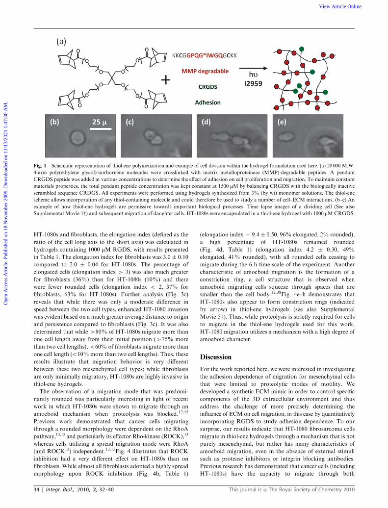
Fig. 1 illustrates the polymerization scheme used for this work.
Hydrogels were synthesized using a thiol-ene photopolymer-
ization mechanism to copolymerize ene-functionalized
poly(ethylene glycol) (PEG) precursors with thiol-containing
peptides. Specifically, a 4-arm PEG-norbornene monomer was
reacted with cysteine-containing peptides to form a functional
biomaterial network. This strategy has recently been
demonstrated as a highly versatile platform for creating
cytocompatible extracellular matrix (ECM) mimics.22 Based
on this strategy, we formed a hydrogel network using a broad
range MMP-degradable crosslinker27,28 and the common
fibronectin-derived adhesion sequence RGDS29,30 to provide
cells with a 3-dimensional environment permissive towards cell
migration. A thiol-ene hydrogel formed using this strategy is
predicted to have a mesh size that is much smaller than the size
of a cell (13 � 1 nm, see ESI,w calculating mesh size31,32),
thereby limiting migration to proteolytic mechanisms.
This polymerization scheme provides a versatile method for
studying specific ECM properties while reducing confounding
interactions caused by changes in materials properties or
biological interactions that may be prevalent in more complex
natural biomaterials. Fig. 1b–e demonstrates that thiol-ene
hydrogels prepared using MMP-degradable crosslinkers and
RGDS for adhesion are permissive towards cell proliferation
and migration (see also Supplemental Movie 1w), similar to
results recently reported for valvular interstitial cells.26
Previous migration studies have demonstrated that inter-
mediate receptor densities promote maximum cell migration
(i.e., migration exhibits a ‘‘biphasic’’ dependence on receptor
density) due to a balance of adhesive forces related to leading
edge attachment and trailing edge detachment for both
2-dimensional and 3-dimensional culture platforms.33–37 For
this work, HT-1080s did not migrate in control gels containing
non-biologically active RDGS (0 mM RGDS) or a non-
degradable crosslinker (SH-PEG-SH instead of the
MMP-degradable peptide), indicating that both adhesion
and degradation are necessary for migration. The dependence
of migration on degradation of the matrix confirms that
the mesh size of the thiol-ene hydrogel as prepared here
(13 � 1 nm, see ESI,w calculating mesh size31,32) is too small
for HT-1080s to migrate via a squeezing amoeboid mechanism.
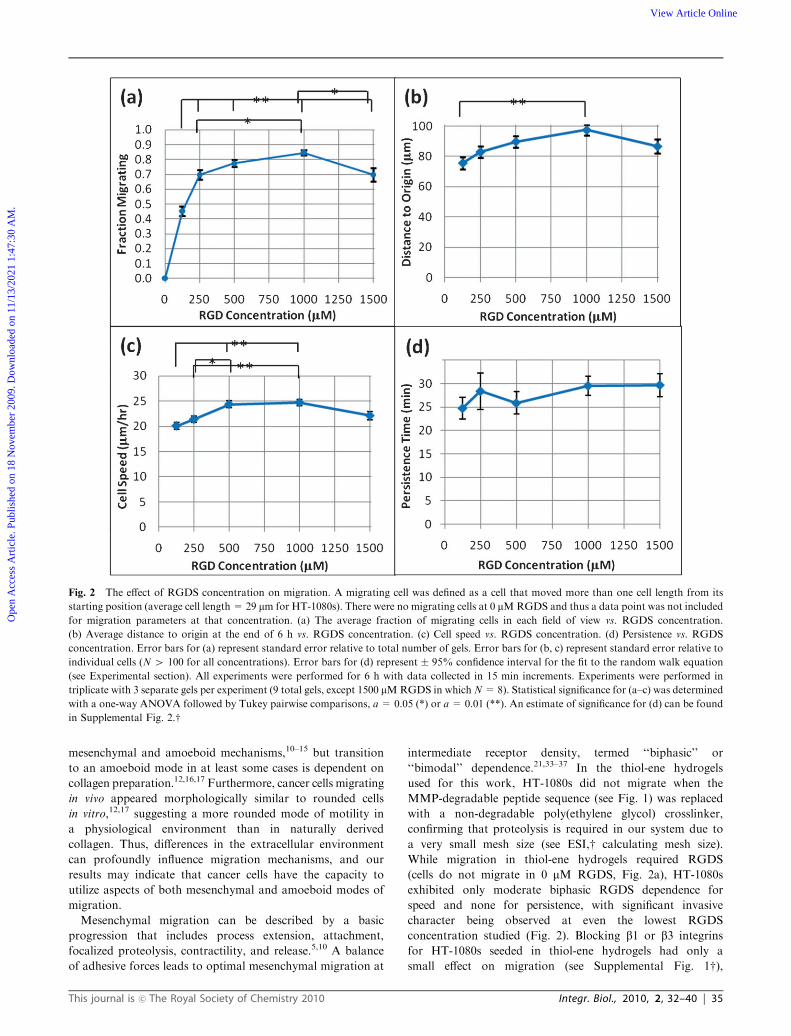
However, the results in Fig. 2 demonstrate that the introduction
of RGDS combined with an MMP-degradable crosslinker
promotes cell migration for all concentrations studied. The
percentage of migrating HT-1080s (Fig. 2a) is particularly
sensitive to RGDS concentration below 250 mM and reaches
a maximum at 1000 mM. However, average distance to origin
(total displacement from the initial starting position, Fig. 2b)
and cell speed (Fig. 2c) were only weakly RGDS dependent
while persistence (a measure of how long a cell migrates before
changing direction) exhibited little or no RGDS dependence
(Fig. 2d).
To further investigate the details of migration in thiol-ene
hydrogels, we compared HT-1080s to dermal fibroblasts, a
well-established mesenchymal cell type, with the results
presented in Fig. 3. Fibroblasts in thiol-ene hydrogels exhibited
a spread morphology with multiple protrusions (Fig. 3b, see
also Supplemental Movie 2w) and migrated in a random
fashion that resulted in cells being only moderately displaced
from their initial position. While some HT-1080s also
migrated with a spread morphology similar to fibroblasts,
the predominant morphology was more rounded and
generally exhibited only a single leading edge protrusion
(Fig. 3a, see also Supplemental Movies 3 and 4w). Migration
of rounded HT-1080s was usually in the direction of the
leading edge protrusion and resulted in the cells following
very long, relatively persistent paths through the synthetic
matrix.
The difference in migration between HT-1080s and
fibroblasts can be quantitatively demonstrated in several ways.
In order to quantify the difference in morphology between
This journal is �c The Royal Society of Chemistry 2010 Integr. Biol., 2010, 2, 32–40 | 33
Ope
n A
cces
s A
rtic
le. P
ublis
hed
on 1
8 N
ovem
ber
2009
. Dow
nloa
ded
on 1
1/13
/202
1 1:
47:3
0 A
M.
View Article Online
HT-1080s and fibroblasts, the elongation index (defined as the
ratio of the cell long axis to the short axis) was calculated in
hydrogels containing 1000 mM RGDS, with results presented
in Table 1. The elongation index for fibroblasts was 3.0 � 0.10
compared to 2.0 � 0.04 for HT-1080s. The percentage of
elongated cells (elongation index 4 3) was also much greater
for fibroblasts (36%) than for HT-1080s (10%) and there
were fewer rounded cells (elongation index o 2, 37% for
fibroblasts, 63% for HT-1080s). Further analysis (Fig. 3c)
reveals that while there was only a moderate difference in
speed between the two cell types, enhanced HT-1080 invasion
was evident based on a much greater average distance to origin
and persistence compared to fibroblasts (Fig. 3c). It was also
determined that while 480% of HT-1080s migrate more than
one cell length away from their initial position (475% more
than two cell lengths),o60% of fibroblasts migrate more than
one cell length (o10%more than two cell lengths). Thus, these
results illustrate that migration behavior is very different
between these two mesenchymal cell types; while fibroblasts
are only minimally migratory, HT-1080s are highly invasive in
thiol-ene hydrogels.
The observation of a migration mode that was predomi-
nantly rounded was particularly interesting in light of recent
work in which HT-1080s were shown to migrate through an
amoeboid mechanism when proteolysis was blocked.12,15
Previous work demonstrated that cancer cells migrating
through a rounded morphology were dependent on the RhoA
pathway,13,15 and particularly its effector Rho-kinase (ROCK),13
whereas cells utilizing a spread migration mode were RhoA
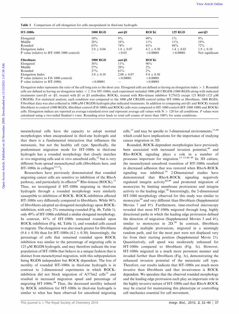
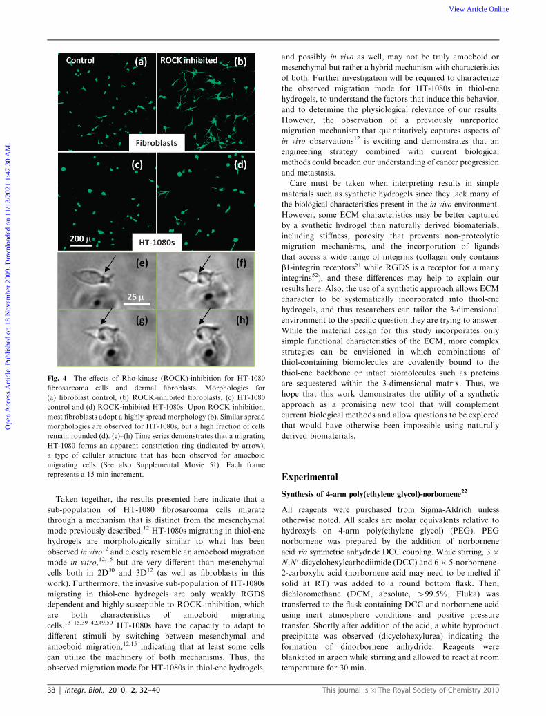
(and ROCK13) independent.13,15Fig. 4 illustrates that ROCK
inhibition had a very different effect on HT-1080s than on
fibroblasts. While almost all fibroblasts adopted a highly spread
morphology upon ROCK inhibition (Fig. 4b, Table 1)
(elongation index = 9.4� 0.50, 96% elongated, 2% rounded),
a high percentage of HT-1080s remained rounded
(Fig. 4d, Table 1) (elongation index 4.2 � 0.30, 49%
elongated, 41% rounded), with all rounded cells ceasing to
migrate during the 6 h time scale of the experiment. Another
characteristic of amoeboid migration is the formation of a
constriction ring, a cell structure that is observed when
amoeboid migrating cells squeeze through spaces that are
smaller than the cell body.12,38Fig. 4e–h demonstrates that
HT-1080s also appear to form constriction rings (indicated
by arrow) in thiol-ene hydrogels (see also Supplemental
Movie 5w). Thus, while proteolysis is strictly required for cells
to migrate in the thiol-ene hydrogels used for this work,
HT-1080 migration utilizes a mechanism with a high degree of
amoeboid character.
Discussion
For the work reported here, we were interested in investigating
the adhesion dependence of migration for mesenchymal cells
that were limited to proteolytic modes of motility. We
developed a synthetic ECM mimic in order to control specific
components of the 3D extracellular environment and thus
address the challenge of more precisely determining the
influence of ECM on cell migration, in this case by quantitatively
incorporating RGDS to study adhesion dependence. To our
surprise, our results indicate that HT-1080 fibrosarcoma cells
migrate in thiol-ene hydrogels through a mechanism that is not
purely mesenchymal, but rather has many characteristics of
amoeboid migration, even in the absence of external stimuli
such as protease inhibitors or integrin blocking antibodies.
Previous research has demonstrated that cancer cells (including
HT-1080s) have the capacity to migrate through both
Fig. 1 Schematic representation of thiol-ene polymerization and example of cell division within the hydrogel formulation used here. (a) 20000 M.W.
4-arm poly(ethylene glycol)-norbornene molecules were crosslinked with matrix metalloproteinase (MMP)-degradable peptides. A pendant
CRGDS peptide was added at various concentrations to determine the effect of adhesion on cell proliferation and migration. To maintain constant
materials properties, the total pendant peptide concentration was kept constant at 1500 mM by balancing CRGDS with the biologically inactive
scrambled sequence CRDGS. All experiments were performed using hydrogels synthesized from 3% (by wt) monomer solutions. The thiol-ene
scheme allows incorporation of any thiol-containing molecule and could therefore be used to study a number of cell–ECM interactions. (b–e) An
example of how thiol-ene hydrogels are permissive towards important biological processes. Time lapse images of a dividing cell (See also
Supplemental Movie 1w) and subsequent migration of daughter cells. HT-1080s were encapsulated in a thiol-ene hydrogel with 1000 mM CRGDS.
34 | Integr. Biol., 2010, 2, 32–40 This journal is �c The Royal Society of Chemistry 2010
Ope
n A
cces
s A
rtic
le. P
ublis
hed
on 1
8 N
ovem
ber
2009
. Dow
nloa
ded
on 1
1/13
/202
1 1:
47:3
0 A
M.
View Article Online
mesenchymal and amoeboid mechanisms,10–15 but transition
to an amoeboid mode in at least some cases is dependent on
collagen preparation.12,16,17 Furthermore, cancer cells migrating
in vivo appeared morphologically similar to rounded cells
in vitro,12,17 suggesting a more rounded mode of motility in
a physiological environment than in naturally derived
collagen. Thus, differences in the extracellular environment
can profoundly influence migration mechanisms, and our
results may indicate that cancer cells have the capacity to
utilize aspects of both mesenchymal and amoeboid modes of
migration.
Mesenchymal migration can be described by a basic
progression that includes process extension, attachment,
focalized proteolysis, contractility, and release.5,10 A balance
of adhesive forces leads to optimal mesenchymal migration at
intermediate receptor density, termed ‘‘biphasic’’ or
‘‘bimodal’’ dependence.21,33–37 In the thiol-ene hydrogels
used for this work, HT-1080s did not migrate when the
MMP-degradable peptide sequence (see Fig. 1) was replaced
with a non-degradable poly(ethylene glycol) crosslinker,
confirming that proteolysis is required in our system due to
a very small mesh size (see ESI,w calculating mesh size).
While migration in thiol-ene hydrogels required RGDS
(cells do not migrate in 0 mM RGDS, Fig. 2a), HT-1080s
exhibited only moderate biphasic RGDS dependence for
speed and none for persistence, with significant invasive
character being observed at even the lowest RGDS
concentration studied (Fig. 2). Blocking b1 or b3 integrins
for HT-1080s seeded in thiol-ene hydrogels had only a
small effect on migration (see Supplemental Fig. 1w),
Fig. 2 The effect of RGDS concentration on migration. A migrating cell was defined as a cell that moved more than one cell length from its
starting position (average cell length = 29 mm for HT-1080s). There were no migrating cells at 0 mMRGDS and thus a data point was not included
for migration parameters at that concentration. (a) The average fraction of migrating cells in each field of view vs. RGDS concentration.
(b) Average distance to origin at the end of 6 h vs. RGDS concentration. (c) Cell speed vs. RGDS concentration. (d) Persistence vs. RGDS
concentration. Error bars for (a) represent standard error relative to total number of gels. Error bars for (b, c) represent standard error relative to
individual cells (N 4 100 for all concentrations). Error bars for (d) represent � 95% confidence interval for the fit to the random walk equation
(see Experimental section). All experiments were performed for 6 h with data collected in 15 min increments. Experiments were performed in
triplicate with 3 separate gels per experiment (9 total gels, except 1500 mMRGDS in whichN=8). Statistical significance for (a–c) was determined
with a one-way ANOVA followed by Tukey pairwise comparisons, a = 0.05 (*) or a = 0.01 (**). An estimate of significance for (d) can be found
in Supplemental Fig. 2.w
This journal is �c The Royal Society of Chemistry 2010 Integr. Biol., 2010, 2, 32–40 | 35
Ope
n A
cces
s A
rtic
le. P
ublis
hed
on 1
8 N
ovem
ber
2009
. Dow
nloa
ded
on 1
1/13
/202
1 1:
47:3
0 A
M.
View Article Online
confirming that integrin dependence is weak. Unlike
mesenchymal migration, amoeboid migrating cells glide
through a porous network very rapidly via a squeezing and
flowing mechanism with weak integrin dependence,39–42 much
like what is observed for adhesion dependence of
HT-1080s in thiol-ene hydrogels. Additionally, constriction
rings, which have been described for neutrophils38 and
HT-1080s migrating through an amoeboid mechanism12 in
natural biomaterials, were also observed for HT-1080s as they
migrated through the 3-dimensional thiol-ene matrix
(Fig. 4e–h, Supplemental Movie 5w). Thus, our results indicatethat HT-1080s migrating in thiol-ene hydrogels exhibit at least
some characteristics that would be expected for amoeboid
migrating cells rather than mesenchymal cells.
A comparison to a well-established mesenchymal cell type,
dermal fibroblasts, further indicates that HT-1080 migration
in a thiol-ene hydrogel is not classically mesenchymal.
While fibroblasts were spread (Fig. 3b and 4a, Supplemental
Movie 2,w Table 1) with multiple protrusions, similar to
what would be expected for a mesenchymal cell type, most
HT-1080s were rounded with a single leading edge protrusion
(Fig. 3a and Fig. 4c, Supplemental Movies 3 and 4,w Table 1).
In order to quantify the degree of spreading, we calculated the
elongation index (the ratio of the cell long axis to the short
axis) for HT-1080s and fibroblasts (Table 1). In gels with
125 mM RGDS, most HT-1080s were rounded (88%, defined
as an elongation index o 2.0) while almost none were
elongated (1%, defined as an elongation index 4 3.0),
demonstrating that significant migration occurs even when a
mesenchymal morphology is not observed. Increasing the
RGDS concentration to 1000 mM led to a slight increase
in elongated HT-1080s (10%) and a significant increase in
elongation index (from 1.4 � 0.03 to 2.0 � 0.04), but the
majority of the cells remained rounded (63%), indicating that
while some cells may behave in a mesenchymal fashion, most
do not. Blocking b1 or b3 integrins for HT-1080s seeded in
thiol-ene hydrogels had only a small effect on morphology
(Table 1), further emphasizing that integrin dependence was
weak for HT-1080s migrating in thiol-ene hydrogels.
Fibroblasts were more spread than HT-1080s (elongation
index of 3.0 � 0.1 for fibroblasts compared to 2.0 � 0.04 for
HT-1080s, Table 1), with more elongated (36% vs. 10%) and
fewer rounded (37% vs. 63%) cells. Blocking b1-integrin had a
much larger influence on morphology for fibroblasts than for
HT-1080s, with a large decrease in elongated cells (36 to 11%)
corresponding to an increase in rounded cells (37 to 61%),
as well as a large decrease in elongation index (3.0 � 0.1 to
2.0 � 0.07). The influence of integrin blocking on fibroblasts
suggests that mesenchymal cells have the capacity to interact
with thiol-ene hydrogels through integrin receptor inter-
actions, and that HT-1080 migration and morphology are
weakly dependent on adhesion due to a fundamental
difference in mechanism rather than lack of appropriate
receptors in the hydrogel. Thus, while there appears to be a
subpopulation of HT-1080s in which spreading is influenced
by adhesion similar to fibroblasts, most retain a morphology
similar to amoeboid cells and behave very differently than the
model mesenchymal cell type.
Of particular interest regarding the potential of our syn-
thetic system for modeling cancer behavior, the elongation
index for HT-1080s migrating in thiol-ene hydrogels
(2.0 � 0.04, Table 1) is similar to amoeboid migrating
HT-1080s in collagen,12 while the fraction of elongated cells
(10% in thiol-ene) was much lower (440% in collagen15). The
HT-1080 strain used here adopted an elongated morphology
in collagen, as well, indicating that the observed difference is
not strain specific (see Supplemental Movie 6w). More
importantly, the elongation index for HT-1080s in thiol-ene
hydrogels was similar to that observed in vivo (based on
reported elongation before proteolytic inhibition12). In
contrast, the elongation index (3.0 � 0.1) and fraction of
elongated cells (36%) for fibroblasts are each very similar
to spread HT-1080s in collagen,12,15 suggesting that
Fig. 3 Comparison of migration for HT-1080s and dermal fibroblasts
in 1000 mM RGDS thiol-ene hydrogels. (a,b) Example of observed
morphologies for (a) HT-1080s and (b) dermal fibroblasts. (a) While
there were several morphologies observed for HT-1080s, most were
rounded with a dynamic leading edge protrusion that defined the
direction of migration (see also Supplemental Movies 3 and 4w)(b) Almost all fibroblasts adopted a typical spread, mesenchymal
morphology (see also Supplemental Movie 2w). (c) Comparison of
relative migration between HT-1080s and fibroblasts. Each parameter
is normalized to HT-1080 values. Experiments were performed in
triplicate with 3 separate gels per experiment (9 total gels). Error bars
for speed and distance to origin represent standard error relative to
individual cells (N 4 100 for all experiments). Statistical significance
was tested using a two-tailed Studen’s t-test (***, P o 0.00001).
Error bars for persistence represent �95% confidence interval for
the fit to the random walk equation (see Experimental section).
See Supplemental Fig. 2w for persistence fits with confidence and
prediction bands.
36 | Integr. Biol., 2010, 2, 32–40 This journal is �c The Royal Society of Chemistry 2010
Ope
n A
cces
s A
rtic
le. P
ublis
hed
on 1
8 N
ovem
ber
2009
. Dow
nloa
ded
on 1
1/13
/202
1 1:
47:3
0 A
M.
View Article Online
mesenchymal cells have the capacity to adopt normal
morphologies when encapsulated in thiol-ene hydrogels and
that there is a fundamental interaction that influences the
metastatic, but not the healthy cell type. Specifically, the
predominant migration mode for HT-1080s in thiol-ene
hydrogels has a rounded morphology that closely matches
in vivo migrating cells and in vitro amoeboid cells,12 but is very
different from spread mesenchymal cells (fibroblasts here, and
HT-1080s in collagen12,15).
Researchers have previously demonstrated that rounded
migrating cancer cells are sensitive to inhibition of the RhoA
pathway, and particularly its effector43 Rho-kinase (ROCK).13
Thus, we investigated if HT-1080s migrating in thiol-ene
hydrogels through a rounded morphology were similarly
susceptible to inhibition of ROCK. ROCK-inhibition affected
HT-1080s very differently compared to fibroblasts. While 96%
of fibroblasts adopted an elongated morphology upon ROCK-
inhibition, with only 2% remaining rounded (Fig. 4b, Table 1),
only 49% of HT-1080s exhibited a similar elongated morphology.
In contrast, 41% of HT-1080s remained rounded upon
ROCK-inhibition (Fig. 4d, Table 1), and rounded cells ceased
to migrate. The elongation was also much greater for fibroblasts
(9.4 � 0.50) than for HT-1080s (4.2 � 0.30). Interestingly, the
percentage of cells that remained rounded upon ROCK
inhibition was similar to the percentage of migrating cells in
125 mM RGDS hydrogels, and may therefore indicate the true
population of HT-1080s that behave in a unique fashion that is
distinct from mesenchymal migration, with this subpopulation
being RGDS independent but ROCK dependent. The loss of
motility of rounded HT-1080s in thiol-ene hydrogels is in
contrast to 2-dimensional experiments in which ROCK-
inhibition did not block migration of A375m2 cells13 and
resulted in increased migration speed and percentage of
migrating HT-1080s.44 Thus, the decreased motility induced
by ROCK inhibition for HT-1080s in thiol-ene hydrogels is
similar to what has been observed for amoeboid migrating
cells,13 and may be specific to 3-dimensional environments,13,44
which could have implications for the importance of studying
cancer migration in 3D.
Rounded, ROCK-dependent morphologies have previously
been associated with increased invasion potential,45 and
Rho-ROCK signaling plays a role in a number of
processes important for migration.13–15,44–49 In 3D culture,
the mesenchymal–amoeboid transition of HT-1080s resulted
in decreased adhesion that was restored when RhoA-ROCK
signaling was inhibited.14 2-Dimensional studies have
demonstrated that RhoA-ROCK signaling negatively
regulated integrin activity44,49 and promoted migration of
monocytes by limiting membrane protrusions and integrin
activity to the leading edge.50 Interestingly, the 2-dimensional
HT-1080 morphology observed for this study was similar to
monocytes50 and very different than fibroblasts (Supplemental
Movies 7 and 8w). Furthermore, time-resolved microscopy
revealed that most HT-1080s migrated through long, highly
directional paths in which the leading edge protrusion defined
the direction of migration (Supplemental Movies 3 and 4w),similar to monocytes in 2D.50 In contrast, fibroblasts
displayed multiple protrusions, migrated in a seemingly
random path, and for the most part were not displaced very
far from their starting position (Supplemental Movie 2w).Quantitatively, cell speed was moderately enhanced for
HT-1080s compared to fibroblasts (Fig. 3c). However,
HT-1080s migrated in a much more persistent manner and
invaded further than fibroblasts (Fig. 3c), demonstrating the
enhanced invasion potential of the metastatic cell type.
Therefore, our results indicate that HT-1080s are much more
invasive than fibroblasts and that invasiveness is ROCK
dependent. We speculate that the observed rounded morphology
and the leading edge protrusion each play an important role in
the highly invasive nature of HT-1080s and that RhoA-ROCK
may be crucial for maintaining this phenotype or controlling
cell mechanics essential for cell movement.
Table 1 Comparison of cell elongation for cells encapsulated in thiol-ene hydrogels
HT-1080s 1000 RGD anti-b1 ROCKi 125 RGD anti-b3
Elongated 10% 9% 49% 1% 9%Middle 28% 18% 11% 11% 19%Rounded 63% 74% 41% 88% 72%Elongation index 2.0 � 0.04 1.8 � 0.07 4.2 � 0.30 1.4 � 0.03 1.9 � 0.10P value (relative to HT-1080 1000 control) — o0.05 o0.00001 o0.00001 Not significant
Fibroblasts 1000 RGD anti-b1 ROCKi
Elongated 36% 11% 96%Middle 27% 28% 2%Rounded 37% 61% 2%Elongation Index 3.0 � 0.10 2.00 � 0.07 9.4 � 0.50P value (relative to Fib 1000 control) — o0.00001 o0.00001P value (relative to HT-1080) o0.00001 — o0.00001
Elongation index represents the ratio of the cell long axis to the short axis. Elongated cells are defined as having an elongation index4 3. Rounded
cells are defined as having an elongation indexo 2. For HT-1080s, each experiment included 1000 mMCRGDS (1000 RGD) along with indicated
treatments (anti-b1 or -b3, treated with b1 or b3 antibodies; ROCKi, treated with Rho-kinase inhibitor Y27632) except 125 RGD (125 mMCRGDS). For statistical analysis, each condition was compared to the 1000 mM CRGDS control (either HT-1080s or fibroblasts, 1000 RGD).
Fibroblast data was also collected in 1000 mMCRGDS hydrogels plus indicated treatments. In addition to comparing anti-b1 and ROCKi treated
fibroblasts to control (1000 RGD), fibroblast control (Fib 1000) and ROCKi cells were compared to HT-1080 control (HT-1080 1000) and ROCKi
cells. Elongation indices are reported as average�standard error and represent average cell values with N 4 120 for all conditions. P values were
calculated using a two-tailed Student’s t-test. Rounding error leads to total cell counts of more than 100% for some conditions.
This journal is �c The Royal Society of Chemistry 2010 Integr. Biol., 2010, 2, 32–40 | 37
Ope
n A
cces
s A
rtic
le. P
ublis
hed
on 1
8 N
ovem
ber
2009
. Dow
nloa
ded
on 1
1/13
/202
1 1:
47:3
0 A
M.
View Article Online
Taken together, the results presented here indicate that a
sub-population of HT-1080 fibrosarcoma cells migrate
through a mechanism that is distinct from the mesenchymal
mode previously described.12 HT-1080s migrating in thiol-ene
hydrogels are morphologically similar to what has been
observed in vivo12 and closely resemble an amoeboid migration
mode in vitro,12,15 but are very different than mesenchymal
cells both in 2D50 and 3D12 (as well as fibroblasts in this
work). Furthermore, the invasive sub-population of HT-1080s
migrating in thiol-ene hydrogels are only weakly RGDS
dependent and highly susceptible to ROCK-inhibition, which
are both characteristics of amoeboid migrating
cells.13–15,39–42,49,50 HT-1080s have the capacity to adapt to
different stimuli by switching between mesenchymal and
amoeboid migration,12,15 indicating that at least some cells
can utilize the machinery of both mechanisms. Thus, the
observed migration mode for HT-1080s in thiol-ene hydrogels,
and possibly in vivo as well, may not be truly amoeboid or
mesenchymal but rather a hybrid mechanism with characteristics
of both. Further investigation will be required to characterize
the observed migration mode for HT-1080s in thiol-ene
hydrogels, to understand the factors that induce this behavior,
and to determine the physiological relevance of our results.
However, the observation of a previously unreported
migration mechanism that quantitatively captures aspects of
in vivo observations12 is exciting and demonstrates that an
engineering strategy combined with current biological
methods could broaden our understanding of cancer progression
and metastasis.
Care must be taken when interpreting results in simple
materials such as synthetic hydrogels since they lack many of
the biological characteristics present in the in vivo environment.
However, some ECM characteristics may be better captured
by a synthetic hydrogel than naturally derived biomaterials,
including stiffness, porosity that prevents non-proteolytic
migration mechanisms, and the incorporation of ligands
that access a wide range of integrins (collagen only contains
b1-integrin receptors51 while RGDS is a receptor for a many
integrins52), and these differences may help to explain our
results here. Also, the use of a synthetic approach allows ECM
character to be systematically incorporated into thiol-ene
hydrogels, and thus researchers can tailor the 3-dimensional
environment to the specific question they are trying to answer.
While the material design for this study incorporates only
simple functional characteristics of the ECM, more complex
strategies can be envisioned in which combinations of
thiol-containing biomolecules are covalently bound to the
thiol-ene backbone or intact biomolecules such as proteins
are sequestered within the 3-dimensional matrix. Thus, we
hope that this work demonstrates the utility of a synthetic
approach as a promising new tool that will complement
current biological methods and allow questions to be explored
that would have otherwise been impossible using naturally
derived biomaterials.
Experimental
Synthesis of 4-arm poly(ethylene glycol)-norbornene22
All reagents were purchased from Sigma-Aldrich unless
otherwise noted. All scales are molar equivalents relative to
hydroxyls on 4-arm poly(ethylene glycol) (PEG). PEG
norbornene was prepared by the addition of norbornene
acid via symmetric anhydride DCC coupling. While stirring, 3 �N,N0-dicyclohexylcarbodiimide (DCC) and 6 � 5-norbornene-
2-carboxylic acid (norbornene acid may need to be melted if
solid at RT) was added to a round bottom flask. Then,
dichloromethane (DCM, absolute, 499.5%, Fluka) was
transferred to the flask containing DCC and norbornene acid
using inert atmosphere conditions and positive pressure
transfer. Shortly after addition of the acid, a white byproduct
precipitate was observed (dicyclohexylurea) indicating the
formation of dinorbornene anhydride. Reagents were
blanketed in argon while stirring and allowed to react at room
temperature for 30 min.
Fig. 4 The effects of Rho-kinase (ROCK)-inhibition for HT-1080
fibrosarcoma cells and dermal fibroblasts. Morphologies for
(a) fibroblast control, (b) ROCK-inhibited fibroblasts, (c) HT-1080
control and (d) ROCK-inhibited HT-1080s. Upon ROCK inhibition,
most fibroblasts adopt a highly spread morphology (b). Similar spread
morphologies are observed for HT-1080s, but a high fraction of cells
remain rounded (d). (e)–(h) Time series demonstrates that a migrating
HT-1080 forms an apparent constriction ring (indicated by arrow),
a type of cellular structure that has been observed for amoeboid
migrating cells (See also Supplemental Movie 5w). Each frame
represents a 15 min increment.
38 | Integr. Biol., 2010, 2, 32–40 This journal is �c The Royal Society of Chemistry 2010
Ope
n A
cces
s A
rtic
le. P
ublis
hed
on 1
8 N
ovem
ber
2009
. Dow
nloa
ded
on 1
1/13
/202
1 1:
47:3
0 A
M.
View Article Online
In a separate reaction vessel, 4-arm PEG (MW 20 000,
JenKemUSA, Allen, TX) was dissolved in DCM (absolute,
499.5%, Fluka) with 5 � pyridine and 0.5 � 4-(dimethyl-
amino)pyridine (DMAP). After the anhydride solution
(first flask) was allowed to stir for 30 min, the PEG solution
(second flask) was added using a syringe. The reaction was
blanketed again with argon and stirred overnight at room
temperature. The reaction mixture was then filtered with a
glass fritted Buchner funnel (medium frit), the filtrate was
washed with 5% sodium bicarbonate solution and the product
was precipitated from the organic phase in ice cold diethyl
ether. The precipitate was filtered and then dried under
vacuum. Proton NMR was used to characterize substitution
and purity of the product. If purity was o95%, washing steps
were repeated.
Peptide synthesis
All peptides were synthesized on solid Rink-amide resin using
Fmoc chemistry on an Applied Biosystems 433A peptide
synthesizer at the 0.25 mmol scale. Lysines were added to
the terminal positions of the peptide sequences to increase
solubility and glycines adjacent to the active sequence were
used as spacers. Peptides were cleaved from resin and
deprotected in trifluoroacetic acid (TFA) with 10% deionized
water, 5% phenol, 5% dithiothreitol (DTT, Research
Products International) and 2% triisopropylsilane (TIPS)
while stirring in the dark for 4 h. Peptides were then
precipitated in 0 1C diethyl ether and centrifuged/washed 5� with
0 1C diethyl ether. After the fifth wash with diethyl ether,
the pellet was resuspended in deionized water, frozen, and
lyophilized to yield a fluffy, white peptide cake. As peptides
prepared by this method often contain substantial amounts
of bound water, the true peptide content was determined by
using the absorbance of the peptide solution at 280 nm with a
molar extinction coefficient for tryptophan of 5500 M�1 cm�1.
To verify the absorbance results and to verify the presence and
content of reduced thiols, an Ellman’s assay (Pierce) was
performed using freshly prepared L-cysteine solutions to
generate a standard curve. Finally, each peptide was analyzed
by reverse phase HPLC and MALDI mass spectrometry to
confirm that purity of the peptide was Z 95%.
Cell culture
HT-1080s were obtained from ATCC and cultured in Alpha
MEM (Lonza) supplemented with 10% Fetal Bovine Serum
(Gibco), 1% Pen. Strep. (Gibco), and 0.2% fungizone (Gibco).
All experiments were performed with HT-1080s passaged
between P6 and P8. Primary neonatal human fibroblasts
(NHF) were kindly provided by Professor R. Rivkah Isseroff,
Department of Dermatology, University of California-Davis
and cultured in Dubellco’s Modified Eagle’s Medium
(DMEM, Gibco) supplemented with 10% FBS, 1% Pen.
Strep., and 0.2% fungizone. Experiments with fibroblasts were
performed with cells between P6 and P9.
Encapsulating cells
Cells were trypsinized and the suspension was mixed well and
counted with a hemacytometer. For experiments in which
receptor density dependence was measured, 1.5 mL of a 3%
by weight solution of PEG-norbornene/MMP-degradable
peptide crosslinker mixture was prepared. 100 mL of the
monomer was added to each of 5 vials. 15 mL total volume of
25 mM RGDS and 25 mM RDGS solutions were added
to the 100 mL monomer solutions, mixed in the proper
ratios to give the reported RGDS concentrations, with total
concentration equal to 1500 mM RGDS + RDGS for all
solutions (uncorrected for volume peptide added). 300 000 cells
were pelleted using a centrifuge and resuspended in the 1.0 mL
remaining monomer solution. Then, 150 mL cell/monomer
suspension was added to each vial and mixed thoroughly
by pipetting. 30 mL aliquots were then added to the cut end
of a 1 mL syringe tip and polymerized under B10 mW cm�2,
352 nm centered UV light (XX series UVP lamp) for 3 min.
The polymerized hydrogels were then suspended in
appropriate media and allowed to swell overnight. For all
other experiments, a similar procedure was followed, but
monomer solutions were prepared as needed in 250 mL aliquots.
For non-degradable control, the MMP-degradable cross-
linker was replaced with SH-PEG-SH (MW = 3400,
Laysan Bio Inc.).
Real-time migration experiments
Hydrogels were prepared as described above. After hydrogels
were allowed to swell overnight, they were placed in the
bottom of 24-well culture insert plates (BD Falcon, Fisher),
immobilized with culture inserts (BD Falcon, Fisher) in which
a 5 mm biopsy punch was used to create a hole in the center,
and suspended in 1.5 mL fresh media. Hydrogels wereB1 mm
thick after swelling. Cells were monitored in real time to
determine migration parameters using a Nikon TE2000-E
microscope with fully automated control of stage position
and image collection, controlled with Metamorph software,
and a Nikon environmental chamber with external heater
(In vivo Scientific) and CO2 regulator (In vivo Scientific) to
control heat and CO2 levels. In order to ensure that no surface
cells were included in the migration analysis, z-stacks of only
the middle 500 mm of the gels were imaged. After being placed
on the microscope, gels were equilibrated for 2 h to allow the
plate to reach the desired temperature and CO2 levels. Images
were then collected every 15 min for 6 h.
Rho-kinase (ROCK) inhibition experiments
Cells were seeded in MMP-degradable hydrogels with
1000 mM RGD as described above and exposed to 10 mMY27632 for 2 h prior to placing samples on the microscope.
For morphology images, cells were incubated in 2 mM calcein
(Invitrogen) for 30 min and images were collected using a Zeiss
Axioplan 2 confocal microscope. 3-Dimensional z-stacked
images were flattened to 2D for display. The percentage
of migrating cells for ROCK inhibition studies was not
determined since only very spread cells migrated upon ROCK
inhibition, and distinguishing the cell body from extending
processes was impossible for most cells. No rounded cells were
observed to migrate in the 6 h time course of the real time
experiments.
This journal is �c The Royal Society of Chemistry 2010 Integr. Biol., 2010, 2, 32–40 | 39
Ope
n A
cces
s A
rtic
le. P
ublis
hed
on 1
8 N
ovem
ber
2009
. Dow
nloa
ded
on 1
1/13
/202
1 1:
47:3
0 A
M.
View Article Online
Image analysis
z-stacks of migrating cells were flattened to 2D images and
cells were tracked using Metamorph software. Cells that inter-
acted, divided, or did not migrate a minimum of one cell length
during the 6 h time course were not included in migration
calculations. One cell length was defined as the average long
axis length of a cell (29 mM for HT-1080s or 39 mM for
fibroblasts). The distance migrated was determined for the
position at all time points during the 6 h experiment, and thus
cells that migrated more than one cell length at any time point
were included even if the final distance was less than the
minimum length. For average distance to origin (reported in
Fig. 3 and 4), only the distance at the final time point was used.
Persistence calculations
A sliding window algorithm53 was used to calculate mean squared
displacements (MSD) at various time intervals (t). Persistence
calculations were performed by fitting mean-squared displacement
data to a persistent random walk model with speed being
unrestricted (calculated speeds were slightly higher than experi-
mentally determined speeds). The randomwalk equation used was:
MSD ¼ 2S2P½t� Pð1� e�tpÞ�
where S is cell speed, P is directional persistence time, and t is the
total time. The error for each data point was calculated by IGOR
software (Wavemetrics) as the square root of the diagonal elements
of the covariance matrix and reported as a �95% confidence
interval. Examples of fitting are provided in Supplemental Fig. 2.w
Acknowledgements
This work was supported by a grant from the National
Institutes of Health (grant #1R01CA132633) and the Howard
Hughes Medical Institute. BDF thanks the Graduate
Assistance in the Areas of National Need (GAANN) program
for a graduate fellowship.
References
1 M. J. Bissell, H. G. Hall and G. Parry, J. Theor. Biol., 1982, 99, 31–68.2 C. M. Nelson and M. J. Bissell, Annu. Rev. Cell Dev. Biol., 2006,22, 287–309.
3 J. T. Erler and V. M. Weaver, Clin. Exp. Metastasis, 2009, 26, 35–49.4 M. J. Bissell and D. Radisky, Nat. Rev. Cancer, 2001, 1, 46–54.5 D. A. Lauffenburger and A. F. Horwitz, Cell, 1996, 84, 359–369.6 L. G. Griffith and M. A. Swartz, Nat. Rev. Mol. Cell Biol., 2006, 7,211–224.
7 E. Cukierman, R. Pankov, D. R. Stevens and K. M. Yamada,Science, 2001, 294, 1708–1712.
8 E. Cukierman, R. Pankov and K. M. Yamada, Curr. Opin. CellBiol., 2002, 14, 633–639.
9 K. B. Hotary, E. D. Allen, P. C. Brooks, N. S. Datta, M. W. Longand S. J. Weiss, Cell, 2003, 114, 33–45.
10 P. Friedl and K. Wolf, Nat. Rev. Cancer, 2003, 3, 362–374.11 Y. Hegerfeldt, M. Tusch, E. B. Brocker and P. Friedl, Cancer Res.,
2002, 62, 2125–2130.12 K. Wolf, I. Mazo, H. Leung, K. Engelke, U. H. von Andrian,
E. I. Deryugina, A. Y. Strongin, E. B. Brocker and P. Friedl,J. Cell Biol., 2003, 160, 267–277.
13 E. Sahai and C. J. Marshall, Nat. Cell Biol., 2003, 5, 711–719.14 N. O. Carragher, S. M. Walker, L. S. A. Carragher, F. Harris,
T. K. Sawyer, V. G. Brunton, B. W. Ozanne and M. C. Frame,Oncogene, 2006, 25, 5726–5740.
15 D. Yamazaki, S. Kurisu and T. Takenawa, Oncogene, 2009, 28,1570–1583.
16 F. Sabeh, I. Ota, K. Holmbeck, H. Birkedal-Hansen, P. Soloway,M. Balbin, C. Lopez-Otin, S. Shapiro, M. Inada, S. Krane, E. Allen,D. Chung and S. J. Weiss, J. Cell Biol., 2004, 167, 769–781.
17 F. Sabeh, R. Shimizu-Hirota and S. J. Weiss, J. Cell Biol., 2009,185, 11–19.
18 M. P. Lutolf and J. A. Hubbell, Nat. Biotechnol., 2005, 23, 47–55.19 M. P. Lutolf, Integr. Biol., 2009, 1, 235–241.20 J. L. West and J. A. Hubbell, Macromolecules, 1999, 32, 241–244.21 M. P. Lutolf, J. L. Lauer-Fields, H. G. Schmoekel, A. T. Metters,
F. E. Weber, G. B. Fields and J. A. Hubbell, Proc. Natl. Acad. Sci.U. S. A., 2003, 100, 5413–5418.
22 B. D. Fairbanks, M. P. Schwartz, A. E. Halevi, C. R.Nuttelman, C. N. Bowman and K. S. Anseth, Adv. Mater., DOI:10.1002/adma.200901808.
23 G. P. Raeber, M. P. Lutolf and J. A. Hubbell, Acta Biomater.,2007, 3, 615–629.
24 G. P. Raeber, M. P. Lutolf and J. A. Hubbell, Biophys. J., 2005, 89,1374–1388.
25 M. R. Lutolf, F. E. Weber, H. G. Schmoekel, J. C. Schense, T. Kohler,R. Muller and J. A. Hubbell, Nat. Biotechnol., 2003, 21, 513–518.
26 J. A. Benton, B. D. Fairbanks and K. S. Anseth, Biomaterials,2009, 30, 6593–6603.
27 S. Netzel-Arnett, G. B. Fields, H. Birkedal-Hansen, H. E.Van Wart and G. Fields, J. Biol. Chem., 1991, 266, 6747–6755.
28 H. Nagase and G. B. Fields, Biopolymers, 1996, 40, 399–416.29 E. Ruoslahti and M. D. Pierschbacher, Science, 1987, 238, 491–497.30 M. D. Pierschbacher and E. Ruoslahti, Nature, 1984, 309, 30–33.31 T. Canal andN. A. Peppas, J. Biomed.Mater. Res., 1989, 23, 1183–1193.32 K. S. Anseth, C. N. Bowman and L. BrannonPeppas, Biomaterials,
1996, 17, 1647–1657.33 S. P. Palecek, J. C. Loftus, M. H. Ginsberg, D. A. Lauffenburger
and A. F. Horwitz, Nature, 1997, 385, 537–540.34 P. A. Dimilla, J. A. Stone, J. A. Quinn, S. M. Albelda and
D. A. Lauffenburger, J. Cell Biol., 1993, 122, 729–737.35 B. T. Burgess, J. L. Myles and R. B. Dickinson, Ann. Biomed. Eng.,
2000, 28, 110–118.36 M. H. Zaman, L. M. Trapani, A. Siemeski, D. MacKellar,
H. Y. Gong, R. D. Kamm, A. Wells, D. A. Lauffenburger andP.Matsudaira,Proc. Natl. Acad. Sci. U. S. A., 2006, 103, 10889–10894.
37 M. H. Zaman, P. Matsudaira and D. A. Lauffenburger,Ann. Biomed. Eng., 2007, 35, 91–100.
38 J. T. H. Mandeville, M. A. Lawson and F. R. Maxfield,J. Leukocyte Biol., 1997, 61, 188–200.
39 P. Friedl, S. Borgmann and E. B. Brocker, J. Leukocyte Biol., 2001,70, 491–509.
40 A. F. Brown, J. Cell Sci., 1982, 58, 455–467.41 W. S. Haston, J. M. Shields and P. C. Wilkinson, J. Cell Biol.,
1982, 92, 747–752.42 T. Lammermann, B. L. Bader, S. J. Monkley, T. Worbs,
R. Wedlich-Soldner, K. Hirsch, M. Keller, R. Forster,D. R. Critchley, R. Fassler and M. Sixt, Nature, 2008, 453, 51–55.
43 T. Ishizaki, M. Maekawa, K. Fujisawa, K. Okawa, A. Iwamatsu,A. Fujita, N. Watanabe, Y. Saito, A. Kakizuka, N. Morii andS. Narumiya, EMBO J., 1996, 15, 1885–1893.
44 V. Niggli, M. Schmid and A. Nievergelt, Biochem. Biophys. Res.Commun., 2006, 343, 602–608.
45 K. Itoh, K. Yoshioka, H. Akedo, M. Uehata, T. Ishizaki andS. Narumiya, Nat. Med., 1999, 5, 221–225.
46 M. Amano, K. Chihara, K. Kimura, Y. Fukata, N. Nakamura,Y. Matsuura and K. Kaibuchi, Science, 1997, 275, 1308–1311.
47 K. Kimura, M. Ito, M. Amano, K. Chihara, Y. Fukata,M. Nakafuku, B. Yamamori, J. H. Feng, T. Nakano, K. Okawa,A. Iwamatsu and K. Kaibuchi, Science, 1996, 273, 245–248.
48 J. Alblas, L. Ulfman, P. Hordijk and L. Koenderman, Mol. Biol.Cell, 2001, 12, 2137–2145.
49 R. A. Worthylake, S. Lemoine, J. M. Watson and K. Burridge,J. Cell Biol., 2001, 154, 147–160.
50 R. A. Worthylake and K. Burridge, J. Biol. Chem., 2003, 278,13578–13584.
51 D. J. White, S. Puranen, M. S. Johnson and J. Heino, Int. J.Biochem. Cell Biol., 2004, 36, 1405–1410.
52 E. Ruoslahti, Annu. Rev. Cell Dev. Biol., 1996, 12, 697–715.53 R. B. Dickinson and R. T. Tranquillo,AIChE J., 1993, 39, 1995–2010.
40 | Integr. Biol., 2010, 2, 32–40 This journal is �c The Royal Society of Chemistry 2010
Ope
n A
cces
s A
rtic
le. P
ublis
hed
on 1
8 N
ovem
ber
2009
. Dow
nloa
ded
on 1
1/13
/202
1 1:
47:3
0 A
M.
View Article Online