Embed Size (px)
Citation preview
A Technique for the Histochemical Demonstration ofPolyphenol Oxidase and its application to Egg-shell
Formation in Helminths and Byssus Formationin Mytilus
By]. D. SMYTH
(From the Dept. of Zoology, Trinity College, Dublin)
With one plate (fig. z)
SUMMARY
1. The distribution of polyphenol oxidase in quinone-tanning systems may bedemonstrated in frozen-dried sections by incubation in 0-2 per cent, aqueous catecholat 400 C. for 15-60 minutes. A red colour develops at the enzyme site.
2. The evidence for the view that the egg-shell in trematodes, in certain cestodegroups, and in turbellarians, is a quinone-tanned protein secreted by the so-called'vitelline' glands, is summarized. The 'vitelline' cells, in addition to giving positivereactions for proteins and phenols, give a strongly positive reaction with the catecholpolyphenol oxidase test.
3. The catechol technique may also be applied to whole helminths fixed in 70 percent, alcohol, and serves as a useful whole mount stain for the shell-producing regionsof the female genitalia.
4. In Mytilus the catechol technique reveals the presence of polyphenol oxidasein an 'upper' or enzyme gland in the foot.
5. It is suggested that in Mytilus the byssus is formed from a phenolic proteinsecreted from the phenol gland, which on contact with polyphenol oxidase canundergo 'auto-quinone tanning'.
INTRODUCTION AND PREVIOUS WORK
WITHIN recent years, particular interest has been shown in the structureand formation of quinone-tanned proteins known to occur in various
invertebrate groups (Brown, 1950). The basis of quinone-tanning appears tobe the interaction of a quinone and a protein, the former being formedenzymatically from an orthodiphenol in the presence of oxygen (Pryor, 1940).Only in the case of the ootheca of Blatta has the diphenol compound beenidentified; in this case, Pryor, Russell, and Todd (1946) isolated protocate-chuic acid. There is evidence to suggest that, in at least one form, Mytilus,the diphenol may be an amino-acid or associated with a protein (Brown, 1952).
Evidence for the existence of a quinone-tanning system is based on:
(a) Macroscopic observation of the actual tanning process on exposure ofthe material to air.
(b) Positive histochemical tests for proteins.(c) Positive histochemical tests for polyphenols.(d) Disappearance of tanning on treatment with sodium hypochlorite.
[Quarterly Journal of Microscopical Science, Vol. 95, part 2, pp. 139-152, June 1954.]2421.2 L
140 Smyth—A Technique for the Histochemical
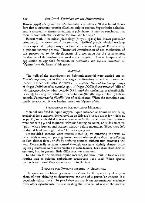
Brown (1950) neatly summarizes the criteria as follows: 'If it is found there-fore that a structural protein dissolves only in sodium hypochlorite solution,and is secreted by tissues containing a polyphenol, it may be concluded thatthere is circumstantial evidence for aromatic tanning.'
Recent work in helminth physiology (Smyth, 1951a) has drawn particularattention to the structure of the so-called 'vitelline' glands which have longbeen suspected to play a major part in the formation of egg-shell material bya quinone-tanning process. Theoretical consideration of the mechanism ofthis process led to the development of a technique for the cytochemicallocalization of the oxidase concerned in such a system. This technique and itsapplication to egg-shell formation in helminths and byssus formation inMytilus form the basis of this paper.
MATERIAL
The bulk of the experiments on helminth material were carried out onFasciola hepatica, but in the later stages confirmatory experiments were ex-tended to other helminths, as follows: Trematoda, Haematoloechus sp. (lungsof frog); Dolichosacchus rastulus (gut of frog); Diclidophora merlangi (gills ofwhiting); pseudophyllidean cestode, Schistocephalns solidus (matured artificiallyin vitro), by using the cellulose tube technique (Smyth, 1954); tetraphyllideancestode, Proteocephalus filicollis (gut of stickleback). When the technique wasfinally established, it was further tested on Mytilus edulis.
PREPARATION OF FROZEN-DRIED MATERIAL
Material was fixed in liquid oxygen (liquid nitrogen or liquid air not beingavailable) for 1 minute, dehydrated in an Edward's tissue dryer for 3 days at—400 C , and embedded in wax in a vacuum by the usual procedure. Sectionswere cut at 7-5 JX and mounted, without floating on water, on slides smearedlightly with albumen and warmed slightly before mounting. Slides were leftto dry, at least overnight, at 40° C. in a drying oven.
Frozen-dried sections were treated either (a) by removing the wax, asusual, with xylene, and passing down the alcohols—sections thus treated beingin fact alcohol-fixed; or (b) by staining sections without first removing thewax. Occasionally sections stained through wax gave slightly sharper cyto-logical pictures or were more reactive to cytochemical tests than alcohol-fixedsections, but, in general, little difference was apparent.
In addition to the freezing-drying method, the usual routine fixatives androutine wax or gelatine embedding procedures were used. When specialmethods were used they are referred to in the text.
EVIDENCE FOR QUINONE-TANNING IN HELMINTHS
The question of obtaining concrete evidence for the specificity of a cyto-chemical test claiming to demonstrate the site of a particular enzyme is apeculiarly difficult one. The proof depends mainly on circumstantial evidencefrom other cytochemical tests indicating the presence of one of the normal

Demonstration of Polyphenol Oxidase 141
substrates of the enzyme, as well as theoretical considerations as to the likeli-hood of the enzyme having the distribution found. It is, therefore, importantto establish that the helminth tissue is positive for the criteria for quinone-tanning systems established by Brown (1952) and mentioned earlier'.
The eggs of the majority of trematodes and pseudophyllidean cestodes arecolourless when laid, but turn brown or 'tan' on exposure to air; such eggsmay be bleached by treatment with sodium hypochlorite. This immediatelyindicates protein tanning of some kind. The pseudophyllidean cestode,Schistocephalns solidus, when matured in vitro under anaerobic conditions,produces normal eggs which tan on exposure to air. When cultured underaerobic conditions (i.e. with a stream of air bubbling through the culturemedium) normal eggs are not produced, the so-called 'vitellaria' become brownin colour and strings of brown material emerge from the uterine pore (Smyth,1950). This result is interpreted as being due to the pre-tanning of the egg-shell material while still within the vitelline glands.
The histochemistry of the vitelline glands of a number of platyhelminthsreveals that these glands are rich in proteins and polyphenols, according tothe work of Vialli (1933, 4), Stephenson (1947), and Nurse (1950). Stephen-son's results with Fasciola have been fully confirmed in this laboratory, byusing mainly frozen-dried material, and applying the most critical histo-chemical tests available. A number of additional observations have also beenmade on other forms. Although the entire range of tests available has notbeen applied in every case, and although many of the tests alone are notspecific, the accumulated evidence as summarized in Table 1 (see end ofpaper) leaves little doubt, on the whole, that the vitelline gland cells are richin proteins and polyphenols. With frozen-dried material, the histochemicaltests for both phenols and proteins which do not require severe techniques(e.g. Millon's) are particularly sharp. An exception is the so-called ferricchloride test for polyphenols, much quoted by previous authors in quinone-tanning studies. In this test the presence of an o-diphenol is supposed to bedemonstrated by the appearance of a green colour with dilute ferric chloride(Lison, 1936), turning red on treatment with 2 per cent, potassium carbonateor purple with dilute ammonia. Microscopically, green is a most unsatis-factory colour to observe, and I have never been able to obtain a colour whichcould be called green with the certainty essential for histochemical tests. Thered colour, after alkaline treatment, is, however, readily obtained and for thisreason the ferric chloride test has been taken to be positive. Of the remainingphenol tests, the argentaffin, chromaffin, ammonium molybdate, and sodiumiodate tests readily give strongly positive reactions. Although no single oneof these is specific, taken as a group, together with a positive ferric chloridereaction (after alkali treatment), they provide strong evidence for the presenceof o-diphenols. Of the protein tests, the Millon works particularly brilliantlywith frozen-dried material, but is •non-specific as the red colour produced isalso given by diphenols. The xanthoproteic test is always positive but, again,is not entirely specific for proteins. The ninhydrin test was first attempted

142 Smyth—A Technique for the Histochemical
on poorly hardened material and was only weakly positive. On the samematerial, the Sakuguchi and Biuret tests were negative. On theoreticalgrounds, this result was not unexpected as a secretory protein of this kind islikely to be in a highly labile and probably soluble state and would easily belost in unhardened material. All these tests proved strongly positive in materialthat had been fixed in 5 per cent, formaldehyde-saline, embedded in gelatine,hardened for 8 weeks in formalin vapour, and cut as frozen sections. Baker'selegant modification (1947) of the Sakuguchi arginine test gave a particularlystrong reaction with gelatine sections. The Biuret test, likewise, gave astrongly positive result, the granules appearing rose-pink, a colour indicativeof lower proteins.
Only in the trematodes have tests for both phenols and proteins beencarried out. In the turbellarians and cestodes, only tests for phenols have beencarried out and positive results reported—except for a few results for Polycelis.The circumstantial evidence for quinone-tanning in these groups is, however,considerable.
THE CATECHOL TECHNIQUE FOR DETECTION OF POLYPHENOL OXIDASE
IN TISSUES
Theoretical basisThe essential reactions in a quinone-tanning system may be summarized
as follows: , , , . ,polyphenol oxidase
protein+diphenol > protein-f-quinone= tanned protein (sclerotin).
In Fasciola rapid auto-tanning of sections does not occur (but see p. 144);so it can be assumed that although the polyphenol, enzyme, and protein con-stituents occur in the same globule (fig. 1), some blocking mechanism prob-ably prevents the interaction of the phenol and enzyme.
Incubation of tissue containing such a system, with catechol as a substrate,should result in the formation of o-quinone at the sites of the enzyme; thisquinone should then rapidly combine with and tan the adjacent protein togive rise to a reddish-brown colour. A prerequisite of such a technique wouldbe that the enzyme should be preserved and the protein left still in a 'tan-nable' condition, i.e. with the imino and amino groups more or less intact.The reddish-brown colour should be very stable and only removable withsodium hypochlorite. The whole reaction should be inhibited by dilutecyanide.
Experimental results
In practice, the method has proved to be remarkably successful. The tech-nique finally adopted for freshly prepared frozen-dried sections of Fasciolawas as follows:
(1) Remove wax and bring down through alcohols to water.

Demonstration of Polyphenol Oxidase 143
(2) Incubate in 0-2 per cent, catechol (freshly prepared) at 400 C. for 15-30minutes.
(3) Dehydrate, as usual, in 70 per cent., 90 per cent., and absolute.(4) Treat with xylene and mount in balsam.
Within 2 minutes of incubation, with this technique, the globules in thevitelline cells became yellow, and after 10-15 minutes pinkish-red. Thevitelline cells so coloured stood out clearly against the remainder of the tissue
yolkglobules
developing shell globules" ' " itelline" globules)
C Mature cell
cytoplasm freeof granules
nucleus phenolic protei enzyme
D Fully developed shell globule (enlarged)
A Immature cell
FIG. 1. Development of the 'vitelline' cells of Fasciola as shown in frozen-dried materialstained in malachite green and counterstained in Gower's carmine. The large black dots
within the shell globules are bright green; nuclei and yolk globules are red.
(fig. 2, B). In sections incubated for long periods (about 12 hours), the vitel-line cells became darkish-brown and the remainder of the tissues greyish-brown, the whole almost having the appearance of a haematoxylin-stainedsection (fig. 2, F). Since, after several hours' exposure to air, catechol solu-tions become partly oxidized to quinone and this quinone solution will thenslightly tan any tissues containing protein, irrespectively of the presence of theenzyme, this result is to be expected. It is essential, therefore, to use freshlyprepared catechol solution for this test; a solution prepared in the morningmay be used throughout the same day, provided it be stored in a reasonablycool place. The development of the red colour in Fasciola sections was in-hibited by the addition of M/1000 KCN to the catechol solution. The redcolour was also destroyed by o-i per cent. NaOCl—both these results beingexpected on theoretical grounds. Under low-power observation the red colourof the vitelline globules appeared sharp, but at oil immersion levels it seldomappeared so, a failing shared with the great majority of cytochemical testsinvolving proteins. The colour occurred only in the larger and mature, or
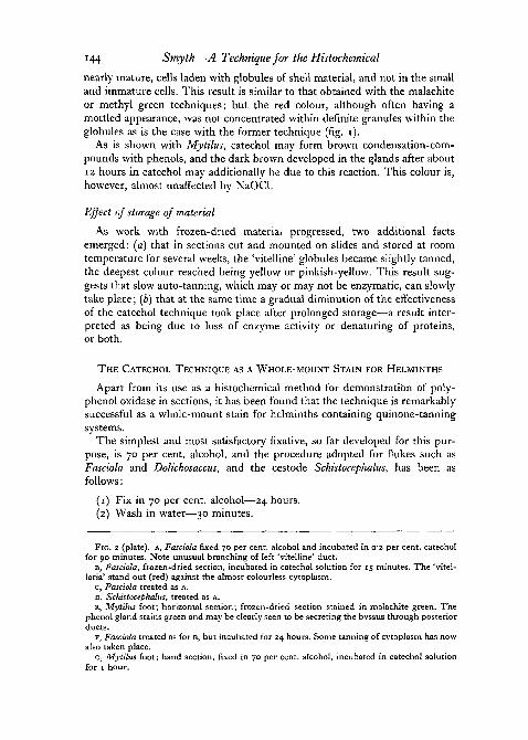
144 Smyth—A Technique for the Histochemical
nearly mature, cells laden with globules of shell material, and not in the smalland immature cells. This result is similar to that obtained with the malachiteor methyl green techniques; but the red colour, although often having amottled appearance, was not concentrated within definite granules within theglobules as is the case with the former technique (fig. i).
As is shown with Mytihis, catechol may form brown condensation-com-pounds with phenols, and the dark brown developed in the glands after about12 hours in catechol may additionally be due to this reaction. This colour is,however, almost unaffected by NaOCl.
Effect of storage of material
As work with frozen-dried material progressed, two additional factsemerged: (a) that in sections cut and mounted on slides and stored at roomtemperature for several weeks, the 'vitelline' globules became slightly tanned,the deepest colour reached being yellow or pinkish-yellow. This result sug-gests that slow auto-tanning, which may or may not be enzymatic, can slowlytake place; (b) that at the same time a gradual diminution of the effectivenessof the catechol technique took place after prolonged storage—a result inter-preted as being due to loss of enzyme activity or denaturing of proteins,or both.
THE CATECHOL TECHNIQUE AS A WHOLE-MOUNT STAIN FOR HELMINTHS
Apart from its use as a histochemical method for demonstration of poly-phenol oxidase in sections, it has been found that the technique is remarkablysuccessful as a whole-mount stain for helminths containing quinone-tanningsystems.
The simplest and most satisfactory fixative, so far developed for this pur-pose, is 70 per cent, alcohol, and the procedure adopted for flukes such asFasciola and Dolichosaccus, and the cestode Schistocephalus, has been asfollows:
(1) Fix in 70 per cent, alcohol—24 hours.(2) Wash in water—30 minutes.
FIG. 2 (plate). A, Fasciola fixed 70 per cent, alcohol and incubated in 02 per cent, catecholfor 90 minutes. Note unusual branching of left 'vitelline' duct.
B, Fasciola, frozen-dried section, incubated in catechol solution for 15 minutes. The 'vitel-laria' stand out (red) against the almost colourless cytoplasm.
c, Fasciola treated as A.D, Schistocephalus, treated as A.E, Mytilus foot; horizontal section; frozen-dried section stained in malachite green. The
phenol gland stains green and may be clearly seen to be secreting the byssus through posteriorducts.
F, Fasciola treated as for B, but incubated for 24 hours. Some tanning of cytoplasm has nowalso taken place.
G, Mytilus foot; hand section, fixed in 70 per cent, alcohol, incubated in catechol solutionfor 1 hour.

FIG. 2
J. D. SMYTH

Demonstration of Polyphenol Oxidase 145
(3) Incubate in 0-2 per cent, catechol (Analar, freshly prepared same day)—30-90 minutes.
(4) Wash in water—30 minutes.(5) Upgrade—70 per cent., 90 per cent., absolute. Pass through xylene
and mount in balsam.
Examples of specimens treated by this method are shown in fig. 2, A, c, D.In Fasciola, the 'vitelline' glands, 'vitelline' reservoir, and even the finest'vitelline' ducts show up with remarkable clarity. Unexpected branching ofthe transverse 'vitelline' ducts are revealed in some specimens (fig. 2, A).Small flukes, such as Dolichosaccus, only require 30 minutes' incubation inthe catechol solution, but Fasciola requires about 90 minutes'. The resultantcolour is red to brown, depending on the incubation time. Since the colouris due to the quinone-tanned protein, it is virtually indestructible in any ofthe reagents commonly employed in staining techniques; it is, of course,easily bleached by NaOCl.
After specimens have been 'tanned' in the above manner by catechol treat-ment, they may be additionally stained (after stage 4) by any of the routinenuclear stains used for whole mounts—borax carmine, paracarmine, &c.—inorder to stain the remainder of the genitalia not containing enzyme. It isrecommended that, as a routine, trematodes should be treated with catecholin this way before normal staining.
The method may also be used for whole mounts of cestodes (fig. 2, D), butit must be emphasized that it is applicable only to groups forming a quinone-tanned shell similar to that of the trematode egg. The histochemistry of thecestodes is not sufficiently well known, as yet, to define these groups. It hasbeen found that Schistocephalus solidus (Pseudophyllidea) and Proteocephalusfilicollis (Tetraphyllidea) tan readily by this method, although there is someevidence to suggest that in Proteocephalus, at least, the enzyme is more difficultto preserve than in Fasciola. The catechol technique greatly simplifies theworking out of the histological details of egg-shell formation. Thus, a hel-minth may be 'tanned1 by the catechol technique, dehydrated and treatedwith xylene, and the actual region where the eggs first appear will becomeapparent. This region may be cut out, embedded, and sectioned. The shell-material, already reddish in colour, may clearly be seen in sections and thecytological details of the egg-shell formation worked out.
CONFIRMATORY EXPERIMENTS ON MYTILUS
Although the results obtained with Fasciola and other helminths wereapparently conclusive, it was considered advisable to test the techniquefurther in an organism which was not a helminth and in which a similarquinone-tanning system was known to occur. Mytilus edulis has proved to bemost satisfactory for this purpose, for its byssus is formed from glandulartissue in the foot by a quinone-tanning process.

146 Smyth—A Technique for the Histochemical
A very full account of byssus formation in Mytilus has recently been givenby Brown (1952). As results obtained here differ in some points from hers,a summary of her findings is given below. For previous work on byssus-formation, reference may be made to her paper.
According to Brown, the byssus is formed in the posterior groove of thefoot from the secretions of two glands: the 'white gland' which suppliesprotein, and the 'purple' gland which supplies the polyphenol constituentresponsible for the tanning; the latter was found to be insoluble in alcohol,and there is evidence that it may be a phenolic amino-acid or protein.
anterior side
ciliated ductdraining glan
.posterior side
site of polyphenol twcidase(enzyme gland)
-white'(protein?)glani
purple (phi
1mm.B
FIG. 3. Histochemical reactions of Mytilus foot. A is a reproduction of a figure by Brown(1952), as re-interpreted here. B is a transverse section at the point X-X in A. c shows
histochemical reactions.
Brown believed that the protein material, after being secreted into thegroove, is coated on the outside by a layer of phenol which forces itselfthrough the groove epithelium 'to form a border to the mass of white glandmaterial lying in the groove'. Unfortunately, the excellent photomicrographsin her paper are not provided with legends, so that it is difficult to obtain acompletely clear picture as to her interpretation of the byssus formationprocess.
Histochemistry of the foot, and application of catechol technique
Histochemical studies of the foot have revealed the presence of three regionsas seen in transverse section. These are shown in fig. 3. Fig. 3, A, is a re-draw-ing of Brown's figure with a re-interpretation. The histochemistry of thesethree regions is as follows:
Upper region. After catechol incubation of frozen-dried sections or alcohol-fixed hand-sections of the foot, a red region appears on each side of theposterior groove (fig. 2, G). The development of this red colour is inhibitedby M/1000 cyanide. The colour is also destroyed by sodium hypochlorite,and it is concluded that it represents the site of polyphenol oxidase activity.

Demonstration of Polyphenol Oxidase 147
According to Brown (1952) this region is merely part of the phenol glandsecreting into the groove. This view is difficult to understand, as there is novisible connexion between it and the phenol gland proper. We agree with herthat this region gives a positive argentaffin reaction, but it also gives a parti-cularly strong and unmistakable reaction with Millon's reagent and also withninhydrin. But since polyphenol oxidase is well known to be a copper protein,this result is to be expected.
Middle region. This is the 'white' or 'protein' gland of Brown. Accordingto her, this gland 'gave all the protein colour reactions but did not give anyof the polyphenol reactions'. Unfortunately, she did not give details as to therange of tests covered by this general statement. All the common proteintests have been carried out on this region, on both frozen-dried material andgelatine-embedded material. The Millon test was completely negative: sincethis test works exceptionally well with frozen-dried material, it may definitelybe concluded that tyrosine is lacking. Of the other protein tests, the Biuret,ninhydrin, and Sakaguchi tests invariably gave negative results or resultswhich were questionable. The strongly positive protein reactions given in thesame section by the upper and lower glands were never obtained. With theresults on Fasciola in mind, the tests were repeated on gelatine-embeddedmaterial hardened in formalin for 2 weeks. Again, the results were consistentlytoo indefinite to be considered positive.
Lower region. This is the 'purple' or 'phenol' gland of Brown. The resultsof histochemical tests in this region agreed with Brown's except that, again,a green colour was not obtained satisfactorily with ferric chloride, althoughafter subsequent alkaline treatment the red colour was produced. In addition,this region gave strongly positive protein tests—the ninhydrin and Millontests giving particularly brilliant reactions. These results thus confirm Brown'sview that the polyphenol is a phenolic protein or phenolic amino-acid. Aftercatechol incubation, this region became brown (in contrast to the enzymeregion, which coloured red), owing to the condensation of the catechol withthe phenol. This brown colour was remarkably stable and not easily affectedby sodium hypochlorite.
According to Brown's account, quoted earlier, the material from the phenolgland forces its way through the epithelium of the groove. That this inter-pretation is incorrect is evident from fig. 2, G, which is a horizontal section ofthe foot, showing clearly that the phenol gland opens by posterior ducts into thegroove in the region of the sucker-like depression. It is also evident from thissection that a large amount of material is being secreted by this gland, whichsuggests that it is playing a major role in the formation of the byssus. Furtheralong the groove the material appears thinned. The interpretation of theseresults is discussed later.
EFFECT OF PHENYLMETHANE DYES ON MYTILUS
It has been shown that the globules in the 'vitelline' glands of Fasciolahave a very marked affinity for methyl green, malachite green, and related

148 Smyth—A Technique for the Histochemical
dyes in neutral solutions, provided that an approved sample of dye is used(Smyth, 1951a, 19516, 1953). It was suspected that this phenomenon was insome way related to properties inherent in quinone-tanning systems, themost likely explanation being that the dye was held either by the protein orthe phenol constituent, which, since they occur in the same globule, couldnot be distinguished in Fasciola. Application of the method to sections ofMytilus have revealed a similar result. The lower (phenol) gland shows avery marked affinity for these dyes (fig. 2, E), the middle ('white') gland hasno affinity at all, and the upper (enzyme) gland has a slight, but constant,affinity. This result has been of considerable use in locating the phenol glandin difficult sections.
DISCUSSION
The catechol technique. From the evidence quoted earlier, i.e. effect ofKCN, NaOCl, and results of phenoland protein tests, there seems littledoubt that the red colour produced after catechol incubation is indicative ofoxidase activity of some kind. Since the catechol is oxidized to quinone, it isjustifiable to term the enzyme a polyphenol oxidase, provided the term is usedin a general sense, i.e. merely as a name for an enzyme that will oxidizecatechol. Such a term would not exclude an enzyme like tyrosinase which isalso capable of oxidizing diphenols under certain conditions (Sizer, 1953).The success of the technique in such widely differing organisms as Fasciolaand Mytilus is particularly satisfying and has provided striking experimentalconfirmation of the soundness of the theoretical background built up byprevious workers.
Since the colour produced by the catechol technique is due to the tanningof protein molecules, it is clear that it can never precisely indicate the enzymesite, but only the nearest protein molecule to such a site, although, on theo-retical grounds, it is likely that the differences between enzyme and proteindistribution would be on a molecular rather than a microscopic level. In anycase, owing possibly to slight diffusion, the colour under oil immersion isnever sufficiently intense to allow of more than general localization withinthe cell, and the red colour seen under low powei; is reduced to a ratherdiffuse apricot colour under oil immersion. For high-power work, then, thetechnique has its limitations.
Egg-shell formation in helminths. The catechol technique, when applied towhole helminths, suggests that a more rational approach to helminth stainingmay be possible. It also provides a biochemical approach to the problem ofinter-relationships between helminths, since forms which produce eggs byquinone-tanning may be very readily detected. There appears to be a widefield here for further investigation, as it is likely that many variations of thebasic principle of quinone-tanning will occur. For example, it is likely thatthere are marked differences between such forms as the anaerobic cestodeSchistocephalus and the trematode Haematoloechus which lives aerobically inthe lungs of the frog. Both tan readily in catechol and clearly produce eggs

Demonstration of Polyphenol Oxidase 149
by quinone-tanning processes. Since in Schistocephalus matured in vitro underaerobic conditions pre-tanning occurs in the vitelline cells, it may be assumedthat normally the phenolic and amino groups and the enzyme are in closecontact, but cannot react during life owing to lack of oxygen. In Haemato-loechus, on the other hand, presumably some inhibitory mechanism must bedeveloped to prevent premature tanning, since oxygen from the lung environ-ment would normally be readily available in life.
From the histochemical results described in this paper, together with someunpublished observations, some idea of the processes occurring in the so-called 'vitelline' cell is now beginning to emerge. This is illustrated in fig. 1.In the mature cell the shell globules are large and occur almost exclusivelyat the periphery. Since these globules react to both protein and polyphenoltests and redden after catechol incubation, it is clear that the protein, phenol,and oxidase constituents occur in the same globule. None of the histochemicaltests for either phenols or proteins has revealed any internal structure to theseglobules. Yet in frozen-dried sections of Fasciola stained in malachite green,it is found that instead of the globules showing a uniform affinity for green,the dye is held in large granules clearly visible within the globules. Thisobservation, which was first made on Fasciola, has been repeated in severalother trematodes with identical results. There is thus also morphologicalevidence for the complex structure of these globules. If, as the evidence fromMytilus suggests, malachite green is held by protein or phenolic material,these groups would appear to occur within definite areas of the globules. Itmay be that in helminths, as is suggested later for Mytilus, the phenol andprotein constituents are the same substance, and the shell substance wouldthus be basically phenolic protein. On this view, the green granules wouldbe phenolic protein and the unstained part of the globules, the oxidase.
Byssus formation in Mytilus. The observations made here suggest that theaccount of byssus formation put forward by Brown may not be correct.According to her interpretation, the 'upper' region of the foot (fig. 3, B) ispart of the phenol gland. It has been shown here that this region reddenson catechol incubation and is, therefore, the site of polyphenol oxidase. Itseems reasonable, therefore, to term this the 'enzyme' gland. Her conclusionthat it was part of the phenol gland is understandable, for near to the pointwhere its ducts open posteriorly into the groove, the phenol gland fills thebulk of the material on each side of the groove, and the 'enzyme' gland,which is large anteriorly, practically disappears (fig. 4).
Histochemical tests on the lower or phenol gland fully confirm Brown'sview that it is a phenolic protein. If the upper gland is secreting an enzymeand the lower gland a phenolic protein, what then is the function of the middlegland—the so-called 'white' gland? In contradiction to Brown, this regionhas been found to give only questionable protein reactions, even in materialsubjected to prolonged hardening, in contrast to the strong protein reactionsof the other two regions. If this region were pouring a protein secretion intothe lower part of the groove, strong protein reactions might be expected. Yet

150 Smyth—Histochemical Demonstration of Polyphenol Oxidase
the cells of this gland appear to some extent to be orientated towards the lowerpart of the transverse groove, which may indicate that they open into it,although even in the best preparations an actual secretion in this region has
byssus sbem
byssus glan(so-called)
posterior groove
enzyme glandsecrebing polyphenoxidase
foob
phenol gland
new byssus bhreadmoving under actionof cilia in groove
phenolic probeinbhread being secreteddown duct fromphenol gland
old byssus threads
line of abtachmentof byssus stem tomuscles
^ecently Formedsingle byssus bhread
attachment disc
.qlands secrebingabtachmenb disc
FIG. 4. Diagram to explain the method of byssus formation in Mytilus. The phenol glandsopen into the distal end of the groove in the foot, into which they pour a thread-like secretionof a phenolic protein. This secretion is drawn forwards along the foot groove by cilary actionand is covered by a secretion of polyphenol oxidase from the enzyme glands on each side ofthe groove. The attachment disk is secreted by cells lining a sucker-line depression near thelower end of the groove. When a byssus thread fills the entire length of the groove, it is
released except at the end attached to the byssus stem.
never been observed. There would appear to be two possible interpretationsof these results. First, the enzyme gland produces polyphenol oxidase, thephenol gland produces phenol, and the 'white' gland produces protein. Apartfrom overlooking the enzyme gland, this interpretation is close to that of Brown.

TA
BL
E I
Res
ult
s o
f his
toch
emic
al
test
s o
n t
he
'vit
ellt
ne'
cel
ls o
f pla
tyh
elm
inth
s.
-)-
= p
osi
tive
. A
b
lan
k in
dic
ate
s te
sts
no
t ca
rrie
d o
ut.
Neg
ati
ve r
esu
lts
ha
ve n
ot
bee
n r
epo
rted
Gro
up
TR
EM
AT
OD
A(D
igen
ea)
TR
EM
AT
OD
A(M
onog
enea
)
TU
RB
EL
LA
RIA
CE
ST
OD
A
Org
anis
m
Fas
ciol
a he
pati
ca
Hae
mat
oloe
chus
sp
.D
icli
doph
ora
mer
lang
i
Den
droc
oelu
m l
acte
um
Pol
ycel
is n
igra
Gun
da s
egm
enta
taL
epto
plan
a tr
emal
lari
s
Tri
aeno
phor
us n
odul
osus
Dip
hyU
obot
hriu
m e
rina
cei
Wor
ker
Via
lli
(193
3)
Ste
phen
son
(194
6)S
myt
h (t
his
pape
r)S
myt
h (t
his
pape
r)R
enni
son
(195
3)
Via
lli
(193
3)N
urse
(19
50)
Via
lli
(193
3)S
myt
h (t
his
pape
r)V
iall
i (1
934)
Via
lli
(193
4)
Via
lli
(193
3)V
iall
i (1
933)
affin
Tes
ts fo
r ph
enol
s
B 2
Tes
ts fo
r pr
otei
ns

152 Smyth—Histochemical Demonstration of Polyphenol Oxidase
If it were true, the 'protein' produced by the white gland would be endowedwith peculiar properties, for it is tyrosine-free, extremely labile, and soluble.This interpretation can be rejected for the reason that a protein sufficientlysoluble in water to disappear readily in histochemical tests is just as likelyto go into solution in sea water. Since the byssus remains insoluble, evenbefore it has become tanned and hardened, this view becomes untenable.
An alternative hypothesis is put forward—that the enzyme gland is pro-ducing a polyphenol oxidase and the phenol gland a phenolic protein, the'white' gland is merely considered to be a developmental stage of the enzymegland. On this view, the bulk of the byssus is secreted from the phenol glandsthrough ducts opening into the distal region of the foot groove. Here thebyssus material is wafted forwards and drawn out into fine threads which aremoulded by the groove and covered by a secretion of polyphenol oxidase enroute to the so-called byssus gland. When a byssus thread is formed alongthe whole length of the foot and an attachment disk secreted, it is released;the attachment disk becomes anchored to a rock and the proximal end of thebyssus thread firmly bound to the byssus stem embedded in the byssus gland(fig. 4). This view essentially suggests that it is a phenolic protein—withoutany further protein being added—that under enzyme action becomes tannedby the combination of the amino-portion and the quinone nucleus of adja-cent molecules. This process might well be termed 'auto-quinone tanning'.
This view that the byssus is made up, not of separate phenol and proteinconstituents, but of one phenolic protein is not unreasonable on theoreticalgrounds. Brown (1950) has already shown that the product of the phenolgland is insoluble in water—exactly the kind of material that would be suit-able for secretion into sea water.
I am grateful to the Medical Research Council of Ireland for a granttowards the expenses of this work.
REFERENCESBAKER, J. R., 1947. Quart. J. micr. Sci., 88, 115.BROWN, C. H., 1950. Ibid., 91, 331.
1952. Ibid., 93, 487.LiSON, L., 1936. Histochimie Animate. Gautier-Villars (Paris).NURSE, F. R., 1950. Nature, 165, 570.PRYOR, M. G. M., 1940. Proc. Roy. Soc. B, 128, 278.PRYOR, M. G. M., RUSSELL, P. B., and TODD, A. R., 1946. Biochem. J., 40, 627.RENNISON, B. D., 1953. Research Thesis (unpublished). University of Dublin.SIZER, I. W., 1953. Advances in enzymology. Interscience Pub. (London).SMYTH, J. D., 1950. J. Parasit., 36, 360.
1951a. Nature, 168, 322.19516. Stain Tech., 26, 255.J9S3- Quart. J. micr. Sci., 94, 243.1954- Exp. Parasitology, 3, 64.
STEPHENSON, W., 1947. Parasitology, 38, 128.VIALLI, M., 1933. Boll. zool. pub. Ital. (Naples), 4, 135.
1934. Ibid., 5. zi.