Embed Size (px)
Citation preview

A TOUR O
F TH
E CELL
CHAPTER 6

ALL ORGANISMS ARE MADE UP OF CELLS (6.1)
Objectives
1. Explain the main ideas of the cell theory.
2. Describe how microscopes aid the study of cells.
3. Compare and contrast animal cells and plant cells.
4. Distinguish between prokaryotic and eukaryotic cells.
Key Terms
cell theory micrograph organelle plasma membrane nucleus cytoplasm cell wall prokaryotic cell eukaryotic cell
2HTTP://WWW.YOUTUBE.COM/WATCH?V=BTICXXXZQA4
Cell song

3

The Cell Theory
Cell theory: generalization that
-all living things are composed of cells
- cells are the basic unit of structure and function in living things
-all cells come from pre-existing cells
Microscopes as Windows to Cells-Light microscopes (100X)
-Electron microscopes (1,000,000)
-Scanning electron microscope (SEM)
-Transmission electron microscope (TEM)
Micrograph A photograph of the view through a microscope
4

5
Pg.111

6
Two major classes of Cells
prokaryotic cell: cell lacking a nucleus and most other organelles
eukaryotic cell: cell with a nucleus (surrounded by its own membrane) and other internal organelles
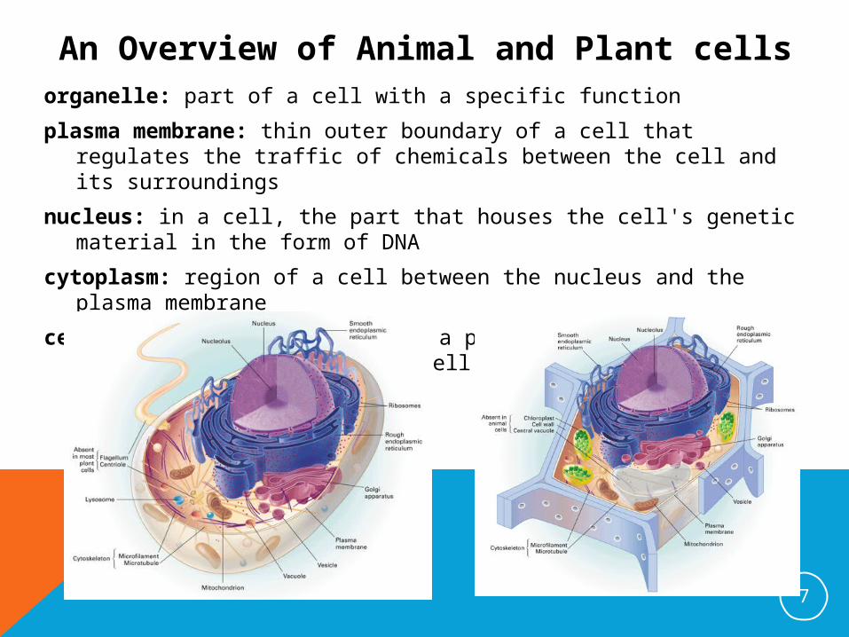
An Overview of Animal and Plant cellsorganelle: part of a cell with a specific function
plasma membrane: thin outer boundary of a cell that regulates the traffic of chemicals between the cell and its surroundings
nucleus: in a cell, the part that houses the cell's genetic material in the form of DNA
cytoplasm: region of a cell between the nucleus and the plasma membrane
cell wall: strong wall outside a plant cell's plasma membrane that protects the cell and maintains its shape
7


MEMBRANES ORGANIZE A CELL’S ACTIVITIES (6.2)
Objectives
1. Describe the structure of cellular membranes.
2. Identify functions of proteins in cellular membranes.
Key Term phospholipid bilayer
10HTTP://WWW.YOUTUBE.COM/WATCH?V=BTICXXXZQA4
Cell song


Membrane Structure-Read paragraph 1
-Membranes help keep the functions of a eukaryotic cell organized
-Membranes regulate the transport of substances across the boundary
-composed mostly of proteins and a type of lipid called phospholipids. |
-Phospholipids two hydrophobic fatty acids at one end (the tail) The other end (the head) of the molecule includes a hydrophilic phosphate group (PO43-)
12
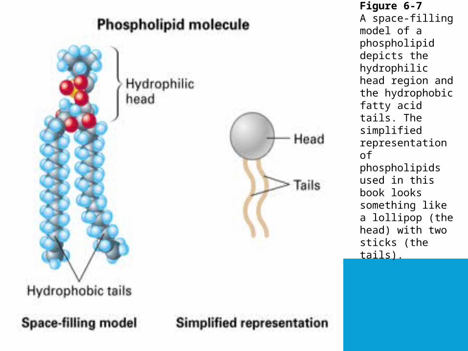
Figure 6-7A space-filling model of a phospholipid depicts the hydrophilic head region and the hydrophobic fatty acid tails. The simplified representation of phospholipids used in this book looks something like a lollipop (the head) with two sticks (the tails).
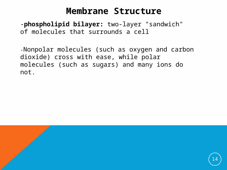
Membrane Structure-phospholipid bilayer: two-layer "sandwich" of molecules that surrounds a cell
-Nonpolar molecules (such as oxygen and carbon dioxide) cross with ease, while polar molecules (such as sugars) and many ions do not.
14


The Many functions of membrane proteins
Figure 6-9Many functions of the plasma membrane involve its embedded proteins. a. Enzymes catalyze reactions of nearby substrates. b. Molecules on the surfaces of other cells are "recognized" by membrane proteins. c. A chemical messenger binds to a membrane protein, causing it to change shape and relay the message inside the cell. d. Transport proteins provide channels for certain solutes.
16

Dissect a plasma membrane.
HTTP://WWW.YOUTUBE.COM/WATCH?V=BTICXXXZQA4Cell song

Monday 11.14.11
Period 1 (9:20-10:00)
1. Quiz
2. Handback Cellquest, Lab sheet
3. Check Egg, soak in colored water
Period 2 (10:00-10:57)
1. Observing Cells Lab
H/W
OUTLINE 6-3 (Do 6-3 online activity)
18

19

MEMBRANES REGULATE THE TRAFFIC OF MOLECULES (6.3)
Objectives1. Relate diffusion and equilibrium.2. Describe how passive transport occurs.3. Relate osmosis to solute concentration.4. Explain how active transport differs from passive
transport.5. Describe how large molecules move across a
membrane.
Key Terms
diffusion equilibrium selectively permeable membrane passive transport facilitated diffusion osmosis hypertonic hypotonic isotonic active transport vesicle exocytosis endocytosis
20
H/W-OUTLINE 6-4 (Do 6-4 online activity)
HTTP://WWW.YOUTUBE.COM/WATCH?V=BTICXXXZQA4Cell song

21

Diffusion-(air freshener)
-diffusion
-equilibrium
22
Figure 6-11Dye molecules diffuse across a membrane. At equilibrium, the concentration of dye is the same throughout the container.

Passive Transport-selectively permeable membrane:
-passive transport:
-facilitated diffusion
23
Figure 6-12Both diffusion and facilitated diffusion are forms of passive transport, as neither process requires the cell to expend energy. In facilitated diffusion, solute particles pass through a channel in a transport

Osmosis-osmosis
-hypertonic
-hypotonic
-isotonic
24
Figure 6-13A selectively permeable membrane (the bag) separates two solutions of different sugar concentrations. Sugar molecules cannot pass through the membrane.
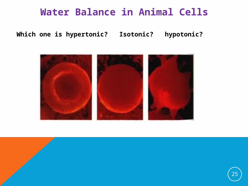
Water Balance in Animal Cells
Which one is hypertonic? Isotonic? hypotonic?
25

Water Balance in PlantCells
26

Active Transport-active transport:
27
Figure 6-16Like an enzyme, a transport protein recognizes a specific solute, molecule or ion. During active transport, the protein uses energy, usually moving the solute in a direction from lesser concentration to greater concentration.

Active Transport
-vesicles:
-exocytosis:
-endocytosis:
28
Figure 6-17Exocytosis (above left) expels molecules from the cell that are too large to pass through the plasma membrane. Endocytosis (below left) brings large molecules into the cell and packages them in vesicles.

29

30

Exit Slip
exocytosis
diffusion
facilitated diffusion
endocytosis
osmosis
31
H/W-OUTLINE 6-4 (Do 6-4 online activity)-Bring in thick liquid for your eggs
THE CELLS BUILDS A DIVERSITY OF PRODUCTS (6.4)
Objectives1. Identify the role of the nucleus in a cell. 2. Describe how the functions of ribosomes, the
endoplasmic reticulum, and the Golgi apparatus are related.
3. Distinguish between the functions of vacuoles and lysosomes.
4. Summarize the path of cellular products through membranes.
Key Terms
nuclear envelope nucleolus ribosome endoplasmic reticulum Golgi apparatus vacuole lysosome
32
H/W-OUTLINE 6.5 AND 6.6 (No online activity)
http://www.ted.com/talks/david_bolinsky_animates_a_cell.htmlCell Animation (6:55)

33

Structure and Function of the Nucleus
-nuclear envelope
-nucleolus
34
Figure 6-18A cell's nucleus contains DNA—information-rich molecules that direct cell activities.

Ribosomes
-Ribosomes
35
Figure 6-19A ribosome is either suspended in the cytoplasm or temporarily attached to the rough endoplasmic reticulum (ER). Though different in structure and function, the two types of ER form a continuous maze of membranes throughout a cell. The ER is also connected to the nuclear envelope..

The Endoplasmic Reticulum
-The Endoplasmic Reticulum
-Smooth ER
-Rough ER
36
Figure 6-19A ribosome is either suspended in the cytoplasm or temporarily attached to the rough endoplasmic reticulum (ER). Though different in structure and function, the two types of ER form a continuous maze of membranes throughout a cell. The ER is also connected to the nuclear envelope..

The Endoplasmic Reticulum
-The Endoplasmic Reticulum
-Rough ER
37
Figure 6-20Some proteins are made by ribosomes (the red structure) on the rough ER and packaged in vesicles. After further processing in other parts of the cell, these proteins will eventually move to other organelles or to the plasma membrane.

The Golgi Apparatus
-The Golgi Apparatus
38
Figure 6-21Golgi stacks receive, modify, and dispatch finished products.

Vacuoles
-Vacuoles
39
Figure 6-22
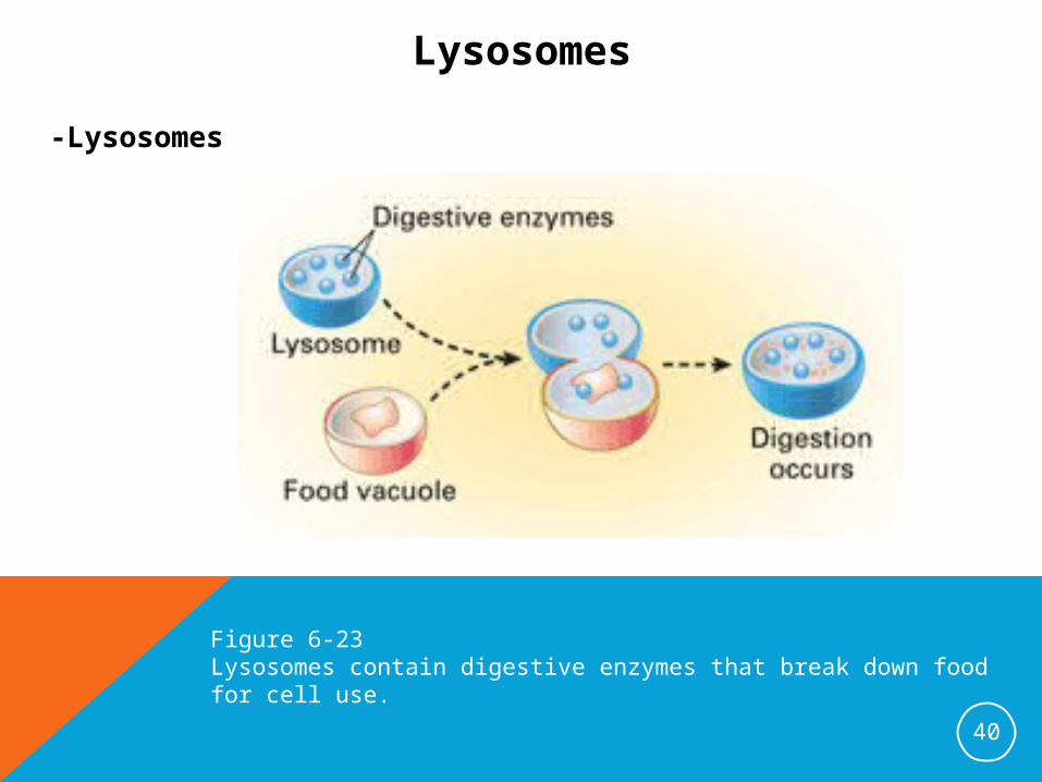
Lysosomes
-Lysosomes
40
Figure 6-23Lysosomes contain digestive enzymes that break down food for cell use.

Membrane Pathways in a Cell
41
Figure 6-24Products made in the ER move through membrane pathways in a cell.

42

Exit Slip
Describe the function of the organelles below
nuclear envelope
nucleolus
ribosome
endoplasmic reticulum
Golgi apparatus
vacuole
lysosome
43
H/W-OUTLINE 6.5 AND 6.6 (No online activity)
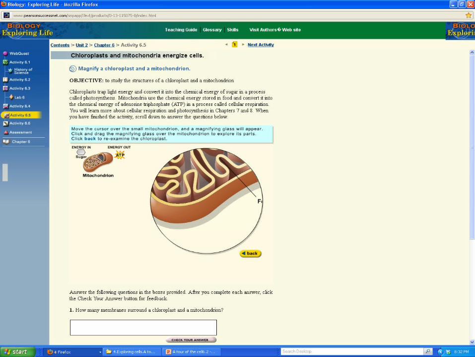
CHLOROPLASTS AND MITOCHONDIA ENERGIZE CELLS (6.5)
Objectives1. Compare and contrast the functions of chloroplasts
and mitochondria.
Key Terms
chloroplast mitochondria ATP
44
H/W-Chapter 6 worksheet-Prepare to present your “egg” periments

45
46
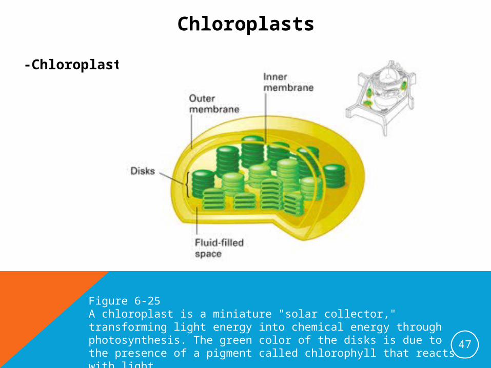
Chloroplasts
-Chloroplasts
47
Figure 6-25A chloroplast is a miniature "solar collector," transforming light energy into chemical energy through photosynthesis. The green color of the disks is due to the presence of a pigment called chlorophyll that reacts with light.

Mitochondria
-Mitochondria
-ATP
48
Figure 6-26Cellular respiration in the mitochondria releases the energy that drives a cell. The many folds of each mitochondrion's inner membrane are the sites of ATP production.

AN INTERNAL SKELETON SUPPORTS THE CELL AND ENABLES MOVEMENT
(6.6)Objectives
1. Describe the role of the cytoskeleton in cell movement.
2. Compare and contrast the functions of flagella and cilia.
3. Explain why a cell can be described as a coordinated unit..
Key Terms
microtubule microfilament flagella cilia
49
H/W-Chapter 6 worksheet-Prepare to present your “egg” periments
HTTP://WWW.YOUTUBE.COM/WATCH?V=BTICXXXZQA4Cell song

50

The Cytoskeleton-microtubules
-microfilaments
51
Figure 6-27

Flagella and Cilia-Flagella
-Cilia
52
Figure 6-28

The Cell as a Coordinated Unit -Flagella
-Cilia
53
Figure 6-29

54

Exit Slip
1.Explain why a cell can be described as a coordinated unit..
55
H/W-Chapter 6 worksheet-Prepare to present your “egg” periments
http://www.youtube.com/watch?v=2wYJKmmLrbQ&feature=fvwrel
Longer animation (music)http://www.youtube.com/watch?
v=uGK9CYetCvM&feature=relatedLonger animation (lecture)