Embed Size (px)
Citation preview

Thomas Jefferson University Haploidentical HSCT for Patients with High Risk Disease Kimmel Cancer Center
1 December 22, 2015 Version 2.3
A Two Step Approach to Allogeneic Hematopoietic Stem Cell Transplantation for High-Risk Hematologic Malignancies Using One Haploidentical Donor
IRB # 13D.352
Principal Investigator: Dolores Grosso, DNP, CRNP
Co-PI: Neal Flomenberg, MD
Co-Investigators: Seyfettin Onder Alpdogan, MD Matthew Carabasi, MD Joanne Filicko-O’Hara, MD Margaret Kasner, MD Ubaldo Martinez, MD John L. Wagner, MD Sameh Gaballa, MD Neil Palmisiano, MD
Radiation Oncologist: Wenyin Shi, MD, PhD Maria Werner-Wasik, MD
Biostatistics: Inna Chernovena, PhD
Protocol Written by: Dolores Grosso, DNP, CRNP
Thomas Jefferson University Haploidentical HSCT for Patients with High Risk Disease Kimmel Cancer Center
2 December 22, 2015 Version 2.3
Table of Contents 1.0 Introduction ……………………………………………………………………………...3 2.0 Objectives ………………….…………………………………………………………..15 3.0 Patient and Donor Selection …………………………………………………………15 4.0 Informed Consent ……………………………………………………………………..17 5.0 Treatment Plan ………………………………………………………………………..18 6.0 Laboratory Studies/Outcome Assessments ………………………………………..24 7.0 Supportive Care ……………………………………………………………………….26 8.0 Drug Information and Administration ………………………………………………..27 9.0 Patient Safety ………………………………………………………………………….31 10.0 Statistical Analysis …………………………………………………………………….33 11.0 References …………………………………………………………………………….34 12.0 Appendices …………………………………………………………………………….38

Thomas Jefferson University Haploidentical HSCT for Patients with High Risk Disease Kimmel Cancer Center
3 December 22, 2015 Version 2.3
1.0 Introduction Allogeneic hematopoietic stem cell transplant (HSCT) is a life saving therapy for patients with hematopoietic malignancies. The ability of HSCT to control an underlying hematologic malignancy is based on three variables, the intrinsic sensitivity/resistance of the malignancy, treatment regimen intensity, and graft versus tumor effects. Transplantation was initially developed as a treatment in patients with resistant leukemia. While the approach achieved short term control in many patients, relapse remained a problem in this patient group such that only 10-20% of patients became long term disease free survivors using matched sibling donors. Even today, long term disease free survival in patients with active leukemia at the time of transplant as reported by the CIBMTR is less than 20%. In contrast, patients transplanted in remission for similar diseases with identical conditioning regimens may achieve long term survivals of greater than 60%. Treatment intensity is near maximal in most transplant regimens. Higher doses of TBI or chemotherapy are associated with lower incidences of relapse, but usually at the price of more regimen related toxicity which limits overall gains in outcome. Disease sensitivity/resistance is not something that can be changed when patients present for treatment, and transplant regimen intensity can be further increased only over a narrow additional range. Consequently, it is difficult to manipulate these variables to effect substantial improvements in this group of high risk HSCT candidates. Graft versus tumor (GVT) effects may thus be the only variable which can be manipulated to address this problem. It is now understood that, in some diseases, a GVT effect, not regimen intensity, is the primary mechanism for long-term disease control after allogeneic transplantation. In other diseases, both treatment intensity and GVT effects contribute to disease eradication. This principle has been firmly established by analysis of transplant outcomes from identical twins, the success of reduced intensity HSCT1 and disease eradication after donor lymphocyte infusions.2 Unfortunately, despite the potent GVT effects associated with HSCT, death due to relapsed disease remains the greatest barrier to long-term survival for patients with resistant disease undergoing matched donor HSCT. As demonstrated in Table 1, the outcomes reported in several trials for patients in this category are dismal with overall survival (OS) rates consistently less than 30% for all the trials, and below 25% in many.

Thomas Jefferson University Haploidentical HSCT for Patients with High Risk Disease Kimmel Cancer Center
4 December 22, 2015 Version 2.3
Table 1 Outcome in Patients with Refractory Disease at the Time of Allo HSCT
One strategy to increase GVT effects for patients with resistant disease is with the use of a mismatched donor. Graft versus tumor effects in man have been elegantly
Disease Trial Overall Survival AML Kim et al., 2013, n=478, median age 38.5, primarily
myeloablative, matched related and URD, minority mismatched URD3
28% at 5 years for patients not in CR at HSCT. ↑relapse cause of death
Oyekunle et al., 2006, n= 25, median age 28, myeloablative, related & URD4
28% @ 5 years Relapse primary cause of death
Kebriaei et al., 2005, n=68, median age 42, primarily myeloablative & matched sib donors5
28% @ 10 years Relapse most frequent cause of death
Wong et al., 2005, n= 93, median age 49.5, myeloablative (41%) and non-myeloablative (59%) related & URD6
28% @ ½ year Relapse primary cause of death
Michallet et al., 2000, n=379, majority adult patients, myeloablative, majority matched related donors7
22% @ 5 years
AML arising from MDS
Alessandrino et al., 2008, n=127, median age 48, myeloablative (67%) and non-myeloablative (33%) matched sib & URD8
25% @ 5 years
MDS Lim et al., 2010, n=1,333, median age 56, 62% RIC, 38% myeloablative, matched URD 39%, matched sib 61%9
At 4 years: Age >60 27% Age 50-60 34% Relapse main cause of death
ALL Garderet et al., 2003, n=102, median age 17-18 years, myeloablative, all matched unrelated donors, 2 groups BM v PBSC10
21-32% leukemia free survival
Ringden et al. , 2009, n=4099 (ALL, AML, CML) 324 with intermediate or advanced ALL, matched related and unrelated donors, median age of whole cohort 37-38 years11
5-27% @ 5 years
Terwey et al., 2008, n=60, median age 29.1 years, primarily myeloablative, matched sib or unrelated donor12
28% @ 5 years
Lymphoma Bertz et al., 2002, n=25, mostly aggressive NHL & Hodgkin, median age 37, myeloablative and non-myeloablative, majority matched related & MUD 13
23% in patient with chemo resistant disease
Hamadani et al., 2009, n=46, aggressive NHL, median age 46, myeloablative, majority matched related 14
38% @ 5 years
Rigacci et al., 2012, n=165 patients relapsed after auto BMT for DLCL. Median age 43, 50% chemorefractory. 65% matched sib, 35% MUD15
Progression-free survival (PFS) at median 21 months was 32%
CLL Dreger et al., 2008, n=90, age <65. 47% Fludarabine refractory. 39% matched related, 61% matched URD, NM HSCT.16
3 year OS 42% whole cohort, chemorefractory lower
Khouri et al., 2002, N=28, myeloablative, majority matched related donors17
PFS 26% for those with refractory CLL @ 5 years
Myeloma Kröger et al., 2009, N=32, multiple myeloma achieving partial remission after HSCT given DLI, median age 50, majority non-myeloablative, related donor and URD 18
Progression-free survival 35% in those not achieving CR @ 56 months

Thomas Jefferson University Haploidentical HSCT for Patients with High Risk Disease Kimmel Cancer Center
5 December 22, 2015 Version 2.3
demonstrated in studies of transplants from identical twins which have shown much higher relapse rates than transplants from HLA identical siblings. The twin is presumably unable to mount a stronger GVT response than the patient him/herself. Conversely, unrelated donor (URD) transplants tend to have lower relapse rates than transplants from matched sibling donors because of the greater mismatching of non-inherited antigens. While HLA antigens and alleles can be matched for in URD HSCT, there is a much greater probability of minor histocompatibility (mHAg) and killer inhibitory receptor (KIR) mismatching resulting in greater recognition of non-self19 by the donor cells. Unfortunately, only about one in three patients will have an available well-matched unrelated donor and for many non Caucasians, the odds of finding a well-matched unrelated donor are considerably worse. HLA mismatching associated with haploidentical HSCT has been shown to potentiate graft versus tumor (GVT) effects because of the high degree of mismatch involved in this type of transplant. It has been demonstrated that the graft versus tumor effects associated with haploidentical HSCT are more potent than those from matched sibling HSCT.20-22 The use of haploidentical donors also broadens the application of HSCT because it is not limited by racial/ethnic HLA diversity. Thus haploidentical HSCT enfranchises segments of the population such as individuals of mixed racial ancestry as well as African Americans who, because of a higher degree of HLA diversity, are often without a well matched donor. Unfortunately, haploidentical HSCT has traditionally been associated with higher mortality rates as compared to transplants from well matched donors, limiting its application. The primary approach to haploidentical BMT has been to rigorously T cell deplete the graft to avoid severe GVHD. T-cell depletion techniques have been successful in decreasing GVHD, but higher rates of relapse, graft rejection and opportunistic infection (OI) due to the lack of T cells in the donor inoculum have resulted in increased mortality.23,24 Outstanding clinical results have been achieved with large doses of rigorously CD34 selected HSC by Ruggeri and associates,25 but their approach has not lent itself to widespread adoption at other centers. Additionally, relapse rates and treatment related mortality in patients entering transplant with active malignancy remain high using this approach. In recent years, administration of cyclophosphamide (CY) after a T replete (ie non T cell depleted) marrow graft in order to preferentially eliminate proliferating alloreactive T cells has been successfully utilized in non-myeloablative haploidentical HSCT.26, 27 With this approach, patients avoid profound immunoincompetence due to the remaining donor T cells which, because they are not alloreactive and proliferating early after transplant, are less affected by CY. While promising, this approach does not allow one to separately control the T cell and stem cell content of the transplant, as the T cells represent a varying number of passenger cells in the graft. In addition, depending on the patient’s age or disease at the time of transplant, a reduced intensity regimen is not always an optimal treatment strategy. An approach to myeloablative haploidentical BMT with low non relapse mortality serves as an ideal platform to explore the GVT effects associated with haploidentical HSCT.

Thomas Jefferson University Haploidentical HSCT for Patients with High Risk Disease Kimmel Cancer Center
6 December 22, 2015 Version 2.3
1.1 2-Step Myeloablative Haploidentical Transplantation To address this issue, we developed a 2-step myeloablative approach to HSCT from haploidentical donors which we have successfully applied to patients with hematologic malignancies. We refer to this as a 2-step approach because the lymphoid and stem cell portions of the graft are collected and administered at different time points during the conditioning regimen. Our approach does not involve ex vivo T cell depletion, but uses CY to tolerize donor lymphocytes. The separation of the myeloid and lymphoid portions of the graft allows us to use a fixed dose of donor T cells creating a consistent platform from which to compare outcomes. We believe this approach fulfills the need for an approach to explore GVT effects in a setting where regimen-related mortality is acceptable. In the initial 2 step trial (IRB #06U.20, 2006-2009), we used TBI (1.5 Gray x 8) and CY (60 mg/kg x 2) for conditioning. Tacrolimus and Mycophenolate Mofetil (MMF) were used as post transplant immunosuppression in a relatively standard fashion. The treatment schema is below. Patient Treatment Schema for Myeloablative Haploidentical BMT
Day -9
Tues -8
Wed -7
Thu -6 Fri
-5 Sat
-4 Sun
-3 Mon
-2 Tues
-1 Wed
0 Thu
AM TBI TBI TBI TBI Rest Rest CY CY Tacrolimus & MMF**
CD 34+ selected HSCT
HSCT Step 2
PM TBI TBI TBI
TBI Donor T
cell Infusion HSCT Step 1
TBI=Total Body Irradiation, DLI=Donor Lymphocyte Infusion, MMF=Mycophenolate Mofetil, HSCT=Hematopoietic Stem Cell Transplant As illustrated in the treatment schema above, donor lymphocytes infusions (DLI) containing a fixed dose of donor T cells, were given after the final fraction of TBI on day -6. In the phase I portion of the trial, the dose of T cells which resulted in consistent engraftment and the avoidance of severe GVHD was 2 x 10⁸/kg. This dose given to all patients in the trial as well as all of the subsequent patients treated on 2 step trials. CY was infused on days -3 and -2 with the intention of leaving at least 60 hours between administration of the DLI and the first dose of CY. This was to allow the alloreactive lymphocytes to become activated and thus more susceptible to elimination by CY but also to enable them to eliminate the remaining vestiges of host immunity and further debulk the malignancy. The HSC portion of the graft was administered 48 hours after the second dose of CY. In total, the regimen was two days longer than it would be if we

Thomas Jefferson University Haploidentical HSCT for Patients with High Risk Disease Kimmel Cancer Center
7 December 22, 2015 Version 2.3
were administering a conventional, one step transplant. For this trial, donors underwent 2 leukophereses for the DLI and then two additional leukophereses the following week for HSC collection. G-CSF was initiated the day after DLI collections were completed to avoid polarization of the T-cells in the DLI product to a TH2 phenotype. We found that donors tolerated the aphereses without appreciable toxicity. In this approach, T cells in the HSC portion of the graft are not exposed to CY and thus are potentially alloreactive. Our goal was to administer a CD34+ cell dose of 2-10x10e6/kg and a CD3+ cell dose of <5x10e4/kg. To minimize T cell content, the HSC portion of the graft was CD34 selected, using the Isolex device (Baxter). After CD34 selection, we incubated the CD34 cells in OKT3 and then washed out any unbound antibody. The goal was to coat any residual T cells with OKT3 which would lead to their lysis in vivo after infusion, but to avoid infusing free OKT3 which would compromise the T cells which had been rendered tolerant by CY. 1.1.1 Results of Our Initial Trial Patient Population: Between 2006 and 2009 twenty-seven patients, median age of 52 years (range 19-67), with high risk hematological malignancies were transplanted from haploidentical donors that were mismatched for HLA-A, B, C, and DR in the GVHD direction at 4 antigens (13), 3 antigens (11), and 2 antigens (2). One patient had no mismatches in the GVHD direction but was a 3 antigen mismatch in the rejection direction due to HLA homozygocity. It was subsequently decided that patients with mismatches only in the HVG direction should be treated on our matched related rather than haploidentical trial (ie that we would consider matching status from the GVH, not HVG perspective). Consequently, the study was amended to add one additional patient to the study group such that 27 patients were treated, rather than the initially planned 26. Diagnoses included MDS (n=2), AML (n=16), ALL (n=4), Biphenotypic Leukemia (n=1), CLL/Richter’s (n=1), NHL (n=2), and Aplastic Anemia (n=1). Fifteen of 27 patients (56%) had evidence of persistent disease at the time of transplant. All 27 had some high risk feature including high risk cytogenetics, secondary leukemia, progressive disease, or second or greater remission. Results: All patients are currently at least 4 years post HSCT and there have been no changes in DFS or OS since 2009. In this group, there were no deaths from GVHD and the cumulative incidence of grade III-IV GVHD was only 7.4%. Only 16% of patients developed chronic GVHD, and in all cases it was mild (NIH consensus criteria score of 1). Cumulative incidence of non-relapse mortality (NRM) was 22.6% with 3 deaths from infection and 3 deaths from regimen-related toxicity. Engraftment was consistent with this approach. Two patients experienced graft failure and died from complications of undergoing two condition regimens in close succession in a second engraftment attempt. These patients account for 2 of the 3 patients that died of regimen-related toxicity. The graft failures occurred in maternal recipients with HLA antibodies to their children/donors’ HLA antigens. The risk of rejection based on HLA antibodies in the myeloablative setting was not widely recognized at the time of this trial. In patients
Thomas Jefferson University Haploidentical HSCT for Patients with High Risk Disease Kimmel Cancer Center
8 December 22, 2015 Version 2.3
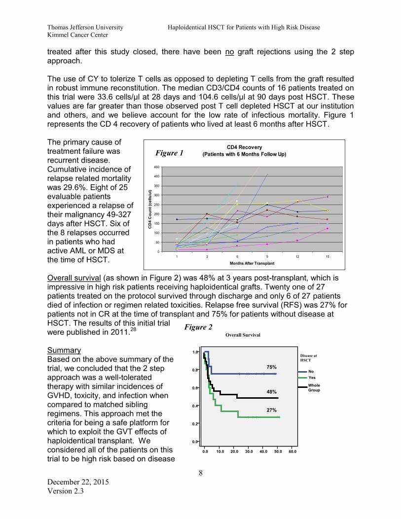
CD4 Recovery(Patients with 6 Months Follow Up)
0
50
100
150
200
250
300
350
400
450
1 3 6 9 12 15
Months After Transplant
CD
4 C
ount
(cel
ls/u
l)
treated after this study closed, there have been no graft rejections using the 2 step approach. The use of CY to tolerize T cells as opposed to depleting T cells from the graft resulted in robust immune reconstitution. The median CD3/CD4 counts of 16 patients treated on this trial were 33.6 cells/µl at 28 days and 104.6 cells/µl at 90 days post HSCT. These values are far greater than those observed post T cell depleted HSCT at our institution and others, and we believe account for the low rate of infectious mortality. Figure 1 represents the CD 4 recovery of patients who lived at least 6 months after HSCT. The primary cause of treatment failure was recurrent disease. Cumulative incidence of relapse related mortality was 29.6%. Eight of 25 evaluable patients experienced a relapse of their malignancy 49-327 days after HSCT. Six of the 8 relapses occurred in patients who had active AML or MDS at the time of HSCT. Overall survival (as shown in Figure 2) was 48% at 3 years post-transplant, which is impressive in high risk patients receiving haploidentical grafts. Twenty one of 27 patients treated on the protocol survived through discharge and only 6 of 27 patients died of infection or regimen related toxicities. Relapse free survival (RFS) was 27% for patients not in CR at the time of transplant and 75% for patients without disease at HSCT. The results of this initial trial were published in 2011.28 Summary Based on the above summary of the trial, we concluded that the 2 step approach was a well-tolerated therapy with similar incidences of GVHD, toxicity, and infection when compared to matched sibling regimens. This approach met the criteria for being a safe platform for which to exploit the GVT effects of haploidentical transplant. We considered all of the patients on this trial to be high risk based on disease
Figure 1
0.0 10.0 20.0 30.0 40.0 50.0 60.0
0.0
0.2
0.4
0.6
0.8
1.0
Cum
Disease at HSCT
No Yes
Overall Survival
27%
75%
48% Whole Group
Figure 2

Thomas Jefferson University Haploidentical HSCT for Patients with High Risk Disease Kimmel Cancer Center
9 December 22, 2015 Version 2.3
at HSCT, chromosomal abnormalities, the presence of secondary disease or chemo-resistance. The patients without disease at the time of HSCT, despite being high risk in other respects, have done very well in terms of relapse free survival which may relate to the degree of mismatch between donor and recipient. A second generation 2 step trial for patients without disease at the time of HSCT (IRB #10D.219) is currently completing accrual. With 26 of 28 patients accrued, the probability of OS is 80% with a median follow-up of 12.5 months (range 1-28 months). This second generation trial confirms the findings of the initial trial. Patients with controlled disease at HSCT have high OS rates after haploidentical HSCT using the 2 Step approach due to the low incidence of NRM. Figure 3 displays the current probability of OS for this second generation trial. 1.2 Relapse in Patients with Disease at the Time of HSCT
Relapse is the primary cause of treatment failure in patients with disease at the time of HSCT. This is a consistent finding in matched related and unrelated HSCT (as reviewed in Table 1) as well as haploidentical HSCT,29-31 including the initial 2 Step trial. In developing strategies to better treat this population we recognized that the ability to intensify the conditioning regimen further is very limited, and for patients presenting with resistant disease, there are little to no options to achieve remission prior to transplant. Therefore, new paradigms in treatment which further exploit an immunological basis for treatment, the only variable which can be further manipulated, were clearly needed.
1.2.1 2nd Generation 2-Step Approach for Patients with Active Disease at HSCT (IRB# 10D.06) To address this issue, we developed a 2nd generation 2 Step approach for patients with resistant disease at HSCT in which the timing of the T cell infusion within the context of the 2 Step platform was modified with the goal of immunologically increasing anti-tumor effects.
5.0 10.0 15.0 20.0 25.0 Months after HSCT
0.0
0.2
0.4
0.6
0.8
1.0
Figure 3- 2nd Generation 2 Step Patients in Remission at HSCT Probability of Survival-#10D.219
80%
% S
urvi
ving
SS
urvi
ving
Rel
apse

Thomas Jefferson University Haploidentical HSCT for Patients with High Risk Disease Kimmel Cancer Center
10 December 22, 2015 Version 2.3
Fever Post DLI
96
97
98
99
100
101
102
103
104
105
106
0 24 48 72 96 120 144
Hours Post DLI
Tem
pera
ture
(F)
CY #1
CY #2
The infusion of 2 x 10⁸/kg haploidentical donor T cells in the 2 Step approach results in their rapid alloactivation demonstrated by the relatively consistent emergence of high fever, diarrhea, and to a lesser extent skin rash within 24 hours of administration of the DLI. For all intents and purposes, this appears to be a hyperacute GVHD which disappears after CY administration as the alloactivated haploidentical lymphocytes die off. A graph of the fever response in 25 patients is shown in Figure 4. This has not been observed in studies using post-transplant CY where smaller doses of T cells were administered.26, 27 However, a similar phenomenon was observed in studies by Colvin and associates where similar doses of haploidentical DLI were administered.32 It is important to note that in the Colvin study, clinical remissions were seen despite the fact that all grafts were ultimately rejected which illustrates the potential of large doses of haploidentical DLI to target normal and malignant lympho-hematopoiesis. As cytotoxic lymphocytes can kill a target cell every 20 minutes, the ability to extend the period of time prior to CY administration by even another 24 hours has the potential to allow for tremendous additional immunologic cytoreduction. This extension may be the most beneficial to patients who because of their lower precursor frequency, experience peak donor T cell effector functions later than others. Therefore, extension of the allogeneic reaction has the potential to regularize and increase the immunological GVT effects of this regimen and provide a second mechanism of enhancing immunological treatment for resistant malignancy. In 2010, the 2nd generation 2 Step trial (IRB #10D.06) for patients with resistant disease at the time of HSCT was opened in which the interval between DLI and CY was increased by 24 hours from the initial Step trial. In previous work with CY tolerization in murine33 and human studies,27 CY was given 72 hours after transplant. Three days appears to be the optimal timeframe for CY administration in mice. In the 2nd generation trial, CY was given approximately 84 hours after HSCT with the rationale that there is a longer time required for cycling of human versus murine lymphocytes. The hypothesis of this trial is that the extra time of alloreactivity would result in greater tumor kill resulting in improved disease-free survival rates in patients with resistant disease. The 2nd generation 2 Step treatment schema for patients with resistant disease at the time of HSCT is shown below:
Figure 4

Thomas Jefferson University Haploidentical HSCT for Patients with High Risk Disease Kimmel Cancer Center
11 December 22, 2015 Version 2.3
TBI=Total Body Irradiation, CY-Cyclophosphamide, MMF=Mycophenolate Mofetil, HSCT=Hematopoietic Stem cell Transplant To date, 19 of 25 planned patients have been treated on this clinical trial. We have observed a 37% NRM rate (7/19) consisting of 4 deaths from infection, 1 death from GVHD, 1 death from veno-occlusive disease, and 1 death from cardiotoxicity related to heavy pretreatment and CY. Death from relapsed disease (RRM) occurred in 5/19 patients (26%), leaving 7/19 patients (37%) alive. All of the patients who are alive are disease free, 4-29 months after HSCT. In addition to the one death from GVHD, there have been two incidences of grade III GVHD (both hepatic), although a large percentage of patients have developed no acute GVHD at all. 1.2.2 Comparison of Outcomes of Patients with Disease at HSCT Initial versus 2nd Generation 2-Step Trial Fifteen patients on the initial trial had evidence of their disease at the time of HSCT. The NRM rate in this group was 33% (5/15), the RRM was 40% (6/15), with 27% (4/15 patients, 2 with lymphoma 2 with AML, alive and well all at least 4 years post HSCT. See below for a tabular comparison of the outcomes of these 2 trials (Table 2).
Because 6 more patients are needed to accrue to the current 2nd generation trial, and because the data from this trial is not mature, comparison to the outcomes from the initial trial are somewhat premature. However, because our investigator group is committed to the performance of HSCT on clinical trials, new clinical trials must be written prior to the existing ones closing. With this philosophy, no patient is treated off study and patient outcomes are universally reported. Therefore, based on the time constraints required for the writing and approval of a new trial, we have performed this interim analysis to guide us in the development of future therapy for this high-risk patient group. While RRM is mildly lower with the new trial, there is not a major difference in overall survival between the two trials. Our conclusion therefore is that a graft versus tumor (GVT) benefit may have been derived from adding
-10
-9
-8
-7
-6
-5
-4 -3
-2
-1
0
AM TBI TBI TBI TBI Rest Rest Rest CY 60 mg/kg
CY 60 mg/kg
Tacrolimus &MMF
CD 34+ selected HSCT PM TBI TBI TBI TBI
DLI
Table 2 Initial 2 Step Trial (TJU IRB #06U.20)
(Patients with Disease at HSCT only n=15) F/U 4-6.5 Years
2nd Generation 2 Step Trial
(TJU IRB # 10D.06) All Patients with Disease at
HSCT, n=19 Accrued to Date F/U 4-29 Months
Non-relapse Mortality 33% 37% Relapse-Related Mortality 40% 26% Disease Free Survival 27% 37%

Thomas Jefferson University Haploidentical HSCT for Patients with High Risk Disease Kimmel Cancer Center
12 December 22, 2015 Version 2.3
the extra day of alloreactivity between the DLI and the CY in the 2 step platform, but there is not sufficient evidence that the 2nd generation trial for patients with disease at HSCT has resulted in significant progress in terms of increasing OS rates for this population. Therefore, alternate strategies are required to increase OS in patients with resistant disease at HSCT. 1.3 Rationale and Hypothesis for the Current Trial A potentially important difference between remission and relapsed patients undergoing transplant is the percentage of GVT versus GVH reactive T cells that are likely to be rapidly activated in vivo and, as a result of this activation, subsequently eliminated by CY. In both remission and relapsed patients, the majority of GVH reactive T cells are likely to encounter an antigen presenting cell capable of activating them, thus rendering them more susceptible to CY. In the remission patient, with a small tumor burden, many GVT reactive T cells may not encounter a tumor cell or an antigen presenting cell capable of presenting tumor antigens during the first few days after infusion. As a result, a smaller percentage of GVT T cells will be activated early on, and, consequently, fewer GVT T cells are likely to be eliminated by CY than their more consistently activated GVH counterparts (Figure 5). As the tumor burden progressively increases, more and more GVT reactive T cells will encounter tumor cells during the first few days after infusion, thus becoming activated and subsequently eliminated by CY as well. The larger the tumor burden at the time of lymphocyte administration, the more the potential of CY to blunt the GVT effect, ultimately eliminating any differential impact compared to GVHD which may occur in remission patients. One simple solution to this problem would be to administer the T Cells closer to the true tumor nadir following the conditioning chemoradiotherapy. Currently, the DLI, containing 2 x 10⁸/kg T cells, is administered immediately after the last fraction of radiation. In this proposed trial, we will administer the T cells two days after radiation ends. This will allow a longer time period for tumor cells and antigen presenting cells capable of presenting tumor antigens to die off prior to the infusion of the DLI. Our hypothesis is that after the infusion, less donor GVT reactive T cells will become activated due to the reduction of these recipient stimulator tumor and antigen presenting cells, and by extension, less GVT reactive donor T cells will be vulnerable to subsequent eradication by CY. According to the original work done by Matsuzaki et al.,34 the induction of peripheral
Figure 5

Thomas Jefferson University Haploidentical HSCT for Patients with High Risk Disease Kimmel Cancer Center
13 December 22, 2015 Version 2.3
tolerance by CY of activated donor T cells results in intrathymic clonal deletion of the activated donor T cell population. While this deletion is favorable in the case of eradication of GVH reactive T cells,33 we wish to avoid the deletion of GVT reactive donor T cells in this trial. In order to estimate when tumor cells were likely to be at nadir, we determined when the maximal effects of TBI could be expected in the 2 Step regimen. Using white blood cell counts as surrogate markers for post HSCT nadir of hematopoietic cells, we examined the historical CBCs of patients with active AML at HSCT undergoing a 2 step HSCT on the current approach. See Table 3. Table 3-Historical WBCs During Conditioning in the Current 2 Step Approach
AM TBI 1.5 Gy
TBI 1.5 Gy
TBI 1.5 Gy
TBI 1.5 Gy
Rest Rest Rest CY 1
PM TBI 1.5 Gy
TBI 1.5 Gy
TBI 1.5 Gy
TBI 1.5 Gy
DLI
Median WBC (x 103)
Early AM COUNTS Patients
Treated on 10D.06 n=15
2.2
(Range 0.6-
10.3)
1.15
(Range 0.2-10.7)
1.0
(Range 0.1-8.1)
0.7
(Range 0.1-4.2)
0.6
(Range 0.1-3.4)
0.6
(Range 0.0-2.8)
0.2
(Range 0.0-0.8)
0.1
(Range 0.0-0.9)
Median WBC (x 103)
Early AM COUNTS Patients
Treated on 06U.20
n=8
0.95
(Range 0.3-1.7)
0.75
(Range 0.1-2.9)
0.35
(Range 0.1-1.2)
0.25
(Range 0.1-1.8)
0.15
(Range 0.1-2.3)
0.05
(Range 0.0-4.2)
X 0.1
(Range 0.0-0.5)
We found that patients treated on both trials had measurable hematopoiesis at the time of DLI infusion, with some patients having normal white counts at the time of the DLI, and all patients with evidence of normal hematopoiesis 2-3 days after DLI. This indicates that the DLI was infused well ahead of the post-TBI immune system nadir and by extension; the T cells were exposed to a potentially significant disease burden. Furthermore, ten of the 23 patients had peripheral blood AML blasts detectable after two days of radiation (6 Gy), and in many cases, the blasts persisted to the day of the DLI or after. All 10 of these patients died of relapsed disease. Conversely, of the remaining 13 patients who had no peripheral blood blasts or who had blasts that disappeared after 2 days of radiation, only 1 patient died of relapsed disease. The decreased relapse rates occurred despite the fact that this latter group had

Thomas Jefferson University Haploidentical HSCT for Patients with High Risk Disease Kimmel Cancer Center
14 December 22, 2015 Version 2.3
demonstrable AML in the marrow just prior to HSCT and all of these patients were at high risk for relapse. As peripheral blood blasts are a reflection of marrow hematopoiesis, the absence of or lack of detectable blasts early in the conditioning regimen for the patients who achieved sustained remission is a reflection of a lower disease burden in these patients. Therefore, the data suggests that even a small decrease in tumor burden at the time of the DLI can improve DFS, possibly due to a parallel decrease in the amount of GVT T cells that become activated by tumor and then later eliminated by CY. 1.4 Summary The goal of this clinical trial is to increase DFS rates in patients with resistant disease at the time of HSCT. In order to do so, a delay period will be inserted into the 2 step platform between the last dose of TBI and the infusion of donor T cells, to allow more of the tumor cells and antigen presenting cells capable of presenting tumor antigens to undergo TBI mediated elimination prior the infusion of donor T cells. This will hypothetically result in less activation of donor GVT reactive T cells and by extension, less vulnerability of these cells to elimination by CY. In the previous 2 Step platforms, 12 Gy of radiation was delivered in 1.5 Gy fractions twice daily over 4 days. In this new protocol, 12 Gy of TBI will be given in 2.0 Gy fractions twice daily over 3 days. This shorter TBI schedule is an alternate, standard TBI regimen for HSCT patients.35-38 The shorter radiation schedule will allow the radiation therapy to be completed in 3 days (Monday-Wednesday). Instead of administering the DLI immediately after the last fraction of radiation as is currently done, the DLI will be administered on Friday afternoon, approximately 48 hours after the last fraction of TBI. Comparison of historical white cell counts of patients treated on the standard 2 step approach (Table 3) to the historical white counts inserted into the proposed 2 step approach (Table 4) shows that the median immune system nadir will occur approximately 12 hours after the DLI in this proposed trial as compared to 60 hours in the historical 2 step approach. Table 4- Historical WBCs During Conditioning as Applied to the Proposed 2 Step Approach
AM TBI 2 Gy
TBI 2 Gy
TBI 2 Gy
Rest Rest
PM TBI 2 Gy
TBI 2 Gy
TBI 2 Gy
DLI
Median WBC (x 103)
Early AM COUNTS Patients
Treated on 10D.06 n=15
2.2
(Range 0.6-
10.3)
1.15
(Range 0.2-10.7)
1.0
(Range 0.1-8.1)
0.7
(Range 0.1-4.2)
0.6
(Range 0.1-3.4)
0.6
(Range 0.0-2.8)
0.2
(Range 0.0-0.8)
0.1
(Range 0.0-0.9)

Thomas Jefferson University Haploidentical HSCT for Patients with High Risk Disease Kimmel Cancer Center
15 December 22, 2015 Version 2.3
Median WBC (x 103)
Early AM COUNTS Patients
Treated on 06U.20
n=8
0.95
(Range 0.3-1.7)
0.75
(Range 0.1-2.9)
0.35
(Range 0.1-1.2)
0.25
(Range 0.1-1.8)
0.15
(Range 0.1-2.3)
0.05
(Range 0.0-4.2)
X 0.1
(Range 0.0-0.5)
We also note that patients with lymphoma will be treated on this protocol. While most of these patients do not have marrow based involvement of their malignancy, the additional time between the radiation and the donor GVT reactive T cell infusion will allow tumor cells in the lymph nodes as well as antigen presenting cells capable of presenting tumor antigens, to die off. As with marrow based disease scenarios, the activation and subsequent elimination by CY of donor GVT reactive T cells will also be avoided. The only difference is that the primary location of the malignant disease is different. 2.0 Objectives The objective of this phase II study is to decrease post HSCT relapse rates in patients with high risk hematological malignancies. Primary Objective:
1. To assess 1 year relapse free survival in high risk patients undergoing HSCT using the TJU 2-step approach with 2 days inserted between the last fraction of TBI and the infusion of donor T cells (DLI)
Secondary Objectives:
1. To assess regimen related toxicity in this updated conditioning regimen, GVHD incidence and severity, and overall survival in patients undergoing treatment on this protocol.
2. To assess the consistency and pace of engraftment 3. To assess the pace of T cell and B cell immune recovery
3.0 Patient and Donor Selection 3.1 Patient Selection Patient Inclusion Criteria:
1. This treatment is for patients with high risk hematologic malignancies, High risk is defined as:
Any patient with a hematologic malignancy with residual disease after treatment with 1 or more chemotherapy regimens in whom achievement of remission with additional chemotherapy is felt to be unlikely.
Patient without morphologic evidence of disease but when high risk features which would predict for relapse despite remission at HCST such as adverse cytogenetics, 3rd or greater CR, or failure to recover peripheral blood counts to normal ranges. While these patients do not have

Thomas Jefferson University Haploidentical HSCT for Patients with High Risk Disease Kimmel Cancer Center
16 December 22, 2015 Version 2.3
detectable disease by current methods, like all patients they have non-detectable disease which in their case is highly aggressive.
2. Patients must have one related donor who is HLA mismatched in the GVHD direction at two or more HLA loci.
3. Patient must have adequate organ function: LVEF of ≥ 50% DLCO (adjusted for hemoglobin) ≥ 50% of predicted and FEV-1 ≥ 50% Adequate liver function as defined by a serum bilirubin ≤ 1.8, AST or ALT
≤ 2.5 x upper limit of normal Creatinine clearance of ≥60 ml/min
4. Karnofsky Performance status of ≥ 80% on the modified KPS tool (see Appendix A)
5. Patients must be willing to use contraception if they have childbearing potential 6. Able to give informed consent
Patient Exclusion Criteria:
1. Modified KPA of < 80% 2. > 5 Comorbidity Points on the HCT-CI Index (See Appendix B) 3. Class I or II antibodies against donor HLA antigens 4. HIV positive 5. Active involvement of the central nervous system with malignancy 6. Psychiatric disorder that would preclude patients from signing an informed
consent 7. Pregnancy, or unwillingness to use contraception if they have childbearing
potential 8. Patients with life expectancy of ≤ 6 months for reasons other than their
underlying hematologic/oncologic disorder 9. Alemtuzumab treatment within 8 weeks of HSCT admission 10. ATG level of ≥ 2 ugm/ml 11. Patients with active inflammatory processes including T max > 101 or active
tissue inflammation are excluded 12. Inability to tolerate cyclophosphamide or undergo total body irradiation at the
doses specified in the treatment plan. The time of the required evaluations for transplant is reviewed in the Jefferson Blood and Marrow Transplant SOP CP:P043. 3.2 Donors Donors will be selected based on which donor in the donor pool is expected to be the most alloreactive. The current version of the donor selection tool will be utilized for the selection. The study binder for each patient will contain the alloreactivity point worksheets for each donor or donor pool, as well as documentation of haplotype analysis. All donors are selected and screened for their ability to provide adequate infection-free apheresis products for the patient in a manner that does not put the donor at risk for
Thomas Jefferson University Haploidentical HSCT for Patients with High Risk Disease Kimmel Cancer Center
17 December 22, 2015 Version 2.3
negative consequences. Donor selection will be in compliance with 21 CFR 1271 and TJU BMT Program SOP CP: P009.03. Specifically, donors will be tested, using the appropriate FDA-licensed and designated screening tests, for:
1. HIV, type 1 2. HIV, type 2 3. HBV (HBsAg, anti-HBc IgC and IgM) 4. HCV 5. Treponema pallidum 6. Human T-lymphotropic virus, types I and II 7. Cytomegalovirus 8. West Nile Virus 9. Trypanosoma cruzi
As per the Jefferson Blood Donor Center Quality Plan, all allogeneic donor testing samples (including HPC donors) will be sent to a laboratory that is FDA and CLIA licensed. Agreements/contracts for these services will be developed according to TJUH policies and all pertinent regulatory requirements will be retained by the Blood Bank. Additional donor testing may be performed as required to assess the possibility of transmission of other infectious and non-infectious diseases. TJUH HPC transplant personnel will discuss the potential for disease transmission from donor to recipient (i.e. the purpose of infectious disease testing) during the donor evaluation. Infectious disease testing must be completed by the time of the recipient’s transplant admission date. As per FACT guidelines, pregnancy will be assessed during the initial donor evaluation and just prior to the initiation of the recipient’s conditioning regiment in female donors of childbearing age. 4.0 Informed Consent Patients referred for the trial will have their eligibility criteria verified. On meeting the eligibility for the trial as outlined, informed consent will be obtained using forms approved by the Thomas Jefferson University Hospital Institutional Review Board and following guidelines related to the use of human subjects in research. The risks and hazards of the procedure, as well as alternative forms of therapy will be presented to the patient in detail. Patients will receive a signed copy of the consent form after the consent interview.

Thomas Jefferson University Haploidentical HSCT for Patients with High Risk Disease Kimmel Cancer Center
18 December 22, 2015 Version 2.3
5.0 Treatment Plan Proposed Schema for Partially-Matched Related HSCT - Patient -10 -9 -8 -7 -6 -5
-4
-3
-2
-1
0
AM 2 Gy TBI
2 Gy TBI
2 Gy TBI
Rest Rest Rest Rest CY 60 mg/kg
CY 60 mg/kg
Tacrolimus &MMF
CD 34+ selected HSCT PM 2 Gy
TBI 2 Gy TBI
2 Gy TBI
DLI
TBI=Total Body Irradiation, CY-Cyclophosphamide, MMF=Mycophenolate Mofetil, HSCT=Hematopoietic Stem Cell Transplant Proposed Schema for Partially-Matched Related HSCT - Donor -7 -6 -5 -4 -3 -2 -1 AM Lymphocyte
Collection Lymphocyte Collection
G-CSF G-CSF G-CSF G-CSF PBSC Collection
G-CSF PBSC Collection
PM G-CSF
G-CSF G-CSF G-CSF
G-CSF=Granulocyte Colony Stimulating Factor, PBSC=Peripheral Blood Stem Cell Collection 5.1 Administration of Immunosuppressive Agents during Conditioning There should be no administration of agents that suppress lymphocyte reactivity from admission until day -1 in this protocol. This includes steroids, calcineurin inhibitors, MMF, or monoclonal antibodies that affect lymphocyte number or function. Patients must be off steroids (aside from premedication for transfusion) for at least 7 days prior to admission. If patients have previously required steroids as a premedication for transfusion, they may receive a dose of steroid equivalent to 5 mg of prednisone on the first day of TBI. After this, no steroids at all should be given through day -1 of the transplant regimen. Diphenhydramine and meperidine may be used if necessary. Any use of steroids after the first day of TBI through day -1 should not be administered without approval from the PI. 5.2 TBI 2 Gy TBI will be administered twice daily for 3 days (6 fractions) on days -10 through -8. The daily fractions of TBI will be minimally separated by 7 hours, but ideally by 8 hours to reduce toxicity. TBI will be utilized for all patients eligible for this protocol. Prior irradiation will be evaluated by the radiation oncologist to define eligibility for this TBI schedule. In additional there may be technical or patient related factors which will require some minor modification in the TBI technique utitlized. Selected patient may require local boosting of certain organ sites prior to conditioning therapy. Deviations from the guidelines described here may only be performed with the approval of the radiation oncologists and the PI. See Appendix C for radiation guidelines.

Thomas Jefferson University Haploidentical HSCT for Patients with High Risk Disease Kimmel Cancer Center
19 December 22, 2015 Version 2.3
5.3 Donor Lymphocyte Infusions The dose of the donor lymphocyte infusion (DLI) will be based on CD3+ T cells per kilogram of recipient body weight. T-cell and progenitor cell doses and cyclophosphamide dosing will be based on adjusted dosing weight (40% the difference between actual and ideal body weight + the actual body weight). The donor T-cells will be collected prior to the use of G-CSF for progenitor cell collection. DLI specimen handling and labeling conventions will be performed in accord with the relevant AABB (American Association of Blood Banks) and/or FACT (Foundation for Accreditation for Cell Therapy) regulations and guidelines. All DLI specimens must be appropriately labeled in accord with these standards to be accepted by the Processing Laboratory. A valid prescription and request form must be submitted by the requesting physician. The following guidelines should be used to calculate the correct volume of blood to be obtained from the donor to achieve the target T-cell dose. An aliquot of the apheresis product will be assessed for CD3 content by flow cytometry. The following cell panel will be used:
FITC PE
IgG1 IgG1
IgG1 IgG2a
CD45 CD14 + CD13
CD3 CD4
CD3 CD8
CD3 CD16 + CD56
CD3 CD19
CD4 CD8
CD4 CD25+ FoxP3+
A gate is drawn around the entire CD45+ population. %WBC/total events = the percentage of CD45+ cells within this gate corrected for the isotype control. CD3 percentages are calculated, corrected for the isotype control, based on the total white cell (CD45+) gate, not based on a “lymphocyte gate”. There are 4 CD3 counts performed in the panel. The two median values are averaged to determine the final raw CD3 count. The raw CD3 count is then corrected for any counted events which are not WBC (i.e. CD45-), as follows:
Corrected %CD3 = (raw CD3 count)/(%WBC/total events). Total T-cells required for the initial infusion = (2x10⁸ T-cells/kg) * (Weight in kg) T-cells/ml of product

Thomas Jefferson University Haploidentical HSCT for Patients with High Risk Disease Kimmel Cancer Center
20 December 22, 2015 Version 2.3
T-cells/ml of product = (WBC) * (Corrected %CD3) Volume to be infused = (Total T-cells required for the initial infusion)/(T-cells/ml of product)
All donors will report for apheresis of lymphocytes the day before the planned DLI. If the targeted amount of lymphocytes is not collected on the first day, the donor will return for a second day of lymphocyte collection on the day of the DLI. Lymphocyte apheresis will be performed at Thomas Jefferson University Hospital or the American Red Cross, by trained apheresis personnel using standard techniques. No hematopoietic growth factors will be administered to apheresis donors prior to lymphocyte collection. The donor will have venous catheters placed in each arm for the purposes of undergoing leukopheresis. Leukocyte collections will be performed using a standard apheresis machine such as the Cobe Spectra apheresis instrument (Cobe Laboratories Inc., Lakewood, CO). For the donor lymphocyte apheresis, total blood volumes to be processed will be determined using the following calculation:
Recipient weight in kg: __________kg Multiply by desired CD3+ cells/kg: x __________x10(7)/kg Total CD3+ cells requested: = __________x10(7) Multiply by 2 x 2 TOTAL mononuclear cells (TMC) = __________x10(7) Divided by 100 x 10(&) TMC/L = _____ Liters processed
During the infusion of the DLI, the patient will be monitored for any untoward reactions. Each infusion will take place in the Bone Marrow Transplant Unit. Donor lymphocyte infusions will be administered by nursing staff experienced in the administration of blood products. 5.3.1 DLI Dosing 2 x 10e8/kg donor T cells will be collected from a single donor and infused approximately 48 hours after the last TBI fraction. Based on minor variations in donor collection and laboratory processing times, an exact time for the DLI infusion will not be prescribed by this protocol. The DLI will be infused in the afternoon on day -6. Because it is anticipated that the last fraction of TBI will be delivered 48 hours before the day of the DLI at approximately 4 PM, the optimal time for DLI infusion on day -6 is 4 PM. Infusion of the DLI prior to 3 PM on day -6, is prohibited. The DLI must be infused before day -5. DLI must NOT be irradiated. DLI should NEVER be administered through a leukocyte depletion filter. If blood filtration is necessary, the filter should be a standard blood product filter with pore size of at least 170 microns.
Thomas Jefferson University Haploidentical HSCT for Patients with High Risk Disease Kimmel Cancer Center
21 December 22, 2015 Version 2.3
5.4 Cyclophosphamide CY 60 mg/kg IV over 2 hours will be administered on days –3 and –2 of the conditioning regimen. Mesna 60 mg/kg continuous IV infusion over 24 hours X 2 doses will be administered on days –3 and -2. Day –1 is a day of rest. Voriconazole can block the conversion of CY to its active metabolite, 4-hydroxycyclophosphamide. For this reason, no voriconazole will be administered to any patient from admission (or the beginning of conditioning) until day -1. Voriconazole may be started on day -1. The data39 regarding Posaconazole, a newer drug is unclear in this regard. Therefore, like voriconazole, no posaconazole will be administered to any patient from admission (or the beginning of conditioning) until day -1. Posaconazole may be started on day -1. There are no restrictions on the use of liposomal amphotericin. 5.5 Collection and Infusion of Progenitor Cells Donors will begin G-CSF, 5µg/kg bid, on day -5, and will return for neupogen-primed progenitor cell collection on days -2 and -1. Each day, 18-27 liters will be processed. 5.5.1 CD 34+ Cell Doses The target dose of donor PBSCs to be infused into the recipient is between 3 –5 x 10⁶ CD34 cells/kg of recipient dosing body weight. The acceptable minimum infusion target of PBSCs will be 1 x 10⁶ CD34 cells/kg. Recipients will receive no more than 10 x 106 CD34 cells/kg, the maximum dose. If less than 50% of the minimum acceptable CD34 cells/kg target dose is obtained after the first collection, one dose of Plerixafor, 0.24 mg/kg (donor actual body weight), may be administered subcutaneously the evening prior to the second collection. Because the meaningful dose of T cells has already been collected and infused by this time, Plerixafor would not have polarization effects on T helper cells. A third day of collection may be performed to meet the minimum cell requirements. Progenitor cell apheresis will be performed at Thomas Jefferson University Hospital or the American Red Cross, by trained apheresis personnel using standard techniques. The donor will have venous catheters placed in each arm or an apheresis catheter for the purposes of undergoing leukopheresis. Leukocyte collections will be performed using a standard apheresis machine such as the Cobe Spectra apheresis instrument (Cobe Laboratories Inc., Lakewood, CO). Handling and labeling of the progenitor cell product will be performed in accord with the relevant AABB (American Association of Blood Banks) and/or FACT (Foundation for Accreditation for Cell Therapy) regulations and guidelines. All donor specimens must be appropriately labeled in accord with these standards to be accepted by the Processing Laboratory. A valid prescription and request form must be submitted by the requesting physician.

Thomas Jefferson University Haploidentical HSCT for Patients with High Risk Disease Kimmel Cancer Center
22 December 22, 2015 Version 2.3
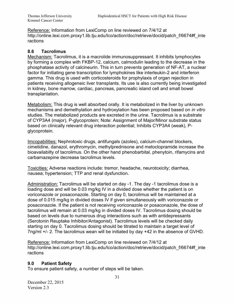
CD34+ cell enrichment will be performed via the closed system method using the CliniMACS® CD34 Reagent System (Miltenyi Biotec Inc., Auburn, CA). The CliniMACS system utilizes super-paramagnetic particles composed of iron oxide and dextran conjugated to monoclonal antibodies. These antibodies bind to target cells with the corresponding cell surface antigen (in this case, CD34). After magnetic labeling, the cells are separated using a high-gradient magnetic separation column. The magnetically labeled cells are retained in the column and separated from the unlabeled cells. Removing the magnetic field from the separation column elutes the retained cells. Eluted cells will be characterized using fluorescent-activated cell sorting (FACS) analysis. All procedures will be performed in a sterile environment with strict adherence to all applicable regulations regarding the processing and use of human stem cells. The use of this device will conform to TJU BMT Laboratory standard operating procedures. In our experience, the ideal amount of T-cells left in the PBSC product is no greater than 5x10⁴/kg, so that every effort will be made to keep T-cell amounts to below this threshold. It is recognized that because of donor heterogeneity, every product will have varying percentages of cells. Thus, patients will be advised during the informed consent process that an excess amount of residual T-lymphocytes in the PBSC product may increase the risk of GVHD. The donor product is infused UNFILTERED or through a filter of at least 170 micron size intravenously through a central catheter. Marrow should only be piggybacked through normal saline and not other intravenous solutions. During the infusion, the patient will be monitored for any untoward reactions. Each infusion will take place in the Bone Marrow Transplant Unit. PBSC infusions will be administered by nursing staff experienced in the administration of blood products. Progenitor cell products must NOT be irradiated. Progenitor cell products should NEVER be administered through a leukocyte depletion filter. If blood filtration is necessary, the filter should be a standard blood product filter with pore size of at least 170 microns. Significant red cell incompatibility between donor and recipient will be managed according to standard operating procedure, CL: Ppp040.05, of the Thomas Jefferson University Hospital Blood and Marrow Transplant Processing Lab. Pre-medications (if any) prior to marrow infusion will be at the discretion of the physician. 5.6 GVHD Prophylaxis Tacrolimus and MMF will be started on day -1. The day -1 tacrolimus dose is a loading dose and will be 0.03 mg/kg IV in a divided dose whether the patient is on voriconazole or posaconazole. Starting on day 0, tacrolimus will be maintained at a dose of 0.015 mg/kg in divided doses IV if given simultaneously with voriconazole or posaconazole. If the patient is not receiving voriconazole or posaconazole, the dose of tacrolimus will remain at 0.03 mg/kg in divided doses IV. Tacrolimus levels will be checked daily

Thomas Jefferson University Haploidentical HSCT for Patients with High Risk Disease Kimmel Cancer Center
23 December 22, 2015 Version 2.3
starting on day 0. Tacrolimus dosing should be titrated to maintain a target level of 8ng/ml +/- 2. MMF will be dosed at 1 gram IV BID beginning on day -1. Tacrolimus oral dosing will be initiated at least 2 to 3 days prior to discharge. This is to assure that stable, therapeutic levels are reached on oral drug prior to discharge. MMF will be discontinued beginning at day +28 +/- 3 days in the absence of GVHD. MMF may be discontinued earlier if there is count suppression thought to be due to the drug. Do not wean MMF. MMF is not tapered. The tacrolimus taper should be initiated by day + 42 in the absence of GVHD. The taper will take place at roughly 15% per week (range 10 to 20% per week). Once tacrolimus levels are less that 5 ng/ml levels, they no longer have to be checked unless there is a clinical concern to do so. Because of the variability in patient outpatient office visit times and the need for GVHD assessment, it is not mandatory that the taper begins exactly day +42, but should begin within 2 weeks of day 42. 5.6.1 Treatment of Patients with Acute GVHD The following steps will be taken if GVHD is suspected:
Begin prednisone at a dose of 2 mg/kg/day (1mg/kg q12h). As soon as manifestations show clear cut evidence of improvement usually 2-3 days after initiation of therapy (and not to be interpreted as complete resolution of all manifestations) reduce the prednisone dose to 1 mg/kg/day (0.5 mg/kg q12h). Thereafter, taper by 20 mg/dose every 2 to 3 days if an inpatient or at least weekly as an outpatient until a 20 mg daily dose is reached.
If patients were on Tacrolimus and/or MMF, they may remain on these mediations to facilitate weaning the steroids. If these medications had been discontinued, they should be restarted only if the patient fails to respond to steroids.
Once prednisone is at 20 mg per day or lower, it may be preferable to taper tacrolimus or discontinue MMF while holding prednisone at the current dose as these other medications may represent a more substantial burden of immunosuppression for the patient at these low prednisone doses. Alternatively, one can complete the steroid wean over another 2-4 weeks.
In the absence of GVHD flare, begin weaning Tacrolimus or discontinue MMF two weeks after the prior medication has been stopped (or immediately after the steroid taper has been paused at a dose of 20 mg/day of prednisone or less.
If patients flare on this sort of taper, medications should be increased to at least the prior dose which achieved control and photopheresis should be initiated 2-3 times per week for patients with skin disease. Efforts should be made to begin a taper again after 2 weeks of photopheresis.
This taper reflects a guide as to the slowest, not the fastest, that immune suppression should be tapered. The pace should be accelerated in patients with significant bacterial, viral, fungal, or other infections. The goal is to find which

Thomas Jefferson University Haploidentical HSCT for Patients with High Risk Disease Kimmel Cancer Center
24 December 22, 2015 Version 2.3
patients will not tolerate a prompt taper of immune suppression and move them to photopheresis rapidly so as to facilitate tapering systemic immunosuppressive medications.
For situations that are not covered by these criteria, discuss the case with the PI. 6.0 Laboratory Studies/Outcome Assessment 6.1 Analyses of Leukemia MHC Gene Content and Expression Since the majority of these patients will be transplanted in relapse, it is permissible, but not mandatory, to obtain and cryopreserve buccal swabs, blood and/or marrow specimens prior to transplant and at the time of relapse if relapse occurs. Specimens will be analyzed using pan-HLA class I and pan-HLA class II antibodies to assess whether overall levels of MHC molecules have declined on the cells surface. Genetic analysis including sequencing of these paired specimens will allow us to assess whether asymmetric loss of one haplotype, such as from uniparental disomy, occurs after transplant on this regimen. In addition to the above studies, SNP or other genetic analyses can be performed in the matched pair samples as hypotheses are generated to test for other mechanisms through which leukemia cells escape GVT. 6.2 Study Measurements All post-allogeneic transplant patients have physical assessments, laboratory studies and pathology studies performed as per the TJUH BMT Guidelines for Post-Transplant Allogeneic Assessments (CP: P035.02) found on the TJUH BMT intranet. Table 5 outlines the mandatory measurements and time points specific to this study. Table 5
Day + 28
Days 28-90
Days 91 -180
Days 180- 270
1 Year GVHD Assessment
Presence and degree of skin rash, presence
and amount of diarrhea, LFT’s
On day +28
Twice Monthly
Monthly
Every 3 Months
At 1 year
Chimerism/
Disease Assessment
Peripheral blood for Total, MNC & CD3+
chimerism
On day
+28
Monthly
Monthly
Every 3 Months
At 1 year
Bone marrow exam (morphology, flow
cytometry, cytogenetics, chimerism)
On day +28
At day +90
At day +180
At day +270
At 1 Year
Flow cytometry for lymphocyte subsets
(IRP)
On day
+28
Monthly
Monthly
Every 2 Months
At 1 year

Thomas Jefferson University Haploidentical HSCT for Patients with High Risk Disease Kimmel Cancer Center
25 December 22, 2015 Version 2.3
The day +28 peripheral blood, marrow studies and the day 28 assessment can be obtained within 1 week of day 28 (i.e. +/- 7 days) to account for scheduling factors. Patient assessments, peripheral blood chimerism and IRP studies, as well as the day +90, +180, +270, and 1 year marrows can be obtained within the time period of 1 month before or 1 month after the targeted time to account for patient scheduling factors. This table represents a minimum recommended sampling and visit strategy. 6.3 Hematopoietic Engraftment Will be defined as:
ANC ≥ 0.5x10⁹/L for at least 30 days Platelet engraftment > 20,000 with no transfusion x 7 days
6.4 Toxicity Criteria Regimen-related toxicity will be graded according to the NCI Common Toxicity Criteria, version 3.0. The NCI Common Toxicity Criteria can also be found at the following WEB address: http://ctep.cancer.gov/reporting/ctc.html 6.5 Disease Response Disease response will be measured according to the National Comprehensive Cancer Network Guidelines (NCCN). The guidelines are disease specific and the guidelines for each disease can be found at: http://www.nccn.org/professionals/physician_gls/f_guidelines.asp#site 6.6 GVHD Scoring GVHD will be graded according to the standard criteria contained in Appendix B. 6.7 Adverse Event Reporting All patients will be followed for adverse experiences (AEs) (serious and non-serious), regardless of relationships to study treatment, from the time of enrollment until day +100 post-transplant. The following events are expected side effects of high-dose chemotherapy and transplant and will not be reported except as noted:
Alopecia, dry skin Emesis from chemotherapy or other agents unless refractory to standard
supportive care, nausea, and anorexia Weight loss, cough, dry mouth, headache Neutropenia/uncomplicated neutropenic fever, grades 1-3 infectious sequellae Thrombocytopenia, petechiae, ecchymoses, minor vaginal bleeding, epistaxis,
hemorrhoidal bleeding, or other similar bleeding events will not be reported. (Bleeding events requiring transfusion and/or intervention such as endoscopy or radiologic evaluation will be reported.)
Anemia Grade I - III Mucositis

Thomas Jefferson University Haploidentical HSCT for Patients with High Risk Disease Kimmel Cancer Center
26 December 22, 2015 Version 2.3
Grade I - III Diarrhea Grades 1-3 allergic or other common reactions to drugs used for supportive care Fluid and electrolyte disturbances not associated with instability
After d + 100, only events that are considered by the investigator to be possibly or probably associated with the treatment regimen will be reported. 6.8 Reports to the Federal Drug Administration (FDA) All grade 3-5 infusion reactions and all unexpected SAEs as defined in 21 CFR 312.32 will be reported to the FDA in an expedited fashion. An annual report will be sent to the FDA regarding the progress to date of patients on the trial. In the report, a separate listing of infusion toxicities and all biological product deviations will be included in addition to the other required elements. 6.9 Study Endpoint The formal endpoint of this study is 1 year post HSCT. Therefore patients will not be followed for the purposes of this clinical trial after this time. However, outcomes for patients undergoing HSCT at TJUH are followed programmatically beyond this study indefinitely. These outcomes include survival, relapse, and GVHD. The study will be eligible for closure when the last patient treated is 1 year post HSCT. 7.0 Supportive Care 7.1 Avoidance of Infection Patients who are post haploidentical transplantation will follow the same guidelines as patients who are neutropenic until advised differently by their attending physician. Infectious prophylaxis and treatment of infection will be as per the “TJUH Guidelines for Infectious Prophylaxis and Management of Febrile Neutropenia”. These guidelines can be found on the TJUH intranet. Central venous catheters will be removed as soon as clinical manageable. IVIG 0.5 g/kg IV will be administered every 4 weeks post transplant to support immune function, until the IgG level is > 500 mg/dL on 2 consecutive monthly measurements. Because there are qualitative defects in humoral immunity for years after HSCT, it is suggested, but not mandated, that IVIG be given monthly for at least 1 year after HSCT, even if there is evidence of quantitative recovery as described above. The first dose will be administered approximately on day +7. It may be given earlier or later if the patient cannot tolerate the large volume on day +7. 7.2 Infectious Prophylaxis-General Guidelines Patients post haploidentical transplantation will be maintained on antifungal (including mold coverage) prophylaxis, usually voriconazole 200 mg BID. It is at the discretion of the treating attending physician to change agents as clinically indicated.

Thomas Jefferson University Haploidentical HSCT for Patients with High Risk Disease Kimmel Cancer Center
27 December 22, 2015 Version 2.3
Patients post haploidentical transplantation will be maintained on HSV prophylaxis, usually Acyclovir 400 mg BID or Valacyclovir 500 mg BID. It is at the discretion of the treating attending physician to change agents based on culture results and sensitivities. Patients post partially-matched related donor transplantation will be maintained on PCP prophylaxis, usually Bactrim DS 1 daily. It is at the discretion of the treating attending physician to change agents based on culture results, drug intolerance. Prophylactic medications may be discontinued when the patient is off immunosuppressive medications for at least 1 month, and/or the CD4 count is > 100/µl. 7.3 Growth Factor and Transfusion Support To prevent inadvertent lymphoid engraftment, all blood cell products must be irradiated to ≥2500cGy. All red cell and platelet products will be leukodepleted to prevent alloimmunization and decrease infectious sequela. Packed red blood cell transfusions will be given as necessary with a goal of keeping the hemoglobin ≥ 8 g/L. Platelet transfusions will be given as necessary with a goal of keeping the morning count ≥ 20x10e9/L, with 10x10e9/L used for situations without an excessive bleeding risk. GM-CSF 250µg/m2 will be administered daily beginning on day +1. GM-CSF will be weaned/discontinued at the discretion of the attending physician. Every effort should be made to keep the ANC > 1000 for all patients post haploidentical transplantation. G-CSF 5µg/m2 can be substituted for GM-CSF in the event of a GM-CSF shortage or withdrawal from market. Red cell growth factors are permissible after transplantation. 8.0 Drug Information and Administration 8.1 Cyclophosphamide Mechanism: A multistep process activates it by conversion to 4-hydroxycyclophosphamide by the liver microsomal oxidase system and to aldophosohamide by tautomerization in the peripheral tissues. Aldophosphamide spontaneously degrades into acrolein and phosporamide mustard, which cause cellular glutathione depletion and DNA alkylation. This results in inhibition of DNA replication and transcription. Cells expressing high levels of aldehyde dehydrogenase (e.g. stem cells, L1210 leukemia cells) resist cyclophosphamide-mediated cytotoxicity as aldophosphamide is inactivated by this enzyme. The drug also does not affect quiescent cells and therefore stem cells are generally protected, an important factor if autologous hematopoietic recovery is relied on in the event of graft failure.

Thomas Jefferson University Haploidentical HSCT for Patients with High Risk Disease Kimmel Cancer Center
28 December 22, 2015 Version 2.3
Metabolism: Cyclophosphamide is broken down as described above and the break down products are excreted by the kidneys. It is a substrate of CYP2A6 (minor), CYP2B6 (major), CYP2C19 (minor), CYP2C9 (minor), CYP3A4 (minor); Note: Assignment of Major/Minor substrate status based on clinically relevant drug interaction potential; Inhibits CYP3A4 (weak); Induces CYP2B6 (weak/moderate), CYP2C9 (weak/moderate). Incompatibilities: Phenobarbital or rifampin may increase the toxicity of cyclophosphamide. Concurrent allopurinol or thiazide diuretics may exaggerate bone marrow depression may prolong neuromuscular blockade from succinylcholine Cardiotoxicity may be additive with other cardiotoxic agents (cytarabine, daunorubicin, doxorubicin). May decrease serum digoxin levels. Additive bone marrow depression with other antineoplastics or radiation therapy. May potentiate the effects of warfarin. May decrease antibody response to live-virus vaccines and increase the risk of adverse reactions. Prolongs the effects of cocaine. Toxicity: Nausea, vomiting, water retention due to inappropriate secretion of anti-diuretic hormone (SIADH), cardiomyopathy with myocardial necrosis and congestive heart failure, hemorrhagic cystitis, alopecia, skin rash, pulmonary fibrosis, sterility and secondary malignancies. Administration: Patients will receive a dose of cyclophosphamide 60 mg/kg IV, on days –3 and -2. The dose of cyclophosphamide will be calculated according to the dosing body weight. The cyclophosphamide dose is dissolved in saline and administered as a 2 hour IV infusion. Patients shall receive hydration consisting of normal saline solution at 3 ml/kg/hour (actual weight) for 2 hours before and 8 hours after the cyclophosphamide infusion. MESNA (sodium-2-mercaptoethane sulfonate) will be administered as a 60 mg/kg/continuous IV infusion over 24 hours starting 30 minutes prior to cyclophosphamide infusion and ending 24 hours after the last dose of cyclophosphamide. The dose of MESNA will also be calculated based on dosing body weight. References: Skeel R & Lachant N. Handbook of Cancer Chemotherapy, 4th Ed. Little, Brown & Co.: Boston. Information from LexiComp on line reviewed on 7/4/12 at http://online.lexi.com.proxy1.lib.tju.edu/lco/action/doc/retrieve/docid/patch_f/6674#f_interactions 8.2 Donor Leukocyte Infusion (DLI) Administration: All patients will receive a dose of CD3+ T cells per kilogram of dosing body weight as outlined in the treatment design. Details of the apheresis procedure to obtain white blood cells, quantification of T cells by flow cytometry, and administration of the white cell product to the recipient are provided in the treatment section. All drugs that may cause lymphocyte suppression are held prior to lymphocyte infusion (day -6), through day 0 as detailed in the treatment section. Every effort will be made to

Thomas Jefferson University Haploidentical HSCT for Patients with High Risk Disease Kimmel Cancer Center
29 December 22, 2015 Version 2.3
administer the donor lymphocytes 48 hours after the last fraction of TBI as described in section 5.0. Moreover the viability of the lymphocytes will be tested by flow cytometry and the number of viable CD3+ T cells will be used to dose the DLI. Toxicity: GVHD, delayed myelosuppression, infusion reactions. 8.3 G-CSF Mechanism: G-CSF is a human granulocyte colony-stimulating factor produced by recombinant DNA technology. It is a glycoprotein which acts on hematopoietic cells by binding to specific cell surface receptors and stimulating proliferation, differentiation, commitment, and some end-cell functions. Activates neutrophils to increase migration andtoxicity. Metabolism: Absorption and clearance of G-CSF follows first-order pharmacokinetic modeling without apparent concentration dependence. The elimination half-life in both normal and cancer patients is 3.5 hours. Incompatibilities: Safety and efficacy of G-CSF when used simultaneously with chemotherapy or radiotherapy has not been evaluated. Donors receiving either of these 2 modalities will not be permitted on study. Toxicities: Allergic reactions consisting of rash, wheezing and tachycardia. Splenic rupture, ARDS, and exacerbation of sickle cell disease have been reported rarely. Adminstration: In this protocol, G-CSF will be administered to healthy donors at a dose of 10 µg/kg (actual weight) subcutaneously on days -5 through day -1. References: Physician’s Desk Reference, Edition 58, 2004. In addition, information from LexiComp on line reviewed on 7/4/12 at http://online.lexi.com.proxy1.lib.tju.edu/lco/action/doc/retrieve/docid/patch_f/6674#f_interactions 8.4 GM-CSF (Sargramostim, Leukine) Mechanism: GM-CSF is a recombinant human granulocyte-colony stimulating factor produced by recombinant DNA technology in a yeast expression system. It supports survival, clonal expansion, and differentiation of hematopoietic cells. GM-CSF is also capable of activating mature granulocytes and macrophages, and is a multilineage factor with effects on the myelomonocytic, erythroid, and megarkaryocytic lines. Metabolism: GM-CSF is detected in the serum at 15 minutes after injection. Peak levels occur about 1 to 3 hours after injection, and it is detectable in the serum for up to 6 hours after injection.

Thomas Jefferson University Haploidentical HSCT for Patients with High Risk Disease Kimmel Cancer Center
30 December 22, 2015 Version 2.3
Incompatibilities: Interactions between GM-CSF and other drugs have not been fully evaluated. Drugs which may potentiate the myeloproliferative effects of GM-CSF, such as lithium and corticosteroids, should be used with caution. Toxicities: Allergic and anaphylactic reactions have been reported. A syndrome characterized by respiratory distress, hypoxia, flushing, hypotension, syncope and or tachycardia has been associated with the first administration of GM-CSF in a cycle. These signs have resolved with treatment. Administration: In this protocol, GM-CSF will be given to the patients beginning on Day +1. The drug should continue until the patient has a self-sustaining ANC of ≥ 1000. References: Physician’s Desk Reference, Edition 58, 2004. In addition, information from LexiComp on line reviewed on 7/4/12 at http://online.lexi.com.prox 8.5 Mycophenolate Mofetil (MMF) Mechanism: Inhibits the enzyme inosine monophosphate dehydrogenase, which is involved in purine synthesis. This inhibition results in suppression of T- and B-lymphocyte proliferation. Metabolism: Following oral and IV administration, mycophenolate is rapidly hydrolyzed to mycophenolic acid (MPA), its active metabolite. Distribution is unknown. MPA is extensively metabolized; <1% excreted unchanged in urine. Some enterohepatic recirculation of MPA occurs. Half Life: MPA¾17.9 hr. Incompatibilities: Combined use with azathioprine is not recommended (effects unknown). Acyclovir and ganciclovir compete with MPA for renal excretion and, in patients with renal failure, may increase each other's toxicity. Magnesium and aluminum hydroxide antacids decrease the absorption of MPA (avoid simultaneous administration). Cholestyramine and colestipol decrease the absorption of MPA (avoid concurrent use). Toxicity may be increased by salicylates. May interfere with the action of oral contraceptives (additional contraceptive method should be used). May decrease the antibody response to and increase risk of adverse reactions from live-virus vaccines, although influenza vaccine may be useful. When administered with food, peak blood levels of MPA are significantly decreased. Toxicities: GI: Bleeding, Diarrhea, Vomiting, Hematopoietic: Leukopenia Miscellaneous: Sepsis, Increased Risk of Malignancy Administration: In this protocol, MMF will be administered at a dose of 1 gram IV BID beginning on day -1. MMF will be discontinued on day +28 in the absence of GVHD. MMF may be stopped earlier if there is count suppression from the drug.
Thomas Jefferson University Haploidentical HSCT for Patients with High Risk Disease Kimmel Cancer Center
31 December 22, 2015 Version 2.3
Reference: Information from LexiComp on line reviewed on 7/4/12 at http://online.lexi.com.proxy1.lib.tju.edu/lco/action/doc/retrieve/docid/patch_f/6674#f_interactions 8.6 Tacrolimus Mechanism: Tacrolimus, it is a macrolide immunosuppressant. It inhibits lymphocytes by forming a complex with FKBP-12, calcium, calmodulin leading to the decrease in the phosphatase activity of calcineurin. This in turn prevents generation of NF-AT, a nuclear factor for initiating gene transcription for lymphokines like interleukin-2 and interferon gamma. This drug is used with corticosteroids for prophylaxis of organ rejection in patients receiving allogeneic liver transplants. Its use is also currently being investigated in kidney, bone marrow, cardiac, pancreas, pancreatic island cell and small bowel transplantation. Metabolism: This drug is well absorbed orally. It is metabolized in the liver by unknown mechanisms and demethylation and hydroxylation has been proposed based on in vitro studies. The metabolized products are excreted in the urine. Tacrolimus is a substrate of CYP3A4 (major), P-glycoprotein; Note: Assignment of Major/Minor substrate status based on clinically relevant drug interaction potential; Inhibits CYP3A4 (weak), P-glycoprotein. Imcopabilities: Nephrotoxic drugs, antifungals (azoles), calcium-channel blockers, cimetidine, danazol, erythromycin, methylprednisone and metoclopramide increase the bioavailabilty of tacrolimus. On the other hand phenobarbital, phenytoin, rifamycins and carbamazepine decrease tacrolimus levels. Toxicities: Adverse reactions include: tremor, headache, neurotoxicity; diarrhea, nausea; hypertension; TTP and renal dysfunction. Administration: Tacrolimus will be started on day -1. The day -1 tacrolimus dose is a loading dose and will be 0.03 mg/kg IV in a divided dose whether the patient is on voriconazole or posaconazole. Starting on day 0, tacrolimus will be maintained at a dose of 0.015 mg/kg in divided doses IV if given simultaneously with voriconazole or posaconazole. If the patient is not receiving voriconazole or posaconazole, the dose of tacrolimus will remain at 0.03 mg/kg in divided doses IV. Tacrolimus dosing should be based on levels due to numerous drug interactions such as with antidepressants (Serotonin Reuptake Inhibitor/Antagonist). Tacrolimus levels will be checked daily starting on day 0. Tacrolimus dosing should be titrated to maintain a target level of 7ng/ml +/- 2. The tacrolimus wean will be initiated by day +42 in the absence of GVHD. Reference: Information from LexiComp on line reviewed on 7/4/12 at http://online.lexi.com.proxy1.lib.tju.edu/lco/action/doc/retrieve/docid/patch_f/6674#f_interactions 9.0 Patient Safety To ensure patient safety, a number of steps will be taken.

Thomas Jefferson University Haploidentical HSCT for Patients with High Risk Disease Kimmel Cancer Center
32 December 22, 2015 Version 2.3
The study will be monitored on an ongoing fashion by the Principal Investigator (PI) and the study medical monitor. Monitoring reports will be submitted to the Clinical Research Management Office (CRMO) for review by the DSMC during their quarterly review. Adverse events and a report summarizing their impact on the conduct of the trial are submitted to the Data Monitoring and Safety Committee (DMSC) quarterly, and the DSMC reports are then submitted to the CCRRC and IRB annually. The PI will submit serious adverse events (SAE) to the TJU IRB utilizing the electronic Kimmel Cancer Center Clinical Trials Adverse Event Reporting system. Due to the nature of the study treatment as outlined in this protocol, expected grade 3 AE/SAEs that occur while receiving standard inpatient protocol treatment may be included on the patient’s AE log for quarterly review by the DSMC rather than be reported via the eSAEy System per the DSM Plan. It is the responsibility of the study Principal Investigator (PI) to report any grade 3 AE/SAE to the DSMC per the DSM Plan should the length of standard protocol treatment hospitalization be extended and/or the grade 3 AE/SAE is more acute than expected as outlined in the informed consent form. Unexpected deaths related to this protocol will be reported within 24 hours. The medical monitor will be a TJU physician who is not a collaborator in this trial. The medical monitor will review all adverse events (in addition to unexpected adverse events), safety data and activity data observed when this trial is ongoing. The medical monitor may recommend reporting adverse events and relevant safety data not previously reported, and may recommend suspension or termination of the trial. The summary of all discussions of adverse events will be submitted to the DSMC after completion and included in the PI's reports to the CCRRC and the TJU IRB as part of the study progress report. The CCRRC, DMSC, and/or the TJU IRB may, based on the monitor's recommendation suspend or terminate of the trial. The quarterly safety and monitoring reports will include a statement as to whether this data has invoked any stopping criteria (dose-limiting toxicities) in the clinical protocol. In addition to the Cancer Center's DMSC, the TJU BMT program members meet weekly to discuss the status of patients on trial and generate discussion regarding the progress of the patients on the trial. Auditing and Inspecting: The investigator will permit study-related monitoring, audits, and inspections by the IRB, the funding sponsor, government regulatory bodies, and University compliance and quality assurance groups of all study related documents (e.g. source documents, regulatory documents, data collection instruments, study data etc.). The investigator will ensure the capability for inspections of applicable study-related facilities. Participation as an investigator in this study implies acceptance of potential inspection by government regulatory authorities and applicable University compliance and quality assurance offices.

Thomas Jefferson University Haploidentical HSCT for Patients with High Risk Disease Kimmel Cancer Center
33 December 22, 2015 Version 2.3
In addition to review by the DSMC, all studies initiated by KCC investigators are audited by an independent auditor once they have achieved 10% of target accrual. However, a study can be audited at any time based on recommendations by the IRB, DSMC, CCRRC and/or the Director of Clinical Investigations, KCC. Studies are re-audited once they have achieved 50% of target accrual. Special audits may be recommended by the IRB, DSMC or CCRRC based on prior findings, allegations of scientific misconduct and where significant irregularities are found through quality control procedures. Any irregularities identified as part of this process would result in a full audit of that study. In addition to the audits at 10 and 50%, the CRMO randomly audits at least 10 percent of all patients entered into therapeutic KCC trials and other trials as necessary, on at least a bi-annual basis, to verify that there is a signed and dated patient consent form, the patient has met the eligibility criteria, and that SAEs are documented and reported to the TJU IRB. All reports are submitted to the DSMC for review and action (when appropriate). A copy of this report and recommended DSMC action is sent to the CCRRC and TJU IRB. The committee regards the scientific review process as dynamic and constructive rather than punitive. The review process is designed to assist Principal Investigators in ensuring the safety of study subjects and the adequacy and accuracy of any data generated. The TJU IRB may, based on the DSMC and auditor’s recommendation, suspend or terminate the trial. 10.0 Statistical Analysis Statistical Analysis Plan: The main objective of this trial is to demonstrate that the disease free survival (DFS) at one year for patients treated on this clinical trial is significantly higher than 25% (the average 1-year DFS in historic controls). The primary hypothesis will be tested using an exact one-sided binomial test with alpha 0.05. The trial will be considered successful if the null hypothesis of 25% 1-year disease free survival (DFS) is rejected. In addition, the exact binomial 95% confidence interval for 1-year DFS will be computed. Sample Size: Sample size computations are based on expectation of ~45% disease free survival (DFS) at one year for patients treated on this clinical trial. This figure is based on the historical literature reviewed in Section 1.0, as well as our current experience with the outcomes of patients with similar disease states treated on the 2 Step approach. Assuming that the true 1 year DFS rate is 45%, we need 36 evaluable patients for an 80% power using a one-sided binomial test with alpha 0.05. Assuming a 10% dropout/incompletion rate, we will need to enroll up to 40 people in order to achieve 36 evaluable subjects. Subject are considered evaluable if…We estimate that it will take 7 years to complete this trial; 6 years to treat 36 patients plus 1 year follow-up. Assessment of Secondary Objectives
Thomas Jefferson University Haploidentical HSCT for Patients with High Risk Disease Kimmel Cancer Center
34 December 22, 2015 Version 2.3
Assessments for the secondary objectives will be reported descriptively. Data pertaining to the secondary objectives will be collected and placed in each patient’s study binder for analysis. Analysis for Safety Patient outcomes are routinely monitored in an ongoing fashion for all patients on investigational trials, beyond their formal endpoints. Based on prior experience using a two step approach similar to that described in this trial, we anticipate that the incidence of graft failure should be less than 10%, the incidence of severe GVHD (grade 3 or 4) should be less than 20%, and the non-relapse mortality should be less than 20% at 100 days. If at any point incidences higher than these thresholds are seen, that would trigger a protocol review to assess whether there are any obvious reasons for the inferior outcomes observed. Depending on the results of the review, enrollment may continue on a limited basis with careful further observation, the protocol may be revised, or the protocol may be terminated. Incidences will be calculated starting after 10 patients are treated on this trial in order to have a sufficient denominator in which to examine outcomes based on percentages. 11.0 References 1. Giralt S, Estey E, Albitar M, et al. Engraftment of allogeneic hematopoietic progenitor cells with purine analog-containing chemotherapy: harnessing graft-versus-leukemia without myeloablative therapy. Blood. 1997;89(12):4531-4536. 2. Bacigalupo A, Soracco M, Vassallo F, et al. Donor lymphocyte infusions (DLI) in patients with chronic myeloid leukemia following allogeneic bone marrow transplantation. Bone Marrow Transplant. 1997;19(9):927-932. 3. Lee S-, Yoon J-, Shin S-, et al. Impact of pre-transplant marrow blasts on survival of allogeneic stem cell transplantation in adult acute myeloid leukemia. Int J Hematol. 2013:1-10. 4. Oyekunle AA, Kroger N, Zabelina T, et al. Allogeneic stem-cell transplantation in patients with refractory acute leukemia: a long-term follow-up. Bone Marrow Transplant. 2006;37(1):45-50. 5. Kebriaei P, Kline J, Stock W, et al. Impact of disease burden at time of allogeneic stem cell transplantation in adults with acute myeloid leukemia and myelodysplastic syndromes. Bone Marrow Transplant. 2005;35(10):965-970. 6. Wong R, Shahjahan M, Wang X, et al. Prognostic factors for outcomes of patients with refractory or relapsed acute myelogenous leukemia or myelodysplastic syndromes undergoing allogeneic progenitor cell transplantation. Biology of Blood & Marrow Transplantation. 2005;11(2):108-114. 7. Michallet M, Thomas X, Vernant JP, et al. Long-term outcome after allogeneic hematopoietic stem cell transplantation for advanced stage acute myeloblastic

Thomas Jefferson University Haploidentical HSCT for Patients with High Risk Disease Kimmel Cancer Center
35 December 22, 2015 Version 2.3
leukemia: a retrospective study of 379 patients reported to the Societe Francaise de Greffe de Moelle (SFGM). Bone Marrow Transplant. 2000;26(11):1157-1163. 8. Alessandrino E, Della Porta M, Bacigalupo A, et al. WHO classification and WPSS predict posttransplantation outcome in patients with myelodysplastic syndrome: a study from the Gruppo Italiano Trapianto di Midollo Osseo (GITMO). Blood. 2008;112(3):895-902. 9. Lim Z, Brand R, Martino R, et al. Allogeneic hematopoietic stem-cell transplantation for patients 50 years or older with myelodysplastic syndromes or secondary acute myeloid leukemia. Journal of Clinical Oncology. 2010;28(3):405-411. 10. Garderet L, Labopin M, Gorin NC, et al. Patients with acute lymphoblastic leukaemia allografted with a matched unrelated donor may have a lower survival with a peripheral blood stem cell graft compared to bone marrow. Bone Marrow Transplant. 2003;31(1):23-29. 11. Ringden O, Pavletic SZ, Anasetti C, et al. The graft-versus-leukemia effect using matched unrelated donors is not superior to HLA-identical siblings for hematopoietic stem cell transplantation. Blood. 2009;113(13):3110-3118. 12. Terwey TH, Massenkeil G, Tamm I, et al. Allogeneic SCT in refractory or relapsed adult ALL is effective without prior reinduction chemotherapy. Bone Marrow Transplant. 2008;42(12):791-798. 13. Bertz H, Illerhaus G, Veelken H, Finke J. Allogeneic hematopoetic stem-cell transplantation for patients with relapsed or refractory lymphomas: comparison of high-dose conventional conditioning versus fludarabine-based reduced-intensity regimens. Annals of Oncology. 2002;13(1):135-139. 14. Hamadani M, Benson Jr. DM, Hofmeister CC, et al. Allogeneic Stem Cell Transplantation for Patients with Relapsed Chemorefractory Aggressive Non-Hodgkin Lymphomas. Biology of Blood and Marrow Transplantation. 2009;15(5):547-553. 15. Rigacci L, Puccini B, Dodero A, et al. Allogeneic hematopoietic stem cell transplantation in patients with diffuse large B cell lymphoma relapsed after autologous stem cell transplantation: A GITMO study. Ann Hematol. 2012;91(6):931-939. 16. Dreger P, Stilgenbauer S, Boettcher S, et al. Prognostic Factors for Outcome of Nonmyeloablative Allogeneic Stem Cell Transplantation (NST) in Poor-Risk Chronic Lymphocytic Leukemia (CLL): Final Results from a Prospective Multicenter Trial (GCLLSG CLL3X study). ASH Annual Meeting Abstracts. 2008;112(11):565. 17. Khouri IF, Keating MJ, Saliba RM, Champlin RE. Long-term follow-up of patients with CLL treated with allogeneic hematopoietic transplantation. Cytotherapy. 2002;4(3):217-221.

Thomas Jefferson University Haploidentical HSCT for Patients with High Risk Disease Kimmel Cancer Center
36 December 22, 2015 Version 2.3
18. Kröger N, Badbaran A, Lioznov M, et al. Post-transplant immunotherapy with donor-lymphocyte infusion and novel agents to upgrade partial into complete and molecular remission in allografted patients with multiple myeloma. Exp Hematol. 2009;37(7):791-798. 19. Farag SS, Bacigalupo A, Eapen M, et al. The Effect of KIR Ligand Incompatibility on the Outcome of Unrelated Donor Transplantation: A Report from the Center for International Blood and Marrow Transplant Research, the European Blood and Marrow Transplant Registry, and the Dutch Registry. Biology of Blood and Marrow Transplantation. 2006;12(8):876-884. 20. Ogawa H, Ikegame K, Kawakami M, et al. Powerful graft-versus-leukemia effects exerted by HLA-haploidentical grafts engrafted with a reduced-intensity regimen for relapse following myeloablative HLA-matched transplantation. Transplantation. 2004;78(3):488-489. 21. Burroughs LM, O'Donnell PV, Sandmaier BM, et al. Comparison of outcomes of HLA-matched related, unrelated, or HLA-haploidentical related hematopoietic cell transplantation following nonmyeloablative conditioning for relapsed or refractory Hodgkin lymphoma. Biology of Blood & Marrow Transplantation. 2008;14(11):1279-1287. 22. Hauer J, Tosi S, Schuster FR, Harbott J, Kolb HJ, Borkhardt A. Graft versus leukemia effect after haploidentical HSCT in a MLL-negative infant AML with HLXB9/ETV6 rearrangement. Pediatric Blood & Cancer. 2008;50(4):921-923. 23. Koh LP, Rizzieri DA, Chao NJ. Allogeneic hematopoietic stem cell transplant using mismatched/haploidentical donors. Biology of Blood & Marrow Transplantation. 2007;13(11):1249-1267. 24. Gill S, Olson JA, Negrin RS. Natural Killer Cells in Allogeneic Transplantation: Effect on Engraftment, Graft- versus-Tumor, and Graft-versus-Host Responses. Biology of Blood and Marrow Transplantation. ;In Press, Corrected Proof. 25. Ruggeri L, Mancusi A, Capanni M, et al. Donor natural killer cell allorecognition of missing self in haploidentical hematopoietic transplantation for acute myeloid leukemia: challenging its predictive value. Blood. 2007;110(1):433-440. 26. Luznik L, O'Donnell PV, Symons HJ, et al. HLA-haploidentical bone marrow transplantation for hematologic malignancies using nonmyeloablative conditioning and high-dose, posttransplantation cyclophosphamide. Biology of Blood & Marrow Transplantation. 2008;14(6):641-650.

Thomas Jefferson University Haploidentical HSCT for Patients with High Risk Disease Kimmel Cancer Center
37 December 22, 2015 Version 2.3
27. O'Donnell PV, Luznik L, Jones RJ, et al. Nonmyeloablative bone marrow transplantation from partially HLA-mismatched related donors using posttransplantation cyclophosphamide. Biology of Blood & Marrow Transplantation. 2002;8(7):377-386. 28. Grosso D, Carabasi M, Filicko-O'Hara J, et al. A2-step approach to myeloablative haploidentical stem cell transplantation: A phase 1/2 trial performed with optimized T-cell dosing. Blood. 2011;118(17):4732-4739. 29. Ruggeri L, Capanni M, Urbani E, et al. Effectiveness of donor natural killer cell alloreactivity in mismatched hematopoietic transplants. Science. 2002;295(5562):2097-2100. 30. Huang X, Liu D, Liu K, et al. Treatment of Acute Leukemia with Unmanipulated HLA-Mismatched/Haploidentical Blood and Bone Marrow Transplantation. Biology of Blood and Marrow Transplantation. 2009;15(2):257-265. 31. Bartolomeo PD, Santarone S, De Angelis G, et al. Haploidentical, unmanipulated, G-CSF-primed bone marrow transplantation for patients with high-risk hematologic malignancies. Blood. 2013;121(5):849-857. 32. Colvin GA, Berz D, Ramanathan M, et al. Nonengraftment Haploidentical Cellular Immunotherapy for Refractory Malignancies: Tumor Responses without Chimerism. Biology of Blood and Marrow Transplantation. 2009;15(4):421-431. 33. Luznik L, Engstrom LW, Iannone R, Fuchs EJ. Posttransplantation cyclophosphamide facilitates engraftment of major histocompatibility complex-identical allogeneic marrow in mice conditioned with low-dose total body irradiation. Biology of Blood and Marrow Transplantation. 2002;8(3):131-138. 34. Matsuzaki G, Yoshikai Y, Ogimoto M, Kishihara K, Nomoto K. Clonal deletion of self-reactive T cells at the early stage of T cell development in thymus of radiation bone marrow chimeras. Journal of Immunology. 1990;145(1):46-51. 35. Kröger N, Zabelina T, Krüger W, et al. Comparison of total body irradiation vs busulfan in combination with cyclophosphamide as conditioning for unrelated stem cell transplantation in CML patients. Bone Marrow Transplant. 2001;27(4):349-354. 36. Mori T, Aisa Y, Kato J, et al. Safety and efficacy of total body irradiation, cyclophosphamide, and cytarabine as a conditioning regimen for allogeneic hematopoietic stem cell transplantation in patients with acute lymphoblastic leukemia. Am J Hematol. 2012;87(4):349-353. 37. Watanabe N, Takahashi Y, Matsumoto K, et al. Total body irradiation and melphalan as a conditioning regimen for children with hematological malignancies undergoing transplantation with stem cells from HLA-identical related donors. Pediatr Transplant. 2011;15(6):642-649.

Thomas Jefferson University Haploidentical HSCT for Patients with High Risk Disease Kimmel Cancer Center
38 December 22, 2015 Version 2.3
38. Zohren F, Czibere A, Bruns I, et al. Fludarabine, amsacrine, high-dose cytarabine and 12Gy total body irradiation followed by allogeneic hematopoietic stem cell transplantation is effective in patients with relapsed or high-risk acute lymphoblastic leukemia. Bone Marrow Transplant. 2009;44(12):785-792. 39. Worth, L. J. et al. Optimizing antifungal drug dosing and monitoring to avoid toxicity and improve outcomes in patients with haematological disorders. Intern. Med. J. 38, 521-537 (2008). 12.0 Appendices
Thomas Jefferson University Haploidentical HSCT for Patients with High Risk Disease Kimmel Cancer Center
39 December 22, 2015 Version 2.3
Appendix A: Modified Karnofsky Performance Scale – Web Based Tool The modified KPS is a Web-based tool developed in our program that builds upon the original KPS.64 The tool was developed to characterize each patient’s performance status in more depth than what was possible with the original KPS. The tool is intended to assist with the assessment and counseling of patients regarding their ability to withstanding the rigors of HSCT. On the tool, the item that most closely reflects the patient’s performance status is selected and the “submit” button is chosen. A performance score is then calculated. The categories “Not on immune suppression” and “At home, not self sufficient” are not further subdivided. The other three categories subdivide into additional questions. An example this further characterization is given below for the “Self sufficient at home” category.

Thomas Jefferson University Haploidentical HSCT for Patients with High Risk Disease Kimmel Cancer Center
40 December 22, 2015 Version 2.3

Thomas Jefferson University Haploidentical HSCT for Patients with High Risk Disease Kimmel Cancer Center
41 December 22, 2015 Version 2.3
Appendix B: The Hematopoietic Cell Transplantation Comorbidity Index (HCT-CI) Comorbidity Definitions of the
Comorbidity HCT-CI
Weighted Scores
Arrhythmia Atrial Fibrillation/Flutter, Sick Sinus Syndrome,
Or Ventricular Arrhythmias
1
Cardiac Coronary Artery Disease, CHF, MI Or EF < 50%
1
Inflammatory Bowel Disease
Crohn Disease or Ulcerative Colitis
1
Diabetes Requiring Treatment with Insulin or Oral Agent but not Diet Alone
1
Cerebral Vascular Disease
Transient Ischemic Attack or Cerebral Vascular Accident
1
Psychiatric Disturbance
Depression or Anxiety Requiring Psychiatric Consult or Treatment
1
Hepatic-Mild Chronic Hepatitis, Bilirubin > ULN to 1.5 X ULN
Or AST/ALT > ULN to 2.5 X ULN
1
Obesity Patients with Body Mass Index > 35 kg/m2 1 Infection Requiring Continuation of Antimicrobial
Treatment after Day 0 1
Rheumatologic SLE, RA, Polymyositis, Mixed CTD Or Polymyalgia Rheumatica
2
Peptic Ulcer Requiring Treatment 2 Moderate/Severe Renal
Serum Creatinine > 2 mg/dL, on Dialysis, Or Prior Renal Transplantation
2
Moderate Pulmonary
DLco and/or FEV1 66%-80%, Or Dyspnea on Slight Activity
2
Prior Solid Tumor Treated at any Time Point in the Patient’s Past
History, Excluding Nonmelanoma Skin Cancer
3
Heart Valve Disease
Except Mitral Valve Prolapse 3
Severe Pulmonary DLco and/or FEV1 < 65%, or Dyspnea At rest or Requiring Oxygen
3
Moderate/Severe Hepatic
Liver Cirrhosis, Bilirubin > 1.5 X ULN, Or AST /ALT > 2.5 X ULN
3
Sorror, M. 65

Thomas Jefferson University Haploidentical HSCT for Patients with High Risk Disease Kimmel Cancer Center
42 December 22, 2015 Version 2.3
Appendix C: Radiation Guidelines Modality: Photon irradiation is to be used for the TBI in all patients. Areas beneath lung blocks will be supplemented with electrons to maintain the homogeneity criteria. Energy: A linear accelerator with energy ≥ 4 MV. Dose to superficial tissues near skin surface will be increased by using a beam “spoiler” lucite plate close to the patient. Since neoplastic infiltrates may be found in the skin, it is necessary for the superficial dose to satisfy the same total dose requirements as other locations. Geometry The treatment configuration shall be such that the patient is entirely included within the treatment beam. It is essential that the correlation between the light field and the radiation field be established and verified for extended TBI distances. Dose Rate A dose rate of 0.05 to 0.25 Gy/minute at the prescription point shall be utilized. The physicist of record, involved with TBI treatments, shall be consulted to achieve correct range of treatment dose rate. Calibration & Beam Data Verification The calibration of the output of the machine, used for this protocol, shall be verified on a daily basis prior to start TBI treatments. All dosimetric parameters, necessary for the calculation of dose delivered during TBI treatments, shall be measured at the appropriate treatment distance. They shall be documented and made available for calculation of every patient treatment. Treatment Volume The patient shall be entirely included within the treatment beam. Care should be taken to guarantee that the entire patient is within the 90% decrement line at each depth. The 90% decrement line is defined as the line in each plane perpendicular to the central axis connecting the points which are 90% of the central axis dose, in that plane. Diagnostic Determination CT scans through the chest and abdomen will be done prior to initiating irradiation. An average chest wall thickness (both anteriorly and posteriorly) will be calculated and used in determination of electron energy for supplementing the chest wall beneath the lung blocks. The abdominal scan, renal ultrasound, or intravenous pyelogram will be used to localize the kidneys for proper placement of renal shielding. Treatment Dose Prescription Point

Thomas Jefferson University Haploidentical HSCT for Patients with High Risk Disease Kimmel Cancer Center
43 December 22, 2015 Version 2.3
The prescription point is defined as the midplane point along the longitudinal axis at the level of the umbilicus. Dose Units All doses shall be specified in Gray (Gy) to muscle tissue. Tissue Inhomogeneity Considerations No inhomogeneity corrections shall be made in the calculation of the dose to the prescription point. Prescription Point Dose The total dose shall be 12.0 Gy. A hyperfractionated regimen over 3 consecutive days shall be used. Time-Dose Considerations Hyperfractionation For patients receiving 2 fractions per day, there is a required minimum time interval of 7 hours between the fractions. Chest Wall Supplement Supplementing the chest wall dose with electrons (both anteriorly and posteriorly) shall be done once a day on 2 treatment days, immediately preceding or following treatment to the entire body. The area beneath the lung blocks shall receive an additional 6.0 Gy to d
max in a total of 2 fractions.
Total Number of Treatment Days There shall be a total 3 consecutive treatment days. Treatment Interruptions An interruption in the radiotherapy regimen shall not be allowed. Dose Homogeneity The total absorbed dose along the patient's head to toe axis(in the midplane of the patient) shall not deviate more than 10% from the prescribed dose. Treatment Technique Treatment Fields Equally weighted parallel opposed portals shall be used. AP/PA fields shall be used. Field Size The collimation and treatment distance shall be such that the patient will be entirely included within the treatment beam and that no part of the patient extends beyond that region. The agreement of the light field and the radiation field should be checked periodically for the extended TBI treatment distance.

Thomas Jefferson University Haploidentical HSCT for Patients with High Risk Disease Kimmel Cancer Center
44 December 22, 2015 Version 2.3
Treatment Position The patient shall be treated in any position that is compatible with the homogeneity requirement, allowing for the reproducibility of the patient setup and dosimetry. Field Shaping Customized blocking to the lungs is required. Customized blocking to the liver and/or kidneys is optional, at the discretion of each participating center with the approval of the coordinating center radiation oncologist. Patient specific, individually fabricated shielding blocks are required for the lung from both the anterior and posterior directions. A partial transmission block corresponding to a total dose of 8.0 Gy at midplane of the patient under the blocks shall be used. No corrections for inhomogeneity shall be used. Patient specific, individually fabricated shielding blocks are optional for the liver from both the anterior and posterior directions. A partial transmission block corresponding to a dose reduction to 90% of the central axis dose shall be utilized. Patient specific, individually fabricated shielding blocks are optional for the kidneys from the posterior direction only. A partial transmission block yielding a total dose of 10.8 Gy +/- 10% to the midplane of the kidney shall be used. Customized electron cut-outs shall also be constructed corresponding to the size of the lung block plus appropriate margins in all directions. Superficial Tissue Supplement Technique The portion of the chest wall shielded by the partial transmission lung blocks will be supplemented with customized (or shaped) low energy electron fields. A total of 6.0 Gy to d
max in 2 fractions will be given to the anterior and posterior chest wall. Electron
energy will be determined by chest wall thickness as determined by a chest CT scan, with the depth of the 90% dose relative to d
max used to determine the electron energy.
The dose prescription point will be at dmax
. Calculations Central Axis Dose It is recommended that the dose calculation method be based upon measurements that are made in a unit density phantom with the following minimum dimensions:
Length equal to top of shoulder to the bottom of the pelvis. Width equal to the patient width at the level of the umbilicus.
Thickness equal to the typical patient thickness at the umbilicus. All measurements should be made at the appropriate extended SSD.
Superficial Dose For the radiation beam with the Plexiglas plate in place, data should be available demonstrating that the skin dose is within 5% of the prescribed dose.

Thomas Jefferson University Haploidentical HSCT for Patients with High Risk Disease Kimmel Cancer Center
45 December 22, 2015 Version 2.3
Normal Tissue Sparing-Lung Dose Lung Dose Each patient must have a calculation performed which shows that with the lung shielding and chest wall supplement, the TBI delivers between 8.0 Gy +/- 10% (defined at midplane at level of carina). Quality Assurance Documentation For purposes of quality assurance the following must be performed on every patient undergoing TBI:
A check of the monitor unit calculation by a second physicist and a radiation oncologist prior to first treatment.
Simulation films documenting lung, liver and kidney blocks in both the anterior and/or posterior projections shall be taken.
Portal films (both AP & PA) verifying the position of the lung, liver and kidney blocks shall be taken and must be approved by the supervising radiation oncologist prior to delivery of the first TBI treatment.

Thomas Jefferson University Haploidentical HSCT for Patients with High Risk Disease Kimmel Cancer Center
46 December 22, 2015 Version 2.3
Appendix D: GVHD Scoring
Clinical Staging of Acute Graft-Versus-Host Disease Stage Skin Liver Gut + Maculopapular
rash < 25% body surface
Bilirubin, 2-3 mg/dl Diarrhea, 500-1,000 ml/day or persistent nausea
++ Maculopapular rash 25-50% body surface
Bilirubin, 3-6 mg/dl Diarrhea, 1,000-1,500 ml/day
+++ Generalized erythroderma
Bilirubin, 6-15 mg/dl
Diarrhea, > 1,500 ml/day
++++ Desquamation and bullae
Bilirubin, > 15 mg/dl
Pain +/- ileus
Clinical Grading of Acute Graft-Versus-Host Disease
Overall Grade Skin Liver Gut Functional
Impairment 0 (none) 0 0 0 0 I (mild) + to ++ 0 0 0 II (moderate) + to +++ + + + III (severe) ++ to +++ ++ to +++ ++ to +++ ++ IV (life-threatening)
++ to ++++ ++ to ++++ ++ to ++++ +++
Tables from Glucksberg H, Storb R, Fefer A, et al. Clinical manifestations of graft-versus-host disease in human recipients of marrow from HL-A-matched sibling donors. Transplantation, 18: 295-304, 1974.