Embed Size (px)
Citation preview

Published OnlineFirst February 7, 2012.Cancer Res Katarzyna Urbanska, Evripidis Lanitis, Mathilde Poussin, et al. through use of a novel T cell antigen receptor.A universal strategy for adoptive immunotherapy of cancer
Updated Version 10.1158/0008-5472.CAN-11-3890doi:
Access the most recent version of this article at:
MaterialSupplementary
http://cancerres.aacrjournals.org/content/suppl/2012/02/07/0008-5472.CAN-11-3890.DC1.htmlAccess the most recent supplemental material at:
ManuscriptAuthor
been edited.Author manuscripts have been peer reviewed and accepted for publication but have not yet
E-mail alerts related to this article or journal.Sign up to receive free email-alerts
SubscriptionsReprints and
[email protected] atTo order reprints of this article or to subscribe to the journal, contact the AACR Publications
To request permission to re-use all or part of this article, contact the AACR Publications
American Association for Cancer Research Copyright © 2012 on March 15, 2012cancerres.aacrjournals.orgDownloaded from
Author manuscripts have been peer reviewed and accepted for publication but have not yet been edited.Author Manuscript Published OnlineFirst on February 7, 2012; DOI:10.1158/0008-5472.CAN-11-3890
Generation of a universal immune receptor.
1
A universal strategy for adoptive immunotherapy of cancer through use of a novel T cell antigen
receptor.
Katarzyna Urbanska (1), Evripidis Lanitis (1), Mathilde Poussin (1), Rachel C. Lynn (1), Brian P. Gavin
(1), Sander Kelderman (1), Jason Yu (1), Nathalie Scholler (1) and Daniel J. Powell Jr (1,2).
1 Ovarian Cancer Research Center, Department of Obstetrics and Gynecology, University of
Pennsylvania, Philadelphia, PA, United States.
2 Perelman School of Medicine, Department of Pathology and Laboratory Medicine, University of
Pennsylvania, Philadelphia, PA, United States.
Running Title: Generation of a universal immune receptor.
Keywords: Adoptive immunotherapy, cancer, chimeric antigen receptor, universal.
Source of funding: This work was supported by grants from the NIH R21-CA152540 (D.J. Powell), the
DOD W81XWH-09-BCRP-IDEA (N. Scholler), and the Joint Fox Chase Cancer Center and University of
Pennsylvania Ovarian Cancer SPORE P50-CA083638 (D.J. Powell).
Corresponding Author: Daniel J. Powell Jr., University of Pennsylvania, Rm. 1313 BRB II/III, 421
Curie Blvd., Philadelphia, PA 19104. Phone: 215-573-4783 Fax: 215-573-7627 Email:
Conflict of Interest: None declared conflict of interest.
Word Counts: Abstract, 200; Body, 4,675. Numbers of figures and tables: Figures, 5; Tables, 0.
American Association for Cancer Research Copyright © 2012 on March 15, 2012cancerres.aacrjournals.orgDownloaded from
Author manuscripts have been peer reviewed and accepted for publication but have not yet been edited.Author Manuscript Published OnlineFirst on February 7, 2012; DOI:10.1158/0008-5472.CAN-11-3890
Generation of a universal immune receptor.
2
ABSTRACT
Adoptive immunotherapies composed of T cells engineered to express a chimeric antigen receptor (CAR)
offer an attractive strategy for treatment of human cancer. However, CARs have a fixed antigen
specificity such that only one tumor-associated antigen (TAA) can be targeted, limiting the efficacy that
can be achieved due to heterogeneous TAA expression. For this reason, a more generalized and effective
application of CAR therapy would benefit from the capability to produce large panels of CARs against
many known TAAs. In this study, we demonstrate a novel strategy to extend the recognition specificity
potential of a bioengineered lymphocyte population, allowing flexible approaches to redirect T cells
against various TAAs. Our strategy employs a biotin-binding immune receptor (BBIR) composed of an
extracellular-modified avidin linked to an intracellular T cell signaling domain. BBIR T cells recognized
and bound exclusively to cancer cells pre-targeted with specific biotinylated molecules. The versatility
afforded by BBIRs permitted sequential or simultaneous targeting of a combination of distinct antigens.
Together, our findings demonstrate that a platform of universal T cell specificity can significantly extend
conventional CAR approaches, permitting the tailored generation of T cells of unlimited antigen
specificity for improving the effectiveness of adoptive T cell immunotherapies for cancer.
American Association for Cancer Research Copyright © 2012 on March 15, 2012cancerres.aacrjournals.orgDownloaded from
Author manuscripts have been peer reviewed and accepted for publication but have not yet been edited.Author Manuscript Published OnlineFirst on February 7, 2012; DOI:10.1158/0008-5472.CAN-11-3890
Generation of a universal immune receptor.
3
INTRODUCTION
Adoptive Cell Transfer (ACT) therapy using genetically modified antigen-specific T cells has increasing
shown promise for the treatment of human malignancies. The development of chimeric antigen receptors
(CARs), which bestow T cells with the capacity to recognize cell surface antigens in an MHC unrestricted
manner and to receive T cell activation and costimulatory signals, allows for the de novo generation of T
cells with potent anti-tumor activity for therapy (1). CAR therapy can lead to profound eradication of
refractory chronic lymphocytic leukemia and advanced follicular lymphoma, where all tumor cells
express, CD19, the target TAA (2, 3). However, human tumors are often heterogeneous in expression of
cell surface antigens, differing markedly not only among individuals but even in the same patient. Further,
tumor cells commonly lose cell surface antigen expression during malignant disease progression. Antigen
loss is one major factor contributing to tumor relapse following specific therapy that had been initially
effective. Alternatively, targeting of TAAs expressed at low levels on normal tissue cells can result in
specific toxicity, leading to the retirement of costly vectors. CARs having fixed antigen specificity which
are capable of targeting only one TAA may therefore be limited in widespread, continued application as
antigen loss variants and toxicity confronted by conventional CAR therapy are not easily addressed by
improving binding affinity, cytolytic activity or survival of redirected T cells. Broad application and
improved success of CARs in the clinic would necessitate a panel of bioengineered T cells with different
specificities, custom-made for each individual. Practically speaking, this approach is technically and
economically challenging (4).
Because current gene-engineered cellular therapy is restricted in antigen specificity, patient
accessibility, and tumor type, we have designed an innovative technological strategy that incorporates
TCR and co-stimulatory signals and allows single transfected T-cells to have near infinite antigen
specificities. For this purpose, we equipped primary human T cells with a universal immune receptor
redirected against biotinylated antigen-specific molecules (Biotin Binding Immune Receptor; BBIR),
American Association for Cancer Research Copyright © 2012 on March 15, 2012cancerres.aacrjournals.orgDownloaded from
Author manuscripts have been peer reviewed and accepted for publication but have not yet been edited.Author Manuscript Published OnlineFirst on February 7, 2012; DOI:10.1158/0008-5472.CAN-11-3890

Generation of a universal immune receptor.
4
including; monoclonal antibodies, scFvs or other tumor specific ligands. This pioneering strategy allows
for the first time flexibility in T cell targeted antigen-specificity.
American Association for Cancer Research Copyright © 2012 on March 15, 2012cancerres.aacrjournals.orgDownloaded from
Author manuscripts have been peer reviewed and accepted for publication but have not yet been edited.Author Manuscript Published OnlineFirst on February 7, 2012; DOI:10.1158/0008-5472.CAN-11-3890

Generation of a universal immune receptor.
5
Materials and Methods
An expanded Methods section is provided in Supplemental Methods.
Biotin-binding immune receptor construction. Monomeric avidin, DNA sequence was amplified from
cDNA obtained from chicken oviduct using primers: 5’-AAAAGCCTAGGATCC-3’ and 5’-
AACCGCGCTAGCAAA-3’. The nucleotide sequence for the dimeric form of chicken avidin (dcAv) was
selected from DDBJ/GenBank™/EBI Data Bank (accessing number AJ616762). After codon
optimization for humans and the insertion of 3’-Bam-H1 and 5’-Nhe-1 restriction, the construct was
purchased from GeneArt and amplified using primers: 5’-AAAGGATCCGCTAGAAAGAGAAC-3’ and
5’-AAAGCTAGCCTCGGAGAACTTCC-3’. PCR products were digested with Bam-HI and NheI
enzymes and ligated into pELNS, a third generation self-inactivating lentiviral expression vector,
containing human CD3z or CD28-CD3z signaling endodomains, under an EF-1a promoter. The resulting
constructs were designated pELNS GFP 2A mcAv. BBIR-z/CD28z and pELNS dcAv.BBIR-z/CD28z,
respectively.
Recombinant lentivirus production. High-titer replication-defective lentiviral vectors were produced
and concentrated as previously described (5, 6). Briefly, 293T human embryonic kidney cells were
transfected with pVSV-G (VSV glycoprotein expression plasmid), pRSV.REV (Rev expression plasmid),
pMDLg/p.RRE (Gag/Pol expression plasmid), and pELNS transfer plasmid using Express Inn (Open
Biosytems). The viral supernatant was harvested at 24 and 48h post-transfection. Viral particles were
concentrated and resuspended in 0.5 ml by ultracentrifugation for 2.5h at 25,000 rpm with a Beckman
SW28 rotor (Beckman Coulter, Fullerton, CA).
T Cells. Primary human CD4+ and CD8+ T cells were isolated from healthy volunteer donors following
leukapheresis by negative selection, and purchased from the Human Immunology Core at University of
Pennsylvania. All specimens were collected under a University Institutional Review Board-approved
protocol, and written informed consent was obtained from each donor. T cells were cultured in complete
media (RPMI 1640 supplemented with 10% heat inactivated fetal bovine serum (FBS), 100 U/ml
penicillin, 100 ug/ml streptomycin sulfate, 10-mM HEPES), and stimulated with anti-CD3 and anti-CD28
American Association for Cancer Research Copyright © 2012 on March 15, 2012cancerres.aacrjournals.orgDownloaded from
Author manuscripts have been peer reviewed and accepted for publication but have not yet been edited.Author Manuscript Published OnlineFirst on February 7, 2012; DOI:10.1158/0008-5472.CAN-11-3890
Generation of a universal immune receptor.
6
mAbs coated beads (Invitrogen) as described. 24hr after activation, T cells were transduced with lentiviral
vectors at MOI of ~5-10. CD4+ and CD8+ T cells used for in vivo experiments were mixed at 1:1 ratio,
activated, and transduced. Human recombinant interleukin-2 (IL-2; Novartis) was added every other day
to 50 IU/ml final concentration and a 0.5-1x10⁶ cells/ml cell density was maintained. Rested engineered T
cells were adjusted for identical transgene expression prior to functional assays.
Cell lines. Lentivirus packaging was performed in the immortalized normal fetal renal 293T cell line
purchased from ATCC. Human cell lines used in immune based assays include the established human
ovarian cancer cell lines A1847, and mouse malignant mesothelioma cell line, AE17, was transduced with
lentivirus to express human mesothelin (AE17-M) or FRα (AE17-FRα). 293T cells and tumor cell lines
were maintained in RPMI-1640 (Invitrogen) supplemented with 10% (v/v) heat-inactivated FBS, 2 mM
L-glutamine, and 100μg/mL penicillin and 100U/mL streptomycin. Functional assays were performed in
biotin free DMEM medium (Invitorgen) supplemented as described above. All cell lines were purchased
from ATCC.
Biotin Binding analysis. Flow cytometry was performed as described above. In brief, 1 x 10⁶
mcAV.BBIR-z, dcAv.BBIR-z or mock-transfected T cells were incubated (30 min, 37˚C) with biotin-
APC (100ng/ml) or P4 Biobody (100ng/ml) in PBS. Cells were washed twice with PBS, and analyzed by
FACS. For each sample 10000 cells were counted and analyzed. Binding of biotinylated antibodies to
biotin binding immune receptor was also assessed by ELISA. 96-well flat-bottomed microtiter plates
(MaxiSorp Immuno microwell plates, Nunc, Roskilde, Denmark) were coated (overnight, 4˚C) with
recombinant human mesothelin (1 µg/ml) in 50 µl coating buffer per well. Plates were washed twice in
PBS and 1x10⁵ BBIR+ or control T cells were administered per well, previously labeled with ani-
mesothelin biotinylated antibodies (as described above for binding assay). After 16h, co-culture
supernatants were assayed for presence of IFNg using an ELISA Kit, according to manufacturer’s
instructions (Biolegend). Values represent the mean of triplicate wells.
Sequential targeting assay. To demonstrate sequential killing of target cells by BBIRs (dcAvBBIR-28z),
ovarian cancer cell line expressing EpCAM and FRα, A1847 was transduced with lentiviral vector
American Association for Cancer Research Copyright © 2012 on March 15, 2012cancerres.aacrjournals.orgDownloaded from
Author manuscripts have been peer reviewed and accepted for publication but have not yet been edited.Author Manuscript Published OnlineFirst on February 7, 2012; DOI:10.1158/0008-5472.CAN-11-3890

Generation of a universal immune receptor.
7
encoding for GFP. Target tumor cell lines A1847/GFP/EpCAM+/FRα+ and AE17/FRα+ were mixed at the
1:1 ratio. For EpCAM redirected killing (first target), tumor cells were incubated with anti-EpCAM
biotinylated antibody (100ng/1x106 cells) for 30min at 37oC, washed and resuspended at 10 x 106 cells/ml
in DMEM medium (Gibco/Invitrogen, Carlsbad, CA). Following 10 hour effector:target (5:1) incubation
at 37oC cells were used for FACS analysis. For sequential redirecting against second target FRα
expressing tumor cells, remaining tumor cells were harvested, washed and anti-FRα biotinylated antibody
was added into the culture (10ng/ml). Following 10 hour remaining cells were harvested and FACS
analysis on CD3 negative population was performed.
Cytokine release assays. Cytokine release assays were performed by co-culture of 1x10⁵ BBIR+T cells
with immobilized Bio-IgG1 or IgG1 as well with Bio-K1, P4 Biobody (100ng/ml) labeled immobilized
recombinant human mesothelin (10ng/well) or 1x10⁵ target cells labeled with antigen specific antibodies
at 100ng/106 cells for 30 min at 4oC, per well in triplicate in 96-well round bottom plates, in a final
volume of 200ul of T cell media. After 16h, co-culture supernatants were assayed for presence of IFNγ
using an ELISA Kit, according to manufacturer’s instructions (Biolegend). Values represent the mean of
triplicate wells. IL-2, IL-4, IL-10, TNF-α and MIP-1a cytokines were measured by flow cytometry using
Cytokine Bead Array, according to manufacturer’s instructions (BD Biosciences).
Cytotoxicity Assays. 51Cr release assays were performed as described. Target cells were labeled with
following antibodies; biotinylated-EpCAM and EpCAM (BioLegends) or biotinylated-K1 and K1 (Bio-
Legends) at 100ng per 106 cells for 30min at 37oC in PBS/2%FBS. Next, antibody-labeled cells were
labeled with 100uCi 100mCi 51Cr at 37°C for 1.5 hours. Target cells were washed three times in PBS,
resuspended in CM at 105 viable cells/mL and 100uL added per well of a 96-well V-bottom plate.
Effector cells were washed twice in CM and added to wells at the given ratios. Plates were quickly
centrifuged to settle cells, and incubated at 37°C in a 5% CO2 incubator for 4 or 18 hours after which time
the supernatants were harvested, transferred to a lumar-plate (Packard) and counted using a 1450
Microbeta Liquid Scintillation Counter (Perkin-Elmer). Spontaneous 51Cr release was evaluated in target
American Association for Cancer Research Copyright © 2012 on March 15, 2012cancerres.aacrjournals.orgDownloaded from
Author manuscripts have been peer reviewed and accepted for publication but have not yet been edited.Author Manuscript Published OnlineFirst on February 7, 2012; DOI:10.1158/0008-5472.CAN-11-3890
Generation of a universal immune receptor.
8
cells incubated with medium alone. Maximal 51Cr release was measured in target cells incubated with
SDS at a final concentration of 2% (v/v). Percent specific lysis was calculated as (experimental -
spontaneous lysis / maximal - spontaneous lysis) times 100.
Xenograft model of ovarian cancer. All animals were obtained from the Stem Cell and Xenograft Core
of the Abramson Cancer Center, University of Pennsylvania. Six to 12-week-old NOD/SCID/γ-chain-/-
(NSG) mice were bred, treated and maintained under pathogen-free conditions in-house under University
of Pennsylvania IACUC approved protocols. For an established ovarian cancer model, 6 to 12-week-old
female NSG mice were inoculated s.c. with 5 × 10⁶ A1847 fLuc+ cells on the flank on day 0. After
tumors become palpable at about 1 month, human primary T cell (CD4+ and CD8+T cells used were
mixed at 1:1 ratio) were activated, and transduced as described above. After 2 weeks T cell expansion,
when the tumor burden was ~150-200 mm3, mice were treated IT with T cells and antibodies (day 45, 48
and 51), or antibodies (100ng/day) only (day 56 and 60). Tumor dimensions were measured with calipers,
and tumor volumes calculated using the formula V = 1/2(length × width2), where length is greatest
longitudinal diameter and width is greatest transverse diameter. In all models, 4 mice were randomized
per group prior to treatment.
Flow cytometric analysis. The following mAbs were used for phenotypic analysis: APC-Cy7 Mouse
Anti-Human CD3; FITC-anti-human CD4; APC-anti-human CD8; (BD Biosciences). Tumor cell surface
expression of FR was detected by Mov18/ZEL antibody (Enzo Life Sciences), mesothelin by biotinylated
P4 Biobody followed by incubation with Strepavidin-APC and/or biotinylated anti-mesothelin K1
antibody (BioLegend), EpCAM by biotinylated ani-EpCAM. CAR expression was detected by FITC-anti-
Avidin antibody (LifeBioscience) at 10ng per 106 cells. PE-conjugated anti-Bcl-XL antibody was
purchased from Southern Biotech. Isotype matched control Abs were used in all analyses. Flow
cytometric data were analyzed by FlowJo software.
Statistical analysis. Data are expressed as mean ± SEM of n experiments. Statistical evaluation was
performed by using 2-tailed Student’s t test. P values less than 0.05 were considered significant.
American Association for Cancer Research Copyright © 2012 on March 15, 2012cancerres.aacrjournals.orgDownloaded from
Author manuscripts have been peer reviewed and accepted for publication but have not yet been edited.Author Manuscript Published OnlineFirst on February 7, 2012; DOI:10.1158/0008-5472.CAN-11-3890
Generation of a universal immune receptor.
9
Results/Discussion
To extend specificity of bioengineered T cells, we developed a universal immune-receptor for flexibility
in targeting multiple and diverse antigens of virtually any specificity. A series of pELNS-based
recombinant lentiviral vectors were generated encoding a biotin binding immune-receptor (BBIR)
comprising extracellular avidin in monomeric (mcAv) or dimeric (dcAv) form, linked to the intracellular
human CD3-z chain signaling domain alone or in tandem with CD28, via a CD8α hinge and
transmembrane region (Figure 1A). Lentiviral vectors encoding an anti-mesothelin CAR containing
CD28/CD3z endodomains (P4-28Z), or GFP were used as antigen specificity controls (7). Surface
expression of the lentivirus encoded vectors in transduced primary human T cells was determined by flow
cytometry. After transduction, BBIR–expressing vectors render efficient transgene expression by
CD3/CD28–activated T cells at a range of 60%–80% (Figure 1B).
To be relevant for tumor therapy, an immune-receptor must be able to redirect the specificity of
primary T cells against antigen. First, we evaluated the ability of BBIR T cells to bind to various
biotinylated antigen-specific molecules, including full length antibodies (Ab) and/or scFvs. Biotin-
redirected dcAv.BBIR T cells secrete IFNγ cytokine when stimulated with immobilized biotinylated
molecules: in vivo biotinylated scFv (referred to as a biobody) (8) or chemically biotinylated-IgG1 (Bio-
IgG1), but not against unlabeled scFv or IgG1 (Figure 1C). In contrast, mcAv.BBIRz and GFP
transduced T cells do not show specific immune-reactivity. The lack of immune-recognition of biotin by
mcAv.BBIR-z is likely due to the known poor affinity between biotin and monomeric avidin (Kd =10-4)
(8) High affinity binding of avidin to biotin is achievable upon avidin dimerization (Kd =10-7) or
tetramization (Kd =10-14)(9). Accordingly, only the dcAv.BBIR retains specificity and affinity sufficient
for immune-recognition, and was utilized for further assays. To determine the level of biotinylated
antibody necessary to trigger BBIR activation, primary T cells transduced with dcAv.BBIR-z or
dcAv.BBIR-28z were stimulated by different concentrations of immobilized biotinylated-IgG1 (Bio-
IgG1). T cells expressing dcAv.BBIR-z or dcAv.BBIR-28z specifically react against immobilized
American Association for Cancer Research Copyright © 2012 on March 15, 2012cancerres.aacrjournals.orgDownloaded from
Author manuscripts have been peer reviewed and accepted for publication but have not yet been edited.Author Manuscript Published OnlineFirst on February 7, 2012; DOI:10.1158/0008-5472.CAN-11-3890
Generation of a universal immune receptor.
10
biotinylated-IgG1 at the 1ng level (Figure 1D). Importantly, incorporation of the CD28 co-stimulatory
module into dcAv.BBIR-28z allows transduced cells to secrete more IFNγ than dcAv-BBIR-z after
immobilized biotin stimulation.
BBIR T cells are also effective in generating specific, but indirect, immune-responses against
immobilized protein antigens via intermediate interaction with bound biotinylated antigen specific Abs or
scFvs. BBIR cells are redirected and produce IFNγ in response to immobilized mesothelin protein-antigen
via engaging biotinylated anti-mesothelin specific molecules, Bio-K1 Ab and P4 Biobody (10, 11),
independently (Figure 2A). Importantly, neither dcAv.BBIR nor control GFP transduced cells react
against mesothelin protein when left unlabeled or painted with non-biotinylated K1 Ab or P4scFv,
demonstrating the need for biotin recognition. Compared to BBIR-z, higher levels of IFNγ are observed in
cultures of stimulated dcAv.BBIR-28z T cells, where CD28 co-stimulation is incorporated (Figure 2A).
This is consistent with the notion that for robust activation, T cells require two simultaneous signals: an
antigen-specific signal provided through TCR/CD3, and a secondary co-stimulatory signal via CD28 co-
receptor ligation (12, 13). Direct stimulation through the TCR/CD3 alone commonly results in anergy, or
antigen induced cell death, and may represent a problem for conventional bispecific-antibodies. Although
BBIRs also require an intermediate biotinylated molecule for redirected antigen specificity, incorporation
of a co-stimulatory domain into BBIR vectors successfully resolves this issue.
Next, we tested the possibility of loading biotinylated antigen-specific molecules onto BBIRs in
order to arm them against selected antigens. Flow cytometric analysis using biotin-APC or anti-
mesothelin P4 Biobody for loading was performed (Figure 2B). Neither mcAv nor dcAv.BBIR cells
retain biotinylated molecules on their surface after loading, indicating that although the affinity of the
dcAv.BBIR permits specific immune-recognition of immobilized biotin, it is insufficient for stable
binding, and postulates the potential use of BBIRs for sequential antigen targeting. Consistent with these
results, dcAv.BBIR T cells loaded with biotinylated molecules and then washed do not produce IFNγ in
response to specific antigen stimulation (Supplementary Figure 1).
American Association for Cancer Research Copyright © 2012 on March 15, 2012cancerres.aacrjournals.orgDownloaded from
Author manuscripts have been peer reviewed and accepted for publication but have not yet been edited.Author Manuscript Published OnlineFirst on February 7, 2012; DOI:10.1158/0008-5472.CAN-11-3890
Generation of a universal immune receptor.
11
An important issue concerning biotin-avidin based therapies is the possible effect of soluble
biotin on the ability of BBIRs to recognize membrane-bound biotinylated-Abs, since biotin is present in
human plasma in levels of 0.2-2nM (14) We evaluated the influence of soluble biotin on BBIRs reactivity
by measurement of IFNγ production against immobilized antigen (Biotinylated-IgG1, or mesothelin
painted with Bio-K1 or P4 Biobody). Immobilized biotinylated-IgG1 as well as recombinant human-
mesothelin painted with P4 Biobody activated dcAv.BBIR-28z T cells, even in the presence of soluble
biotin at the concentration 20 times higher than physiological, 40nM (Figure 2C). Notably, soluble biotin
alone did not cause antigen-independent activation of BBIRs even at supraphysiological levels.
We next examined whether BBIR modified T cells are effective in generating specific immune-
responses against TAAs expressed on the tumor cell surface by culturing BBIRs with the human ovarian
cancer cell line, A1847, painted with Bio-EpCAM Ab. In the co-culture with EpCAM-positive A1847
cells, dcAv.BBIR-28z T cell activation is induced when biotinylated anti-EpCAM Ab is added in a dose-
dependent fashion (Figure 2D). Moreover, a linear correlation exists between the levels of attached
biotinylated Ab, presented as specific MFI, and the level of IFNγ secretion by BBIR, but not GFP, T cells
(Figure 2D). Specific recognition and reactivity against A1847 is detectable when targeted against a
single antigen using Bio-EpCAM Ab, even at 0.1ng/ml concentration. Consistent with enhanced effector
function (Figures 1D and 2A), increased T cell survival is observed in cultures of antigen-stimulated
dcAv.BBIR-28z T cells, where CD28 co-stimulation is incorporated, compared to BBIR-z
(Supplementary Figure 2).
We theorized that the universality of the BBIR platform would allow BBIR–modified T cells to
generate specific immune response against variable TAAs expressed on the cancer cell surface. BBIR T
cells were tested for function against a panel of established cancer cell lines that express varying cell
surface antigens, including A1847 (mesothelin+, folate binding protein/FRα+, EpCAM+); antigen‐negative
AE17 mouse mesothelial cells non-modified or transduced to express either human mesothelin or human
FRα (Supplementary Figure 3). Binding of biotinylated Abs to mesothelin, FRα (Bio-MOV18) or
American Association for Cancer Research Copyright © 2012 on March 15, 2012cancerres.aacrjournals.orgDownloaded from
Author manuscripts have been peer reviewed and accepted for publication but have not yet been edited.Author Manuscript Published OnlineFirst on February 7, 2012; DOI:10.1158/0008-5472.CAN-11-3890
Generation of a universal immune receptor.
12
EpCAM on the respective tumor cell surface enables specific immune-recognition of various tumor cells
with non-overlapping antigen expression in an MHC-independent manner and triggers secretion of IFNγ
by BBIR T cells (Figure 3A). To further evaluate the flexibility of BBIR platform, we tested whether
BBIRs can be sequentially redirected from one antigen to another antigen of distinct specificity. To test
this, GFP-transduced A1847 cells were mixed at the ratio 1:1 with the EpCAM-negative AE17/FRα+ cells
and then co-cultured with BBIR T cells. Here, BBIR T cell specificity can be redirected from first
targeting EpCAM+ tumors (A1847/GFP) via Bio-EpCAM Ab, to additionally targeting tumor cells
expressing FRα but not EpCAM (AE17/FRα+), by secondarily adding a biotinylated Ab with FRα
specificity (Bio-Mov18) to culture (Figure 3B). Similar results were observed after redirecting BBIRs in
the reverse sequence, targeting FRα first then EpCAM (data not shown). These observations underscore
the versatility of the BBIR platform.
We next compared the in vitro anti-cancer immune response of primary human T lymphocytes
expressing a conventional CAR to those retargeted with dcAv.BBIR and biotinylated molecules. Anti-
mesothelin P4-28z CAR+ T cells stimulated with ovarian cancer cells expressing mesothelin (A1847)
preferentially secrete high levels of Th1 cytokines including IFNγ, TNFα, and IL-2 upon tumor encounter
(7). Here, T cells expressing conventional anti-mesothelin P4-28z CAR or dcAv.BBIR-28z redirected
against mesothelin via Bio-K1 (anti-mesothelin) Ab tumor cell labeling secrete Th1 cytokines at similar
levels in co-cultures with A1847 (Figure 4A). In line with our previous experiments (Figure 3A), BBIR
T cells exhibit immune-recognition of A1847 cell line upon engaging biotinylated Abs specific to either
human mesothelin or EpCAM on the cancer cell surface.
To interrogate antigen-specific cytolytic potential, dcAv-BBIR-28z T cells were co-cultured with
mesothelin+ EpCAM+ A1847 cancer cells painted with biotinylated or non-biotinylated Abs specific to
these molecules. In chromium release assays, BBIRs specifically lyse A1847 cancer cells when painted
with either Bio-K1 or Bio-EpCAM Abs but not non-biotinylated counterparts (Figure 4B). Thus, human
T cells expressing dcAv.BBIR specifically can recognize various painted antigens and exert cytotoxic
American Association for Cancer Research Copyright © 2012 on March 15, 2012cancerres.aacrjournals.orgDownloaded from
Author manuscripts have been peer reviewed and accepted for publication but have not yet been edited.Author Manuscript Published OnlineFirst on February 7, 2012; DOI:10.1158/0008-5472.CAN-11-3890
Generation of a universal immune receptor.
13
activity in vitro. Control GFP transduced cells exhibit no substantial cytotoxic activity against the same
target cells, excluding possibility of nonspecific lysis.
Lastly, the antitumor efficacy of BBIR T cells was evaluated in a xenograft model of large,
established human cancer. Immunodeficient NOD/SCID/IL-2Rγcnull (NSG) mice were inoculated s.c.
with firefly luciferase (fLuc) transfected EpCAM+ A1847 human ovarian cancer cells on the flank and
received intratumoral injections of BBIR T cells and biotinylated Ab when tumors were ≥150 mm3 in
size. Tumors progressed beyond the time of T cell transfer in mice receiving injections of a control
biotinylated antibody, Bio-IgG1, whereas tumor growth was significantly delayed in similarly treated
mice receiving Bio-EpCAM Ab, establishing the concept that introduction of antigen-specific biotinylated
antibody induces anti-tumor activity of BBIR T cells in vivo (Figure 5).
Further refinement of this approach in the preclinical setting is warranted, particularly identifying
the optimal antibody dose required for efficient tumor-labeling and BBIR recognition, as well as
determining the impact of BBIR affinity to targets on the antitumor activity. Our finding that preloading
or arming of BBIR+ T cells with soluble biotinylated scFV (or biotin-APC) is not sufficient for immune
recognition, represents a possible advantageous feature of the BBIr system particularly given the presence
of natural biotin present in human plasma that might otherwise preclude antigen-independent activation of
BBIRs. Another important issue is the potential host immune recognition and responses against avidin
regions of the BBIR molecule. Such responses have been observed in some clinical trials of adoptive
immunotherapy, when T cells are engineered to express xenogeneic transgenes (15, 16). However,
therapy applied in a favorable preconditioned environment resulting from host lymphodepletion where
severe immunosuppression occurs, can minimize the risk of developing inhibitory immunogenicity.
Indeed, cancer regression and high level T cell persistence has been observed in patients receiving
autologous transfer of T cells engineered to express a xenogeneic TCR or CAR when combined with host
lymphodepleting preconditioning (2, 3, 17). Importantly, chicken avidin is reported to have low
immunogenic potential, though conflicting reports exist in the literature (18, 19).
American Association for Cancer Research Copyright © 2012 on March 15, 2012cancerres.aacrjournals.orgDownloaded from
Author manuscripts have been peer reviewed and accepted for publication but have not yet been edited.Author Manuscript Published OnlineFirst on February 7, 2012; DOI:10.1158/0008-5472.CAN-11-3890
Generation of a universal immune receptor.
14
To the best of our knowledge, the BBIR platform represents the first “universal immune receptor”
approach for the targeting of gene-modified T cells to diverse and multiple antigens via interaction with
antigen bound biotinylated molecules, either simultaneously or sequentially. We provide evidence that
BBIR expressing T cells generate robust immune responses in vitro against immobilized or cell surface
expressed mesothelin marked with biotinylated anti-mesothelin P4scFv, indicating utility of the BBIR
platform in the screening of Ab and scFv candidates for possible CAR construction. Of note, both BBIR
with P4 Biobody and conventional P4scFv-based CAR exhibit reactivity in vitro. Though validated with
biotinylated Ab and scFvs as antigen targeting molecules here, the platform may be broadened in
application to include ligand/receptors, oligonucleotides, and/or single chain TCRs. Additionally, the
binding partners themselves may be substituted for those with higher affinity or more specific binding to
the targeting molecule. Theoretically, BBIR can redirect T cell function against virtually any antigen for
which a specific targeting agent exists.
Finally, our proof-of-concept findings, coupled with recent results showing that CAR redirected
allogeneic T cells can be used as universal “off-the-shelf” effectors for cancer therapy, offer the potential
to substantially broaden availability of highly personalized, potent redirected T cells to patients in future
cancer immunotherapy trials.
Acknowledgements
The authors thank Dr. Gwenn Danet-Desnoyers and members of his team in the Stem Cell and Xenograft
Core, and the Human Immunology Core in the Abramson Cancer Center for their continued service. We
also thank Shree Joshi for technical help.
American Association for Cancer Research Copyright © 2012 on March 15, 2012cancerres.aacrjournals.orgDownloaded from
Author manuscripts have been peer reviewed and accepted for publication but have not yet been edited.Author Manuscript Published OnlineFirst on February 7, 2012; DOI:10.1158/0008-5472.CAN-11-3890

Generation of a universal immune receptor.
15
References
1. Eshhar Z, Waks T, Gross G, Schindler DG. Specific activation and targeting of cytotoxic lymphocytes through chimeric single chains consisting of antibody-binding domains and the gamma or zeta subunits of the immunoglobulin and T-cell receptors. Proc Natl Acad Sci U S A 1993;90:720-4. 2. Kochenderfer JN, Wilson WH, Janik JE, Dudley ME, Stetler-Stevenson M, Feldman SA, et al. Eradication of B-lineage cells and regression of lymphoma in a patient treated with autologous T cells genetically engineered to recognize CD19. Blood 2010;116:4099-102. 3. Porter DL, Levine BL, Kalos M, Bagg A, June CH. Chimeric antigen receptor-modified T cells in chronic lymphoid leukemia. N Engl J Med 2011;365:725-33. 4. Kohn DB, Dotti G, Brentjens R, Savoldo B, Jensen M, Cooper LJ, et al. CARs on track in the clinic. Mol Ther;19:432-8. 5. Song DG, Ye Q, Carpenito C, Poussin M, Wang LP, Ji C, et al. In Vivo Persistence, Tumor Localization, and Antitumor Activity of CAR-Engineered T Cells Is Enhanced by Costimulatory Signaling through CD137 (4-1BB). Cancer Res;71:4617-27. 6. Perez EE, Riley JL, Carroll RG, von Laer D, June CH. Suppression of HIV-1 infection in primary CD4 T cells transduced with a self-inactivating lentiviral vector encoding a membrane expressed gp41-derived fusion inhibitor. Clin Immunol 2005;115:26-32. 7. Lanitis E, Poussin M, Hagemann IS, Coukos G, Sandaltzopoulos R, Scholler N, et al. Redirected Antitumor Activity of Primary Human Lymphocytes Transduced With a Fully Human Anti-mesothelin Chimeric Receptor. Mol Ther 2011. 8. Green NM, Toms EJ. The properties of subunits of avidin coupled to sepharose. Biochem J 1973;133:687-700. 9. Laitinen OH, Marttila AT, Airenne KJ, Kulik T, Livnah O, Bayer EA, et al. Biotin induces tetramerization of a recombinant monomeric avidin. A model for protein-protein interactions. J Biol Chem 2001;276:8219-24. 10. Scholler N, Garvik B, Quarles T, Jiang S, Urban N. Method for generation of in vivo biotinylated recombinant antibodies by yeast mating. J Immunol Methods 2006;317:132-43. 11. Bergan L, Gross JA, Nevin B, Urban N, Scholler N. Development and in vitro validation of anti-mesothelin biobodies that prevent CA125/Mesothelin-dependent cell attachment. Cancer Lett 2007;255:263-74. 12. Salomon B, Bluestone JA. Complexities of CD28/B7: CTLA-4 costimulatory pathways in autoimmunity and transplantation. Annu Rev Immunol 2001;19:225-52. 13. Koehler H, Kofler D, Hombach A, Abken H. CD28 costimulation overcomes transforming growth factor-beta-mediated repression of proliferation of redirected human CD4+ and CD8+ T cells in an antitumor cell attack. Cancer Res 2007;67:2265-73. 14. Stratton SL, Horvath TD, Bogusiewicz A, Matthews NI, Henrich CL, Spencer HJ, et al. Plasma concentration of 3-hydroxyisovaleryl carnitine is an early and sensitive indicator of marginal biotin deficiency in humans. Am J Clin Nutr;92:1399-405. 15. Kershaw MH, Westwood JA, Parker LL, Wang G, Eshhar Z, Mavroukakis SA, et al. A phase I study on adoptive immunotherapy using gene-modified T cells for ovarian cancer. Clin Cancer Res 2006;12:6106-15. 16. Lamers CH, Willemsen R, van Elzakker P, van Steenbergen-Langeveld S, Broertjes M, Oosterwijk-Wakka J, et al. Immune responses to transgene and retroviral vector in patients treated with ex vivo-engineered T cells. Blood;117:72-82. 17. Johnson LA, Heemskerk B, Powell DJ, Jr., Cohen CJ, Morgan RA, Dudley ME, et al. Gene transfer of tumor-reactive TCR confers both high avidity and tumor reactivity to nonreactive peripheral blood mononuclear cells and tumor-infiltrating lymphocytes. J Immunol 2006;177:6548-59.
American Association for Cancer Research Copyright © 2012 on March 15, 2012cancerres.aacrjournals.orgDownloaded from
Author manuscripts have been peer reviewed and accepted for publication but have not yet been edited.Author Manuscript Published OnlineFirst on February 7, 2012; DOI:10.1158/0008-5472.CAN-11-3890

Generation of a universal immune receptor.
16
18. Samuel A, Paganelli G, Chiesa R, Sudati F, Calvitto M, Melissano G, et al. Detection of prosthetic vascular graft infection using avidin/indium-111-biotin scintigraphy. J Nucl Med 1996;37:55-61. 19. Paganelli G, Magnani P, Zito F, Villa E, Sudati F, Lopalco L, et al. Three-step monoclonal antibody tumor targeting in carcinoembryonic antigen-positive patients. Cancer Res 1991;51:5960-6.
American Association for Cancer Research Copyright © 2012 on March 15, 2012cancerres.aacrjournals.orgDownloaded from
Author manuscripts have been peer reviewed and accepted for publication but have not yet been edited.Author Manuscript Published OnlineFirst on February 7, 2012; DOI:10.1158/0008-5472.CAN-11-3890
Generation of a universal immune receptor.
17
Figure Legends
Figure 1. Generation and specific immune recognition by BBIR transduced human T cells in vitro. A.
Schematic representation of avidin based Immune Receptor gene constructs containing extracellular
avidin as a monomer (mcAV) or dimer (dcAv) fused to the human CD3z cytosolic domain alone (BBIR-
z) or in combination with the CD28 co-stimulatory module (BBIR-28z). B. BBIR expression (open
histograms) was detected via GFP expression for mcAv constructs, or anti-avidin antibody for dcAV
constructs. Staining was performed 5 days after transduction with lentivirus and compared to
untransduced T cells (grey filled histograms). Percent CAR transduction is indicated. C. Biotin re-directed
dcAV but not mcAV.BBIR T cells secrete IFNγ in response to plate-bound biotinylated, but not non-
biotinylated, antibody or scFv (10ng) in overnight culture. Concentration of IFNγ was expressed as mean
± SEM in pg/ml from triplicate wells. D. dcAv.BBIR-z and dcAv.BBIR-28z transduced T cells
specifically react against immobilized biotinylated-IgG1. Biotin re-directed dcAv.BBIR-z and
dcAv.BBIR-28z T cells secrete IFNγ in response to plate-bound biotinylated antibody in overnight culture
at the lowest concentration of 1ng/well. dcAv.BBIR-z, dcAv.BBIR-28z T cells or control GFP cells (105
cells/well) were incubated with plate-immobilized antibody at a concentration range 0 – 100ng/well.
Concentration of IFNγ is expressed in pg/ml (means ± SEM; n = 3).
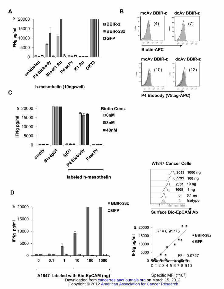
Figure 2. BBIR+ T cells exhibit specific effector functions. A. BBIRs respond against immobilized
human mesothelin protein when redirected with biotinylated anti-mesothelin scFv or antibody (P4
Biobody and Bio-K1 Ab, respectively). dcAv.BBIR-z, dcAv.BBIR-28z T cells or control GFP cells (105
cells/well) were incubated with 10ng of plate-immobilized mesothelin and with either biotinylated or not,
anti-mesothelin antibodies or scFvs (0.1μg/ml). Overnight culture supernatants were analyzed for human
IFNγ cytokine by ELISA. Data represent the means ± SD for 3 different experiments. B. Biotinylated
specific molecules retention on the BBIR T cell surface was assessed by flow cytometry. BBIR+ T cells
were incubated with 10 ng biotinylated reagents Biotin-APC or P4 Biobody (open histograms), and
American Association for Cancer Research Copyright © 2012 on March 15, 2012cancerres.aacrjournals.orgDownloaded from
Author manuscripts have been peer reviewed and accepted for publication but have not yet been edited.Author Manuscript Published OnlineFirst on February 7, 2012; DOI:10.1158/0008-5472.CAN-11-3890
Generation of a universal immune receptor.
18
compared to untransduced control T cells (grey). C. BBIRs exhibit effector functions in the presence of
free biotin at physiological concentration. BBIR T cells were incubated overnight with Bio-K1 Ab or P4
Biobody painted immobilized mesothelin protein or only with plate-bound biotinylated Abs in the
presence of the indicated concentration of biotin. Concentration of IFNγ is expressed as mean ± SEM in
pg/ml from triplicate wells. D. BBIR+ T cells exhibit effector functions against painted cell surface tumor
antigens in the presence of antigen-specific biotinylated antibodies. Left, BBIR T cells respond against
painted EpCAM on A1847 cancer cell surface. dcAv.BBIR-28z+ or control GFP+ T cells (105) were
cultured with an equal number of human A1847 unlabeled or labeled with biotinylated anti-EpCAM Ab
(0 up to 1000 ng). After overnight incubation, cell-free supernatants were analyzed for human IFNγ by
ELISA. Results depict the mean ± SEM of triplicate wells. Upper Right, Detectable surface EpCAM
expression (open histograms) after labeling with different concentrations of biotinylated EpCAM Ab was
evaluated by flow cytometry. Lower Right, Correlation of detectable Bio-EpCAM mean fluorescence
intensity (MFI) on EpCAM+ tumors was plotted vs. the production of IFNγ by BBIR-28z T cells when co-
cultured with labeled cancer cells.
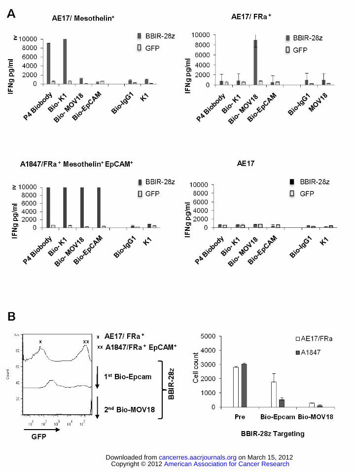
Figure 3. BBIR+ T cells exhibit effector functions against various painted cell surface tumor antigens in
the presence of antigen-specific biotinylated antibodies. A. BBIR+ T cells exhibit effector functions
against multiple antigen specificities. BBIR or GFP transduced T cells were cultured overnight with an
equal number of antigen-negative AE17, AE17/mesothelin+, AE17/Folate binding protein (FRa)+, or
A1847 cancer cells. Cell-free supernatant from three independent cultures was harvested after overnight
incubation and IFNγ levels were measured by ELISA. Mean IFNγ concentration ± SEM (pg/ml) is shown.
B. BBIR T cells can be redirected towards different antigens sequentially. BBIR T cells were cultured
with GFP transduced EpCAM+ A1847 and AE17/FRa+ cell lines at a 1:1:1 ratio. After addition of Bio-
EpCAM Ab to cultures for 10 hours, CD3-negative cells were analyzed by FACS to detect for the
presence of GFP transduced EpCAM+ A1847 cells. A second Bio-MOV18Ab (anti-FRa) was then added
American Association for Cancer Research Copyright © 2012 on March 15, 2012cancerres.aacrjournals.orgDownloaded from
Author manuscripts have been peer reviewed and accepted for publication but have not yet been edited.Author Manuscript Published OnlineFirst on February 7, 2012; DOI:10.1158/0008-5472.CAN-11-3890
Generation of a universal immune receptor.
19
to culture for an additional 10 hours, and FACS was repeated to measure for remaining CD3-negative,
GFP-negative AE17/FRa+ cells. Left, Histograms are shown. Right, Results of tumor cell count analysis
of pretreated cultures (pre) and after sequential Bio-EpCAM Ab and Bio-MOV18 Ab targeting of A1847
and AE17/FRa+ cells, respectively.
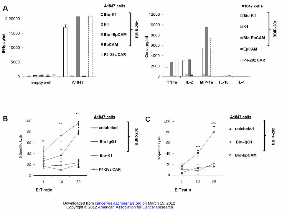
Figure 4. Activity of dcAv.BBIR-28z engineered T cells. A. dcAv.BBIR-28z+ T lymphocytes produce
inflammatory cytokines in response to painted A1847 tumor cells with biotinylated antibodies: anti-
mesothelin (Bio-K1) and/or anti-EpCAM (Bio-EpCAM). BBIR+ T cells produced equal levels of (Right)
IFNγ, and (Left) Th1 cytokines in response to painted A1847 cells compared with conventional anti-
mesothelin P4-28z CAR+ T cells. Left, Overnight culture supernatants were analyzed for human IFNγ
cytokine by ELISA. Concentration of IFNγ is expressed as mean ± SEM in pg/ml from triplicate wells.
Right, Cytokine bead-array analysis of cytokine production by dcAv.BBIR-28z+ T cells or P4-28z CAR+
T cells. Supernatants from three independent cultures were pooled and assessed after 16h. B. Antigen-
specific tumor killing by mesothelin or EpCAM redirected BBIRs. Primary human T cells transduced to
express P4-28z CAR or dcAv.BBIR-28z were co-cultured with Cr51-labeled A1847 cells with painted
mesothelin (Bio-K1) or (C) EpCAM (Bio-EpCAM) for 17hrs at the indicated effector to target ratio.
Percent specific target cell lysis was calculated as (experimental - spontaneous release) ÷ (maximal -
spontaneous release) x 100. Data represent the means ± SD for 3 different experiments. *P ≤ .005
comparing BBIR+ / Bio-K1 and BBIR+ / Bio-IgG1 T cells. **P ≤ 0.005 comparing BBIR+ and P4 CAR+
T cells and ***P ≤ .005 comparing BBIR+ / Bio-EpCAM and BBIR+ / Bio-IgG1 T cells. The difference
between the cytotoxic activity was statistically significant at given E:T ratio.
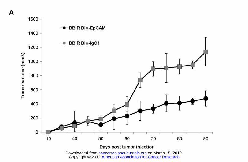
Figure 5. dcAv.BBIR-28z+ T cells control tumor growth in an ovarian cancer xenograft model. 5×10⁶
A1847 tumor cells were inoculated subcutaneously in the flank of NSG mice. To test the therapeutic
efficacy of BBIR+ T cells, mice bearing an established tumor (≥150mm3) were inoculated IT with 6×10⁶
American Association for Cancer Research Copyright © 2012 on March 15, 2012cancerres.aacrjournals.orgDownloaded from
Author manuscripts have been peer reviewed and accepted for publication but have not yet been edited.Author Manuscript Published OnlineFirst on February 7, 2012; DOI:10.1158/0008-5472.CAN-11-3890

Generation of a universal immune receptor.
20
BBIR+ T cells and Bio-EpCAM Ab (100ng) or BBIR+ T cells and Bio-IgG1 Ab (100ng) on day 45, 48
and 51. Additional antibody only injections (100ng) were performed on day 56 and 60. Tumor growth
was then monitored as tumor diameter per day. Data represent the means ± SD of 4 mice for each panel
presented. P ≤ .005 comparing BBIR+/Bio-EpCAM and BBIR+ / Bio-IgG1 group.
American Association for Cancer Research Copyright © 2012 on March 15, 2012cancerres.aacrjournals.orgDownloaded from
Author manuscripts have been peer reviewed and accepted for publication but have not yet been edited.Author Manuscript Published OnlineFirst on February 7, 2012; DOI:10.1158/0008-5472.CAN-11-3890


![Adoptive immunotherapy for Epstein-Barr virus-associated ... · gene, thereby inhibiting apoptosis of infected cells [59]. Recently, it has been recog- nized that LMP-1 is a viral](https://img.pdfslide.net/doc/110x75/5fb9b381ddf8ad35374fdda3/adoptive-immunotherapy-for-epstein-barr-virus-associated-gene-thereby-inhibiting.jpg)


![NK Cell Metabolism and the Potential Offered for Cancer Immunotherapy … · 2019. 6. 12. · immunotherapy is the advent of CAR-T adoptive cell therapies [2]. Most research efforts](https://img.pdfslide.net/doc/110x75/5ff1356f30eba7460d671eb6/nk-cell-metabolism-and-the-potential-offered-for-cancer-immunotherapy-2019-6-12.jpg)

![Immune escape after adoptive T cell therapy for malignant ......2020/08/11 · Introduction Immunotherapy has revolutionized cancer care [1, 2]. However, tumor escape is common and](https://img.pdfslide.net/doc/110x75/601ba983b3dd8949660f7303/immune-escape-after-adoptive-t-cell-therapy-for-malignant-20200811-.jpg)