Embed Size (px)
Citation preview
A Universal Stress Protein Involved in Oxidative StressIs a Phosphorylation Target for Protein Kinase CIPK61
Emilio Gutiérrez-Beltrán, José María Personat, Fernando de la Torre, and Olga del Pozo
Instituto de Bioquímica Vegetal y Fotosíntesis, Universidad de Sevilla/Consejo Superior de InvestigacionesCientíficas, 41092 Sevilla, Spain
ORCID IDs: 0000-0001-7978-3164 (E.G.B.); 0000-0003-2273-5000 (J.M.P.); 0000-0002-5143-3709 (F.d.l.T.); 0000-0001-9852-0431 (O.d.P.).
Calcineurin B-like interacting protein kinases (CIPKs) decode calcium signals upon interaction with the calcium sensorscalcineurin B like proteins into phosphorylation events that result into adaptation to environmental stresses. Fewphosphorylation targets of CIPKs are known and therefore the molecular mechanisms underlying their downstream outputresponses are not fully understood. Tomato (Solanum lycopersicum) Cipk6 regulates immune and susceptible Programmed celldeath in immunity transforming Ca2+ signals into reactive oxygen species (ROS) signaling. To investigate SlCipk6-inducedmolecular mechanisms and identify putative substrates, a yeast two-hybrid approach was carried on and a protein wasidentified that contained a Universal stress protein (Usp) domain present in bacteria, protozoa and plants, which we named“SlRd2”. SlRd2 was an ATP-binding protein that formed homodimers in planta. SlCipk6 and SlRd2 interacted usingcoimmunoprecipitation and bimolecular fluorescence complementation (BiFC) assays in Nicotiana benthamiana leaves and thecomplex localized in the cytosol. SlCipk6 phosphorylated SlRd2 in vitro, thus defining, to our knowledge, a novel target forCIPKs. Heterologous SlRd2 overexpression in yeast conferred resistance to highly toxic LiCl, whereas SlRd2 expression inEscherichia coli UspA mutant restored bacterial viability in response to H2O2 treatment. Finally, transient expression ofSlCipk6 in transgenic N. benthamiana SlRd2 overexpressors resulted in reduced ROS accumulation as compared to wild-typeplants. Taken together, our results establish that SlRd2, a tomato UspA, is, to our knowledge, a novel interactor andphosphorylation target of a member of the CIPK family, SlCipk6, and functionally regulates SlCipk6-mediated ROS generation.
Environmental factors, especially those imposingstress, stimulate endogenous cellular cues, which initi-ate protective responses in plants. Among the concur-rent events during stress are changes in the intracellularCa2+ concentration, which activate an overlapping setof downstream responses. Ca2+ changes are perceivedand decoded by an array of Ca2+ sensors including cal-modulins or calmodulin-related proteins, Ca2+-dependentprotein kinases (CDPKs, CPKs), and calcineurin B-like
proteins (CBLs; Dodd et al., 2010). Particularly, the CBLfamily has been shown to play a crucial role in differentCa2+-dependent processes in plants (Sanyal et al., 2015).CBL proteins present homology to the regulatoryB-subunit of calcineurin and the neuronal calcium sen-sor proteins from animals and yeast (Luan, 2009). Theoverall structure of CBLs consists of four EF-hands.Spacing of EF-hands is invariable, while the C- andN-terminal extension of CBL proteins varies in length.Posttranslational modifications of CBLs, including pro-tein phosphorylation and lipid modifications affect theirsubcellular localization and their stability to interact withother proteins (Sanyal et al., 2015; Nagae et al., 2003).Thus, phosphorylation of the conserved Ser residue inthe C-terminal PFPF motif of the CBL proteins enhancesthe interaction with CBL-interacting protein kinases(CIPKs; Du et al., 2011; Hashimoto et al., 2012).
Upon Ca2+ binding, CBLs physically interact withCIPKs, Ser/Thr kinases that structurally belong to Sucnonfermenting 1-related kinases, group 3, also calledprotein kinases related to SOS2 (PKS; Gong et al., 2004;Yu et al., 2014). CIPKs are constituted of a C-terminal orregulatory domain and a conserved kinase catalyticdomain at the N terminus. Within the divergent regu-latory domain, CIPKs contain an autoinhibitory NAF/FISL motif and a type 2C protein phosphatase bindingsite called the “PPI motif”. It is well established thatbinding of CBLs to the NAF/FISL motif releases theC-terminal (autoinhibitory) domain from the kinasedomain, thus leading the kinase into an active state
1 This work was funded in part by the European Regional Devel-opment Fund through the Ministerio de Economía y Competitividad(grant nos. BIO2005-02136, BIO2009-08648, and BIO2013-44750R)and by the Junta de Andalucía, Spain (grant no. P07-CVI-03171, toO.d.P.); O.d.P. was also supported in part by the Junta de Andalucía,Spain (Programa de Retorno de Investigadores); F.d.l.T. was sup-ported by Marie Curie Programme through the International Reinte-gration grants (MIRG-CT-2005-031174) and a Juan de la Ciervacontract (Ministerio de Ciencia e Innovación, Spain); E.G.-B. was a re-cipient of a Formación de Personal Investigador fellowship (Ministeriode Educación, Spain); and J.M.P. was supported by grant no.BIO2013-44750R.
* Address correspondence to [email protected] author responsible for distribution of materials integral to the
findings presented in this article in accordance with the policy de-scribed in the Instructions for Authors (www.plantphysiol.org) is:Olga del Pozo ([email protected]).
E.G.B., O.d.P., and F.d.l.T. designed experiments and analyzed thedata; O.d.P. wrote the manuscript and E.G.B. helpedwith the writing;E.G.B. performed most of experiments; J.M.P. generated Figure 7A.
www.plantphysiol.org/cgi/doi/10.1104/pp.16.00949
836 Plant Physiology�, January 2017, Vol. 173, pp. 836–852, www.plantphysiol.org � 2017 American Society of Plant Biologists. All Rights Reserved. www.plantphysiol.orgon June 15, 2020 - Published by Downloaded from
Copyright © 2017 American Society of Plant Biologists. All rights reserved.
(Guo et al., 2001; Chaves-Sanjuan et al., 2014). In Ara-bidopsis (Arabidopsis thaliana), there are 10 CBL and26CIPK homologs (Yu et al., 2014). By yeast two-hybrid(Y2H) and bimolecular fluorescence complementation(BiFC) assays it has been determined that CBLs show alevel of specificity in targeting different CIPKs. On theother hand, a specific CIPK can also interact with dif-ferent CBLs, thus allowing a single CIPK to access dif-ferent cellular compartments and hence differentsubstrates (Kim et al., 2000, 2007). It is believed that thespecificity of the response to a given stimulus is ach-ieved by decoding specific Ca2+ profiles by CBLs fol-lowed by the subsequent formation of different CBL/CIPK complexes in planta, and finally by phosphoryl-ation of CIPK-specific substrates that contribute to thespecific output response (Batistic et al., 2010).At the moment, the most numerous and best charac-
terized interactors or substrates for CBL/CIPK com-plexes are membrane proteins, which include salt overlysensitive 1 (SOS1; Quintero et al., 2002; Katiyar-Agarwalet al., 2006), H+-ATPase 2 (He et al., 2004), nitrate trans-porter (Ho et al., 2009), K+ transporter 1 (AKT1; Xu et al.,2006), high-affinity K+ transporter 5 (Ragel et al., 2015),and the respiratory burst oxidase homolog F (Drerupet al., 2013). Additionally, CIPKs have also been shown tointeract with nonmembrane proteins; for example, SOS2-like protein kinase 5 interacts with the chaperone DnaJ(He et al., 2004; Yang et al., 2010); AtCIPK24 interactswith GIGANTEA (Kim et al., 2013), nucleoside diphos-phate kinase 2, the catalases CAT2 and CAT3 (Verslueset al., 2007) andwith ABA-insensitive 2 (ABI2), a type 2CSer/Thr phosphatase (Guo et al., 2002; Ohta et al., 2003);and CIPK26 interacts with the RING-type E3 ligase“Keep on Going” and with ABI1, ABI2 (Lyzenga et al.,2013). Although it appears that CBL/CIPK complexescould interact with several proteins, at present only fewCIPK phosphorylation targets have been identified.Previously, our group demonstrated, to our knowl-
edge, a novel role for tomato (Solanum lycopersicum) Cipk6(SlCipk6) in plant innate immunity, thus functionallyimplicating for the first time the participation of a CBL/CIPK module in biotic stress signaling in plants (de laTorre et al., 2013). Other studies demonstrated the par-ticipation of Cipk6 orthologs from different plant speciesin diverse abiotic stress responses (Chen et al., 2012, 2013;Tsou et al., 2012; Tripathi et al., 2009). As a first step toinvestigate SlCipk6 downstream signaling molecularmechanisms, we set to identify SlCipk6-interacting pro-teins using a Y2H approach. We discovered that tomatoResponsive to desiccation 2 (SlRd2), which contains aUniversal stress protein (Usp) domain [Pfam (http://pfam.xfam.org/) accession no. PF00582], interactedwith SlCipk6 and by means of a BiFC approach, wefound that the complex SlCipk6/SlRd2 is localized in thecytoplasm. In addition, we demonstrated that SlRd2 is aphosphorylation substrate of SlCipk6, thus expandingthe previously described substrates for the CIPK family.Interestingly, SlRd2 is anATP-binding protein that formshomodimers, which is required for its biological role andfor interacting with SlCipk6.
The universal stress protein A (UspA) superfamilywas originally discovered in Escherichia coli, where itsexpression drastically increased in response to multiplestress conditions and to starvation (Vanbogelen et al.,1990). Importantly, UspA protein accumulation wasnecessary for bacterial survival at the stationary phase(Nyström and Neidhardt, 1994). It was found later thatE. coli has six usp genes (uspA, uspC, uspD, uspF, uspF,and uspG); however, UspA set the nomenclature for theorthologous groups of proteins. UspA family membersare classified into two major groups according to theirATP binding capability. The first group is constitutedby ATP-binding proteins and is represented by Mj0577from Methanococcus jannaschii (Zarembinski et al., 1998).Members of the second group have no ATP-binding ca-pability and are represented by Haemophilus influenzaeand E. coliUspAs (Sousa andMcKay, 2001). BothMj0577and HiUspA form homodimers in vivo (Zarembinskiet al., 1998). At present, more than 2000 UspA (or Uspcontaining domain) proteins have been identified from awide range of organisms such as bacteria, archaea, fungi,protozoa and plants, constituting an ancient and con-served group of proteins (Aravind et al., 2002). In Ara-bidopsis, at least 44 proteins were found to contain anUsp domain, all of which resemble ATP-binding Mj0577protein (Kerk et al., 2003) and several plantUspAs seemedto be involved in abiotic stress. In Arabidopsis, two UspAproteins, AtPHOS32 and AtPHOS34, were phosphory-lated by AtMPK3 and AtMPK6 in response to bacterialelicitors in cell suspension cultures (Merkouropoulos et al.,2008). Other reports described several UspAmembers aseffectors of low water potential (Merkouropoulos et al.,2008). Several UspA proteins have been characterized inrice (Sauter et al., 2002), tomato (Zegzouti et al., 1999;Loukehaich et al., 2012) legumes (Becker et al., 2001;Hohnjec et al., 2000), Salicornia (Udawat et al., 2016), andcotton (Zahur et al., 2009). Still, the precise structure,regulation, biochemical function, or mechanism offunction of UspA proteins in planta, are largely un-known.
RESULTS
Identification of SlRd2 as a SlCipk6-Interacting Protein
To identify SlCipk6-interacting proteins (CIPs), wecarried out a Y2H approach in two separate screens,using a tomato cDNAprey library previously developed(Zhou et al., 1995) and SlCipk6 and a mutant derivative,SlCipk6(T172D), as baits (Fig. 1A). SlCipk6(T172D)displayed enhanced kinase and autophosphorylationactivity compared to SlCipk6 (de la Torre et al., 2013),and we hypothesized that using either SlCipk6 orSlCipk6(T172D) as a bait, might facilitate the identi-fication of putative regulatory proteins or phosphor-ylation substrates respectively, because constitutiveactive kinase versions stabilize the interaction withtheir substrates (Uno et al., 2009). Approximately 1.13 103 and 4.53 103 yeast transformants were screenedfor SlCipk6- and SlCipk6(T172D)-interacting proteins
Plant Physiol. Vol. 173, 2017 837
SlRd2 Phosphorylation and Role in Stress Defense
www.plantphysiol.orgon June 15, 2020 - Published by Downloaded from Copyright © 2017 American Society of Plant Biologists. All rights reserved.
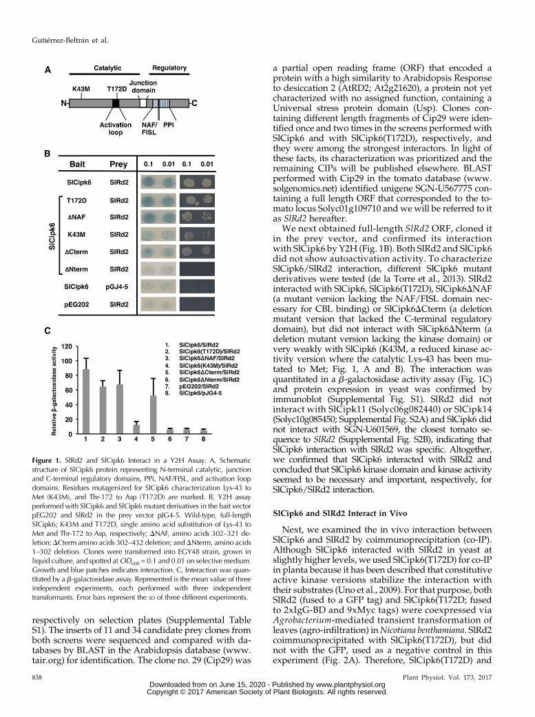
respectively on selection plates (Supplemental TableS1). The inserts of 11 and 34 candidate prey clones fromboth screens were sequenced and compared with da-tabases by BLAST in the Arabidopsis database (www.tair.org) for identification. The clone no. 29 (Cip29) was
a partial open reading frame (ORF) that encoded aprotein with a high similarity to Arabidopsis Responseto desiccation 2 (AtRD2; At2g21620), a protein not yetcharacterized with no assigned function, containing aUniversal stress protein domain (Usp). Clones con-taining different length fragments of Cip29 were iden-tified once and two times in the screens performed withSlCipk6 and with SlCipk6(T172D), respectively, andthey were among the strongest interactors. In light ofthese facts, its characterization was prioritized and theremaining CIPs will be published elsewhere. BLASTperformed with Cip29 in the tomato database (www.solgenomics.net) identified unigene SGN-U567775 con-taining a full length ORF that corresponded to the to-mato locus Solyc01g109710 and we will be referred to itas SlRd2 hereafter.
We next obtained full-length SlRd2 ORF, cloned itin the prey vector, and confirmed its interactionwith SlCipk6 by Y2H (Fig. 1B). Both SlRd2 and SlCipk6did not show autoactivation activity. To characterizeSlCipk6/SlRd2 interaction, different SlCipk6 mutantderivatives were tested (de la Torre et al., 2013). SlRd2interacted with SlCipk6, SlCipk6(T172D), SlCipk6DNAF(a mutant version lacking the NAF/FISL domain nec-essary for CBL binding) or SlCipk6DCterm (a deletionmutant version that lacked the C-terminal regulatorydomain), but did not interact with SlCipk6DNterm (adeletion mutant version lacking the kinase domain) orvery weakly with SlCipk6 (K43M, a reduced kinase ac-tivity version where the catalytic Lys-43 has been mu-tated to Met; Fig. 1, A and B). The interaction wasquantitated in a b-galactosidase activity assay (Fig. 1C)and protein expression in yeast was confirmed byimmunoblot (Supplemental Fig. S1). SlRd2 did notinteract with SlCipk11 (Solyc06g082440) or SlCipk14(Solyc10g085450; Supplemental Fig. S2A) and SlCipk6 didnot interact with SGN-U601569, the closest tomato se-quence to SlRd2 (Supplemental Fig. S2B), indicating thatSlCipk6 interaction with SlRd2 was specific. Altogether,we confirmed that SlCipk6 interacted with SlRd2 andconcluded that SlCipk6 kinase domain and kinase activityseemed to be necessary and important, respectively, forSlCipk6/SlRd2 interaction.
SlCipk6 and SlRd2 Interact in Vivo
Next, we examined the in vivo interaction betweenSlCipk6 and SlRd2 by coimmunoprecipitation (co-IP).Although SlCipk6 interacted with SlRd2 in yeast atslightly higher levels, we used SlCipk6(T172D) for co-IPin planta because it has been described that constitutiveactive kinase versions stabilize the interaction withtheir substrates (Uno et al., 2009). For that purpose, bothSlRd2 (fused to a GFP tag) and SlCipk6(T172D; fusedto 2xIgG-BD and 9xMyc tags) were coexpressed viaAgrobacterium-mediated transient transformation ofleaves (agro-infiltration) inNicotiana benthamiana. SlRd2coimmunoprecipitated with SlCipk6(T172D), but didnot with the GFP, used as a negative control in thisexperiment (Fig. 2A). Therefore, SlCipk6(T172D) and
Figure 1. SlRd2 and SlCipk6 Interact in a Y2H Assay. A, Schematicstructure of SlCipk6 protein representing N-terminal catalytic, junctionand C-terminal regulatory domains, PPi, NAF/FISL, and activation loopdomains. Residues mutagenized for SlCipk6 characterization Lys-43 toMet (K43M), and Thr-172 to Asp (T172D) are marked. B, Y2H assayperformed with SlCipk6 and SlCipk6 mutant derivatives in the bait vectorpEG202 and SlRd2 in the prey vector pJG4-5. Wild-type, full-lengthSlCipk6; K43M and T172D, single amino acid substitution of Lys-43 toMet and Thr-172 to Asp, respectively; DNAF, amino acids 302–321 de-letion;DCterm amino acids 302–432 deletion; andDNterm, amino acids1–302 deletion. Clones were transformed into EGY48 strain, grown inliquid culture, and spotted atOD600 = 0.1 and 0.01 on selective medium.Growth and blue patches indicates interaction. C, Interaction was quan-titated by a b-galactosidase assay. Represented is the mean value of threeindependent experiments, each performed with three independenttransformants. Error bars represent the SD of three different experiments.
838 Plant Physiol. Vol. 173, 2017
Gutiérrez-Beltrán et al.
www.plantphysiol.orgon June 15, 2020 - Published by Downloaded from Copyright © 2017 American Society of Plant Biologists. All rights reserved.
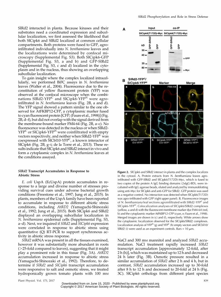
SlRd2 interacted in planta. Because kinases and theirsubstrates need a coordinated expression and subcel-lular localization, we first assessed the likelihood thatboth SlCipk6 and SlRd2 localized at common cellularcompartments. Both proteins were fused to GFP, agro-infiltrated individually into N. benthamiana leaves andthe localizations were determined by confocal mi-croscopy (Supplemental Fig. S3). Both SlCipk6-GFP(Supplemental Fig. S3, a and b) and GFP-SlRd2(Supplemental Fig. S3, c and d) localized in the cyto-plasm and in the nucleus, thus showing an overlappingsubcellular localization.To gain insight where the complex localized intracel-
lularly, we performed BiFC assays in N. benthamianaleaves (Walter et al., 2004). Fluorescence due to the re-constitution of yellow fluorescent protein (YFP) wasobserved at the confocal microscope when the combi-nations SlRd2-YFPC and SlCipk6-YFPN were agro-infiltrated in N. benthamiana leaves (Fig. 2B, a and d).The YFP signal showed a pattern similar to the one ob-served for AtFKBP12-CFP, a cytoplasmic marker fusedto cyanfluorescent protein [CFP; (Faure et al., 1998)] (Fig.2B, d–f), but did not overlapwith the signal derived fromthe membrane-bound marker FM4-64 (Fig. 2B, a–c). Nofluorescence was detected in the nucleus or when SlRd2-YFPC or SlCipk6-YFPN were coinfiltrated with emptyvectors respectively, and neither when SlRd2-YFPCwascoexpressed with SlCbl10-YFPC, a known interactor ofSlCipk6 (Fig. 2B, g–i; de la Torre et al., 2013). These re-sults indicate that SlCipk6 and SlRd2 interact in vivo andform a cytoplasmic complex in N. benthamiana leaves atthe conditions assayed.
SlRd2 Transcript Accumulates in Response toAbiotic Stress
E. coli UspA (EcUspA) protein accumulates in re-sponse to a large and diverse number of stresses pro-viding survival cues under adverse bacterial growthconditions (Freestone et al., 1997; Jung et al., 2015). Inplants, members of the UspA family have been reportedto accumulate in response to different abiotic stressconditions, including AtRD2 (Yamaguchi-Shinozakiet al., 1992; Jung et al., 2015). Both SlCipk6 and SlRd2displayed an overlapping subcellular localization inN. benthamiana epidermal cells (Supplemental Fig. S3,a–d). Next, we inquired if SlRd2 and SlCipk6 expressionwere correlated in response to abiotic stress usingquantitative (Q) RT-PCR to support synchronous ac-tivity in abiotic stress response.SlRd2mRNAwas present in all the tissues examined,
however it was substantially more abundant in roots(.25-fold compared to leaves), suggesting that it mightplay a prominent role in this tissue (Fig. 3A). AtRD2accumulation increased in response to abiotic stress(Yamaguchi-Shinozaki et al., 1992). Therefore, to de-termine if SlRd2 and SlCipk6 transcript accumulationwere responsive to salt and osmotic stress, we treatedhydroponically grown tomato plants with 100 mM
NaCl and 300 mM mannitol and analyzed SlRd2 accu-mulation. NaCl treatment rapidly increased SlRd2transcript accumulation [approximately 12-fold, after2 h (h)], whichwasmaintained up to 12 h and decreased24 h later (Fig. 3B). Osmotic pressure resulted in asimilar accumulation of SlRd2 after 2 h and 4 h, but incontrast, SlRd2 accumulation increased up to 30-foldafter 8 h to 12 h and decreased to 20-fold at 24 h (Fig.3C). SlCipk6 orthologs from different plant species
Figure 2. SlCipk6 and SlRd2 interact in planta and the complex localizesin the cytosol. A, Protein extracts from N. benthamiana leaves agro-infiltrated with GFP-SlRd2 and SlCipk6(T172D)-Myc, which is fused totwo copies of the protein A IgG binding domains (2xIgG-BD), were in-cubatedwith IgG agarose beads, eluted and analyzed by immunoblottingusing anti-Myc for SlCipk6 and anti-GFP for SlRd2. GFP protein was usedas a negative control. No interaction was detected when SlCipk6(T172D)was agro-infiltrated with GFP (right upper panel). B, Fluorescence imagesof N. benthamiana leaf sections agroinfiltrated with SlRd2-YFPC andSlCipk6-YFPN. Colocalization analyses of SlCipk6/SlRd2 complexes(yellow, a and d) with the fluorescent membranemarker dye FM4-64 (red,b) and the cytoplasmicmarker AtFKBP12-CFP (cyan, e; Faure et al., 1998).Merged images are shown in (c) and (f), respectively. White arrows showthe cytoplasmic localization observed for the SlCipk6/SlRd2 complexes.Localization analysis of YFPN (g) and YFPC (h) empty vectors and SlCbl10/SlRd2 (i) were used as an experiment controls. Bars = 10 mm.
Plant Physiol. Vol. 173, 2017 839
SlRd2 Phosphorylation and Role in Stress Defense
www.plantphysiol.orgon June 15, 2020 - Published by Downloaded from Copyright © 2017 American Society of Plant Biologists. All rights reserved.

have been described to participate in different abioticstress responses, and we tested if SlCipk6 transcriptaccumulated also in response to abiotic stress. For thatpurpose, we treated tomato plants with NaCl andmannitol, using the same conditions as describedabove. After NaCl treatment, accumulation of SlCipk6
mRNA increased 6-fold to 8-fold after 2 h and wasmaintained at the same levels up to 8 h, decreasinggradually thereafter, reaching a 2-fold increase at 24 h(Fig. 3D). Osmotic pressure also resulted in an in-creased SlCipk6 mRNA accumulation; however, fol-lowed a different pattern compared to SlRd2: 2 h afterSlCipk6 transcripts increased 6-fold and steadily in-creased up to 15 fold 24 h after (Fig. 3E). Both SlCipk6and SlRd2 transcript accumulation pattern showedsimilar kinetics, thus supporting the possibility of acoordinated role for SlCipk6 and SlRd2 in abiotic stressresponses in tomato.
SlRd2 Belongs to the Universal Stress ProteinA Superfamily
The translated sequence of SlRd2 ORF yields a177 amino acid protein, with a Mr of 19.5 kD and a pI of5.98. We aligned the Usp domain of SlRd2 with those ofN. benthamiana and Arabidopsis orthologs NbRd2 andAtRD2 and other plant proteins containing the Usp do-main, including tomato LeER6 (Zegzouti et al., 1999), rice(Oryza sativa) OsUSP1 (Sauter et al., 2002), and Vicia favaEnod18 (Becker et al., 2001) along with bacterial proteinsbelonging to the UspA family, including E. coli (Nyströmand Neidhardt, 1992), H. influenzae (Sousa and McKay,2001) and M. jannaschii (Zarembinski et al., 1998;Supplemental Fig. S4). Amino acid sequence alignmentrevealed that SlRd2 contained the residues involved inATP binding including the Walker motif A or P-loop,(G-2X-G-9X-G(S/T)), also present in Mj0577 and UspAplant representatives but absent in E. coli paralogs (Sousaand McKay, 2001). This observation suggests that plantUspA proteins might also be functional ATP-bindingproteins (Supplemental Figs. S4 and 5A). Overall, allUspA proteins (plant and bacterial) shared conservationwithin the dimerization subdomain (Supplemental Fig.S4), thus raising the possibility that plant UspAs could bepresent as dimers in the cell. In addition to the conservedUsp domain, SlRd2 has an N-terminal domain (aminoacids 1 to 38) and a C-terminal extension (amino acids168 to 177) with unknown function, which is sharedwiththe Arabidopsis and N. benthamiana homologs (Figs. 4Aand S5). Subsequently, a phylogenetic analysis was per-formed using E. coli UspG and the proteins included inthe alignment shown in Supplemental Fig. S4. SlRd2 andNbRd2 were located in the same clade as AtRD2, thussupporting their orthology (Fig. 4B). Moreover, SlRd2,and all plant UspAs, are more related to Mj0577 sub-family and E. coliUspG than to the E. coliUspA (Fig. 4B).
SlRd2 Is an ATP-binding Protein and Forms Homodimersin Yeast, Bacteria, and Plants
In light of the high degree of conservation of SlRd2amino acids putatively involved in nucleotide binding(see alignments in Figs. 5A and S4), we next inquired ifSlRd2 also had the functional competence to bind ATP,as described for bacterial Mj0577. For that purpose, we
Figure 3. SlRd2 and SlCipk6 transcripts highly accumulate after nacl andosmotic stress in tomato. A to E, y axis represents mean values of QRT-PCR of three experiments, with three biological replicates in each.Error bars represent the SE. The expression levels were normalized toSlActin2. Gene induction (fold increase) in infected or treated plants wascompared with the expression level of control or mock inoculated plantsat 0 h and is shown as relative expression, excepting in (A). A, SlRd2transcript accumulates in tomato leaves, petioles, stems, flowers, androots. Relative expression on y axiswas comparedwith expression level inleaves. B and C, SlRd2; D and E, SlCipk6 mRNA accumulation in leavesduring NaCl (B and D) and osmotic stress (C and E). Hydroponicallygrown tomato plants were treatedwith 100mMNaCl (B andD) or 300mM
mannitol (C and E). OS, osmotic stress.
840 Plant Physiol. Vol. 173, 2017
Gutiérrez-Beltrán et al.
www.plantphysiol.orgon June 15, 2020 - Published by Downloaded from Copyright © 2017 American Society of Plant Biologists. All rights reserved.
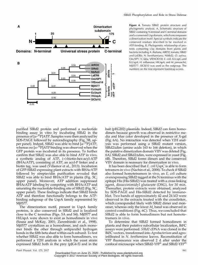
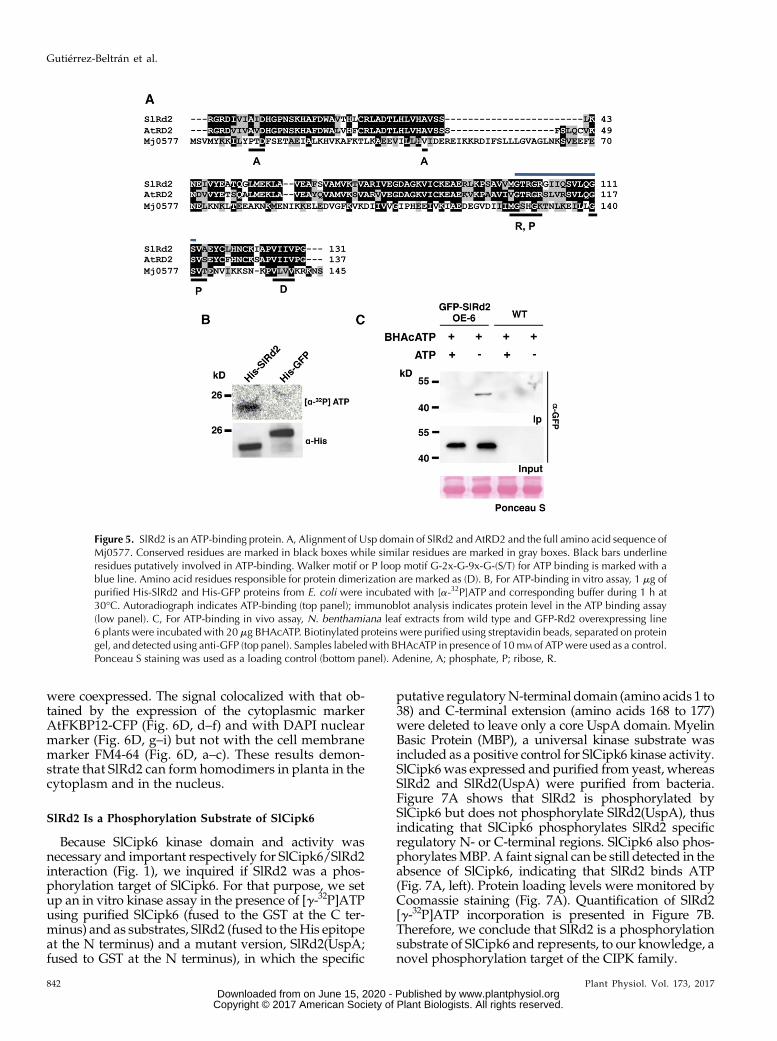
purified SlRd2 protein and performed a nucleotidebinding assay in vitro by incubating SlRd2 in thepresence of [a-32P]ATP. Samples were then analyzed bySDS-PAGE followed by autoradiography (Fig. 5B, up-per panel). Indeed, SlRd2 was able to bind [a-32P]ATP,whereas no [a-32P]ATP binding was observed when theGFP protein was incubated in its presence. To furtherconfirm that SlRd2 was also able to bind ATP in vivo,a synthetic analog of ATP, (+)-biotin-hex-acyl-ATP(BHAcATP), consisting of ATP, an acyl-P linker and abiotin tag, was used (Villamor et al., 2013). Incubationof GFP-SlRd2 expressing plant extracts with BHAcATPfollowed by streptavidin purification revealed thatSlRd2 was able to bind BHAcATP in planta (Fig. 5C,upper panel). Moreover, ATP addition suppressedBHAcATP labeling by competing with BHAcATP andsaturating the nucleotide-binding site of SlRd2 (Fig. 5C,upper panel). These findings indicate that SlRd2 bindsATP and therefore functionally belongs to the ATP-binding subgroup of the UspA family represented byMj0577.The dimerization motif, present in UspA family
proteins, is also conserved in SlRd2 and is localizedclose to the C terminus (Figs. 5A and S4). Mj0577 andHiUspA were shown to exist as homodimers in vivo(Sousa and McKay, 2001; Zarembinski et al., 1998).Mj0577 crystallizes as a homodimer, and each mono-mer binds the other through antiparallel hydrogenbonds in the fifth beta sheet within each subunit. To testwhether SlRd2 was also able to form homodimers, weperformed a Y2H analysis in which the yeast strainexpressed SlRd2 both in the prey (pJG4-5) and in the
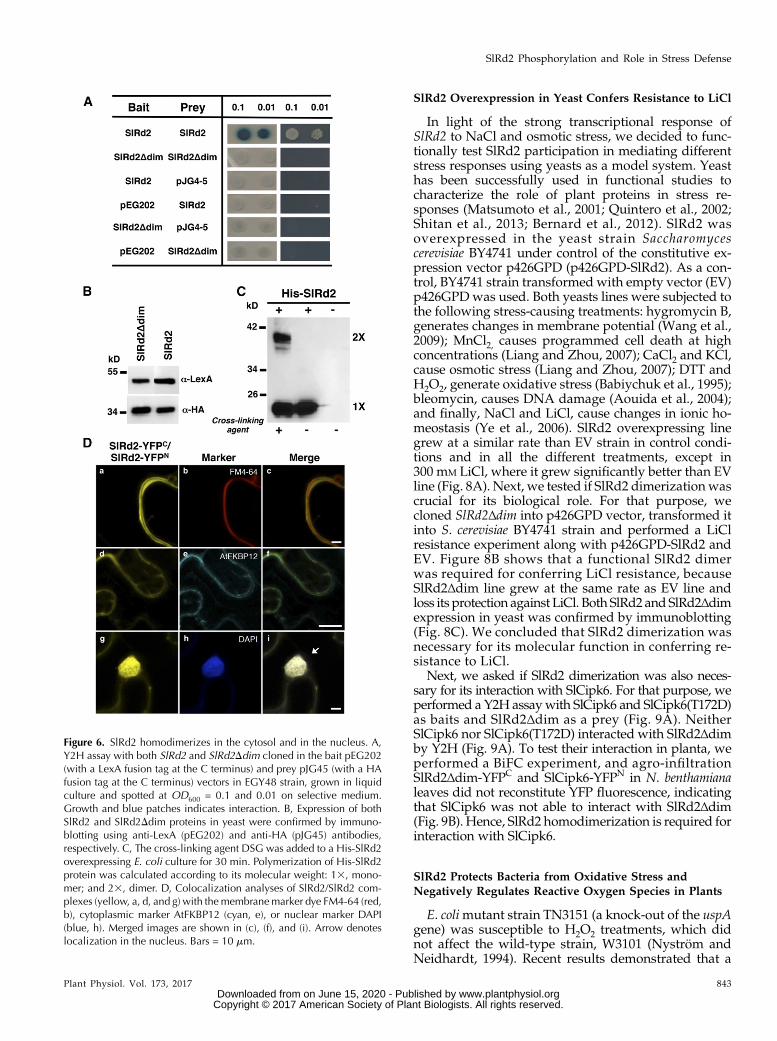
bait (pEG202) plasmids. Indeed, SlRd2 can form homo-dimers because growth was observed in restrictive me-dia and blue color developed in the presence of X-gal(Fig. 6A). No interaction was detected when Y2H anal-ysis was performed using a SlRd2 mutant version,SlRd2Δdim (amino acids 163 to 166 deletion), in whichthe putative dimerization domain VIIVwas deleted (Fig.6A). SlRd2 and SlRd2Δdim,were expressed in yeast (Fig.6B). Therefore, SlRd2 forms dimers and the conservedVIIV domain is necessary for dimerization in vivo.
It has been described that E. coli UspC is able to formtetramers in vivo (Nachin et al., 2008). To check if SlRd2also formed homotetramers in vivo, an E. coli cultureoverexpressing SlRd2 tagged at theN terminuswith theepitope His (His-SlRd2) was treated with a cross-linkingagent, disuccinimidyl glutarate (DSG), for 30 min.Thereafter, protein extracts were obtained, analyzedby SDS-PAGE and His-SlRd2 detected by immuno-blot. Two bands of approximately 42 and 24 kD wereobserved in the extracts treated with the crosslinker,which corresponded likely with SlRd2 dimer and mon-omer, whereas only the lower Mr band was observed incontrol conditions (Fig. 6C). Thus, we concluded thatSlRd2 is able to form homodimers but not homote-tramers in vivo.
To determine that SlRd2 formed homodimers inplanta and their putative subcellular localization, BiFCassays were performed. SlRd2 cDNA was cloned in theBiFC vectors, transformed into Agrobacterium and agro-infiltrated in N. benthamiana leaves. Reconstitution ofYFP fluorescence was observed 2 d after under theconfocal microscope when SlRd2-YFPC and SlRd2-YFPN
Figure 4. Tomato SlRd2 protein structure andphylogenetic analysis. A, Schematic structure ofSlRd2 containing N-terminal and C-terminal domainsand a conservedUsp domain, which encompassesa dimerization motif. Special symbols indicate theconserved residues described to be involved inATP-binding. B, Phylogenetic relationship of pro-teins containing Usp domains from plants andbacteria includingA. thaliana, AtRD2; tomato, SlRd2and LeER6; N. benthamiana, NbRd2; O. sativa,OsUSP1; V. faba, VfENOD18; E. coli, EcUspG andEcUspA;H. influenzae, HiUspA; andM. jannaschii,Mj0577. SlCbl10 was used as the outgroup. Thenumbers on the tree represent bootstrap scores.
Plant Physiol. Vol. 173, 2017 841
SlRd2 Phosphorylation and Role in Stress Defense
www.plantphysiol.orgon June 15, 2020 - Published by Downloaded from Copyright © 2017 American Society of Plant Biologists. All rights reserved.
were coexpressed. The signal colocalized with that ob-tained by the expression of the cytoplasmic markerAtFKBP12-CFP (Fig. 6D, d–f) and with DAPI nuclearmarker (Fig. 6D, g–i) but not with the cell membranemarker FM4-64 (Fig. 6D, a–c). These results demon-strate that SlRd2 can form homodimers in planta in thecytoplasm and in the nucleus.
SlRd2 Is a Phosphorylation Substrate of SlCipk6
Because SlCipk6 kinase domain and activity wasnecessary and important respectively for SlCipk6/SlRd2interaction (Fig. 1), we inquired if SlRd2 was a phos-phorylation target of SlCipk6. For that purpose, we setup an in vitro kinase assay in the presence of [g-32P]ATPusing purified SlCipk6 (fused to the GST at the C ter-minus) and as substrates, SlRd2 (fused to theHis epitopeat the N terminus) and a mutant version, SlRd2(UspA;fused to GST at the N terminus), in which the specific
putative regulatoryN-terminal domain (amino acids 1 to38) and C-terminal extension (amino acids 168 to 177)were deleted to leave only a core UspA domain. MyelinBasic Protein (MBP), a universal kinase substrate wasincluded as a positive control for SlCipk6 kinase activity.SlCipk6was expressed and purified fromyeast, whereasSlRd2 and SlRd2(UspA) were purified from bacteria.Figure 7A shows that SlRd2 is phosphorylated bySlCipk6 but does not phosphorylate SlRd2(UspA), thusindicating that SlCipk6 phosphorylates SlRd2 specificregulatory N- or C-terminal regions. SlCipk6 also phos-phorylatesMBP. A faint signal can be still detected in theabsence of SlCipk6, indicating that SlRd2 binds ATP(Fig. 7A, left). Protein loading levels were monitored byCoomassie staining (Fig. 7A). Quantification of SlRd2[g-32P]ATP incorporation is presented in Figure 7B.Therefore, we conclude that SlRd2 is a phosphorylationsubstrate of SlCipk6 and represents, to our knowledge, anovel phosphorylation target of the CIPK family.
Figure 5. SlRd2 is an ATP-binding protein. A, Alignment of Usp domain of SlRd2 and AtRD2 and the full amino acid sequence ofMj0577. Conserved residues are marked in black boxes while similar residues are marked in gray boxes. Black bars underlineresidues putatively involved in ATP-binding. Walker motif or P loop motif G-2x-G-9x-G-(S/T) for ATP binding is marked with ablue line. Amino acid residues responsible for protein dimerization are marked as (D). B, For ATP-binding in vitro assay, 1 mg ofpurified His-SlRd2 and His-GFP proteins from E. coli were incubated with [a-32P]ATP and corresponding buffer during 1 h at30°C. Autoradiograph indicates ATP-binding (top panel); immunoblot analysis indicates protein level in the ATP binding assay(low panel). C, For ATP-binding in vivo assay, N. benthamiana leaf extracts from wild type and GFP-Rd2 overexpressing line6 plants were incubated with 20 mg BHAcATP. Biotinylated proteins were purified using streptavidin beads, separated on proteingel, and detected using anti-GFP (top panel). Samples labeledwith BHAcATP in presence of 10mM of ATPwere used as a control.Ponceau S staining was used as a loading control (bottom panel). Adenine, A; phosphate, P; ribose, R.
842 Plant Physiol. Vol. 173, 2017
Gutiérrez-Beltrán et al.
www.plantphysiol.orgon June 15, 2020 - Published by Downloaded from Copyright © 2017 American Society of Plant Biologists. All rights reserved.
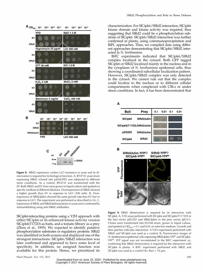
SlRd2 Overexpression in Yeast Confers Resistance to LiCl
In light of the strong transcriptional response ofSlRd2 to NaCl and osmotic stress, we decided to func-tionally test SlRd2 participation in mediating differentstress responses using yeasts as a model system. Yeasthas been successfully used in functional studies tocharacterize the role of plant proteins in stress re-sponses (Matsumoto et al., 2001; Quintero et al., 2002;Shitan et al., 2013; Bernard et al., 2012). SlRd2 wasoverexpressed in the yeast strain Saccharomycescerevisiae BY4741 under control of the constitutive ex-pression vector p426GPD (p426GPD-SlRd2). As a con-trol, BY4741 strain transformed with empty vector (EV)p426GPDwas used. Both yeasts lines were subjected tothe following stress-causing treatments: hygromycin B,generates changes in membrane potential (Wang et al.,2009); MnCl2, causes programmed cell death at highconcentrations (Liang and Zhou, 2007); CaCl2 and KCl,cause osmotic stress (Liang and Zhou, 2007); DTT andH2O2, generate oxidative stress (Babiychuk et al., 1995);bleomycin, causes DNA damage (Aouida et al., 2004);and finally, NaCl and LiCl, cause changes in ionic ho-meostasis (Ye et al., 2006). SlRd2 overexpressing linegrew at a similar rate than EV strain in control condi-tions and in all the different treatments, except in300 mM LiCl, where it grew significantly better than EVline (Fig. 8A). Next, we tested if SlRd2 dimerizationwascrucial for its biological role. For that purpose, wecloned SlRd2Δdim into p426GPD vector, transformed itinto S. cerevisiae BY4741 strain and performed a LiClresistance experiment along with p426GPD-SlRd2 andEV. Figure 8B shows that a functional SlRd2 dimerwas required for conferring LiCl resistance, becauseSlRd2Δdim line grew at the same rate as EV line andloss its protection against LiCl. Both SlRd2 and SlRd2Δdimexpression in yeast was confirmed by immunoblotting(Fig. 8C). We concluded that SlRd2 dimerization wasnecessary for its molecular function in conferring re-sistance to LiCl.
Next, we asked if SlRd2 dimerization was also neces-sary for its interaction with SlCipk6. For that purpose, weperformed a Y2H assaywith SlCipk6 and SlCipk6(T172D)as baits and SlRd2Δdim as a prey (Fig. 9A). NeitherSlCipk6 nor SlCipk6(T172D) interacted with SlRd2Δdimby Y2H (Fig. 9A). To test their interaction in planta, weperformed a BiFC experiment, and agro-infiltrationSlRd2Δdim-YFPC and SlCipk6-YFPN in N. benthamianaleaves did not reconstitute YFP fluorescence, indicatingthat SlCipk6 was not able to interact with SlRd2Δdim(Fig. 9B). Hence, SlRd2 homodimerization is required forinteraction with SlCipk6.
SlRd2 Protects Bacteria from Oxidative Stress andNegatively Regulates Reactive Oxygen Species in Plants
E. colimutant strain TN3151 (a knock-out of the uspAgene) was susceptible to H2O2 treatments, which didnot affect the wild-type strain, W3101 (Nyström andNeidhardt, 1994). Recent results demonstrated that a
Figure 6. SlRd2 homodimerizes in the cytosol and in the nucleus. A,Y2H assay with both SlRd2 and SlRd2Ddim cloned in the bait pEG202(with a LexA fusion tag at the C terminus) and prey pJG45 (with a HAfusion tag at the C terminus) vectors in EGY48 strain, grown in liquidculture and spotted at OD600 = 0.1 and 0.01 on selective medium.Growth and blue patches indicates interaction. B, Expression of bothSlRd2 and SlRd2Ddim proteins in yeast were confirmed by immuno-blotting using anti-LexA (pEG202) and anti-HA (pJG45) antibodies,respectively. C, The cross-linking agent DSG was added to a His-SlRd2overexpressing E. coli culture for 30 min. Polymerization of His-SlRd2protein was calculated according to its molecular weight: 13, mono-mer; and 23, dimer. D, Colocalization analyses of SlRd2/SlRd2 com-plexes (yellow, a, d, and g) with themembranemarker dye FM4-64 (red,b), cytoplasmic marker AtFKBP12 (cyan, e), or nuclear marker DAPI(blue, h). Merged images are shown in (c), (f), and (i). Arrow denoteslocalization in the nucleus. Bars = 10 mm.
Plant Physiol. Vol. 173, 2017 843
SlRd2 Phosphorylation and Role in Stress Defense
www.plantphysiol.orgon June 15, 2020 - Published by Downloaded from Copyright © 2017 American Society of Plant Biologists. All rights reserved.

UspAprotein from the pathogenic bacteriaMycobacteriumtuberculosis provided protection for the parasiteagainst host reactive oxygen species (ROS) generatedby mammalian macrophages defense (Drumm et al.,2009). SlCipk6 was demonstrated to participate in ROSgeneration during plant responses to bacterial patho-gen attack, which was dependent on the NADPH oxi-dase, RbohB (de la Torre et al., 2013). Given the striking
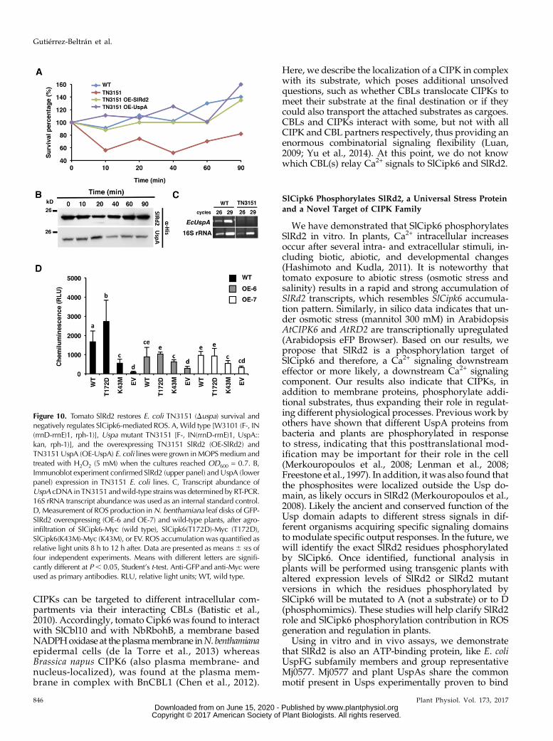
conserved structural and functional features betweenSlRd2 and bacterial UspA proteins, we decided to test ifSlRd2 could functionally complement TN3151 in pro-tecting bacteria in response to H2O2 treatment. E. coliwild-type W3101 (wild type), uspamutant TN3151 andTN3151 complemented with either SlRd2 (TN3151-SlRd2) or with the native UspA gene (TN3151-UspA)were grown in minimal media (morpholine propanesulphonic acid, MOPS) until OD600 reached 0.7. Then,H2O2 (5 mM) was added and survival was measured atdifferent time points after (Fig. 10A). A strong growthinhibition was observed for TN3151 strain after H2O2treatment, as previously described (Nyström andNeidhardt, 1994); 40 min after H2O2 addition, only 45%of TN3151 survived whereas 100% TN3151-SlRd2 sur-vived, showing a similar survival rate as either TN3151-EcUspA or wild-type W3101. SlRd2 and EcUspAexpression in TN3151was confirmed by immunoblotting(Fig. 10B) and lack of UspA gene expression in TN3151was confirmed by PCR (Fig. 10C). In light of these re-sults, we concluded that tomato SlRd2 functionallycomplements TN3151 mutant strain in protecting bac-teria against oxidative stress damage and thus SlRd2and E. coli UspA are functionally conserved in theirROS protection role in bacteria.
We have previously shown that kinase activity ofSlCipk6 is associated with ROS production in N.benthamiana (de la Torre et al., 2013), so we next in-quired if SlRd2 is required for SlCipk6-mediated ROSgeneration. To this end, c-Myc tagged versions ofSlCipk6, SlCipk6(T172D) and SlCipk6(K43M; clonedinto pTAPa-pYL436 vector) or EV were agro-infiltratedin N. benthamiana wild-type and overexpressing GFP-SlRd2 (OE-6, OE-7) leaves and production of ROS wasquantifiedbya chemiluminescence assay in a luminometer(Fig. 10D). As expected, SlCipk6 and SlCipk6(T172D)expression in wild-type leaf discs resulted in ROS gener-ation, which was significantly reduced in SlCipk6(K43M)expressing leaf discs (de la Torre et al., 2013). However,SlCipk6 and SlCipk6(T172D) agro-infiltration in OE-6and OE-7 plants resulted in a significant reductionof ROS (Fig. 10D) whereas SlCipk6(K43M)-inducedROS levels remained as in wild-type discs. SlCipk6,SlCipk6(T172D), SlCipk6(K43M), and GFP-SlRd2 expres-sion were confirmed by immunoblotting (SupplementalFig. S6). This result clearly indicated that overexpressionof SlRd2 negatively regulates SlCipk6-mediated ROSgeneration and that a functional link exists betweenboth proteins.
DISCUSSION
SlRd2, a Member of the Universal Stress Protein Family,Interacts with SlCipk6
The identification and characterization of SlCipk6targets and regulatory components is an importantstep for understanding how downstream SlCipk6signaling specificity is achieved and to identify thepathways it regulates. As a first step, we set to identify
Figure 7. SlCipk6 Phosphorylates SlRd2. A, SlCipk6, SlRd2, SlRd2(UspA)and MBPalone and with SlCipk6 were incubated in the presence of [g-32
P] ATPand kinase buffer, electrophoresed on SDS polyacrylamide gel andautoradiographed. They were expressed and purified as fusion proteins asfollows: SlCipk6-GST, His-SlRd2, GST-SlRd2(UspA). Autoradiographs in-dicate SlCipk6 autophosphorylation (upper panels) and SlRd2 phospho-rylation (lower panels); Coomassie protein staining indicates SlRd2,SlRd2(UspA), MBP, and SlCipk6 protein loads. Protein amounts loaded:0.3 mg for SlCipk6, 2.5 mg for SlRd2 and SlRd2(UspA), and 0.5 mg forMBP. B, Phosphorylation was quantified using Cyclone PhosphorimagerOptiquant software (Packard Bioscience, Perkin Elmer). Signal from MBPphosphorylation was not normalized to protein content to better quantifySlRd2 phosphorylation and ATP binding. Similar results were obtained intwo additional experiments. a.u., arbitrary units.
844 Plant Physiol. Vol. 173, 2017
Gutiérrez-Beltrán et al.
www.plantphysiol.orgon June 15, 2020 - Published by Downloaded from Copyright © 2017 American Society of Plant Biologists. All rights reserved.
SlCipk6-interacting proteins using a Y2H approach witheither SlCipk6 or its enhanced kinase activity versionSlCipk6(T172D) as baits, and a tomato library as a prey(Zhou et al., 1995). We expected to identify putativephosphorylation substrates or regulatory proteins. SlRd2was identified in both screens and displayed one of thestrongest interactions. SlCipk6/SlRd2 interaction waslater confirmed and appeared to have some level ofspecificity. In addition, no assigned function wasavailable for this protein. Hence, we prioritized its
characterization. For SlCipk6/SlRd2 interaction, SlCipk6kinase domain and kinase activity was required, thussuggesting that SlRd2 could be a phosphorylation sub-strate of SlCipk6. SlCipk6/SlRd2 interaction was furtherconfirmed in planta, using coimmunoprecipitation andBiFC approaches. Thus, we compiled data using differ-ent approaches demonstrating that SlCipk6/SlRd2 inter-acted in N. benthamiana.
BiFC experiments indicated that SlCipk6/SlRd2complex localized in the cytosol. Both GFP taggedSlCipk6 or SlRd2 localized mainly in the nucleus and inthe cytoplasm of N. benthamiana epidermal cells, thusshowing a coordinated subcellular localization pattern.However, SlCipk6/SlRd2 complex was only detectedin the cytosol. We cannot rule out that the complexcould localize to the nucleus or to different cellularcompartments when complexed with CBLs or understress conditions. In fact, it has been demonstrated that
Figure 8. SlRd2 expression confers LiCl resistance in yeast and its di-merization is required for its biological function. A, BY4741 yeast strainexpressing SlRd2 (cloned into p426GPD) was subjected to differentstress conditions. As a control, BY4741 was transformed with theEV. Both SlRd2 and EV lineswere grown in liquid culture and spotted onspecificmedium at different dilutions. Overexpression of SlRd2 showeda higher growth than EV in response to LiCl (300 mM). B, Over-expression of SlRd2Ddim showed the same growth rate than EV line inresponse to LiCl. The experiment was performed as described in (A). C,Expression of SlRd2 and SlRd2Ddimproteins in yeast were confirmed byimmunoblotting using anti-SlRd2 antibodies.
Figure 9. SlRd2 dimerization is required for its interaction withSlCipk6. A, Y2H assay performedwith SlCipk6 and SlCipk6(T1172D) inthe bait vector pEG202 and SlRd2Ddim in the prey vector pJG4-5.Clones were transformed into EGY48 strain, grown in liquid culture,and spotted atOD600 = 0.1 and 0.01 on selective medium. Growth andblue patches indicates interaction. A Y2H experiment performed withSlRd2 and SlCipk6 was used as a control. B, Fluorescence images ofN. benthamiana epidermal cells expressing SlRd2Ddim-YFPC and SlCipk6-YFPN. YFP signal was not reconstituted in the BiFC experiment (a),confirming that SlRd2 dimerization is required for the interaction withSlCipk6 in planta. A BiFC experiment performed with SlRd2 andSlCipk6 was used as a control (b). Bars = 10 mm.
Plant Physiol. Vol. 173, 2017 845
SlRd2 Phosphorylation and Role in Stress Defense
www.plantphysiol.orgon June 15, 2020 - Published by Downloaded from Copyright © 2017 American Society of Plant Biologists. All rights reserved.
CIPKs can be targeted to different intracellular com-partments via their interacting CBLs (Batistic et al.,2010). Accordingly, tomato Cipk6 was found to interactwith SlCbl10 and with NbRbohB, a membrane basedNADPHoxidase at theplasmamembrane inN. benthamianaepidermal cells (de la Torre et al., 2013) whereasBrassica napus CIPK6 (also plasma membrane- andnucleus-localized), was found at the plasma mem-brane in complex with BnCBL1 (Chen et al., 2012).
Here, we describe the localization of a CIPK in complexwith its substrate, which poses additional unsolvedquestions, such as whether CBLs translocate CIPKs tomeet their substrate at the final destination or if theycould also transport the attached substrates as cargoes.CBLs and CIPKs interact with some, but not with allCIPK and CBL partners respectively, thus providing anenormous combinatorial signaling flexibility (Luan,2009; Yu et al., 2014). At this point, we do not knowwhich CBL(s) relay Ca2+ signals to SlCipk6 and SlRd2.
SlCipk6 Phosphorylates SlRd2, a Universal Stress Proteinand a Novel Target of CIPK Family
We have demonstrated that SlCipk6 phosphorylatesSlRd2 in vitro. In plants, Ca2+ intracellular increasesoccur after several intra- and extracellular stimuli, in-cluding biotic, abiotic, and developmental changes(Hashimoto and Kudla, 2011). It is noteworthy thattomato exposure to abiotic stress (osmotic stress andsalinity) results in a rapid and strong accumulation ofSlRd2 transcripts, which resembles SlCipk6 accumula-tion pattern. Similarly, in silico data indicates that un-der osmotic stress (mannitol 300 mM) in ArabidopsisAtCIPK6 and AtRD2 are transcriptionally upregulated(Arabidopsis eFP Browser). Based on our results, wepropose that SlRd2 is a phosphorylation target ofSlCipk6 and therefore, a Ca2+ signaling downstreameffector or more likely, a downstream Ca2+ signalingcomponent. Our results also indicate that CIPKs, inaddition to membrane proteins, phosphorylate addi-tional substrates, thus expanding their role in regulat-ing different physiological processes. Previous work byothers have shown that different UspA proteins frombacteria and plants are phosphorylated in responseto stress, indicating that this posttranslational mod-ification may be important for their role in the cell(Merkouropoulos et al., 2008; Lenman et al., 2008;Freestone et al., 1997). In addition, it was also found thatthe phosphosites were localized outside the Usp do-main, as likely occurs in SlRd2 (Merkouropoulos et al.,2008). Likely the ancient and conserved function of theUsp domain adapts to different stress signals in dif-ferent organisms acquiring specific signaling domainsto modulate specific output responses. In the future, wewill identify the exact SlRd2 residues phosphorylatedby SlCipk6. Once identified, functional analysis inplants will be performed using transgenic plants withaltered expression levels of SlRd2 or SlRd2 mutantversions in which the residues phosphorylated bySlCipk6 will be mutated to A (not a substrate) or to D(phosphomimics). These studies will help clarify SlRd2role and SlCipk6 phosphorylation contribution in ROSgeneration and regulation in plants.
Using in vitro and in vivo assays, we demonstratethat SlRd2 is also an ATP-binding protein, like E. coliUspFG subfamily members and group representativeMj0577. Mj0577 and plant UspAs share the commonmotif present in Usps experimentally proven to bind
Figure 10. Tomato SlRd2 restores E. coli TN3151 (Duspa) survival andnegatively regulates SlCipk6-mediated ROS. A,Wild type [W3101 (F-, IN(rrnD-rrnE)1, rph-1)], Uspa mutant TN3151 [F-, IN(rrnD-rrnE)1, UspA::kan, rph-1)], and the overexpressing TN3151 SlRd2 (OE-SlRd2) andTN3151UspA (OE-UspA) E. coli lines were grown inMOPSmedium andtreated with H2O2 (5 mM) when the cultures reached OD600 = 0.7. B,Immunoblot experiment confirmed SlRd2 (upper panel) and UspA (lowerpanel) expression in TN3151 E. coli lines. C, Transcript abundance ofUspA cDNA in TN3151 andwild-type strainswas determined by RT-PCR.16S rRNA transcript abundance was used as an internal standard control.D, Measurement of ROS production inN. benthamiana leaf disks of GFP-SlRd2 overexpressing (OE-6 and OE-7) and wild-type plants, after agro-infiltration of SlCipk6-Myc (wild type), SlCipk6(T172D)-Myc (T172D),SlCipk6(K43M)-Myc (K43M), or EV. ROS accumulation was quantified asrelative light units 8 h to 12 h after. Data are presented as means6 SEs offour independent experiments. Means with different letters are signifi-cantly different at P, 0.05, Student’s t-test. Anti-GFPand anti-Myc wereused as primary antibodies. RLU, relative light units; WT, wild type.
846 Plant Physiol. Vol. 173, 2017
Gutiérrez-Beltrán et al.
www.plantphysiol.orgon June 15, 2020 - Published by Downloaded from Copyright © 2017 American Society of Plant Biologists. All rights reserved.
ATP (Kvint et al., 2003). Obtaining SlRd2 nucleotidebinding impaired mutant versions will help to under-stand the physiological relevance of this feature inplants under stress responses. The ATP binding capa-bility in some members of the UspA family has led tothe speculation that nucleotide binding Usps couldfunction as molecular switches by sensing ATP levelsduring stress signaling detecting cellular energy ormetabolic status (O’Toole et al., 2003; Persson et al.,2007; Drumm et al., 2009). In fact, autoadenylation isobserved in bacteria in late stationary phase (Weberand Jung, 2006) and it has been shown to be a key factorin microbial survival under O2 depletion, duringgrowth arrest and in virulence. In this line, it has beendescribed that the ability of UspA protein Rv2623 fromM. tuberculosis to regulate its growth and latency in thehost, is dependent on its ATP-binding activity (Drummet al., 2009). Understanding the molecular mechanismsby which Usp-proteins act has broader implications inhuman health, because they contribute to humanpathogen’s virulence and survival in the host (SeifartGomes et al., 2011; Liu et al., 2007).Although several reports described that UspA proteins
contain a dimerization domain in its sequence, little isknown about its functional implication. It has been de-scribed that EcUsp proteins have the capability to formhomodimers and/or heterodimers in vivo leading to ahigher adaptation to stress (Nachin et al., 2008;Heermannet al., 2009). In this work, we have found that SlRd2forms homodimers in vivo at the cytoplasm and nucleus.However, we cannot discard that SlRd2 might form het-erodimers with other UspA proteins in the plant cell. In-terestingly we found that the dimerization of SlRd2 isrequired for both its biological stress-protection role inyeast and its interaction with SlCipk6. Similarly, Weberand Jung (2006) described that the dimerization ofEcUspG was necessary for its cellular function. Unlikeplants, the importance of UspA dimerization is welldocumented in E. coli. Thus, the Usp domain of KdpD(K+ transport system) functions as a binding surface forEcUspC and it is essential for its signaling role (Heermannet al., 2009). Recently it has been found that ArabidopsisAtUSP is able to switch from low Mr species to high Mrcomplexes, suggesting a chaperone function in stresstolerance to heat shock and oxidative stress (Jung et al.,2015). Notably, dimerization of Hypoxia ResponsiveUniversal Stress Protein 1 is also important for ROSregulation and apparently for subcellular localization(Gonzali et al., 2015). An important question is whetherSlRd2 dimerization is also required for ATP binding,which could in turn regulate the interaction with SlCipk6.These aspects deserve further analyses in the future.
SlRd2 Protects Bacteria against ROS and RegulatesSlCipk6-mediated ROS in Plants
Despite UspA proteins are widely represented inplants [with 48 members in Arabidopsis (Kerk et al.,2003; Isokpehi et al., 2011)], very little is known abouttheir function, regulation, molecular mechanisms or
their participation in physiological responses. Previ-ously, two Arabidopsis UspA proteins (At5g54430,At4g27320), were identified as differential phospho-rylation substrates in response to pathogen derivedelicitors (Lenman et al., 2008; Merkouropoulos et al.,2008). In vitro kinase assays identified AtPHOS32(At5g54430) as a mitogen-activated protein kinasesAtMPK3 and AtMPK6 substrate (Merkouropouloset al., 2008). This observation along with SlRd2 beingphosphorylated by SlCipk6 (a Ca2+-regulated kinase)indicates that plant UspA members might be regulateddistinctly in response to a plethora of stimuli thus re-ceiving and integrating signals from different path-ways.
An interesting observation is that overexpression ofSlRd2 in S. cerevisiae results in an increased tolerance toLiCl. In yeast, increased LiCl tolerance is promoted by arise of activity of the K+ transporter Trk1/2, which is inturn controlled by phosphorylation and dephospho-rylation modifications (Zaidi et al., 2012; Yenush et al.,2005). In plants CIPK/CBL complexes regulate thecellular K+
flux by interaction with the transporterAKT1 (Li et al., 2014). According to the yeast data, it istempting to speculate that SlCipk6 and SlRd2 might acttogether in plants to regulate the activity of the plant K+
transporter AKT1. In fact, in Arabidopsis, AtCIPK6interacts with different CBLs to regulate AKT1 andAKT2 (Lee et al., 2007; Lan et al., 2011; Held et al., 2011).However, further studies should be carried out to figureout this functional implication. Significantly, we havefound that SlRd2 complements an E. coli Uspa mutantline restoring bacterial viability to wild-type levels tootherwise lethal doses of H2O2 for the mutant. BecauseEcUspAdoes not containATP-binding regionswhereasSlRd2 can bind ATP, both proteins might play a com-mon and conserved molecular role that does not implyATP binding, at least in bacteria (Weber and Jung,2006). The functional complementation of E. coli Uspamutant by the expression of tomato Rd2 protein indi-cates that themolecular function of SlRd2, a plant UspAprotein in protecting cells against the toxic effects ofoxidative stress in bacteria have been conserved in ev-olution.
A rapid ROS burst have been implicated in differentphysiological responses. Recently, we reported thatSlCipk6 contributed to ROS generation during bioticstress response in N. benthamiana, which largely de-pends on RbohB (de la Torre et al., 2013). Also, SlCipk6overexpression resulted in ROS generation, which wasdependent on SlCipk6 kinase activity but did not occurin RbohB silenced N. benthamiana plants (de la Torreet al., 2013). The fact that overexpression of SlRd2 re-sults in reduced SlCipk6-mediated ROS in plantaclearly indicates a functional relationship between bothproteins in which SlRd2 negatively regulates SlCipk6-mediated ROS output. Similarly, overexpression ofSalicornia brachiata Usp in tobacco plants resulted inreduced accumulation of ROS during stresses (Udawatet al., 2016). Because RBOHs require posttranslationalmodifications for their activation (Kobayashi et al.,
Plant Physiol. Vol. 173, 2017 847
SlRd2 Phosphorylation and Role in Stress Defense
www.plantphysiol.orgon June 15, 2020 - Published by Downloaded from Copyright © 2017 American Society of Plant Biologists. All rights reserved.
2012), we propose that SlCipk6 phosphorylates theN-terminal regulatory domain of RbohB and SlRd2directly or indirectly modulates this event, thus affect-ing ROS output. Because SlRd2 cancelled SlCipk6-dependent ROS, it might work in a negative feedbackloop tempering the SlCipk6-RbohB signaling. If thiswas true, it will be important to determine whetherSlRd2 inhibited SlCipk6 in a phosphorylation assayusing MBP or RbohB as substrates. On the other hand,similar to UspA family members, the small GTP-binding proteins (Rac/Rop) have been postulated to actas molecular switches regulating a wide variety of im-portant physiological functions in cells (Nibau et al.,2006; Xu et al., 2010). In this context, OsRac1 was re-quired to activate OsRbohB in N. benthamiana cells(Wong et al., 2007). Interestingly, HRU1 has been foundto interact with the GTPase ROP2 and RbohD, partici-pating in the modulation of ROS levels under anoxia(Gonzali et al., 2015). Similarly, SlRd2 might act as aregulatory element of RbohB by affecting SlCipk6 ac-tivity. In the future, the molecular mechanism under-lying SlRd2 regulation of SlCipk6-mediated ROS willbe studied in higher detail.
MATERIALS AND METHODS
Bacteria, Yeast, and Plant Materials
Agrobacterium (Agrobacterium tumefaciens) strain C58C1 was grown at 30°Cin Luria-Bertani (LB) mediumwith appropriate antibiotics. Yeast strains EGY48(Mata trp1 his3 ura3 leu2::6LexAop-LEU2), BY4741 (Matamet15D0 his3D1 ura3D1leu2D0) andGRF-167 (MATa his3D200 ura3167) were grown at 30°C in syntheticdropout medium with Glc as a carbon source. Escherichia coli strain W3101 [F-,galT22, l-, IN (rrnD-rrnE)1, rph-1] and TN3151 [F-, l-, IN (rrnD-rrnE)1, uspA1::kan, rph-1] were grown at 37°C in liquid morpholine propane sulphonic acid(MOPS) supplemented with Glc 0.4% (w/v). Nicotiana benthamiana) was grownin the greenhouse with 16 h of light and at 24°C (d) and 22°C (night). Tomatoes(Solanum lycopersicum) line Rio Grande-PtoR (Pto/Pto, Prf/Prf) was grown inhydroponic culture (Hoagland medium) in a growth chamber at the samegrowth conditions as N. benthamiana. The ORF of SlRd2 was cloned into thebinary vector pGWB6 under the control of a cauliflower mosaic virus 35Spromoter. Transgenic N. benthamiana plants were obtained according to theprocedures of Rajput et al. (2014) and Park et al. (2013), and the homozygoustransgenic lines 6 and 7 from T3 progeny were used (OE-6 and OE-7). Salt andosmotic shock treatments were performed adding NaCl or mannitol to final100 mM or 300 mM concentration respectively to the media on 4-week-old to-mato plants. For ROS detection and measurement, N. benthamiana leaf disks(0.28 cm2) were floated on 100 mL of distilled water in a 96-well white-bottomplate overnight at room temperature. Water was later replaced with 50 mL ofdistilled water and then incubated for 8 to 12 h at room temperature. For ROSdetection, 50 mL of a 23 solution containing 100 mM luminol (Sigma-Aldrich)and 1 mg of horseradish peroxidase was quickly added to each well and ROSwere measured in vivo as luminescence using a Varioskan Flash MultimodeReader.
SlRd2 Open Reading Frame cDNA Cloning and DeletionMutant Generation
SlRd2 open reading frame was amplified from a tomato cDNA libraryprepared from leaf tissue by RT-PCR using primers OPS291 (59-ATG-GAAACGGTTATGGA-39) / OPS292 (59-TTAAATCACAGAGACTT-39) andcloned into Gateway entry vector pDONR207 (Invitrogen). PCR-based sitemutagenesis was performed to generate deletion of dimerization domain inSlRd2 using the QuikChange kit (Stratagene) using OPS405 (59-CACAACTG-TAAGATAGCACCGCCTGGAAAAGAAGCTGGGG-39) and OPS406(59-CCCCAGCTTCTTTTCCAGGCGGTGCTATCTTACAGTTGTG-39) primers.
Y2H Assay
Yeast strain EGY48 (containing pS18-34 vector) was used for Y2H assays.SlCipk6 cDNA and its mutant derivatives (de la Torre et al., 2013) were clonedinto the bait vector pEG202, and SlRd2 cDNA was cloned into the prey vectorpJG4-5. To generate DNterm mutant derivate in SlCipk6, a PCR reaction wasperformed using OPS501 (59-ATGTTGAATGCTTTTCATATCATTTC-39) andOPS165 (59-CGGAATTCATGGGGACAGAAGAAAAATGTGC-39) primers.Yeast transformation was performed by LiAc/PEG method as described inYeast Protocols Handbook (Clontech). Growth and blue colonies on SD (X-Gal/Gal/-Ura,-His,-Trp,-Leu) plates indicated positive interaction. Finally, b-galactosidase assay was performed as described in Yeast Protocols Handbook.Expression of bait and prey fusion proteins was verified by immunoblottingusing anti-LexA mouse monoclonal antibody (Santa Cruz Biotechnology) oranti-HA rat monoclonal antibody (Roche).
Quantitative Real-Time PCR
Total RNAwas isolated from tomato leaves using TRIZOL reagent (Invitrogen)andsubjected toDNAse treatmentusingTURBODNA-free (Ambion).Aquantityof2 mg of total RNA was used to synthesize cDNA using random primers and Su-perscript II reverse transcriptase (Invitrogen) following the manufacturer’s proto-col. Quantitative real-time (Q RT) PCR was performed with SsoFast EvaGreenSupermix (BIORAD) and a BIORAD real-time PCR system. The thermal cycle usedwas: 95°C for 10 min; 40 cycles of 95°C for 30 s, 60°C for 30 s, and 72°C for 30 s.Quantitative real-time PCR reactions were carried out with the following oligo-nucleotides: OPS389 (59-AGCAAACACGCTTTTGATTGGGC-39)/OPS390 (59-CACTGTCTTCACCATAGCAACC-39) for SlRd2, OPS305 (59-ATCCATGCACT-TAATATCTTCC-39) / OPS306 (59-GCAATGATGGGTATCTGATAGCG-39) forSlCipk6 and OPS281 (59-AGCCACACAGTTCCCATCTAC-39) / OPS282(59-AACTTCTCCTTCACTCCCTA-39) for SlActin2 as an internal standard. Rel-ative expression levels were determined as described previously in de la Torreet al. (2013).
Co-IP
For the co-IP experiment, SlRd2 and SlCipk6(T172D) coding sequences werecloned into pMDC43 (with a N-terminal GFP epitope) and pTAPa-pYL436(with C-terminal 2xIgG-BD and 9xMyc tags) vectors, respectively, by gate-way technology (Invitrogen; Rubio et al., 2005). Agrobacterium strains C58C1carrying GFP-SlRd2 and SlCipk6(T172D)-Myc, respectively, were coinfiltratedinto N. benthamiana leaves. Coinfiltration Agrobacterium C58C1 cultures carry-ing GFP and SlCipk6(T172D) were used as a negative control. Two d after,leaves were collected, frozen and grounded in liquid nitrogen and resuspendedin three volumes of extraction buffer [50 mM Tris-HCl pH8, 0.1% NP-40, 13Complete protease inhibitors (Roche)] and centrifuged at 14,000 g for 20 min at4°C. Supernatant was filtered through two layers of Miracloth (Calbiochem).2 mL was incubated with 50 mL IgG beads (Amersham Biosciences) for 2 h at4°C with gentle rotation. Beads were washed five times with 2 mL of washingbuffer (50 mM Tris-HCl pH 8, 0.1% NP-40). Elution from the IgG beads wasperformed by boiling the samples with 13 Laemmli buffer.
ATP-Binding Assay
For in vitroATP binding assay, 1mg of SlRd2 andGFPproteins purified fromE. coliwere incubated in binding buffer (20 mM Tris-HCl pH 7.5, 15 mM MgCl2,1 mM DTT), 50 mM ATP and 10 mCi of [a-32P]ATP [Amersham; 3000 Ci/mmol(1 Ci = 37 GBq)] during 60 min at 30°C. Then, the reactions were stopped byadding 10 mL of 43 Laemmli loading buffer. The samples were denatured byboiling 5 min and later were run on 12% SDS-PAGE gel. ATP binding was vi-sualized by autoradiography. Protein levels were verified by immunoblot usinganti-His mouse monoclonal antibody (Roche). The in vivo ATP-binding assaywas performed as described in Villamor et al. (2013). N. benthamiana leaf sam-ples (1 g fresh weight) expressing GFP-SlRd2 were ground in liquid nitrogenand thawed in 2 volumes of extraction buffer (50 mM Tris pH 7.5). The lysatewas later cleared via centrifugation and subjected to gel filtration using DG10columns (Bio-Rad). Labeling was performed adding 10mmMgCl2 and 20mM ofBHAcATP (Thermo Scientific) to each sample and then incubated at roomtemperature for 1 h. For inhibition experiments, the lysate was incubated with10 mM ATP for 30 min before labeling with BHAcATP. Biotinylated proteinswere affinity purified by incubating the samples with Streptavidin beads(Thermo Scientific) for 1 h at room temperature. The beads were washed threetimes with 6 M urea. Finally, the purified proteins were boiled in 43 Laemmli
848 Plant Physiol. Vol. 173, 2017
Gutiérrez-Beltrán et al.
www.plantphysiol.orgon June 15, 2020 - Published by Downloaded from Copyright © 2017 American Society of Plant Biologists. All rights reserved.
buffer and analyzed by SDS-PAGE proteins gel and immunoblotted using anti-GFP mouse monoclonal antibody.
Cross-Linking Assay
The cross-linking assay was performed as described in Nachin et al. (2008).Fresh LB containing 50 mg/mL ampicillin was inoculated to a final OD600 of 0.3with an overnight culture of E. coli harboring pDEST17-SlRd2 construct andgrown at 37°C. After 1 h, protein expression was induced by adding 1 mM
isopropyl b-D-1-thiogalactopyranoside. After 4 h at 37°C, cells were harvestedby centrifugation and resuspended in phosphate-buffered saline 13 to a finalOD600 of 0.7. Subsequently, 0.5 mM of cross-linking agent DSG was added to200 mL of E. coli culture during 30 min at room temperature. The reaction wasstopped by adding 40 mL of TS (200 mM Tris-HCl pH 8.8, 5 mM EDTA, 1 M Suc,and 0.05% (w/v) bromophenol blue)/TD (18% SDS and 0.3 M DTT; ratio 2:1).Finally, the samples were denatured by boiling 5min and thenwere run on 12%SDS-PAGE gel. Protein Mr was determined by immunoblot using anti-Hismouse monoclonal antibody (Roche).
In Vitro Phosphorylation Assays
For protein kinase assays, SlCipk6 full length was amplified by PCR usingthe primers OPS606 (59-TCTAGACATGGGGACAGAAGAAAAATGT-39) andOPS607 (59-GTCGACCTCAAGCAATTGTTGGATTCTC-39) and cloned as SalI/XbaI fragment in the yeast expression vector pEG(KT) (Mitchell et al., 1993), andthen purified from yeast (strain GRF-167) using Glutathione Sepharose 4B affinityresin (GE Healthcare). SlRd2 cDNA was cloned into pET28a and purified fromE. coli using a His column kit (GE Healthcare). SlRd2(UspA) was amplified byPCR using primers OPS675 (59-AAAAAGCAGGCTCTATGGGCCGTGATA-TAGTGATC-39) and OPS676 (59-AGAAAGCTGGGTTTAAGGAACTATGAT-GACCGG-39), cloned into pDEST15 vector (Invitrogen), and purified usingGlutathione Sepharose 4B affinity beads. For kinase assays, 0.3 mg of SlCipk6proteins and 2.5 mg of SlRd2, 2.5 mg of SlRd2(UspA) or 0.5 mg of MBP proteinswere incubated in afinal volume of 30mL in kinase buffer (50mMTris-HClpH7.5,2 mM MnCl2, 2 mM DTT), 10 mM ATP and 10 mCi of [g-32P]ATP [Amersham;3000Ci/mmol (1Ci = 37GBq)] during 60min at 30°C. The reactionswere stoppedby adding 10 mL of 43 Laemmli loading buffer. The samples were denatured byboiling 5 min and then were run on 12% SDS-PAGE gel. Kinase activity was vi-sualized by autoradiography. Protein levels were verified by colloidal CoomassieBrilliant Blue G-250 staining.
BiFC Assay
For BiFC assays, SlRd2 and SlCipk6 coding sequences were cloned into pYFPC
(C-terminal YFP fragment) and pYFPN (N-terminal YFP fragment) respectively bygateway technology (Invitrogen). Agrobacterium strains C58C1 carrying SlRd2-YFPC and SlCipk6-YFPN were coinfiltrated into N. benthamiana leaves. Agro-bacterium cultures carrying YFPC, YFPN empty vectors and the mix SlRd2-YFPC/SlCbl10-YFPNwere used as negative controls. Staining ofN. benthamiana cells withFM4-64 was performed as described in Bolte et al. (2004). Fluorescence imageswere obtained 48 h after infiltration using a Leica TCS Sp2/DMRE confocal mi-croscope with excitation wavelengths of 514 nm (YFP), 543 nm (FM4-64), and440 nm (CFP). Transient expression of proteins inN. benthamiana leaves via agro-infiltration was performed as described in He et al. (2004).
Yeast Stress Tolerance Assays
For stress tolerance assays, yeast strain BY4741 harboring p426GPD EV andp426GPD-SlRd2 constructsweregrown in liquidSDmedium lackingUracil (SD-Ura) containing 1% Glc (w/v) during 24 h at 30°C. Subsequently, they werediluted to the same concentrations (OD600 = 1021, 1022, 1023, and 1024) and10 mL of each dilution was spotted onto solid YPDmedium supplemented withthe different stress agents. Finally, yeast were grown at 30°C during 3 d andphotographed.
Preparation and Purification of SlRd2 Antibody
SlRd2 open reading frame was cloned into pET-28a in frame with anN-terminal His-tag). The pET-28a-SlRd2 construct was transformed in BL21(DE3) RIL (Stratagene) E. coli cells. Preparation of recombinant protein wasperformed as described in San-Miguel et al. (2013). Briefly, protein expression
was induced at OD600 = 0.5 by adding of 1 mM isopropyl b-D-1-thio-galactopyranoside to LB medium supplemented with 50 mg mL ampicillin.Cells were harvested by centrifugation at 3000 g for 10min at room temperatureand frozen overnight at280°C. Preparation of His-tagged recombinant proteinfrom pET-28a-SlRd2 was performed according to the manufacturer’s instruc-tions (Qiagen). The antiserum was raised in rabbits using the full length SlRd2as the antigen.
Oxidative Stress Complementation Assay in E. coli
The complementation assay was performed as described by Nyström andNeidhardt (1994). Fresh MOPS medium containing 50 mg/mL ampicillin wasinoculated with overnight cultures of W3101 [F-, galT22, l-, IN (rrnD-rrnE)1,rph-1] and TN3151 [F-, l-, IN (rrnD-rrnE)1, uspA1::kan, rph-1] strains harbor-ing pDEST17-SlRd2 and pDEST17-UspA constructs at an OD600 of 0.1 andgrown at 37°C until they reached an OD600 = 0.7. A final concentration of 5 mM
H2O2 was then added. 1 mL of each culture was harvested to perform thegrowth curve. Viability is expressed as the number of colony forming unitsat time divided by the number of colony forming units before the impo-sition of stress. Expression of SlRd2 and UspA proteins was determined byimmunoblotting using anti-His mouse monoclonal antibody (Roche).mRNA UspA and 16S rRNA abundance were verified by RT-PCR usinggene specific primers: OPS446 (59-ATGGCTTATAAACACATTCTC-39)and OPS448 (59-TTATTCTTCTTCGTCGCGCAGC-39) for UspA and OPS600(59-CTCCTACGGGAGGCAGCAG-39) andOPS601 (59-ATTACCGCGGCKGCTG-39) for 16S rRNA.
Accession Numbers
Sequence data from this article can be found in the GenBank/EMBL datalibraries under the following accession numbers: SlRd2 (KP843662), SlCipk6(JF831200), SlCipk11 (JF831201), and SlCipk14 (JF831202).
Supplemental Data
The following supplemental materials are available.
Supplemental Figure S1. SlRd2, SlCipk6, and SlCipk6 mutant derivativeproteins are expressed in yeast.
Supplemental Figure S2. Specificity of SlCipk6/SlRd2 interaction.
Supplemental Figure S3. Both SlCipk6 and SlRd2 are localized in the cy-tosol and in the nucleus.
Supplemental Figure S4. SlRd2 protein sequence alignment.
Supplemental Figure S5. SlRd2 Usp domain protein sequence alignment.
Supplemental Figure S6. SlCipk6-Myc (wild type), SlCipk6(T172D)-Myc(T172D), and SlCipk6(K43M)-Myc (K43M) are expressed in N. benthami-ana GFP-SlRd2 overexpressing (OE-6 and OE-7) and wild-type plants.
Supplemental Table S1. Summary of the Y2H screen.
ACKNOWLEDGMENTS
We thank Jose M. Pardo (Instituto de Bioquímica Vegetal y Fotosíntesis,Consejo Superior de Investigaciones Científicas, Sevilla, Spain) for criticallyreading the manuscript.
Received June 15, 2016; accepted November 26, 2016; published November 29,2016.
LITERATURE CITED
Aouida M, Tounekti O, Leduc A, Belhadj O, Mir L, Ramotar D (2004)Isolation and characterization of Saccharomyces cerevisiae mutants withenhanced resistance to the anticancer drug bleomycin. Curr Genet 45:265–272
Aravind L, Anantharaman V, Koonin EV (2002) Monophyly of class Iaminoacyl tRNA synthetase, USPA, ETFP, photolyase, and PP-ATPasenucleotide-binding domains: implications for protein evolution in theRNA. Proteins 48: 1–14
Plant Physiol. Vol. 173, 2017 849
SlRd2 Phosphorylation and Role in Stress Defense
www.plantphysiol.orgon June 15, 2020 - Published by Downloaded from Copyright © 2017 American Society of Plant Biologists. All rights reserved.
Babiychuk E, Kushnir S, Belles-Boix E, Van Montagu M, Inzé D (1995)Arabidopsis thaliana NADPH oxidoreductase homologs confer toleranceof yeasts toward the thiol-oxidizing drug diamide. J Biol Chem 270:26224–26231
Batistic O, Waadt R, Steinhorst L, Held K, Kudla J (2010) CBL-mediatedtargeting of CIPKs facilitates the decoding of calcium signals emanatingfrom distinct cellular stores. Plant J 61: 211–222
Becker JD, Moreira LM, Kapp D, Frosch SC, Pühler A, Perlic AM (2001)The nodulin vfENOD18 is an ATP-binding protein in infected cells ofVicia faba L. nodules. Plant Mol Biol 47: 749–759
Bernard A, Domergue F, Pascal S, Jetter R, Renne C, Faure JD, HaslamRP, Napier JA, Lessire R, Joubès J (2012) Reconstitution of plant alkanebiosynthesis in yeast demonstrates that Arabidopsis ECERIFERUM1 andECERIFERUM3 are core components of a very-long-chain alkane syn-thesis complex. Plant Cell 24: 3106–3118
Bolte S, Talbot C, Boutte Y, Catrice O, Read ND, Satiat-Jeunemaitre B(2004) FM-dyes as experimental probes for dissecting vesicle traffickingin living plant cells. J Microsc 214: 159–173
Chaves-Sanjuan A, Sanchez-Barrena MJ, Gonzalez-Rubio JM, MorenoM, Ragel P, Jimenez M, Pardo JM, Martinez-Ripoll M, Quintero FJ,Albert A (2014) Structural basis of the regulatory mechanism of theplant CIPK family of protein kinases controlling ion homeostasis andabiotic stress. Proc Natl Acad Sci USA 111: E4532–E4541
Chen L, Ren F, Zhou L, Wang QQ, Zhong H, Li XB (2012) The Brassicanapus calcineurin B-Like 1/CBL-interacting protein kinase 6 (CBL1/CIPK6) component is involved in the plant response to abiotic stress andABA signalling. J Exp Bot 63: 6211–6222
Chen L, Wang QQ, Zhou L, Ren F, Li DD, Li XB (2013) Arabidopsis CBL-interacting protein kinase (CIPK6) is involved in plant response to salt/osmotic stress and ABA. Mol Biol Rep 40: 4759–4767
de la Torre F, Gutiérrez-Beltrán E, Pareja-Jaime Y, Chakravarthy S,Martin GB, del Pozo O (2013) The tomato calcium sensor Cbl10 and itsinteracting protein kinase Cipk6 define a signaling pathway in plantimmunity. Plant Cell 25: 2748–2764
Dodd AN, Kudla J, Sanders D (2010) The language of calcium signaling.Annu Rev Plant Biol 61: 593–620
Drerup MM, Schlücking K, Hashimoto K, Manishankar P, Steinhorst L,Kuchitsu K, Kudla J (2013) The Calcineurin B-like calcium sensors CBL1and CBL9 together with their interacting protein kinase CIPK26 regulatethe Arabidopsis NADPH oxidase RBOHF. Mol Plant 6: 559–569
Drumm JE, Mi K, Bilder P, Sun M, Lim J, Bielefeldt-Ohmann H, BasarabaR, So M, Zhu G, Tufariello JM, Izzo AA, Orme IM, et al (2009) My-cobacterium tuberculosis universal stress protein Rv2623 regulates bacillarygrowth by ATP-binding: requirement for establishing chronic persistent in-fection. PLoS Pathog 5: e1000460
Du W, Lin H, Chen S, Wu Y, Zhang J, Fuglsang AT, Palmgren MG, WuW,Guo Y (2011) Phosphorylation of SOS3-like calcium-binding proteins bytheir interacting SOS2-like protein kinases is a common regulatorymechanism in Arabidopsis. Plant Physiol 156: 2235–2243
Faure JD, Gingerich D, Howell SH (1998) An Arabidopsis immunophilin,AtFKBP12, binds to AtFIP37 (FKBP interacting protein) in an interactionthat is disrupted by FK506. Plant J 15: 783–789
Freestone P, Nyström T, Trinei M, Norris V (1997) The universal stressprotein, UspA, of Escherichia coli is phosphorylated in response to stasis.J Mol Biol 274: 318–324
Gong D, Guo Y, Schumaker KS, Zhu JK (2004) The SOS3 family of calciumsensors and SOS2 family of protein kinases in Arabidopsis. Plant Physiol134: 919–926
Gonzali S, Loreti E, Cardarelli F, Novi G, Parlanti S, Pucciariello C,Bassolino L, Banti V, Licausi F, Perata P (2015) Universal stress proteinHRU1 mediates ROS homeostasis under anoxia. Nat Plants 1: 15151
Guo Y, Halfter U, Ishitani M, Zhu JK (2001) Molecular characterization offunctional domains in the protein kinase SOS2 that is required for plantsalt tolerance. Plant Cell 13: 1383–1400
Guo Y, Xiong L, Song CP, Gong D, Halfter U, Zhu JK (2002) A calciumsensor and its interacting protein kinase are global regulators of abscisicacid signaling in Arabidopsis. Dev Cell 3: 233–244
Hashimoto K, Eckert C, Anschütz U, Scholz M, Held K, Waadt R, ReyerA, Hippler M, Becker D, Kudla J (2012) Phosphorylation of calcineurinB-like (CBL) calcium sensor proteins by their CBL-interacting proteinkinases (CIPKs) is required for full activity of CBL-CIPK complexes to-ward their target proteins. J Biol Chem 287: 7956–7968
Hashimoto K, Kudla J (2011) Calcium decoding mechanisms in plants.Biochimie 93: 2054–2059
He X, Anderson JC, del Pozo O, Gu YQ, Tang X, Martin GB (2004) Si-lencing of subfamily I of protein phosphatase 2A catalytic subunits re-sults in activation of plant defense responses and localized cell death.Plant J 38: 563–577
Heermann R, Weber A, Mayer B, Ott M, Hauser E, Gabriel G, Pirch T,Jung K (2009) The universal stress protein UspC scaffolds the KdpD/KdpE signaling cascade of Escherichia coli under salt stress. J Mol Biol386: 134–148
Held K, Pascaud F, Eckert C, Gajdanowicz P, Hashimoto K, Corratgé-Faillie C, Offenborn JN, Lacombe B, Dreyer I, Thibaud JB, Kudla J(2011) Calcium-dependent modulation and plasma membrane targetingof the AKT2 potassium channel by the CBL4/CIPK6 calcium sensor/protein kinase complex. Cell Res 21: 1116–1130
Ho CH, Lin SH, Hu HC, Tsay YF (2009) CHL1 functions as a nitrate sensorin plants. Cell 138: 1184–1194
Hohnjec N, Küster H, Albus U, Frosch SC, Becker JD, Pühler A, PerlickAM, FrühlingM (2000) The broad bean nodulin VfENOD18 is a member of anovel family of plant proteins with homologies to the bacterial MJ0577 su-perfamily. Mol Gen Genet 264: 241–250
Isokpehi RD, Simmons SS, Cohly HH, Ekunwe SI, Begonia GB, AyensuWK (2011) Identification of drought-responsive universal stress proteinsin viridiplantae. Bioinform Biol Insights 5: 41–58
Jung YJ, Melencion SM, Lee ES, Park JH, Alinapon CV, Oh HT, Yun DJ,Chi YH, Lee SY (2015) Universal stress protein exhibits a redox-dependent chaperone function in Arabidopsis and enhances plant toler-ance to heat shock and oxidative stress. Front Plant Sci 6: 1141
Katiyar-Agarwal S, Zhu J, Kim K, Agarwal M, Fu X, Huang A, Zhu JK(2006) The plasma membrane Na+/H+ antiporter SOS1 interacts withRCD1 and functions in oxidative stress tolerance in Arabidopsis. ProcNatl Acad Sci USA 103: 18816–18821
Kerk D, Bulgrien J, Smith DW, Gribskov M (2003) Arabidopsis proteinscontaining similarity to the universal stress protein domain of bacteria.Plant Physiol 131: 1209–1219
Kim BG, Waadt R, Cheong YH, Pandey GK, Dominguez-Solis JR,Schültke S, Lee SC, Kudla J, Luan S (2007) The calcium sensor CBL10mediates salt tolerance by regulating ion homeostasis in Arabidopsis.Plant J 52: 473–484
Kim KN, Cheong YH, Gupta R, Luan S (2000) Interaction specificity ofArabidopsis calcineurin B-like calcium sensors and their target kinases.Plant Physiol 124: 1844–1853
Kim WY, Ali Z, Park HJ, Park SJ, Cha JY, Perez-Hormaeche J, QuinteroFJ, Shin G, Kim MR, Qiang Z, Ning L, Park HC, et al (2013) Release ofSOS2 kinase from sequestration with GIGANTEA determines salt tol-erance in Arabidopsis. Nat Commun 4: 1352
Kobayashi M, Yoshioka M, Asai S, Nomura H, Kuchimura K, Mori H,Doke N, Yoshioka H (2012) StCDPK5 confers resistance to late blightpathogen but increases susceptibility to early blight pathogen in potatovia reactive oxygen species burst. New Phytol 196: 223–237
Kvint K, Nachin L, Diez A, Nyström T (2003) The bacterial universal stressprotein: function and regulation. Curr Opin Microbiol 6: 140–145
Lan WZ, Lee SC, Che YF, Jiang YQ, Luan S (2011) Mechanistic analysis ofAKT1 regulation by the CBL-CIPK-PP2CA interactions. Mol Plant 4:527–536
Lee SC, Lan WZ, Kim BG, Li L, Cheong YH, Pandey GK, Lu G, BuchananBB, Luan S (2007) A protein phosphorylation/dephosphorylation net-work regulates a plant potassium channel. Proc Natl Acad Sci USA 104:15959–15964
Lenman M, Sörensson C, Andreasson E (2008) Enrichment of phospho-proteins and phosphopeptide derivatization identify universal stressproteins in elicitor-treated Arabidopsis. Mol Plant Microbe Interact 21:1275–1284
Li J, Long Y, Qi GN, Li J, Xu ZJ, Wu WH, Wang Y (2014) The Os-AKT1channel is critical for K+ uptake in rice roots and is modulated by the riceCBL1-CIPK23 complex. Plant Cell 26: 3387–3402
Liang Q, Zhou B (2007) Copper and manganese induce yeast apoptosis viadifferent pathways. Mol Biol Cell 18: 4741–4749
Liu WT, Karavolos MH, Bulmer DM, Allaoui A, Hormaeche RD, Lee JJ,Khan CM (2007) Role of the universal stress protein UspA of Salmonellain growth arrest, stress and virulence. Microb Pathog 42: 2–10
850 Plant Physiol. Vol. 173, 2017
Gutiérrez-Beltrán et al.
www.plantphysiol.orgon June 15, 2020 - Published by Downloaded from Copyright © 2017 American Society of Plant Biologists. All rights reserved.
Loukehaich R, Wang T, Ouyang B, Ziaf K, Li H, Zhang J, Lu Y, Ye Z(2012) SpUSP, an annexin-interacting universal stress protein, enhancesdrought tolerance in tomato. J Exp Bot 63: 5593–5606
Luan S (2009) The CBL-CIPK network in plant calcium signaling. TrendsPlant Sci 14: 37–42
Lyzenga WJ, Liu H, Schofield A, Muise-Hennessey A, Stone SL (2013)Arabidopsis CIPK26 interacts with KEG, components of the ABA sig-nalling network and is degraded by the ubiquitin-proteasome system. JExp Bot 64: 2779–2791
Matsumoto TK, Pardo JM, Takeda S, Bressan RA, Hasegawa PM (2001)Tobacco and Arabidopsis SLT1 mediate salt tolerance of yeast. Plant MolBiol 45: 489–500
Merkouropoulos G, Andreasson E, Hess D, Boller T, Peck SC (2008) AnArabidopsis protein phosphorylated in response to microbial elicitation,AtPHOS32, is a substrate of MAP kinases 3 and 6. J Biol Chem 283:10493–10499
Mitchell DA, Marshall TK, Deschenes RJ (1993) Vectors for the inducibleoverexpression of glutathione S-transferase fusion proteins in yeast.Yeast 9: 715–722
Nachin L, Brive L, Persson KC, Svensson P, Nyström T (2008) Hetero-dimer formation within universal stress protein classes revealed by anin silico and experimental approach. J Mol Biol 380: 340–350
Nagae M, Nozawa A, Koizumi N, Sano H, Hashimoto H, Sato M, ShimizuT (2003) The crystal structure of the novel calcium-binding proteinAtCBL2 from Arabidopsis thaliana. J Biol Chem 278: 42240–42246
Nibau C, Wu HM, Cheung AY (2006) RAC/ROP GTPases: ‘hubs’ for signalintegration and diversification in plants. Trends Plant Sci 11: 309–315
Nyström T, Neidhardt FC (1992) Cloning, mapping and nucleotide se-quencing of a gene encoding a universal stress protein in Escherichia coli.Mol Microbiol 6: 3187–3198
Nyström T, Neidhardt FC (1994) Expression and role of the universal stressprotein, UspA, of Escherichia coli during growth arrest. Mol Microbiol 11:537–544
O’Toole R, Smeulders MJ, Blokpoel MC, Kay EJ, Lougheed K, WilliamsHD (2003) A two-component regulator of universal stress protein ex-pression and adaptation to oxygen starvation in Mycobacterium smeg-matis. J Bacteriol 185: 1543–1554
Ohta M, Guo Y, Halfter U, Zhu JK (2003) A novel domain in the proteinkinase SOS2 mediates interaction with the protein phosphatase 2C ABI2.Proc Natl Acad Sci USA 100: 11771–11776
Park HJ, Kim WY, Yun DJ (2013) A role for GIGANTEA: keeping thebalance between flowering and salinity stress tolerance. Plant SignalBehav 8: e24820
Persson O, Valadi A, Nyström T, Farewell A (2007) Metabolic control ofthe Escherichia coli universal stress protein response through fructose-6-phosphate. Mol Microbiol 65: 968–978
Quintero FJ, Ohta M, Shi H, Zhu JK, Pardo JM (2002) Reconstitution inyeast of the Arabidopsis SOS signaling pathway for Na+ homeostasis.Proc Natl Acad Sci USA 99: 9061–9066
Ragel P, Ródenas R, García-Martín E, Andrés Z, Villalta I, Nieves-Cordones M, Rivero RM, Martínez V, Pardo JM, Quintero FJ, Rubio F(2015) The CBL-interacting protein kinase CIPK23 regulates HAK5-mediated high-affinity K+ uptake in Arabidopsis roots. Plant Physiol169: 2863–2873
Rajput NA, Zhang M, Ru Y, Liu T, Xu J, Liu L, Mafurah JJ, Dou D (2014)Phytophthora sojae effector PsCRN70 suppresses plant defenses in Nico-tiana benthamiana. PLoS One 9: e98114
Rubio V, Shen Y, Saijo Y, Liu Y, Gusmaroli G, Dinesh-Kumar SP, DengXW (2005) An alternative tandem affinity purification strategy appliedto Arabidopsis protein complex isolation. Plant J 41: 767–778
San-Miguel T, Pérez-Bermúdez P, Gavidia I (2013) Production of solubleeukaryotic recombinant proteins in E. coli is favoured in early log-phasecultures induced at low temperature. Springerplus 2: 89
Sanyal SK, Pandey A, Pandey GK (2015) The CBL-CIPK signaling modulein plants: a mechanistic perspective. Physiol Plant 155: 89–108.
Sauter M, Rzewuski G, Marwedel T, Lorbiecke R (2002) The novelethylene-regulated gene OsUsp1 from rice encodes a member of a plantprotein family related to prokaryotic universal stress proteins. J Exp Bot53: 2325–2331
Seifart Gomes C, Izar B, Pazan F, Mohamed W, Mraheil MA, MukherjeeK, Billion A, Aharonowitz Y, Chakraborty T, Hain T (2011) Universalstress proteins are important for oxidative and acid stress resistance and
growth of Listeria monocytogenes EGD-e in vitro and in vivo. PLoS One 6:e24965
Shitan N, Dalmas F, Dan K, Kato N, Ueda K, Sato F, Forestier C, Yazaki K(2013) Characterization of Coptis japonica CjABCB2, an ATP-binding cassetteprotein involved in alkaloid transport. Phytochemistry 91: 109–116
Sousa MC, McKay DB (2001) Structure of the universal stress protein ofHaemophilus influenzae. Structure 9: 1135–1141
Tripathi V, Parasuraman B, Laxmi A, Chattopadhyay D (2009) CIPK6, aCBL-interacting protein kinase is required for development and salttolerance in plants. Plant J 58: 778–790
Tsou PL, Lee SY, Allen NS, Winter-Sederoff H, Robertson D (2012) AnER-targeted calcium-binding peptide confers salt and drought tolerancemediated by CIPK6 in Arabidopsis. Planta 235: 539–552
Udawat P, Jha RK, Sinha D, Mishra A, Jha B (2016) Overexpression of acytosolic abiotic stress responsive universal stress protein (SbUSP)mitigates salt and osmotic stress in transgenic tobacco plants. FrontPlant Sci 7: 518
Uno Y, Rodriguez Milla MA, Maher E, Cushman JC (2009) Identificationof proteins that interact with catalytically active calcium-dependentprotein kinases from Arabidopsis. Mol Genet Genomics 281: 375–390
Vanbogelen RA, Hutton ME, Neidhardt FC (1990) Gene-Protein databaseof Escherichia coli K-12: Edition 3. Electrophoresis 11: 1131–1166.
Verslues PE, Batelli G, Grillo S, Agius F, Kim YS, Zhu J, Agarwal M,Katiyar-Agarwal S, Zhu JK (2007) Interaction of SOS2 with nucleosidediphosphate kinase 2 and catalases reveals a point of connection be-tween salt stress and H2O2 signaling in Arabidopsis thaliana. Mol CellBiol 27: 7771–7780
Villamor JG, Kaschani F, Colby T, Oeljeklaus J, Zhao D, Kaiser M,Patricelli MP, van der Hoorn RA (2013) Profiling protein kinases andother ATP binding proteins in Arabidopsis using Acyl-ATP probes. MolCell Proteomics 12: 2481–2496
Walter M, Chaban C, Schütze K, Batistic O, Weckermann K, Näke C,Blazevic D, Grefen C, Schumacher K, Oecking C, Harter K, Kudla J(2004) Visualization of protein interactions in living plant cells usingbimolecular fluorescence complementation. Plant J 40: 428–438
Wang L, Tsuda K, Sato M, Cohen JD, Katagiri F, Glazebrook J (2009)Arabidopsis CaM binding protein CBP60g contributes to MAMP-inducedSA accumulation and is involved in disease resistance against Pseudo-monas syringae. PLoS Pathog 5: e1000301
Weber A, Jung K (2006) Biochemical properties of UspG, a universal stressprotein of Escherichia coli. Biochemistry 45: 1620–1628
Wong HL, Pinontoan R, Hayashi K, Tabata R, Yaeno T, Hasegawa K,Kojima C, Yoshioka H, Iba K, Kawasaki T, Shimamoto K (2007)Regulation of rice NADPH oxidase by binding of Rac GTPase to itsN-terminal extension. Plant Cell 19: 4022–4034
Xu J, Li HD, Chen LQ, Wang Y, Liu LL, He L, Wu WH (2006) A proteinkinase, interacting with two calcineurin B-like proteins, regulates K+
transporter AKT1 in Arabidopsis. Cell 125: 1347–1360Xu T, Wen M, Nagawa S, Fu Y, Chen JG, Wu MJ, Perrot-Rechenmann C,
Friml J, Jones AM, Yang Z (2010) Cell surface- and rho GTPase-basedauxin signaling controls cellular interdigitation in Arabidopsis. Cell 143:99–110
Yamaguchi-Shinozaki K, Koizumi M, Urao S, Shinozaki K (1992) Mo-lecular cloning and characterization of 9 cDNAs for genes that are re-sponsive to desiccation in Arabidopsis thaliana: sequence analysis of onecDNA clone that encode a putative transmembrane channel protein.Plant Cell Physiol 33: 217–224
Yang Y, Qin Y, Xie C, Zhao F, Zhao J, Liu D, Chen S, Fuglsang AT,Palmgren MG, Schumaker KS, Deng XW, Guo Y (2010) The Arabidopsischaperone J3 regulates the plasma membrane H+-ATPase through in-teraction with the PKS5 kinase. Plant Cell 22: 1313–1332
Ye T, García-Salcedo R, Ramos J, Hohmann S (2006) Gis4, a new com-ponent of the ion homeostasis system in the yeast Saccharomyces cer-evisiae. Eukaryot Cell 5: 1611–1621
Yenush L, Merchan S, Holmes J, Serrano R (2005) pH-Responsive, post-translational regulation of the Trk1 potassium transporter by the type1-related Ppz1 phosphatase. Mol Cell Biol 25: 8683–8692
Yu Q, An L, Li W (2014) The CBL-CIPK network mediates different sig-naling pathways in plants. Plant Cell Rep 33: 203–214
Zahur M, Maqbool A, Ifran M, Barozai MY, Rashid B, Riazuddin S,Husnain T (2009) Isolation and functional analysis of cotton universalstress protein promoter in response to phytohormones and abioticstresses. Mol Biol (Mosk) 43: 628–635
Plant Physiol. Vol. 173, 2017 851
SlRd2 Phosphorylation and Role in Stress Defense
www.plantphysiol.orgon June 15, 2020 - Published by Downloaded from Copyright © 2017 American Society of Plant Biologists. All rights reserved.

Zaidi I, González A, Touzri M, Alvarez MC, Ramos J, Masmoudi K,Ariño J, Hanin M (2012) The wheat MAP kinase phosphatase 1 confershigher lithium tolerance in yeast. FEMS Yeast Res 12: 774–784
Zarembinski TI, Hung LW, Mueller-Dieckmann HJ, Kim KK, Yokota H,Kim R, Kim SH (1998) Structure-based assignment of the biochemicalfunction of a hypothetical protein: a test case of structural genomics.Proc Natl Acad Sci USA 95: 15189–15193
Zegzouti H, Jones B, Frasse P, Marty C, Maitre B, Latch A, Pech JC,Bouzayen M (1999) Ethylene-regulated gene expression in tomato fruit:characterization of novel ethylene-responsive and ripening-relatedgenes isolated by differential display. Plant J 18: 589–600
Zhou J, Loh YT, Bressan RA, Martin GB (1995) The tomato gene Pti1 en-codes a serine/threonine kinase that is phosphorylated by Pto and isinvolved in the hypersensitive response. Cell 83: 925–935
852 Plant Physiol. Vol. 173, 2017
Gutiérrez-Beltrán et al.
www.plantphysiol.orgon June 15, 2020 - Published by Downloaded from Copyright © 2017 American Society of Plant Biologists. All rights reserved.