Embed Size (px)
Citation preview


A very special thank you to our sponsors...
Gold Star Sponsor
Silver Star Sponsor

Exhibitors

2014 Yale Epilepsy Comprehensive Research Retreat
The Yale Epilepsy Research Retreat is a two day meeting in which clinical and basic science
researchers from Yale and collaborators from other institutions will discuss the latest
advances in cutting-edge epilepsy research. In addition, Dr. James McNamara, an
outstanding leader in epilepsy research, concentrates on mechanisms of "epileptogenesis" --
the process by which a normal brain becomes epileptic, will speak at the Retreat, provide
feedback and guidance, and serve as an external Moderator and reviewer for the research
program. The Retreat will consist of investigator slide presentations, poster session, and
discussions on new research approaches and collaborations.

2014 EPILEPSY RETREAT GUEST SPEAKER
James McNamara, MD
Carl R. Deane Professor of Neuroscience
Duke University School of Medicine
Jim McNamara is a Duke School of Medicine Professor in the Departments of Neurobiology
and Neurology. His research focuses on the mechanisms of epileptogenesis. A member of the
Institute of Medicine of the National Academy of Sciences and a recipient of the American
Epilepsy Society Research Recognition Award, Jim has received two National Institutes of
Health Jacob Javits Neuroscience Investigator Awards, an American Epilepsy Society Research
Recognition Award, and a Freedom to Discover award from Bristol-Myers Squibb. Jim
graduated from Marquette University and from the University of Michigan Medical School
where he was elected to Alpha Omega Alpha. He served as chief resident in neurology and
completed his postdoctoral work in neuroscience at Duke.


2014 YALE EPILEPSY COMPREHENSIVE
RESEARCH RETREAT
AGENDA
April 3 - 4, 2014
Madison Beach Hotel, Madison, CT
Thursday, April 3rd
10:00-11:00 a.m. Registration, coffee and cookies, Poster Display
11:00-12:20 p.m. Slide Session I: Neuroimaging Moderator: Dennis Spencer, MD
11:00-11:20 a.m. EEG and fMRI Predicts Behavioral Impairment in Absence
Seizures Robert Kim, Jennifer N. Guo, Wendy Xiao, Erin Feeney, Stephen Jhun, Hetal
Mistry, Adam Kundishora, Xiaoxiao Bai, Michiro Negishi, Michael J. Crowley
Linda C. Mayes, R.T. Constable, H. Blumenfeld
11:20-11:40 a.m. fMRI Changes in a Ferret Model of 3-4 Hz Spike-Wave Seizures Mark W. Youngblood, William C. Chen, Asht M. Mishra, Sheila Enamandram,
Basavaraju G. Sanganahalli, Joshua E. Motelow, Harrison X. Bai, Flavio
Frohlich, Alexandra Gribizis, Alexis Lighten, Fahmeed Hyder, Hal Blumenfeld
11:40-12:00 p.m. Multiphoton Microscopy of Cellular and Network Brain Activity
in Awake Mouse During Epileptic Convulsion Michael Levene, Markus Wolfel, Hal Blumenfeld
12:00-12:20 p.m. Mechanisms of Cortical fMRI Decreases in Absence Seizures
Investigated in the Awake Mouse
Florian Serout, Jeanette Chin, Hiam Naiditch, Qiong Zhan, Abhijeet
Gummadavelli, Eric Chen, Peter Herman, Basavaraju G. Sanganahalli, Fahmeed
Hyder, Hal Blumenfeld
12:20-1:20 p.m. Lunch and Annual Yale Comprehensive Epilepsy Center Clinical,
Research, and Surgical Updates Lawrence Hirsch, MD; Hal Blumenfeld, MD, PhD; Dennis Spencer, MD
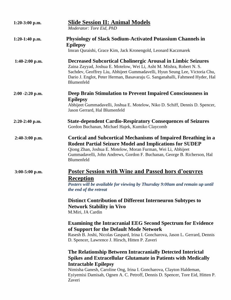
1:20-3:00 p.m. Slide Session II: Animal Models Moderator: Tore Eid, PhD
1:20-1:40 p.m. Physiology of Slack Sodium-Activated Potassium Channels in
Epilepsy
Imran Quraishi, Grace Kim, Jack Kronengold, Leonard Kaczmarek
1:40-2:00 p.m. Decreased Subcortical Cholinergic Arousal in Limbic Seizures
Zaina Zayyad, Joshua E. Motelow, Wei Li, Asht M. Mishra, Robert N. S.
Sachdev, Geoffrey Liu, Abhijeet Gummadavelli, Hyun Seung Lee, Victoria Chu,
Dario J. Englot, Peter Herman, Basavaraju G. Sanganahalli, Fahmeed Hyder, Hal
Blumenfeld
2:00 -2:20 p.m. Deep Brain Stimulation to Prevent Impaired Consciousness in
Epilepsy
Abhijeet Gummadavelli, Joshua E. Motelow, Niko D. Schiff, Dennis D. Spencer,
Jason Gerrard, Hal Blumenfeld
2:20-2:40 p.m. State-dependent Cardio-Respiratory Consequences of Seizures
Gordon Buchanan, Michael Hajek, Kumiko Claycomb
2:40-3:00 p.m. Cortical and Subcortical Mechanisms of Impaired Breathing in a
Rodent Partial Seizure Model and Implications for SUDEP
Qiong Zhan, Joshua E. Motelow, Moran Furman, Wei Li, Abhijeet
Gummadavelli, John Andrews, Gordon F. Buchanan, George B. Richerson, Hal
Blumenfeld
3:00-5:00 p.m. Poster Session with Wine and Passed hors d’oeuvres
Reception
Posters will be available for viewing by Thursday 9:00am and remain up until
the end of the retreat
Distinct Contribution of Different Interneuron Subtypes to
Network Stability in Vivo M.Miri, JA Cardin
Examining the Intracranial EEG Second Spectrum for Evidence
of Support for the Default Mode Network Rasesh B. Joshi, Nicolas Gaspard, Irina I. Goncharova, Jason L. Gerrard, Dennis
D. Spencer, Lawrence J. Hirsch, Hitten P. Zaveri
The Relationship Between Intracranially Detected Interictal
Spikes and Extracellular Glutamate in Patients with Medically
Intractable Epilepsy Nimisha Ganesh, Caroline Ong, Irina I. Goncharova, Clayton Haldeman,
Eyiyemisi Damisah, Ognen A. C. Petroff, Dennis D. Spencer, Tore Eid, Hitten P.
Zaveri

Factors Influencing Driving Impairment in Persons with
Refractory Epilepsy Vineet Punia, Pue Farooque , William Chen, Lawrence J. Hirsch, Anne T. Berg,
the Multi-Center Study of Epilepsy Surgery, Hal Blumenfeld
Reliability of Patient Report of Impaired Consciousness During
Seizures Yang Si, Adeolu Morawu, Yigit Baykara, Rahiwa Gebre, William Chen, Petr
Vitkovskiy, Ningcheng Li, Michelle Johnson, Eric Chen, Dan Kluger, Hal
Blumenfeld
Cosmetic Adverse Effects of Anti Epileptic Drugs in Adults With
Epilepsy Baibing Chen, Kamil Detyniecki, Richard Buchsbaum, Hyunmi Choi, Asif Javed,
Lawrence Hirsch
Seizure Control for Intracranial Arteriovenous Malformations is
Directly Related to Treatment Modality: A Meta-Analysis Jacob F Baranoski, Ryan A Grant, Lawrence J Hirsch, Paul Visintainer, Jason L
Gerrard, Murat Günel, Charles C Matouk, Dennis D Spencer, and Ketan R
Bulsara
Advances in Neuropsychological Testing in Epilepsy; the
Importance of Non-Neurological Variables While Interpreting
Tests Franklin C. Brown
Glutamine Synthetase inhibition and the Methionine-Sulfoximine
Rat-Model of Epilepsy Edgar Perez, Helen Wang, Hitten Zaveri, Ronnie Dhaher, Tore Eid
5:00-5:30 p.m. General Discussion and Day 1 Summary
Moderator: James McNamara, MD
5:45-6:30 p.m. Group Beach Run or Kayaking
7:00-11:00 p.m. Dinner and Social Event

Friday, April 4th
7:00- 8:30 a.m. Breakfast
8:30-10:30 a.m. Slide Session III: Clinical/Electrophysiology Moderator: Lawrence J. Hirsch, MD
8:30- 8:50 a.m. Characteristics of Patients with "Poorly - Localized" Ictal Onset
on Intracranial EEG Adithya Sivaraju, MD, MHA; Hitten Zaveri, PhD; Irina Goncharova, PhD;
Jeremy J. Moeller, MD; Robert B. Duckrow, MD
8:50- 9:10 a.m. Investigating Consciousness Using Human Intracranial
Recordings
Wendy Xiao, Reba Watsky, William Chen, Erik Levinsohn, Dennis D. Spencer,
Jason Gerrard, Hal Blumenfeld
9:10- 9:30 a.m. Functional Connectivity, the Default Mode Network, and the
Seizure Onset Area Hitten P. Zaveri, Steven M. Pincus, Irina I. Goncharova, Nicolas Gaspard, Jason
Gerrard, Lawrence J. Hirsch, Dennis D. Spencer
9:30- 9:50 a.m. The Failure of More Than a Dozen new AEDs to Improve Total
Seizure Control Richard Mattson
9:50-10:10 a.m. Progressive Change in Sleep Quality During Intracranial EEG
Monitoring and the Impact of Seizures and AEDs Rasesh B. Joshi, Nicolas Gaspard, Irina I. Goncharova, Milena Pavlova, Jason L.
Gerrard, Dennis D. Spencer, Lawrence J. Hirsch, Hitten P. Zaveri
10:10-10:30 a.m. Prospective Evaluation of Driving During Seizures Using Driving
Simulation on the Video/EEG Monitoring Unit William C. Chen, Andrew Bauerschmidt, Mark W. Youngblood, Courtney
Cunningham, Cel Ezeani, Zachary Kratochvil, Jared Bronen, James Thomson,
Dan Kluger, Ningcheng Li, Katherine Riordan, Ji Yeoun Yoo, Romina Shirka,
Louis Manganas, Andres Fernandez, Anupama Alareddy, Rup Sainju, Adithya
Sivaraju, Imran Quraishi, Lawrence J. Hirsch, Hal Blumenfeld
10:30-11:00 a.m. Coffee Break

11:00-12:00 p.m. Slide Session IV: New Directions aka “Data Free Zone”
11:00-11:15 a.m. Auditory Discrimination and Physiology in an Awake Behaving
Mouse Absence Seizure Model Eric Chen, Alex Kwan, Jeanette Chin, Hal Blumenfeld
11:15-11:30 a.m. Gut-Brain Axis in Epilepsy: Role of Microbiota on Seizures
Eid Tore
11:30-11:45 a.m. Defining the Risk Factors, Prevalence, and Consequences of
Seizure Clusters
Tenzin Choezom, Jennifer Bonito, Lawrence J. Hirsch, and Kamil Detyniecki
11:45-12:00 p.m. EEG Sonification for the Detection of Seizures Psyche Loui, Mark Frick, Matan Koplin-Green, Mike Massone.
12:00- 1:30 p.m. Lunch Buffet and Final Discussion
THANK YOU ALL FOR PARTICIPATING IN THE THIRD ANNUAL EPILEPSY RESEARCH RETREAT
A SPECIAL THANKS TO DR. JAMES MCNAMARA

Neurocritical Care Monitoring
Mini Retreat
1:30-1:45 p.m. Welcome and introduction
Emily J Gilmore and Nicolas Gaspard
1:45-2:10 p.m. EEG Biomarkers of Sleep and Delirium in the Critically Ill Melissa Knauert
2:10-2:35 p.m. Pathophysiology of Sepsis Associated Brain Dysfunction
Emily J Gilmore
2:35-3:00 p.m. The Value of CEEG for Predicting Outcome after Cardiac Arrest Adithya Sivaraju
3:00-3:15 p.m. BREAK
3:15-3:40 p.m. Metabolomics Approach to Vasospasm
Ketan Bulsara
3:40-4:05 p.m. Towards the Next Generation of Delayed Cerebral Ischemia
Detection
M Brandon Westover
4:05-4:30 p.m. EEG Biomarkers of Epileptogenesis After Acute Brain Injury
Rup Sainju
4:30-4:55 p.m. The Treatment of Refractory Status Epilepticus
Andres Fernandez
4:55-5:20 p.m. The Spectrum of New-Onset Refractory Status Epilepticus
Nicolas Gaspard
5:20-5:30 p.m. Closing remarks

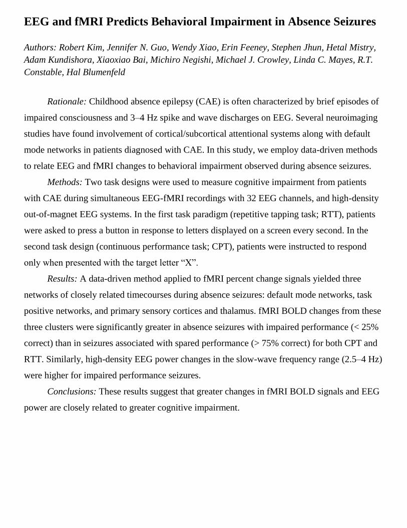
EEG and fMRI Predicts Behavioral Impairment in Absence Seizures
Authors: Robert Kim, Jennifer N. Guo, Wendy Xiao, Erin Feeney, Stephen Jhun, Hetal Mistry,
Adam Kundishora, Xiaoxiao Bai, Michiro Negishi, Michael J. Crowley, Linda C. Mayes, R.T.
Constable, Hal Blumenfeld
Rationale: Childhood absence epilepsy (CAE) is often characterized by brief episodes of
impaired consciousness and 3–4 Hz spike and wave discharges on EEG. Several neuroimaging
studies have found involvement of cortical/subcortical attentional systems along with default
mode networks in patients diagnosed with CAE. In this study, we employ data-driven methods
to relate EEG and fMRI changes to behavioral impairment observed during absence seizures.
Methods: Two task designs were used to measure cognitive impairment from patients
with CAE during simultaneous EEG-fMRI recordings with 32 EEG channels, and high-density
out-of-magnet EEG systems. In the first task paradigm (repetitive tapping task; RTT), patients
were asked to press a button in response to letters displayed on a screen every second. In the
second task design (continuous performance task; CPT), patients were instructed to respond
only when presented with the target letter “X”.
Results: A data-driven method applied to fMRI percent change signals yielded three
networks of closely related timecourses during absence seizures: default mode networks, task
positive networks, and primary sensory cortices and thalamus. fMRI BOLD changes from these
three clusters were significantly greater in absence seizures with impaired performance (< 25%
correct) than in seizures associated with spared performance (> 75% correct) for both CPT and
RTT. Similarly, high-density EEG power changes in the slow-wave frequency range (2.5–4 Hz)
were higher for impaired performance seizures.
Conclusions: These results suggest that greater changes in fMRI BOLD signals and EEG
power are closely related to greater cognitive impairment.

fMRI Changes in a Ferret Model of 3-4 Hz Spike-Wave Seizures
Authors: Mark W.Youngblood, William C. Chen, Asht M. Mishra, Sheila Enamandram,
Basavaraju G. Sanganahalli, Joshua E. Motelow, Harrison X. Bai, Flavio Frohlich, Alexandra
Gribizis, Alexis Lighten, Fahmeed Hyder, Hal Blumenfeld
Functional neuroimaging studies have provided key insights on the changes in neurovascular
activity associated with human absence seizures. Previous work has identified widespread
cortical decreases in blood oxygen-level dependent (BOLD) signal during absence seizures,
however the meaning of these decreases remains to be elucidated. Rodent models of epilepsy
have failed to replicate this finding, which could be due to several important differences
between human and rodent seizures. Human absence seizures occur at a frequency of 3-4 Hz
and involve changes in developed cortical regions. Pharmacologic and genetic rodent models
produce seizures occurring at 7-8 Hz, and lack a developed cortex capable of replicating human
findings. To further investigate the meaning of ictal decreases in BOLD signal, we developed a
ferret model of generalized epilepsy. This species contains a gyrated cortex, and previous in-
vitro studies have shown ferret thalamic slices are capable of generating a 3-4 Hz rhythm in
response to bicuculline. We used 9.4 T BOLD imaging with simultaneous recording of EEG to
characterize changes in neurovascular activity during ictal periods. To our surprise, the presence
of 3-4 Hz discharge in this model was not sufficient to replicate the BOLD decreases seen in
humans. This finding was consistent across seizures of different duration, frequency, and with
various analytical techniques. However this model was capable of generating previously
described post-ictal decreases during tonic-clonic seizures, reducing the likelihood that
technical factors played a role. The mechanism and meaning of ictal decreases during absence
seizures will require further study.

Multiphoton Microscopy of Cellular and Network Brain Activity in
Awake Mouse During Epileptic Convulsion
Authors: Michael Levene, Markus Wolfel, Hal Blumenfeld
Imaging of neuronal activity in the brain in vivo has provided fundamental insight into
information processing in single cells and neural networks. However, forceful movement during
epileptic convulsions prevents conventional imaging due to excessive motion artifacts and risk
of injury. Therefore, we designed a suspension platform for motion-stabilized imaging of brain
activity with sub-cellular resolution. With the animals head fixed in place using an implanted
head mount, the animal rests on a suspension platform underneath. This protects the animal
from force exerted against the implant, and removes motion artifacts in the recorded images.
The stabilization system was combined with micro-prism implants to image neurons throughout
the full cortical depth. We were able to record activity of neurons throughout tonic-clonic
seizures in the awake, convulsing mouse. Single cells stayed in place during recording sessions,
and no further motion correction was needed to track cells over time.

Mechanisms of Cortical fMRI Decreases in Absence Seizures
Investigated in the Awake Mouse
Authors: Florian Serout, Jeanette Chin, Hiam Naiditch, Qiong Zhan, Abhijeet
Gummadavelli, Eric Chen, Peter Herman, Basavaraju G. Sanganahalli, Fahmeed Hyder,
Hal Blumenfeld
Numerous animal models of childhood absence epilepsy exist but none have shown perfect
similarity to human patients, especially regarding the fMRI changes associated with spike-
wave seizures. Previous work on children with absence epilepsy has shown a marked decrease
in the cortical fMRI signal persisting long after the seizure’s end. However, experiments in
rat, ferret and non-human primate animal models have shown mainly cortical fMRI increases
in spike-wave seizures. Our hypothesis is that the anesthesia is responsible for the differences
between animal models and human absence seizures. Anesthetic agents may prevent a
normal physiological response to absence seizures and could mask the resulting fMRI
decreases in the cortex. In order to assess this hypothesis and provide an animal model of
absence seizures more similar to the human condition, we have begun to perform EEG-fMRI,
as well as Laser Doppler Flowmetry and Multi-Unit Activity (MUA) recordings on awake
C3H/HeJ mice, a relatively pure absence seizure model. Initial results have shown the
feasibility of obtaining spike-wave seizures in head-fixed C3H/HeJ mice both in the high field
fMRI setting with simultaneous EEG, as well as the opportunity to directly record neuronal
physiology and cerebral blood flow through bench experiments. Ongoing investigations with
this model may determine whether the fMRI decreases observed in human patients in absence
seizures are associated with decreased electrical activity of cortical neurons, or another
mechanism for altered cerebral blood flow following seizures.


Physiology of Slack Sodium-Activated Potassium Channels in
Epilepsy
Authors: Imran Quraishi, Grace Kim, Jack Kronengold, Leonard Kaczmarek
I will present the current state of our research into how sodium-activated potassium channels
contribute to epilepsy. Slack (Kcnt1) is a potassium channel that is unique because of its sodium
activation and unusually large C-terminal cytoplasmic domain that interacts with Fragile X
Mental Retardation Protein (FMRP), which is essential for normal cognitive development.
Slack mutations have been identified in patients and families with a variety of epilepsy
syndromes, including migrating malignant partial seizures of infancy (MMPSI), Ohtahara
syndrome, West syndrome, and autosomal dominant nocturnal frontal lobe epilepsy
(ADNFLE). Slack-associated epilepsies may lead to severe epileptic encephalopathy, epilepsy
with neuropsychiatric features, or less commonly epilepsy without cognitive or psychiatric
manifestations. All of the epilepsy-associated mutations are associated with gain of function as
determined by increased Slack conductance in a heterologous expression system (Xenopus
oocytes). Single channel conductance is either unchanged, increased, or decreased by different
mutations. The increased whole cell conductance may be explained in part by positive
cooperativity between neighboring channels and by constitutive phosphorylation behavior.
Slack knockout mice display marked learning deficits and a small reduction in maximum
electroshock seizure thresholds as compared to wild type. Future studies will address the
influence of mutated channels in neuronal culture, animal models, and simulated epileptic
cortical networks. We believe these studies of Slack physiology will provide insights into the
mechanisms of epileptogenesis and cortical development, as well as a potential new therapeutic
target for antiseizure medications.

Decreased Subcortical Cholinergic Arousal in Limbic Seizures
Authors: Zaina Zayyad, Joshua E. Motelow, Wei Li, Asht M. Mishra, Robert N. S. Sachdev,
Geoffrey Liu, Abhijeet Gummadavelli, Hyun Seung Lee, Victoria Chu, Dario J. Englot, Peter
Herman, Basavaraju G. Sanganahalli, Fahmeed Hyder, Hal Blumenfeld
Subcortical arousal systems play a critical role in the normal operation of the waking brain.
During loss of consciousness in deep sleep and coma, slow waves arise in the cortex,
accompanied by reduced subcortical arousal. These same phenomena are observed during
complex partial limbic seizures arising in the temporal lobe. We are interested in understanding
the mechanism for impaired consciousness and cortical slow oscillation during these focal
seizures with impaired consciousness. In experiments using blood oxygen-level dependent
(BOLD) functional magnetic resonance imaging (fMRI), we observed broad suppression of
arousal systems during hippocampal seizures in a rodent model. We conducted juxtacellular
recordings in the brainstem and observed reduced firing of cholinergic neurons in focal limbic
partial seizures. Furthermore, using enzyme-based amperometry, we found reduced choline
levels in the cortex and thalamus. Consequently, the mechanism for impaired consciousness
during complex partial limbic seizures resembles slow-wave sleep initiation as opposed to
seizure propagation to the cortex.

Deep Brain Stimulation to Prevent Impaired Consciousness in
Epilepsy
Authors: Abhijeet Gummadavelli, Joshua E. Motelow, Nicholas D. Schiff, Dennis D. Spencer,
Jason Gerrard, Hal Blumenfeld
Abstract: For patients with medically refractory epilepsy, the ability to remain conscious
despite seizure occurrence would provide a dramatic improvement in quality of life, morbidity
and mortality. Our lab developed a rodent model of complex partial seizures which mimics the
human cortical electroencephalographic signature of ictal neocortical slow waves (0-2 Hz) and
behavioral arrest associated with loss of consciousness (Englot et al, 2008). Using this model,
we measured suppression of subcortical arousal systems including the brainstem cholinergic
and intralaminar thalamic nuclei (Motelow et al, 2014). Stimulation of central lateral thalamic
nuclei has improved measures of consciousness in a patient from minimally conscious state
after traumatic brain injury (Schiff et al, 2007). We show restored measures of consciousness in
the anesthetized and awake-behaving animal model by stimulating the central lateral thalamic
nuclei under anesthesia and during seizures. We stereotactically implanted electrodes and
confirmed localization with histology. We stimulated at varying current intensities (ranging
from 0.1 µA to 3mA, unilaterally and bilaterally) at 100 Hz while synchronously recording
behavior with video and electrophysiology with electrodes in the lateral orbitofrontal cortex and
hippocampus. Thalamic stimulation under anesthesia shows loss of cortical slow waves in a
current dose-responsive fashion, and behavioral arousal with head, tail and limb movement.
Ictal and post-ictal stimulation shows similar decrease in cortical slowing, associated
behaviorally with increased arousal. These data suggest a novel therapeutic approach for
restoring consciousness during seizures. If paired with responsive neurostimulation, it may lead
to rapid implementation of a therapy for preventing impaired consciousness in patients.

State-Dependent Cardio-Respiratory Consequences of Seizures
Authors: Gordon F. Buchanan, Michael A. Hajek, Daniel A. Rappoport and
Kumiko I. Claycomb
Sudden unexpected death in epilepsy (SUDEP) is the leading cause of death in refractory
epilepsy patients. In most cases the direct cause of death is likely seizure-induced respiratory
arrest, though cardiac arrest or suppression of cerebral activity may be responsible in some
patients. Intriguingly, SUDEP tends to occur at night. Why this is the case is not known, but
there is a complex relationship between seizures, sleep-wake state, circadian phase, regulation
of breathing, cardiac control, and regulation of the neurotransmitters involved in modulation of
all of these. Here we aimed to determine whether there are state-dependent effects of seizures
on cardiac and respiratory function that may contribute to seizure-related death. Seizures were
induced with maximal electroshock (MES) in seizure naïve mice or amygdala stimulation (AS)
in kindled mice. EEG, EMG, EKG, breathing and video were recorded before, during and after
seizures. Vigilance state was determined in real-time using EEG, EMG and behavioral
parameters. Preliminarily, we have found that seizures induced via MES during sleep were
more likely to be fatal, especially those induced during REM sleep. Mice that survived seizures
induced by MES or AS displayed increased post-ictal respiratory rate variability and a greater
number of apneas when seizures were induced during NREM sleep. Furthermore, mice that
eventually died when seizures were induced during sleep tended to have more irregular baseline
breathing. There was no state-dependent difference in effects of seizures on cardiac activity.
These data support that there are state-dependent effects on breathing that may contribute to
seizure-related death.

Cortical and Subcortical Mechanisms of Impaired Breathing in a
Rodent Partial Seizure Model and Implications for SUDEP
Authors: Qiong Zhan, Joshua E. Motelow, Abhijeet Gummadavelli, Moran Furman, Wei Li,
John Andrews, Gordon F. Buchanan, George B. Richerson, Hal Blumenfeld
Seizures have both local and remote effects on nervous system function. Focal seizures can
propagate from the site of onset to engage a larger network and induce severe consequences
including impaired arousal, cardiorespiratory changes and in some cases death. Impaired
breathing during and following seizures is increasingly recognized as an important cause of
sudden unexpected death in epilepsy (SUDEP). Impaired breathing in seizures has been
compared to sudden infant death syndrome (SIDS), in which deficits in serotonergic arousal
may contribute to impaired respiratory drive and impaired arousal in response to CO2 retention
while lying face down, leading to asphyxia and death. We now utilize an acute rat model of
limbic seizures to measure changes in breathing and in the firing patterns of brainstem nuclei
using extracellular multiunit and juxtacellular recordings. We found that during limbic seizure
the breathing is markedly impaired, possibly related to seizure propagation beyond the site of
onset. The firing of neurons in the midbrain and medullary raphe nuclei is dramatically
decreased during the ictal and post-ictal period. Based on these results, we suggest that seizures
may propagate to inhibitory circuits which depress the ascending and descending arousal
systems. Failure of the ascending arousal system to maintain consciousness and of the
descending arousal system to stimulate the respiratory network may both be important factors in
SUDEP. Improved mechanistic understanding of these pathways may lead to improved
treatments to prevent this significant cause of death in people living with epilepsy.


Distinct Contribution of Different Interneuron Subtypes to Network
Stability in Vivo
Author: M. MIRI*, J. A. CARDIN
GABAergic inhibition is critical for regulation of excitation and is thought to maintain
neural network stability through a precise balancing process. Loss or dysfunction of
inhibitory interneurons is thought to lead to abnormal activity patterns and ultimately to
seizure initiation. Two major interneuron classes are the Parvalbumin-expressing (PV),
fast-spiking cells and Somatostatin-expressing (SOM), low-threshold spiking cells. Due
to differing physiology, biophysical properties, and synaptic targeting, they may contribute
differently to ongoing computations in the surrounding local network. Previous studies
have examined preferential interneuron vulnerability leading to or resulting from seizures,
but there the roles of specific sources of inhibition in seizure generation remain
unclear. Using combined optical and electrophysiological tools, we can investigate network
dynamics during the development of seizures by monitoring the activity of individual
interneurons and synchronization between multiple hippocampal cell types.
Here we used two transgenic mouse lines: PV-Cre and SOM-Cre. Using AAVDIO-
ChR2-mCherry, we selectively targeted optogenetic tools to PV or SOM interneurons
in CA1. During experiments, we used stereotrode arrays to extracellularly record
the activity of many simultaneous cells in hippocampal CA1 in animals lightly anesthetized
with ketamine/xylazine. We identified targeted ChR2-expressing PV and SOM
interneurons in the recorded population by stimulating with brief pulses of blue light at
473nm. Light pulses were continued throughout the experiment to track identified neurons
without altering the pattern of spontaneous activity. Using a pharmacological model
of seizure induction (PTZ), we performed measurements of neural activity during three
periods: 1) baseline 2) early preictal and 3) late preictal. We first assessed the recruitment
of local excitatory neurons by ictal activity. We further assessed the temporal pattern
of identified interneuron activity during each period and the relationship between
interneuron and excitatory neuron activity.
To examine the relationship between local network activity and interneuron spiking,
we calculated the spike-triggered LFP average for each cell during each period. We
find that PV+ cells have a lower spike probability in response to blue light pulses that
progressively decreases from baseline to late preictal stages. Additionally, we find that
average local synaptic activity increases surrounding spike times of identified RS cells
during both early and late preictal periods, as compared to baseline. These preliminary
results demonstrate that these combined approaches provide a powerful set of tools for
dissecting network activity during seizure initiation.

Examining the Intracranial EEG Second Spectrum for Evidence of
Support for the Default Mode Network
Authors: Rasesh B. Joshi, Nicolas Gaspard, Irina I. Goncharova, Jason L. Gerrard, Dennis D.
Spencer, Lawrence J. Hirsch, Hitten P. Zaveri
It has been shown previously that the intracranial EEG (icEEG) relationship within the
default mode network (DMN) was not significantly greater than between DMN locations and a
control location, indicating a lack of support for the DMN in the icEEG. However, recent
literature suggests that the electrophysiological basis of these networks may lie in coherent slow
amplitude modulations of different frequency bands, i.e. in the second spectrum, rather than in
synchronization of the icEEG. In particular, studies performed with resting state
magnetoencephalography (MEG) suggest that the electrophysiological correlates of the DMN
can be observed in the alpha band second spectrum. We examined the icEEG second spectrum
for evidence of support for the DMN.
We first sought to characterize the magnitude-squared coherence (MSC) estimator to
determine a threshold for when an MSC value can be considered significantly nonzero and to
determine the ideal parameters to be used in the MSC calculation. We studied a total of 12
patients. For each patient, we selected a one-hour icEEG epoch when the patient appeared to be
resting quietly with eyes open. For each one-hour segment, we calculated the running power, at
a one-second resolution, in different frequency bands (delta, theta, alpha, beta, and gamma). For
each band power time-series, we then calculated the average coherence in frequencies less than
0.15 Hz for every possible electrode contact pair. We aggregated these values for all patients
and examined how second spectrum connectivity changes with distance, second spectrum
relationships between different lobes of the brain, as well as whether DMN-specific contacts
exhibit enhanced connectivity in the second spectrum.
Interestingly, we found that second spectrum coherence is relatively high in the delta
band for short distances (less than 5 cm). Furthermore, we observed that second spectrum
coherence decreases in higher frequency bands, with the lowest values in the gamma band.
Analysis of DMN-specific contacts did not show any evidence in support of the DMN. Second
spectrum coherence in the DMN contacts was significantly nonzero only in the delta, theta, and
alpha bands. In these bands, there was no significant difference between relationships within the
DMN locations and between the DMN location and a control location.

The Relationship Between Intracranially Detected Interictal Spikes
and Extracellular Glutamate in Patients with Medically Intractable
Epilepsy
Authors: Nimisha Ganesh, Caroline Ong, Irina I. Goncharova, Clayton Haldeman, Eyiyemisi
Damisah, Ognen A. C. Petroff, Dennis D. Spencer, Tore Eid, Hitten P. Zaveri
Abstract: Recent research from our laboratory suggests that interictal spikes may represent
inhibition rather than excitation. Research from our center has also established the presence of
elevated basal levels of extracellular glutamate in the epileptic brain. Here we sought to
correlate interictal spikes and basal extracellular glutamate concentrations in patients (n=20)
undergoing intracranial EEG (icEEG) monitoring for epilepsy surgery. Spike counts were
documented for the two icEEG contacts on either side of the microdialysis membrane, all
contacts within 3cm of each probe, and all contacts within the region surrounding the probe.
Additionally, basal glutamate and glutamine values were correlated with the probe distance
from the seizure onset area. The basal glutamate and basal glutamine values were negatively
correlated with the probe distance to seizure onset area with correlation coefficient values of 0.2
(p=0.06) and 0.4 (p=0.001), respectively. We observed a slightly negative correlation between
the basal glutamate values and spike counts. The correlation coefficient values for the probe
contacts, contacts within 3cm, and regional contacts were 0.07, 0.08, and 0.14, respectively. In
areas of high spiking, basal glutamate values were particularly low. The higher basal glutamate
and glutamine values closer to the seizure onset area suggest a correlation between increased
glutamate and glutamine levels and the seizure onset area. The slight negative correlation
observed between basal glutamate level and spike counts, moreover, support the suggestion that
spikes may instead be a measure of brain inhibition. This study suggests considerable value for
co-local in-vivo measurements of electrophysiology ad neurochemistry to improve our
understanding of the pathophysiology of medically intractable epilepsy.

Factors Influencing Driving Impairment in Persons with Refractory
Epilepsy
Authors: Vineet Punia, Pue Farooque, William Chen, Lawrence J. Hirsch, Anne T. Berg, the
Multi-Center Study of Epilepsy Surgery, Hal Blumenfeld
Objectives: To evaluate factors related to seizures that potentially impair driving leading
to motor vehicle accidents (MVA) in people with medically refractory epilepsy.
Methods: We used the Multicenter Study of Epilepsy Surgery (MSES) database to
identify patients who reported having seizures while driving. We divided this population in two
groups: ones in whom seizures while driving led to a MVA (“Accident” group) and those who
had seizures while driving that did not lead to a MVA(“No Accident” group). We then
compared these two sub-groups for different variables that could potentially affect the risk of
MVA.
Results: 215 patients out of a total of 553 patients reported having seizure(s) while
driving. 74 formed part of the “No Accident” group and 141 were in the “Accident” group.
The two groups did not differ in terms of age, sex distribution, seizure localization, ictal
duration, motor impairment during seizure and AED history. Interestingly, they also did not
differ (p = 0.76; OR = 0.89, CI 0.49 – 1.61) based on presence of reliable auras. We found that
patients were more likely to have MVA due to a seizure if they had a history of complex partial
seizure (CPS)(p = 0.029; OR = 2.83, CI 1.14-7.09) or if they had one or more CPS on an
average each month for last 3 months prior to interview (p = 0.01; OR = 2.52, CI 1.22-5.21).
Patient in “No accident” group were found to have longer duration (>1 minute) of post-ictal
state (p = 0.05; OR = 2.53, CI 1.04 – 6.19).
Conclusions: Our study shows that reliable auras do not provide protective benefit
against MVA, which questions the long held belief that the reliability of auras is a favorable
modifier in preventing MVAs. We also found that CPS, possibly implying impaired
consciousness, may be the most salient feature of seizures affecting the ability to drive safely.

Reliability of Patient Report of Impaired Consciousness During
Seizures
Authors: Yang Si, Adeolu Morawu, Yigit Baykara, Rahiwa Gebre, William Chen, Petr
Vitkovskiy, Ningcheng Li, Michelle Johnson, Eric Chen, Dan Kluger, Hal Blumenfeld
Epileptic seizures often cause loss of consciousness (LOC). However, patient report of their
mental state during seizures can be unreliable, making diagnosis of epilepsy and guidance on
patient safety issues difficult for clinicians. The present study used a daily questionnaire
administered to epilepsy inpatient undergoing continuous video-EEG monitoring to study the
reliability of patient report of their consciousness during seizures. Patients with episodes during
the monitoring were questioned whether they were aware of their surroundings, were
responsive, and were able to speak during their seizures. These subjective reports were
compared with their actual ictal behavior on video EEG. A total of 37 seizures in 17 patients
were analyzed. Among those with ictal unconsciousness, self-report of loss of awareness,
inability to respond and speak during the events was highly accurate. Among those with spared
consciousness, self-report was not always consistent with the real ictal performance and tended
to overestimate ictal impairment of consciousness. These findings indicated that epilepsy
patients with ictal LOC may be able to report this fact correctly.

Cosmetic Adverse Effects of Antiepileptic Drugs In Adults With
Epilepsy
Authors: Chen B, Hirsch LJ, Detyniecki K, Moeller J, Javed A, Kato K, Legge A,
Buchsbaum R, Choi H.
Introduction Adverse cosmetic effects (ACEs) can occur in patients taking antiepileptic
drugs (AEDs). The objective of the study was to compare the ACE profiles among both older
and newer AEDs.
Methods We reviewed patient records including demographics, medical history, AED
use, and side effects for 1903 adult epilepsy patients (age ≥ 16 years). ACEs included were
acne, gingival hyperplasia, hair loss, hirsutism, and weight gain. We compared the AED-
attributed ACEs and intolerable ACE (i.e. ACE that led to AED dosage reduction or
discontinuation) in patients newly started on an AED after controlling for gender.
Results Overall, ACEs occurred in 110/1903 (5.8%) patients, and led to intolerability in
70/1903 (3.7%) patients. 37.2% of patients who had at least one ACE developed additional
ACEs. Female patients (7.0%) reported more ACEs than male patients (4.3%; P < 0.05).
Weight gain was more commonly reported in patients using pregabalin (8.4%; P < 0.001) and
valproic acid (13.0%; P < 0.001), hair loss was more commonly seen in patients taking valproic
acid (8.9%; P < 0.001), and more patients taking phenytoin developed gingival hyperplasia
(2.5%; P < 0.001) compared to average.
Conclusion There is significant variability amongst all AEDs in terms of their ACE
profiles. Particular attention should be paid to pregabalin, phenytoin, and valproic acid when
considering ACEs. Patients who have had an ACE to an AED and/or are female may be more
susceptible to AED-attributed ACEs. Our findings may help clinicians to better assess the
risk/benefit ratios in the AED selection process.

Table 1. Comparison of AED-attributed ACEs in adults with epilepsy newly started on an AED.
AED n ACE
% (n) ‡P-value ‡OR
IACE
% (n) ‡P-value ‡OR
CBZ 326 1.5 (5) 0.031* 0.40 (0.18, 0.92) 0.6 (2) 0.045* 0.31 (0.10, 0.97)
CLB 80 0.0 (0) NS - 0.0 (0) NS -
FBM 53 1.9 (1) NS 0.89 (0.21, 3.64) 1.9 (1) NS 1.37 (0.33, (5.72)
GBP 251 2.8 (7) NS 0.74 (0.36, 1.52) 1.2 (3) NS 0.55 (0.20, 1.52)
LCM 86 0.0 (0) NS - 0.0 (0) NS -
LEV 524 1.3 (7) 0.002* 0.32 (0.15, 0.65) 1.0 (5) 0.019* 0.37 (0.16,0.85)
LTG 521 1.9 (10) 0.012* 0.45 (0.24, 0.84) 1.0 (5) 0.020* 0.37 (0.16,0.86)
OXC 160 0.6 (1) NS 0.28 (0.07, 1.13) 0.6 (1) NS 0.43 (0.11, 1.78)
PB 98 0.0 (0) NS - 0.0 (0) NS -
PGB 143 9.8 (14) < 0.001** 2.85 (1.63, 5.01) 5.6 (8) 0.011* 2.51 (1.24, 5.09)
PHT 404 4.2 (17) NS 1.07(0.65, 1.78) 3.2 (13) NS 1.32 (0.74, 2.34)
PRM 14 0.0 (0) NS - 0.0 (0) NS -
TGB 25 0.0 (0) NS - 0.0 (0) NS -
TPM 230 1.7 (4) NS 0.49 (0.20, 1.20) 1.7 (4) NS 0.77 (0.31, 1.92)
VGB 32 3.1 (1) NS 1.48 (0.35, 6.26) 0.0 (0) NS -
VPA 270 21.9 (59) < 0.001** 10.40 (7.26, 14.88) 13.3 (36) < 0.001** 8.55 (5.54, 13.18)
ZNS 230 0.9 (2) 0.033* 0.29 (0.09, 0.91) 0.0 (0) NS -
Average 3.7 2.3
Abbreviations: AED: Antiepileptic Drug; ACE: Adverse Cosmetic Effects; CBZ: carbamazepine; CI: Confidence Interval; CLB: clobazam; FBM:
felbamate; GBP: gabapentin; IACE: Intolerable Adverse Cosmetic Effects; LCM: lacosamide; LEV: levetiracetam; LTG: lamotrigine; OR: Odds
Ratio; OXC: oxcarbazepine; PB: phenobarbital; PGB: pregabalin; PHT: phenytoin; PRM: primidone; TGB: tiagabine; TPM: topiramate; VGB:
vigabatrin; VPA: valproate; ZNS: zonisamide
P < 0.003 (0.05/17=0.003)
** Significant: p < 0.003
* Trend: 0.003 < p < 0.05
Rate is higher than the
average rate of other AEDs
Rate is lower than the
average rate of other AED
‡ controlled for gender

Seizure Control for Intracranial Arteriovenous Malformations is
Directly Related to Treatment Modality: A Meta-analysis
Authors: Jacob F. Baranoski, B.S., Ryan A. Grant, M.D., M.S., Lawrence J. Hirsch, M.D., Paul
Visintainer, Ph.D., Jason L. Gerrard, M.D., Ph.D, Murat Günel M.D., Charles C. Matouk,
M.D., Ketan R. Bulsara, M.D.
Object – Seizures are a common presenting sign of intracranial arteriovenous
malformations (AVMs). The object of this meta-analysis was to determine if the modality
selected to treat AVMs affects the rate of seizure-free outcomes.
Methods – All published data describing seizure status as an outcome goal over the last
20 years were included in this study. Seizure outcomes following microsurgery (MS),
endovascular embolization for cure (EVE), or stereotactic radiosurgery (SRS) were compared
using a validated random-effect logistic regression approach.
Results – Twenty-four studies, with a total of 1157 patients, were analyzed. Overall, the
microsurgical group had the best seizure control (p<0.01), with the relative predicted rates of
seizure-free outcome as follows: MS 78.3% [95% CI, 70.1-85.8%]; SRS 62.8% [95% CI, 55.0-
70.0%]; EVE 49.3% [95% CI, 32.1-66.6%]. However, further analysis revealed that for patients
with unruptured AVMs, MS and SRS were equally efficacious. Patients in the SRS group who
had complete obliteration of their AVMs achieved the highest rate of seizure-free outcomes at
85.2% [95% CI, 79.1-91.2%] (p<0.01). The development of new-onset seizures occurred more
frequently in patients undergoing EVE (39.4% [95% CI, 8.1-67.8%]) compared to MS (9.1%
[95% CI, 5.0-13.1%]) and SRS (5.4% [95% CI, 3.0-7.8%]) (p<0.3 and p<0.01, respectively).
Conclusions – This is the first meta-analysis designed to study relative rates of seizure-
free outcomes following the currently utilized AVM treatment modalities. In general,
microsurgery results in the highest proportion of seizure-free outcomes. However, if stereotactic
radiosurgery results in successful obliteration of the AVM, then this modality is the most
effective in achieving seizure-control.


Advances in Neuropsychological Testing in Epilepsy; the Importance
of Non-Neurological Variables While Interpreting Tests
Author: Franklin C. Brown
Neuropsychological evaluations are a important part of presurgical evaluations. In addition to
being useful to better understand cognitive correlates with epileptogenic and irritative zones. In
addition, presurgical memory tends to be as good, if not better, a predictor of postsurgical
memory. However, such tests are only useful if the performance is a valid representation of
neurological functioning, and not due to confounding variables. This presentation discusses
some advances in studying factors that can affect performance such as medications, anxiety, age
effects, and the degree to which tests can actually measure their purported construct.
Furthermore, several published studies and case reports will be provided to demonstrate the
impact that such variables have on performance.

Glutamine Synthetase inhibition and the Methionine-Sulfoximine
Rat-Model of Epilepsy
Authors: Edgar Perez, Helen Wang, Hitten Zaveri, Ronnie Dhaher, Tore Eid
Glutamine synthetase (GS) has been found to be deficient in the hippocampus of patients with
MTLE. The role of glutamine synthetase deficiency in the generation of seizures is being
explored in a newly developed rat model of MTLE involving infusion of methionine
sulfoximine (MSO), an inhibitor of GS, in the entorhinal cortex. We are assessing the effects of
MSO after a chronic infusion (28 days) and after an acute infusion (72 hours) on seizure
outcome and pathology. The goal is to assess whether restoration of GS leads to cessation of
seizures in the presence of extensive brain pathology. In a subset of rats infused for 72 hours,
seizures were still observed after an extensive latent period, suggesting a possible secondary
process involved in epileptogenesis assuming that GS has been restored to normal. We hope
that we can leverage this knowledge to determine time-sensitive perturbations leading to
epilepsy and when it might be appropriate to intervene in terms of treatment.


Characteristics of Patients with “Poorly – Localized” Ictal Onset on
Intracranial EEG Authors: Adithya Sivaraju; Jeremy J. Moeller; Irina Goncharova; Hitten Zaveri; Robert B.
Duckrow
Introduction: Intracranial EEG monitoring offers a unique and precise way of localizing
seizure onset in humans. Various patterns of seizure onset & propagation have been well
described in the literature. However, most of this data has been devoted to describing well-
defined ictal onset patterns/seizure onset zones (SOZ). We seek to identify & describe the
characteristics of patients with poorly localized ictal onset on Intracranial EEG. The extensive
degree of electrode coverage at our institution offers us a unique opportunity to conduct this
study.
Methods: Retrospective chart review of prospectively collected patient data. This
includes all patients who underwent intracranial EEG monitoring for seizure localization at
Yale New Haven Hospital, from January 2002 to March 2014. The raw EEG data was reviewed
by three independent board certified neurophysiologists, with extensive experience in
interpreting intracranial EEG’s. 9 Zones were defined as Medial frontal (MF), Dorsolateral
frontal (DF), Orbito frontal (OF), Lateral temporal (LT), Medial temporal (MT), Inferior
temporal (IT), Lateral parietal (LP), Medial parietal (MP), & Occipital (O).We defined poorly
localized as involving ≥ 2 of the above defined zones (contiguous or non-contiguous) with a
minimum of 10 electrode contacts at onset; and without a clear lead in of at-least 200 ms.
After patient identification based on EEG findings, we reviewed clinical, radiologic and electro-
physiologic data (including scalp EEG) for each patient.
Results and Conclusion: Pending

Investigating Consciousness using Human Intracranial Recordings
Authors: Wendy Xiao, Reba Watsky, William Chen, Erik Levinsohn, Dennis D. Spencer, Jason
Gerrard, Hal Blumenfeld
The mechanism by which perceived stimuli are processed and encoded into consciousness has
been extensively studied with behavioral tasks. The addition of imaging and electrophysiology
methods can elucidate the temporal and spatial location of the sequences necessary to study the
process by which some stimuli are consciously perceived and later reportable while others are
not. We hypothesize that this selection mechanism is related to memory circuits interacting with
sensory and attentional circuitry. We have developed a behavioral task paradigm with which to
study the mechanism of conscious report by showing a face stimulus image titrated to threshold
perception levels of transparency. Subjects validate their subjective perception of the stimulus
by also objectively reporting the location of the face. While subjects report perceiving the face
about 48% of the time, they are able to locate the stimulus 90% of the time when they do
perceive it. We have collected intracranial EEG data from two subjects, and have been able to
successfully elicit an N170 event-related potential (ERP) following the face stimulus in the
fusiform face area as well as visual cortex ERPs after the presentation of the stimulus. We plan
to use this behavioral task in conjunction with imaging methods such as functional MRI and
intracranial EEG to elucidate the brain activity patterns in memory and attention regions
associated with conscious experience.

Functional Connectivity, the Default Mode Network, and the Seizure
Onset Area
Authors: Hitten P. Zaveri, Steven M. Pincus, Irina I. Goncharova, Nicolas Gaspard, Jason
Gerrard, Lawrence J. Hirsch, Dennis D. Spencer
Purpose: To determine the relationship between band-related local functional
connectivity (LFC) of the background intracranial EEG (icEEG) and the seizure onset area
(SOA).
Method: This study was conducted on 1 hr icEEG epochs recorded from 18 unselected
adult patients with localization related epilepsy (LRE) undergoing monitoring for possible
epilepsy surgery. The LFC of each electrode contact was estimated as the average coherence
between it and all electrode contacts within a specified distance of it. In a separate study, we
tested if a relationship between distant parts of the default mode network (DMN), a resting state
network defined by fMRI studies, could be observed in the icEEG.
Key Findings: A graded relationship was observed between band-related LFC and
distance to the SOA such that electrode contacts with the greatest connectivity were closest to
the SOA and those with the lowest connectivity were at a distance of several cm from the SOA.
This relationship between distance to the SOA and connectivity was present primarily in the
theta, alpha, beta, gamma and high frequency bands, with lesser effect being seen in the delta
frequency band. We did not observe a relationship in the icEEG between distant parts of the
DMN, suggesting a lack of icEEG support for the DMN.
Significance: There is altered band-related LFC, spatially co-localized with the SOA, in
the background icEEG of patients with LRE. This altered band-related LFC may be a marker of
the SOA in LRE, and may suggest a role for aberrant connectivity in the generation of seizures.

Antiepileptic Drug Development:
Failure?
Author: Richard H. Mattson, M.D.
More than a century and a half ago bromides were discovered to be effective in preventing
seizures in symptomatic epilepsy with focal (partial) onset seizures. Twenty one (21)
compounds are currently available to treat the problem, but no clear evidence shows one drug
possesses significantly more efficacy. A third of patients can not be controlled. Serendipity,
“brute force” and designer methods have not changed this fact. Drugs decreasing neuronal
excitability or enhancing inhibition via multiple mechanistic targets all have similar results. A
final pathway may limit the efficacy due to adverse effects.
New approaches are needed. Methods to prevent tolerance, new models, genetic manipulation
and non pharmacological tools may help.

Progressive Change in Sleep Quality During Intracranial EEG
Monitoring and the Impact of Seizures and Antiepileptic Drugs
Authors: Rasesh B. Joshi, Nicolas Gaspard, Irina I. Goncharova, Milena Pavlova,
Jason L. Gerrard, Dennis D. Spencer, Lawrence J. Hirsch, Hitten P. Zaveri
Objective: We sought to understand how sleep structure changes during intracranial EEG
(icEEG) monitoring of epilepsy patients and the effect of seizures and antiepileptic drugs
(AEDs) on these changes.
Methods: We developed a metric of sleep structure calculated from relative delta power
to evaluate sleep quality during continuous icEEG monitoring. We used this metric to analyze
trends in sleep quality over multiple nights in the icEEG of 15 patients. We then analyzed
subsets of these patients to determine the effect of seizures and AEDs on these trends.
Results: There was a significant increase in sleep quality over the term of icEEG
monitoring. We further observed that seizures and AEDs did not have a significant effect on
this trend.
Conclusions: Our results suggest that sleep quality improves during icEEG monitoring,
regardless of seizures and AEDs. It is possible that this improvement is due to the patient
recovering from surgical implantation of intracranial electrodes and acclimating to the epilepsy
monitoring unit (EMU), and that the effect of this adjustment outweighs the detrimental effects
of seizures and AEDs.
Significance: Our study suggests the presence of a considerable normalizing trend in
sleep during long-term icEEG monitoring. This suggests that icEEG epochs for sleep analysis
should be selected from later in the term of monitoring, when the patient is more likely to be
sleeping properly. This observation may help guide analysis of interictal spikes and other
epileptiform discharges, the study of high-frequency oscillations, and the evaluation of the
relationship between sleep and epilepsy, all of which depend directly on patient state.

Prospective Evaluation of Driving During Seizures Using Driving
Simulation on the Video/EEG Monitoring Unit
Authors: William C. Chen, Andrew Bauerschmidt, Mark W. Youngblood, Courtney
Cunningham, Cel Ezeani, Zachary Kratochvil, Jared Bronen, James Thomson, Dan Kluger,
Ningcheng Li, Katherine Riordan, Ji Yeoun Yoo, Romina Shirka, Louis Manganas, Andres
Fernandez, Anupama Alareddy, Rup Sainju, Adithya Sivaraju, Imran Quraishi, Lawrence J.
Hirsch, Hal Blumenfeld
Restrictions to driving due to a diagnosis of epilepsy can have significant impact on the
everyday lives of people living with epilepsy. There is a need for prospective studies of driving
ability in people with epilepsy to inform decision making by patients, health providers, and
regulators. In this study, telemetry data from a driving video game played by patients
undergoing continuous video/EEG monitoring was captured. 19 patients had a total of 30
seizures during driving gameplay. Quantitative criteria and video review were used to determine
the presence or absence of impairment in driving, consciousness, and motor control before,
during and after seizures. Seizures causing loss of consciousness were invariably associated
with severe driving impairment and crash. 100% (8/8) of seizures with loss of
consciousness and 36% (4/11) of seizures without loss of consciousness were associated with
driving impairment (p≤0.05, Fisher’s exact test). In addition, 57% (4/7) of seizures with
disruptive motor impairment and 28% (5/18) of seizures with non-disruptive or no motor
impairment were associated with severe driving impairment. This difference was not significant
(p≥0.2, Fisher’s exact test). However, seizures causing severe driving impairment tended to
have a greater degree of motor impairment. Among seizures with any motor impairment, those
associated with severe driving impairment tended to cause impairment in a greater number of
body parts (2.7±0.5 vs 1.0±0.0, p≤0.05, two-tailed Mann-Whitney U-test). These findings and
further prospective evaluation of driving ability during seizures may ultimately provide
improved evidence-based guidelines for driving restrictions in people with epilepsy.

Auditory Discrimination and Physiology in an Awake Behaving
Mouse Absence Seizure Model
Authors: Eric Chen, Alex Kwan, Jeanette Chin, Hal Blumenfeld
Absence seizures are brief periods when the patient loses normal consciousness and is “absent.”
These seizures are categorized by generalized spike-wave discharges (SWD) around 10 seconds
in duration with abrupt onsets and offsets. Behavioral studies have shown that severe deficits
are observed when complex decisions are required while simple repetitive actions are not
affected. Electrical recordings and fMRI studies in rodent models have confirmed that deficits
are only seen in some specific areas while other areas are spared. To study the effects of absence
seizures on behavior, the C3H/HeJ mouse strain can be used due to its spkw1 allele having a
mutation in Gria4 (an AMPA glutamate receptor subunit), making the strain prone to absence
seizures. The C3H/HeJ mice are trained to complete a licking auditory discrimination task. By
running physiological tests concurrently with the behavioral task, we can not only determine the
effects of absence seizures on behavior, but also establish which groups of neurons are impaired
and which are spared during absence seizures. Preliminary results show that the C3H/HeJ mice
can learn the discrimination task and that EEG can be recorded simultaneously to determine
seizure durations. It appears that the mice cease licking during absence seizures (Fig. 1). Future
direction will involve looking at whether the seizures affect discrimination between the auditory
stimuli. We also hope to study how the severity of seizures can affect behavior and to refine the
spatial characterization of SWDs to determine the exact subsets of neurons involved in absence
seizures.

Figure 1. Lick rate before and after seizures.

Gut-Brain Axis: A Role for Microbiota in Epilepsy?
Author: Tore Eid, M.D., Ph.D.
The human gastrointestinal tract is colonized by over 100 trillion microorganisms, many of
which are bacteria (also known as the gut microbiota). Colonization occurs at birth with modest
changes in the bacterial composition over the lifespan of the individual. More dramatic changes
can occur in response to antibiotic treatment, infection, diet, and stress. The microbiota has
important physiological roles in metabolism of nutrients and drugs prior to entry into the
systemic circulation, in production of vitamins, and in modulating the immune system.
Furthermore, the microbiota has been implicated in an increasing number of pathological
conditions such as pseudomembranous colitis, type 2 diabetes, obesity, and autism. We now
propose the novel idea that perturbations in the gut microbiota may alter brain excitability and
trigger epileptic seizures. We will discuss three possible mechanisms for such a scenario: (1)
bacterial synthesis of neuroactive molecules which enter the CNS and modulate synaptic
function; (2) effects of bacterial metabolites on the enteric nervous system with signaling to the
CNS via the vagus nerve; and (3) immune modulation.

Defining the Risk Factors, Prevalence, and Consequences of Seizure
Clusters
Authors: Tenzin Choezom, Jennifer Bonito, Lawrence J. Hirsch, and Kamil Detyniecki
Seizure clusters often occur as a component of a patient’s seizure disorder. Seizure clustering is
a phenomenon described as the occurrence of repetitive seizures over a brief period of time, but
there is a lack of consensus on a uniform definition. This is a prospective observational study to
define the prevalence and characteristics of seizure clustering in the Yale Epilepsy Center
patient population.
Patients are categorized according to their reported seizure frequency over the year prior to
enrollment into one of the three groups: high risk (prior clusters), Intermediate risk (active
epilepsy) and low risk (seizure free). We anticipate recruiting 100 patients in each group for a
total of 300 patients. Patients record their seizure descriptions, frequency, episodes of
medication non-compliance, use of rescue medications and ER visits using a paper or electronic
diary and are followed for a period of one year. Quality of Life in Epilepsy Inventory (QOLIE-
31) and seizure severity Questionnaire (SSQ) are administered at baseline and every 6 months.
Quality of Life questionnaire (QOLIE-31) is scored out of 100 points where higher scores
indicated better function and seizure severity questionnaire (SSQ) is measured on a 7-point
scale, where higher scores indicate more severity.
We present preliminary results at baseline for the pattern of clustering, use of rescue
medications, seizure severity, and quality of life on the 148 patients recruited so far. Overall, 48
out of 148 (32.4%) patients reported seizure clustering in the year prior to enrollment and of
those, 15 (31.3%) had prescribed rescue medication whereas 33 (69.7%) did not. There was no
significant difference in the mean total QOLIE-31score (p=0.881) and the total SSQ score
(p=0.062) between the prior clusters group (57.2, SD 20.3; 3.95, SD 1.36) and the active
epilepsy group (56.5, SD 18.2; 4.15, SD 1.56). The seizure free group (72.5, SD 16.5) had a
significantly better mean total QOLIE-31 score compared to the prior clusters (p=0.01) and the
active epilepsy groups (p=0.003).
Seizure clustering is not uncommon among patients with epilepsy and rescue medications are
underutilized for those patients. This prospective study is ongoing to define risk factors, the use
of rescue medications, prevalence and consequences of seizure clusters.

EEG Sonification for the Detection of Seizures
Authors: Psyche Loui, Mark Frick, Matan Koplin-Green, Mike Massone
Abstract: The ability to detect seizures automatically using EEG may present a route towards
biofeedback-based clinical interventions for epilepsy. However, in their daily lives patients with
epilepsy may rarely use biofeedback due to lack of access and inability to interpret their own
EEGs. We propose that sonifying EEG signals (i.e. converting EEG signals into musical
sounds) may enable patients to monitor their own EEG signals through the auditory system,
thereby providing a biofeedback-based intervention for epilepsy.
Our overall hypothesis is that patients with epilepsy may be able to use auditory biofeedback of
their own EEG signals to detect and eventually control their seizures. Here we present some
new algorithms to sonify EEG recordings. We define a mapping system to convert EEG to
music, and apply this mapping on EEG data from epilepsy patients. In this talk we will present
music that is converted from EEG data from epilepsy patients, which contain baseline activity
as well as episodes of seizures, obtained from a publicly available archive at from the
Children’s Hospital Boston-Massachusetts Institute of Technology Scalp EEG Database
(Shoeb, 2009). These data are sonified via a mapping algorithm that combines music theory and
signal processing to create music from frequency-domain and time-domain information in EEG
signals. This mapping system is implemented in the Max/MSP programming environment,
combined with MATLAB-based data access routines. Preliminary testing shows that listeners
who are unskilled in reading EEGs are nevertheless able to distinguish ictal activity from
baseline activity by listening to sonified EEGs alone.


![Gravitational Anomalies (in) matter - TU Wienquark.itp.tuwien.ac.at/~grumil/ESI2017/talks/landsteiner.pdf · Gravitational Anomalies (in) matter ... [Alekseev, Chainanov, Frohlich]](https://img.pdfslide.net/doc/110x75/5ab9f5da7f8b9ad3038eb153/gravitational-anomalies-in-matter-tu-grumilesi2017talkslandsteinerpdfgravitational.jpg)