Embed Size (px)
Citation preview

INTUBATION OF DOGS AND CATS
A&A Page 245

Intubation
Page 2
Flexible tube, placed inside trachea of an anesthetized patient, used to transfer gases directly from anesthesia machine to patient’s lungs.
Usually after induction

Advantages Patient airway is assured
◦ Free from obstruction Artificial ventilation can be provided
◦ Flow of O2 and iso from the machine will fill the reservoir bag, which can be used to provide a breath
◦ When should this be done other than emergencies?
Dead air space reduced increase efficiency of gas exchange◦ Dead air space describes the breathing passages that
contain air but no gas exchange can occur
3

“Cuffed” ET tubes reduces the risk of vomit/saliva/water being aspirated◦ Where would water come from? Vomit?
Secretions can be removed with suction catheter through the ET tube
Efficient delivery of inhalant anesthetics◦ Gas rates can be lowered (safe personnel)◦ Anesthetic gas stays in the system, not the sx
suite Drugs can be easily administered in
emergency◦ Must give double the IV dosage
More advantages

PROBLEMS Difficult to intubate certain patients
◦ Brachycephalic, tiny animals, bull dogs Overzealous efforts to intubate can
damage larynx, pharynx, soft palate◦ Cats especially with small glottis
Blind intubation (esophagus) Tubes can be inserted too far
Ventilation of only one lung Pressure necrosis from over inflation

Types of ET Tubes PVC, red rubber, or silicone Non-cuffed
◦ Used in birds/reptiles due to their tracheal rings
Cuffed◦ Balloon type structure on the lower half of tube◦ Inflated with air in a syringe AFTER tube
has been placed in the trachea◦ Need a designated “cuff inflator”
◦ Murphy eye-beveled, with a side hole
Purpose of Cuff on Tube
Positive pressure ventilationEasier achievement of anesthesia◦P isn’t breathing in room air too
Prevent foreign material from entering lungs
Less waste gases in room*Inflating the cuff should not take the place of using a larger tube

Laryngoscope
Light source
Blade
Handle
Responsible for maintenance:
batteries/charging and light bulbs
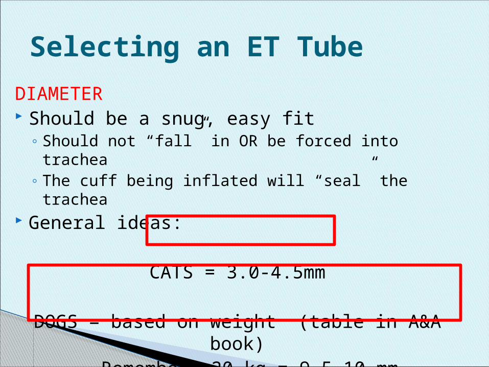
DIAMETER Should be a snug, easy fit
◦ Should not “fall” in OR be forced into trachea◦ The cuff being inflated will “seal” the trachea
General ideas:
CATS = 3.0-4.5mm
DOGS = based on weight (table in A&A book)Remember: 20 kg = 9.5-10 mm
Selecting an ET Tube

Weight based is a guideline Always prep 3 tubes (choice,1 smaller, 1 bigger) Brachycephalics may need smaller than you
think◦ Long soft palate with extra tissue and narrow tracheas
Tips for Tube Size
Use width of space between thenostrils as a guide

LENGTH Extend from the tip of the nose to the thoracic
inlet◦ ABOVE THE TRACHEAL BIFURCATION
If you extend into only one lung:◦ Hypoventilation and hypoxemia ???
If you extend tube too far past patient’s nose
Increased dead space
Selecting an ET Tube

Prior to Intubation Check several tubes for loose connectors,
excessive wear, cuff leaks, debrisCuff leak check: inflate cuff fully and let it sit Remember to deflate cuff completely prior to intubation
Apply lubrication- very small amount and optional
◦ Larger tubes – KY jelly or saliva Do not allow it to dry on tube
◦ Smaller tubes (<4.0mm) – water or saliva Check patient jaw tone
◦ Swallow reflex

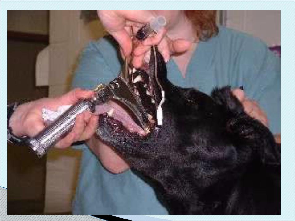
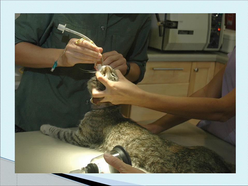
Intubation Techniques1. Visual Preferred technique for dog and cat Direct visualization of larynx minimizes
possibility of traumatic or improper intubation
Position: Sternal, Dorsal, Lateral recumbency◦ Position is preference

Visual Technique Assistant holds hand placed on the muzzle
with fingers behind front canine teeth (like you would for pilling) pulling upward to open the mouth
Neck should be slightly extended and in line with body
Pull out tongue to visualize back of throat
◦ May need gauze to hold tongue- slippery
14

Tongue is pulled forward and off to the side, then held in place with thumb – moves epiglottis forward and down◦ Tongue can be held by you or restrainer
Move soft palate up and out of the way with ET tube and at the same time…
Move epiglottis down out of the way with the tube – this brings tracheal opening into view
Under direct vision, tube is passed through tracheal opening
Tech note: You may need to wait for a breath or stimulate animal’s body to inhale to see opening

With laryngoscope
RIGHT HANDED PERSON
Hold laryngoscope w/ left hand Hold ET tube in right hand Press blade against pulled out tongue,
exposing trachea Can be used to hold epiglottis down
◦ Lightly! Blade too far forward will obstruct view

Page 18

With Stylet Stylet runs inside the ET tube Made of strong wire/metal
◦ Stiffens the tube and molds the tube Sticks out past the ET tube
◦ Provides smaller, blunt point to first pass through the vocal cords
◦ Allows larger ET tube to slide into trachea Stylet should be longer than ET tube!
◦ Why?
19
Intubation Techniques
2. Blind Used in dogs and horses NOT suitable for cats, very small dogs, or patients
with edema, swelling or trauma Lateral recumbency (RIGHT lateral in dogs) Head and neck extended and mouth open Advance tube with bevel parallel to vocal cords Move soft palate up and out of the way and
epiglottis down Advance upon expiration Rotating tube to follow curve and point tip down

Intubation Techniques
3. Tactile Cattle, large exotics, a few large dogs
Finger holds down the epiglottis Slide tube into trachea using your finger as a guide
More difficult Small mouth and sensitive larynx Dome shaped vocal cords tend to close
and push tube to side Swallowing reflex or contact with end of
tube causes laryngospasm
Feline Intubation

Laryngeal sensitivity◦Can be reduced by application of topical
anesthetic
Apply 0.1 cc of 2% Lidocaine soln. on glottis (without the needle!)
◦ Or use cotton swab Can also coat the end of the tube w/
lidocaine gel
Feline Intubation
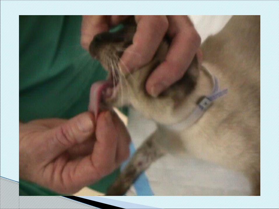


Tips for cats
Sternal recumbency Good light source Use direct visualization technique-must be
able to see vocal cord opening before inserting tube
Stylet is helpful! Cats often cough – don’t let go!


How Do You Know You’re In?
Condensation seen in ET tube Feel air through tube
◦ Place something light or metalat end of ET tube
Palpate throat◦ One tube – you’re in◦ Two tubes – you’re in esophagus
Normal breathing sounds◦ No gurgling
Patient can not vocalize

Using Machines
Give a breath = chest should rise (stomach should NOT)◦ Listen to BOTH lung sounds
Rebreathing bag and flutter valve should move with respirations
Capnograph should give appropriate reading
Radiographs
How Do You Know You’re In?

Page 32
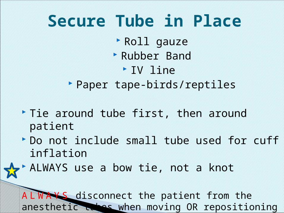
Secure Tube in Place Roll gauze
Rubber Band IV line
Paper tape-birds/reptiles
Tie around tube first, then around patient Do not include small tube used for cuff
inflation ALWAYS use a bow tie, not a knot
A LW A Y S disconnect the patient from the anesthetic tubes when moving OR repositioning

Page 34

Cuff Inflation
Cats: 1 – 2 cc of air Dogs: 2 – 10 cc of air
◦Valve port should inflate, but not be maximally full of air
◦If more than 10 cc needed: Leak or need a larger ET
tube
Cuff Inflation
Recheck every 30 min of surgery – especially after moving or repositioning patient
If you are running anesthesia for longer than 2 hours reposition the tube slightly so pressure necrosis does not happen.◦ Must deflate cuff before moving tube!

Extubation Your patient will be in recovery
◦ Sternal or lateral recumbency ◦ Head and neck extended
Deflate the cuff when the patient shows signs of waking up Remove ET tube after un-stimulated swallowing has
returned Prevent obstruction of airway with tongue by pulling
tongue forward during and after pulling the tube*Waiting too long can cause patient to bite tube in half*

Brachycephalic animals – should be head up, chewing on tube before it is pulled
Extubation Note
Post-op Advice to Owner
Patients may cough for 1 – 2 days post operatively
Should not be severe or continue to get worse
Advising owner will avoid phone calls and later explanations!

ET Tube Cleaning Inflate cuff and leave inflated until dry
Wash inside AND outside of endotracheal tube Use warm soapy water to get mucus off
◦ Commercial brushes available, cotton swabs, pipe cleaners
Rinse Disinfect for minimum of 15 minutes in Ultra
Sonic Cleaning soln. with DILUTE chlorhexidine Rinse VERY well Hang upright to dry over night Deflate cuff