Embed Size (px)
Citation preview
Aalborg Universitet
Physiological models of gas exchange in decision support of mechanical ventilation
Karbing, Dan Stieper
Publication date:2009
Document VersionPublisher's PDF, also known as Version of record
Link to publication from Aalborg University
Citation for published version (APA):Karbing, D. S. (2009). Physiological models of gas exchange in decision support of mechanical ventilation:prospective evaluation in an intensive care unit. Aalborg: Center for Model-based Medical Decision Support.Department of Health Science and Technology. Aalborg University.
General rightsCopyright and moral rights for the publications made accessible in the public portal are retained by the authors and/or other copyright ownersand it is a condition of accessing publications that users recognise and abide by the legal requirements associated with these rights.
? Users may download and print one copy of any publication from the public portal for the purpose of private study or research. ? You may not further distribute the material or use it for any profit-making activity or commercial gain ? You may freely distribute the URL identifying the publication in the public portal ?
Take down policyIf you believe that this document breaches copyright please contact us at [email protected] providing details, and we will remove access tothe work immediately and investigate your claim.
Downloaded from vbn.aau.dk on: september 04, 2018
Physiological models of gas exchange in decision
support of mechanical ventilation
Prospective evaluation in an intensive care unit
By: Dan Stieper Karbing
Center for Model-based Medical Decision Support
Department of Health Science and Technology
Aalborg University
2009

ISBN (print edition): 978-87-7094-048-1
ISBN (electronic edition): 978-87-7094-049-8
Supervisors Stephen E. Rees, PhD, Center for Model-based Medical Decision Support,
Department of Health Science and Technology, Aalborg University.
Steen Andreassen, Dr. Tech., PhD, Center for Model-based Medical Decision
Support, Department of Health Science and Technology, Aalborg University.
Søren Kjærgaard, PhD, Anaesthesia and Intensive Care, Region North Jutland,
Aalborg Hospital, Aarhus University.
3
Physiological models of gas exchange in decision support of
mechanical ventilation – prospective evaluation in an intensive care
unit
Abstract of thesis defended 27 November 2009
Introduction: Management of mechanical ventilation is a complex process of finding
the right balance between conflicting goals, where clinicians must make timely
decisions in unfavorable circumstances. Minimal models of pulmonary gas exchange
may be used at the bedside in the intensive care unit to help in this process providing a
deeper understanding of the patient‟s gas exchange status. The aim of this PhD project
was to build and evaluate minimal models of gas exchange, and prospectively
evaluate a minimal model-based decision support system.
Methods: Three retrospective studies were performed using data from various patient
types including intensive care patients: comparing a hypoxemia index and model of
O2 gas exchange available in clinical practice with a two parameter minimal model;
evaluating a decision support system for suggestions of inspired O2 fraction; and
investigating three minimal models of varying complexity for their ability to describe
gas exchange of both O2 and CO2. A prospective study was performed in an intensive
care unit to compare decision support system suggestions of inspired O2 and resulting
oxygenation with those selected by attending clinicians.
Results: The often used hypoxemia index, PaO2/FiO2 ratio, varies significantly with
changes in inspired O2, a common change in therapy. The clinically available shunt
only model of gas exchange can not accurately describe this variation, a two
parameter minimal model describing shunt and ventilation-perfusion mismatch can.
The decision support system provides appropriate suggestions of inspired O2 fraction
retrospectively, and prospectively. A three parameter minimal modeling complexity is
necessary for an accurate description of gas exchange of both O2 and CO2.
Conclusions: A minimal model-based decision support system can be used to provide
a deeper understanding of the individual patient‟s gas exchange status, and to provide
appropriate suggestions on inspired O2 fraction freeing the focus of clinicians for
more challenging therapies.
4
List of Papers
The thesis is based on the four listed papers, which will be referred to in the text by
their corresponding roman numerals:
I Karbing DS, Kjærgaard S, Smith BW, Espersen K, Allerød C, Andreassen S ,
Rees SE.
Variation in the PaO2/FiO2 ratio with FiO2: mathematical and experimental
description, and clinical relevance.
Critical Care 2007, 11:R118.
Commented in:
Critical Care 2007; 11(6):182.
Critical Care 2008; 12(1):407; Author reply 407.
II Karbing DS, Kjærgaard S, Smith BW, Allerød C, Espersen K, Andreassen S,
Rees SE.
Decision support of inspired oxygen fraction using a model of oxygen
transport.
IFAC PapersOnLine, Proceedings of the 2008 Congress of the International
Federation of Automatic Control, Seoul, Korea, July 6 – 11, Vol. 17(1) (DOI:
10.3182/20080706-5-KR-1001.2130)
III Karbing DS, Allerød C, Thorgaard P, Carius A, Frilev L, Andreassen S,
Kjærgaard S, Rees SE.
Prospective evaluation of a decision support system for setting inspired
oxygen in intensive care patients.
In press. Journal of Critical Care (DOI: 10.1016/j.jcrc.2009.12.013)
IV Karbing DS, Kjærgaard S, Andreassen S, Espersen K, Rees SE.
The minimal model approach to quantification of pulmonary gas exchange of
oxygen and carbon dioxide.
In preparation.
5
Contents
List of Papers ................................................................................................................. 4
Contents ......................................................................................................................... 5
Abbreviations and symbols ............................................................................................ 7
1. Clinical and technical background of the project ...................................................... 9
1.1 Introduction .......................................................................................................... 9
1.2 Managing mechanical ventilation in the ICU .................................................... 10
Acute lung injury and the acute respiratory distress syndrome ........................... 10
Ventilator induced lung injury ............................................................................. 11
Lung-protective ventilator strategies ................................................................... 12
1.3 Decision support systems for mechanical ventilation ........................................ 14
Rule-based systems .............................................................................................. 14
Model-based decision support in ventilator management ................................... 17
1.4 Mathematical models of gas exchange .............................................................. 21
Measurements and models available in clinical practice ..................................... 21
The Multiple Inert Gas Elimination Technique ................................................... 22
Minimal modeling of pulmonary gas exchange ................................................... 23
1.5 Aims of the project ............................................................................................. 24
2. Gas exchange models and decision support system ................................................ 27
2.1 Minimal models of pulmonary gas exchange .................................................... 27
2.2 Estimation of model parameters ........................................................................ 28
2.3 Decision support system .................................................................................... 33
2.4 ICARE system and database .............................................................................. 35
3. Summary of Papers .................................................................................................. 37
3.1 Paper I ................................................................................................................ 37
Aim ...................................................................................................................... 37
Methods ................................................................................................................ 37
Results .................................................................................................................. 38
Conclusions .......................................................................................................... 40
3.2 Paper II ............................................................................................................... 41
Aim ...................................................................................................................... 41
Methods ................................................................................................................ 41

6
Results .................................................................................................................. 42
Conclusions .......................................................................................................... 43
3.3 Paper III ............................................................................................................. 44
Aim ...................................................................................................................... 44
Methods ................................................................................................................ 44
Results .................................................................................................................. 45
Conclusions .......................................................................................................... 48
3.4 Paper IV ............................................................................................................. 49
Aim ...................................................................................................................... 49
Methods ................................................................................................................ 49
Results .................................................................................................................. 50
Conclusions .......................................................................................................... 52
4. Discussion ................................................................................................................ 53
4.1 The major findings of this thesis ........................................................................ 53
4.2 Model-based or rule-based decision support systems? ...................................... 55
4.3 Current status of model-based decision support of mechanical ventilation....... 57
4.4 Future work ........................................................................................................ 59
Advice on FiO2, Vt, and f .................................................................................... 60
Advice on PEEP ................................................................................................... 61
Clinical integration ............................................................................................... 63
4.5 Model limitations ............................................................................................... 65
4.6 Clinical perspectives .......................................................................................... 66
5. General Conclusions ................................................................................................ 69
Acknowledgements ...................................................................................................... 70
References .................................................................................................................... 71
Summary ...................................................................................................................... 86
Danish summary .......................................................................................................... 88
7
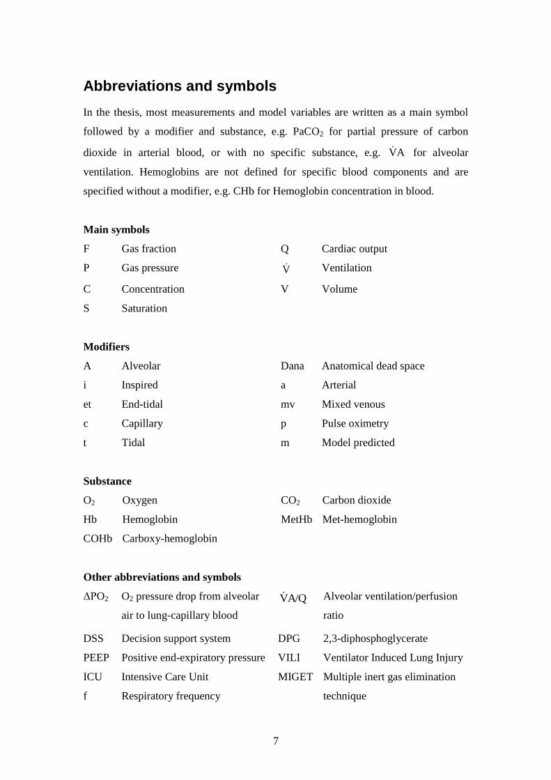
Abbreviations and symbols
In the thesis, most measurements and model variables are written as a main symbol
followed by a modifier and substance, e.g. PaCO2 for partial pressure of carbon
dioxide in arterial blood, or with no specific substance, e.g. AV for alveolar
ventilation. Hemoglobins are not defined for specific blood components and are
specified without a modifier, e.g. CHb for Hemoglobin concentration in blood.
Main symbols
F Gas fraction Q Cardiac output
P Gas pressure V Ventilation
C Concentration V Volume
S Saturation
Modifiers
A Alveolar Dana Anatomical dead space
i Inspired a Arterial
et End-tidal mv Mixed venous
c Capillary p Pulse oximetry
t Tidal m Model predicted
Substance
O2 Oxygen CO2 Carbon dioxide
Hb Hemoglobin MetHb Met-hemoglobin
COHb Carboxy-hemoglobin
Other abbreviations and symbols
ΔPO2
O2 pressure drop from alveolar
air to lung-capillary blood
A/QV
Alveolar ventilation/perfusion
ratio
DSS Decision support system DPG 2,3-diphosphoglycerate
PEEP Positive end-expiratory pressure VILI Ventilator Induced Lung Injury
ICU
f
Intensive Care Unit
Respiratory frequency
MIGET Multiple inert gas elimination
technique
8
… modelling is assuming a more prominent role in mainstream
anaesthesia and critical care research, becoming an accepted
methodology and an ever-more useful part of the research
process.
... Modelling runs through all of our endeavours, and we stand
to benefit hugely by becoming acquainted with this powerful
device.
J. G. Hardman and J. J. Ross
Editorial in British Journal of Anaesthesia 2006
Vol 97, pages 589-92
9
1. Clinical and technical background of the project
1.1 Introduction
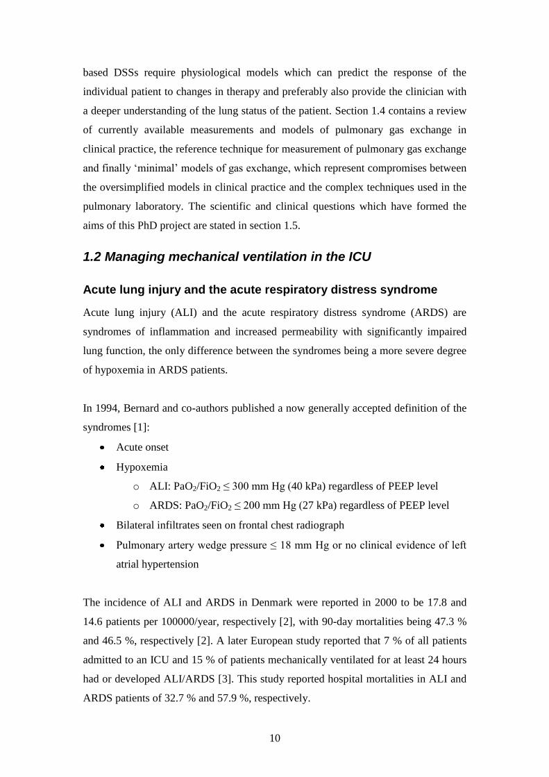
Mechanical ventilation is a life-sustaining therapy used to secure sufficient
oxygenation and carbon dioxide elimination and spare patients‟ energy allowing them
to cope with underlying diseases and recover from surgery or trauma. Managing
mechanical ventilator settings for ventilator therapy of the common postoperative
patient is generally a simple task mainly comprised of weaning the patient from
ventilator support, i.e. stepwise reduction in ventilator support until the patient alone
is driving ventilation. However, in critically ill patients presenting in the intensive
care unit (ICU), with failure of one or more organ systems often including the lungs,
managing mechanical ventilation is a complex task. In these patients, selecting the
appropriate ventilator settings can be considered as a search for the optimal
compromise of conflicting goals. Such a search would preferably be performed based
on a good understanding of the patient‟s lung function. However, this is often difficult
using the vast number of relatively simple measurements currently available in the
ICU. Mathematical models of pulmonary gas exchange may be used to integrate
simple measurements and provide a deeper understanding of the patient‟s underlying
physiology and pathophysiology. Implementing such models in decision support
systems (DSSs) to calculate suggestions on therapy and provide physiological
understanding may provide a valuable tool for clinicians, when deciding on
appropriate therapy.
To illustrate the need for DSSs in mechanical ventilation, section 1.2 will present the
clinical background of the project. The syndromes acute lung injury and acute
respiratory distress syndrome are introduced. Ventilator induced lung injury (VILI)
will be presented constituting the background of recent approaches to mechanical
ventilation, termed lung protective ventilator strategies. Recent studies on ventilator
strategies will also be introduced illustrating the lack of consensus on how to properly
mechanically ventilate patients with severe lung disorders. In section 1.3 the literature
on decision support systems is reviewed covering rule-based systems representing the
most prevalent type of DSS, and model-based DSS. The focus of this PhD is decision
support of mechanical ventilation using models of pulmonary gas exchange. Model-
10
based DSSs require physiological models which can predict the response of the
individual patient to changes in therapy and preferably also provide the clinician with
a deeper understanding of the lung status of the patient. Section 1.4 contains a review
of currently available measurements and models of pulmonary gas exchange in
clinical practice, the reference technique for measurement of pulmonary gas exchange
and finally „minimal‟ models of gas exchange, which represent compromises between
the oversimplified models in clinical practice and the complex techniques used in the
pulmonary laboratory. The scientific and clinical questions which have formed the
aims of this PhD project are stated in section 1.5.
1.2 Managing mechanical ventilation in the ICU
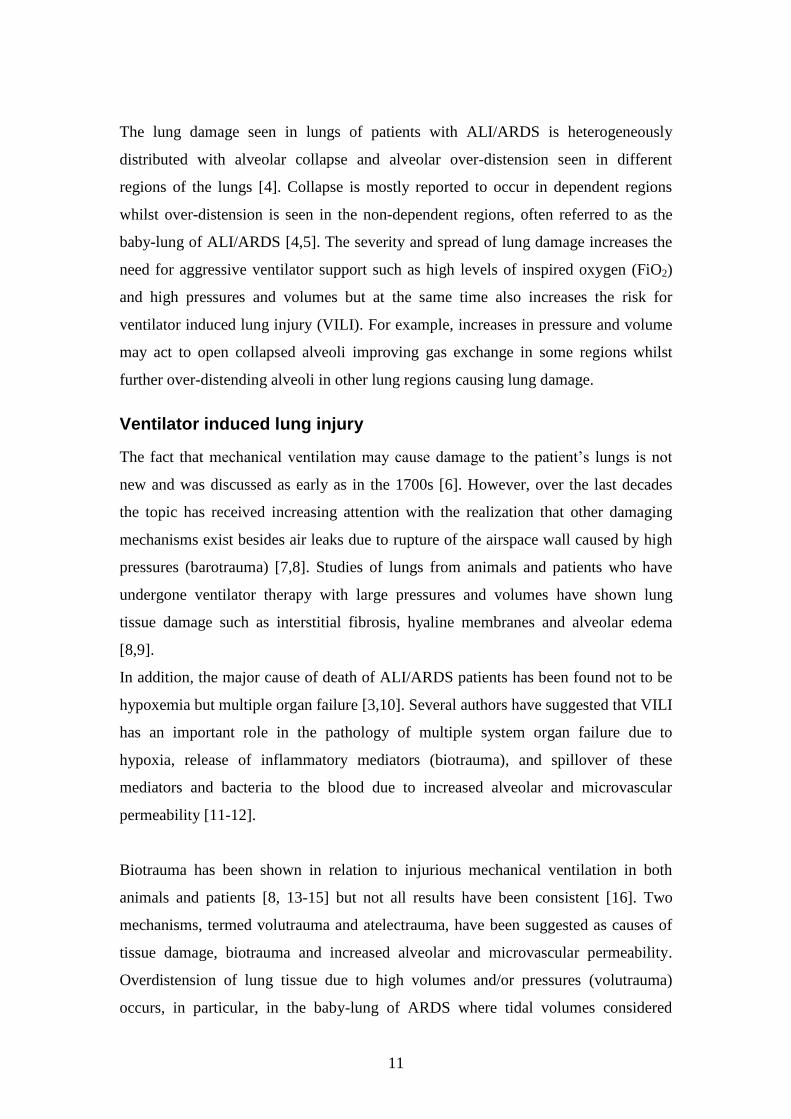
Acute lung injury and the acute respiratory distress syndrome
Acute lung injury (ALI) and the acute respiratory distress syndrome (ARDS) are
syndromes of inflammation and increased permeability with significantly impaired
lung function, the only difference between the syndromes being a more severe degree
of hypoxemia in ARDS patients.
In 1994, Bernard and co-authors published a now generally accepted definition of the
syndromes [1]:
Acute onset
Hypoxemia
o ALI: PaO2/FiO2 ≤ 300 mm Hg (40 kPa) regardless of PEEP level
o ARDS: PaO2/FiO2 ≤ 200 mm Hg (27 kPa) regardless of PEEP level
Bilateral infiltrates seen on frontal chest radiograph
Pulmonary artery wedge pressure ≤ 18 mm Hg or no clinical evidence of left
atrial hypertension
The incidence of ALI and ARDS in Denmark were reported in 2000 to be 17.8 and
14.6 patients per 100000/year, respectively [2], with 90-day mortalities being 47.3 %
and 46.5 %, respectively [2]. A later European study reported that 7 % of all patients
admitted to an ICU and 15 % of patients mechanically ventilated for at least 24 hours
had or developed ALI/ARDS [3]. This study reported hospital mortalities in ALI and
ARDS patients of 32.7 % and 57.9 %, respectively.
11
The lung damage seen in lungs of patients with ALI/ARDS is heterogeneously
distributed with alveolar collapse and alveolar over-distension seen in different
regions of the lungs [4]. Collapse is mostly reported to occur in dependent regions
whilst over-distension is seen in the non-dependent regions, often referred to as the
baby-lung of ALI/ARDS [4,5]. The severity and spread of lung damage increases the
need for aggressive ventilator support such as high levels of inspired oxygen (FiO2)
and high pressures and volumes but at the same time also increases the risk for
ventilator induced lung injury (VILI). For example, increases in pressure and volume
may act to open collapsed alveoli improving gas exchange in some regions whilst
further over-distending alveoli in other lung regions causing lung damage.
Ventilator induced lung injury
The fact that mechanical ventilation may cause damage to the patient‟s lungs is not
new and was discussed as early as in the 1700s [6]. However, over the last decades
the topic has received increasing attention with the realization that other damaging
mechanisms exist besides air leaks due to rupture of the airspace wall caused by high
pressures (barotrauma) [7,8]. Studies of lungs from animals and patients who have
undergone ventilator therapy with large pressures and volumes have shown lung
tissue damage such as interstitial fibrosis, hyaline membranes and alveolar edema
[8,9].
In addition, the major cause of death of ALI/ARDS patients has been found not to be
hypoxemia but multiple organ failure [3,10]. Several authors have suggested that VILI
has an important role in the pathology of multiple system organ failure due to
hypoxia, release of inflammatory mediators (biotrauma), and spillover of these
mediators and bacteria to the blood due to increased alveolar and microvascular
permeability [11-12].
Biotrauma has been shown in relation to injurious mechanical ventilation in both
animals and patients [8, 13-15] but not all results have been consistent [16]. Two
mechanisms, termed volutrauma and atelectrauma, have been suggested as causes of
tissue damage, biotrauma and increased alveolar and microvascular permeability.
Overdistension of lung tissue due to high volumes and/or pressures (volutrauma)
occurs, in particular, in the baby-lung of ARDS where tidal volumes considered

12
normal in healthy lungs are deleterious [5]. Repeated recruitment and de-recruitment
of atelectic lung regions (atelectrauma) has been suggested to cause stress and strain
in the junctions between adjacent alveoli [8,17]. The physical stress and strain
involved in volutrauma and atelectrauma may lead to epithelial damage and increased
alveolar and microvascular permeability causing pulmonary edema [8]. Volutrauma
and atelectrauma may also impair the function of pulmonary surfactant [18].
Surfactant is a chemical compound which acts on the air/water interface inside the
alveolar epithelium to reduce surface tension lowering work of breathing, maintaining
fluid balance across the alveolar membrane and preventing alveolar collapse [18].
High fractions of oxygen in the inspired air (FiO2) can also affect lung status leading
to gas-exchange impairment or tissue damage. High levels of FiO2 can cause
atelectasis in regions with low ventilation/perfusion ratios [19-21] and cause toxic
effects [22-23].
Lung-protective ventilator strategies
The role of mechanical ventilation as a major cause of patient mortality has spurred
numerous experimental investigations and clinical trials addressing how to properly
manage mechanical ventilation, in particular in ALI/ARDS patients. In the following,
some of the major studies within lung-protective ventilation are described.
In 1998, Amato and coworkers described a statistically significant improvement in
28-day mortality in 53 ARDS patients by using a strategy consisting of: recruitment
maneuvers i.e. short periods of large pressures to open atelectic lung regions; positive
end expiratory pressure (PEEP) to keep recruited alveoli open; and small tidal
volumes to reduce lung tissue stress [24]. After 28 days the mortality of the lung
protective group was 38 % compared to 71 % in the conventionally treated group.
However, several studies with similar strategies and number of patients did not find
significant differences in mortality between low and high tidal volumes [25-27].
A large multicenter study conducted by the Acute Respiratory Distress Syndrome
Network (ARDSNet) followed the trial by Amato et al. comparing the use of small
and large tidal volumes [28]. This study showed that a strategy comprising tidal
volumes small (Vt = 6 ml/kg) in comparison to previously common tidal volumes and
13
peak inspiratory pressures less than 30 cmH2O resulted in improved mortality
compared to large tidal volumes (Vt = 12 ml/kg) and peak inspiratory pressures less
than 50 cmH2O.
A later study conducted by the ARDSNet investigated the use of low versus high
PEEP, maintaining a Vt of 6 ml/kg in both groups [29]. The study did not find any
significant difference in mortality between the two groups. However, later analysis
has indicated that this study might not have had large enough differences in PEEP
levels between the two groups to demonstrate a significant difference in mortality
[18].
Larger differences between PEEP levels in two patient groups (13.4 ±2.6 cmH2O in
53 patients vs. 9.8 ± 2.8 cmH2O in 50 patients) were reported in a recent study to
produce significant improvement in mortality [30]. However, the two groups were
ventilated with different tidal volumes preventing the authors from drawing
conclusions on the importance of PEEP levels on mortality [30]. In addition, two
recently published multicenter trials compared two groups with equal low tidal
volumes but with lower and higher PEEP levels [31,32]. Neither of these two studies
could demonstrate significant differences in mortality between the studied patient
groups.
The focus of current ventilator strategies is on preventing VILI by lowering volumes
and pressures. However, FiO2 should not be increased indiscriminately to secure
oxygenation [19-23], and several authors have pointed out that low tidal volumes may
lead to low ventilation/perfusion ( A/QV ) regions in the lungs, which limits gas
exchange and are highly susceptible to adsorption atelectasis due to hyperoxia
[33,34]. Although not the focus of recent debate, the vast majority of clinical trials
have included limitation of FiO2 either directly or through goals for oxygenation in
their ventilator strategies [15, 24-29, 31,32].
Whilst there is a general consensus that the lungs should be ventilated with caution,
there is also a general consensus that the understanding of the different types of lung
damage and the mechanisms involved is not complete [16-18,33-35]. Furthermore the

14
varying results from clinical trials indicate that the perfect ventilation strategy, if there
is such a thing, has yet to be found. It has been speculated, that the heterogeneity of
ALI/ARDS patients requires that every patient should be treated on an individual
basis [17], which is supported by recent clinical studies [36,37]
This leaves intensive care clinicians with a far from straightforward task, which needs
to be performed in a timely manner, based on interpretation of large amounts of data.
The increasing complexity and available options on modern ventilators do not help to
alleviate the problem. These circumstances work against human nature. The human
brain can process a limited amount of information when making decisions [38], which
combined with the stressful environment of the ICU have been suggested as
augmenting factors for errors committed by health care professionals in the ICU [39,
40]. These points illustrate that DSS for mechanical ventilation may be beneficial.
1.3 Decision support systems for mechanical ventilation
Decision support systems (DSSs) may be categorized with regards to several aspects:
open or closed loop; approach to data integration and analysis; approach to decisions,
e.g. rule-based, utility theory, etc.; and the settings optimized by the system. In the
following, the literature is reviewed categorizing published DSS for mechanical
ventilation into rule-based systems and model-based systems. Rule-based systems will
refer to systems that are mimicking experts in the field or clinical guidelines
performing data integration and analysis using the clinical measurements directly
without physiological models.
Rule-based systems
The vast majority of developed DSS for ventilator management have been rule-based
systems [41-63]. These systems have often been developed for specific subproblems
of ventilator management such as weaning patients from mechanical ventilation [49].
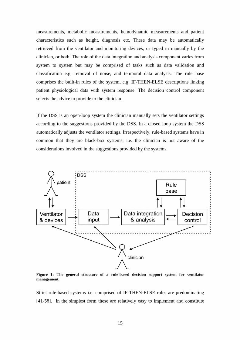
Figure 1 shows the general overall structure of such systems. Rule-based systems
typically include 4 overall components: data input; data integration and analysis; rule
base; and decision control (often also called inference engine). The interaction and
integration of these components may vary from system to system. Data input can
consist of ventilator settings, lung mechanic measurements, gas exchange
15
measurements, metabolic measurements, hemodynamic measurements and patient
characteristics such as height, diagnosis etc. These data may be automatically
retrieved from the ventilator and monitoring devices, or typed in manually by the
clinician, or both. The role of the data integration and analysis component varies from
system to system but may be comprised of tasks such as data validation and
classification e.g. removal of noise, and temporal data analysis. The rule base
comprises the built-in rules of the system, e.g. IF-THEN-ELSE descriptions linking
patient physiological data with system response. The decision control component
selects the advice to provide to the clinician.
If the DSS is an open-loop system the clinician manually sets the ventilator settings
according to the suggestions provided by the DSS. In a closed-loop system the DSS
automatically adjusts the ventilator settings. Irrespectively, rule-based systems have in
common that they are black-box systems, i.e. the clinician is not aware of the
considerations involved in the suggestions provided by the systems.
Figure 1: The general structure of a rule-based decision support system for ventilator
management.
Strict rule-based systems i.e. comprised of IF-THEN-ELSE rules are predominating
[41-58]. In the simplest form these are relatively easy to implement and constitute
16
electronic versions of paper based clinical guidelines [e.g. 45-48]. Almost all
published DSS based on this simple structure have been prospectively evaluated [41-
53]. Two of these studies have been large multicenter randomized trials [48, 53]. The
study reported by East et al. investigated a clinical guideline for managing ARDS
patients, and although the implemented clinical guideline did not result in statistically
significant improvement in mortality, the study demonstrated the feasibility of
implementing a DSS across several institutions [48]. The study by Lellouche et al.
compared weaning of patients using a closed-loop DSS (GANESH) [49] with
weaning using written clinical guidelines [53]. The patient group weaned using
GANESH had lower duration of weaning, shorter duration of mechanical ventilation
and shorter ICU stay [53]. GANESH has also been implemented as part of a
commercial system, termed SmartCareTM
/PS by Dräger Medical [64].
Adaptive support ventilation (ASV) is another commercially available closed loop
DSS, implemented in Hamilton ventilators [65]. When using ASV the clinician
defines a desired minute volume and the system automatically adjusts respiratory
frequency, tidal volume and inspiratory pressure and switches between support and
control behavior using rules according to measurements of the patient‟s lung
mechanics [65]. Several studies have been performed using ASV, for example a
multicenter study comparing ASV with controlled ventilator modes in patients with
acute respiratory failure [66]. The study showed that ASV could maintain similar
PaCO2 as clinicians but with lower peak airway pressures.
In addition to SmartCareTM
and ASV several advanced ventilator modes have been
developed which have elements in common with DSS. Most notable are proportional
assist ventilation (PAV), and neurally adjusted ventilatory assistance (NAVA) which
can be considered advanced versions of the pressure support mode. These systems
determine the level of pressure support using a gain factor adjusted by the clinician
combined with either measured inspiratory flow (PAV) or the electromyographic
activity from the diaphragm (NAVA) [67].
Different research groups have taken alternative approaches to capture the heuristics
of critical care experts [59-63]. These approaches include knowledge bases using
automated knowledge acquisition [59], and fuzzy logic for temporal data

17
classification [60, 63], to derive the rule base [62], and to mimic human decision-
making [61]. One of these systems have been implemented and prospectively
evaluated in 7 neonates showing agreement between clinicians and provided advice in
more than 90% of cases [63].
The black-box approach shared by all the presented rule-based DSS is also one of the
major weaknesses of these systems, as they do not provide the clinician with a deeper
understanding of the individual patient‟s status. If changes in settings alter the status
of the patients this may require reevaluations leading to new changes. As such, the
rule-based systems may require a trial and error approach. Model-based DSS may
solve both of these problems. Parameters of physiological models may provide a
deeper physiological understanding of the patient. In addition, once model parameters
have been tuned to fit the individual patient data, models can predict patient response
to changes in ventilator settings allowing the clinician quick evaluation of therapy
changes, thereby eliminating the need for the trial and error approach [68].
Model-based decision support in ventilator management
Figure 2 shows the overall general structure of model-based DSS in ventilator
management. Five overall components are generally included: data input;
physiological models; parameter identification; model prediction; and decision
control. The data input component is conceptually identical to that of the rule-based
systems. The physiological model component constitutes the physiological models
used in a model-based DSS such as models of pulmonary gas exchange. Parameter
estimation often constitutes measurement of patient response to small variations in
therapy to allow tuning of model parameters to fit the physiological models to the
individual patient. This process can be manual with the system interacting with the
user during the process or it can be automated. Parameter estimation will often
encompass the data integration in the system. Once fitted to patient data the models
can be used to simulate patient response to changes in therapy, e.g. changes in
oxygenation upon changes in inspired oxygen fraction. This can involve the clinician,
by letting the clinician test different changes in settings without involving the patient
or it can be done by the DSS to calculate optimal therapy. This can be performed by a
decision control component using mathematical functions associating different
strategies with corresponding utilities, i.e. models of clinical preferences. Hybrid

18
systems have also been developed where physiological models are combined with a
rule base to decide suggestions on therapy.
Figure 2: General structure of model-based decision support systems for ventilator management.
Model-based DSSs may solve two problems, i.e. providing a deeper understanding of
patient physiology and preventing trial and error approach to ventilator management.
However, they may also introduce two overall limitations. When physiological
models are integrated into the calculation of new advice, model-based DSS depend on
the implemented models to accurately predict patient response to changes in ventilator
settings. In addition, in order to allow patient specific predictions, the model
parameters must be tuned to fit patient specific data before suggestions on therapy can
be calculated, and this may be a time-demanding process. These limitations have been
dealt with in different ways by the different model-based DSS [69-74].
The first reported DSS using physiological models was the open-loop system
OPTPROG [69]. OPTPROG found the combination of respiratory frequency (f), tidal
volume (Vt) and PEEP which resulted in the minimal peak respiratory power (PRP),
which was defined as an index of lung trauma. The system also maintained PaO2 and
PaCO2 within limits defined by a clinician. OPTPROG was prospectively evaluated in
5 patients with various pulmonary diseases and 7 post operative coronary artery
bypass graft patients showing that the system was able to minimize PRP whilst
19
maintaining adequate PaO2 and PaCO2 values [69, 70]. However, OPTPROG had a
couple of significant limitations. OPTPROG was based on linear programming, and
the model parameters had no physiological interpretation [68]. As such, the system
did not provide clinicians with a deeper physiological understanding of the patient. In
addition, estimation of model parameters required a time-demanding experiment
taking approximately one hour involving frequent sampling of arterial blood beyond
that of routine clinical practice [68].
The VentPlan system [71] used a model of pulmonary gas exchange to provide open-
loop decision support of FiO2, Vt and f. The implemented model was a classical three
compartment model with model parameters having physiological interpretations [75].
The model includes two parameters: a shunt parameter which quantifies the fraction
of pulmonary perfusion not reaching ventilated alveoli; and a parameter describing the
amount of physiological dead space, i.e. the amount of ventilation not participating in
gas exchange. Parameter estimation was performed as a combination of a Bayesian
belief network and patient specific measurements [71]. The belief network was
implemented to enable parameter estimation in cases when measurement data were
insufficient to allow a unique numerical solution when estimating model parameters.
Advice was calculated based on a combination of model simulations and utility theory
[76], using penalty functions to model clinical preferences. VentPlan was
retrospectively evaluated using data from 10 ICU patients indicating potential of the
system [71]. However, to the best of my knowledge, VentPlan development stopped
before any prospective evaluation could be performed.
The Sheffield Intelligent Ventilator Advisor (SIVA) uses a physiological model
describing the same factors as that of VentPlan, i.e. shunt and physiological dead
space [72]. SIVA uses the ratio between the alveolar-arterial oxygen difference to
PaO2 (PA-aO2 / PaO2) as an input to an Adaptive-Network-based Fuzzy-Inference
System [62] to estimate the shunt parameter. To estimate the physiological dead space
the system requires invasive measurement data using a pulmonary artery catheter and
a numerical method, which the authors report has often convergence problems [72].
Alternatively physiological dead space could be estimated by the clinician. SIVA is a
hybrid system and uses fuzzy rule-bases in combination with models to provide open-
loop decision support of FiO2, PEEP, inspiratory pressure (Pinsp) and f, although

20
without modeling the effect of PEEP. Evaluations of SIVA have so far been limited to
simulation studies [72].
The open-loop system INVENT presented by Rees et al. uses a two parameter
physiological model of gas exchange in combination with a model of the acid-base
chemistry of blood as well as a simple model of lung mechanics [73]. The gas
exchange model describes shunt and ventilation/perfusion mismatch, the two major
factors affecting pulmonary gas exchange in patients with respiratory failure [77]. The
parameters of the gas exchange model are estimated using a method comprised of
varying inspired oxygen fraction in 4-6 steps and measuring the oxygen contents of
the expired gas as well as pulse oximetry oxygen saturation (SpO2), this process
taking approximately 10-15 minutes [78]. The lung mechanics model requires input of
PEEP and respiratory compliance and the blood model and the gas exchange model
also require a single arterial blood gas measurement. The INVENT system provides
advice on FiO2, Vt and f using utility theory in the form of penalty functions
combined with the three models [73]. At the beginning of this PhD project evaluations
of the system had not been published.
The most recently introduced model-based DSS is the FLEX hybrid system, which
can act both as an open-loop and a closed-loop system [74]. The approach of the
FLEX system has similarities to the OPTPROG system with the implemented models
being empirical by nature and mainly using model parameters without physiological
interpretation. FLEX incorporates a large number of these simple models in
combination with a rule base to calculate suggested levels of FiO2, PEEP, f, I:E-ratio,
PIP, and Vt as well as to wean patients [74]. In this process the system aims at
minimizing the work of breathing using a modified version of an empirically derived
equation [79]. The FLEX system does not require any parameter estimation
procedures, but use readily available measurement data or parameters which are not
fitted to the individual patient. So far, the system has been limited to retrospective
evaluations, showing the suggestions of the system to be in general agreement with
decisions taken by clinicians in ICU patients [74] and neonates [80].
21
1.4 Mathematical models of gas exchange
Measurements and models available in clinical practice
Several measurements are available in clinical practice which may provide some
information regarding the gas exchange status of a patient. For oxygenation,
measurement of arterial blood gases yields arterial partial pressure of O2 (PaO2) and
arterial oxygen saturation (SaO2). An arterial blood sample can also be analyzed to
measure hemoglobins (Hb, MetHb and COhb) providing information regarding the
oxygen carrying capacity of the blood. Hemoglobin concentration, PaO2 and SaO2 can
also be used to calculate the total contents of O2 in arterial blood. Oxygen
concentrations and pressures can also be obtained from samples of central or mixed
venous blood, yielding information regarding the use of oxygen by the organs and
peripheral tissues, i.e. the general ischemic status of the body. Mixed and central
venous blood samples, however, require catheters in the pulmonary artery or one of
the larger veins (e.g. the internal jugular vein), respectively, and are not part of routine
clinical care in all ICUs. These measurements need to be related to the ventilation and
FiO2 to be interpreted with regards to the lung status of the patient.
A range of oxygen tension based indices have been developed to aid in interpretation
of oxygenation with regards to ventilator settings. The ratio between oxygen partial
pressure in arterial blood to FiO2 (PaO2/FiO2) is probably the most common index of
hypoxemia, especially in clinical studies, and is part of the definition of ALI/ARDS.
Another frequently used tension based index is the alveolar-arterial oxygen partial
pressure difference (PA-aO2) [81]. This index provides an estimate of the total drop in
oxygen partial pressure through the pulmonary system. However, the index requires
calculation of the alveolar partial pressure of oxygen (PAO2) using the alveolar air
equation requiring measurement of, or an assumed value of the respiratory quotient
[81]. All these oxygenation measures and indices vary with one or more
extrapulmonary factors such as ventilation and variation in FiO2 which are common
therapeutic interventions in mechanically ventilated patients and affect oxygenation
but not the underlying physiology or pathophysiology of the patient [77,].
The standard method for evaluating pulmonary gas exchange of CO2 is to measure the
partial pressure of CO2 in arterial blood (PaCO2). In addition, capnography can be
22
used to evaluate the CO2 contents in the expired gas in relation to either time or
expired volume, although this is not commonly applied in the ICU [83]. Capnography
allows measurement of end-tidal partial pressure or fraction of CO2 (PetCO2 or
FetCO2). When both PetCO2 and PaCO2 are available it is possible to calculate the
PetCO2-PaCO2 difference which will increase with A/QV mismatching and to a
lesser degree venous admixture [84]. The anatomical and alveolar dead space volumes
can also be calculated from the capnogram, the latter if a PaCO2 measurement is
available. Alternatively the physiological dead space can be calculated using
Enghoff‟s modification of the Bohr equation [85] requiring PaCO2 and measurement
or calculation of the partial pressure of CO2 in the mixed expired gas.
The current state of the art for quantifying pulmonary gas exchange in clinical
practice is measurement of intrapulmonary shunt [81]. When measured at an FiO2 less
than 100% the value is termed venous admixture and describes the patient‟s
pulmonary gas exchange abnormality as due to alveoli being perfused but not
ventilated. It has been shown that the measurement of intrapulmonary shunt is
inadequate to describe changes in oxygenation with variation in FiO2, and that it is
necessary to separate oxygenation problems into that caused by pulmonary shunt and
that due to an alveolar-lung capillary drop in partial pressure of oxygen [86-88]. By
measuring intrapulmonary shunt at FiO2=100%, the intrapulmonary shunt can be
measured without the effects of an alveolar-lung capillary drop in partial pressure of
oxygen. However, inspiration of pure oxygen may cause absorption atelectasis
thereby giving an overestimate of the true shunt value [19,89] and the method still
gives no information regarding the presence of an alveolar-lung capillary drop in
partial pressure of oxygen, such as due to A/QV mismatching in the lungs.
The Multiple Inert Gas Elimination Technique
To appreciate the concept of minimal modeling and the compromises made, one
should first look at the current reference technique for quantifying gas exchange in the
pulmonary laboratory, which is the multiple inert gas elimination technique (MIGET)
[90]. MIGET relies on measurement of retention and excretion of 6 inert gases
sampling blood and expired gas data. MIGET uses a model of pulmonary gas
exchange comprised of 50 compartments with different A/QV relationships
23
accounting for an alveolar-lung capillary drop in partial pressure of oxygen. The
model also includes shunt being the one extreme of the A/QV range ( A/QV = 0) and
alveolar dead space being the other extreme ( A/QV = ∞). This model is fitted to the
retention and excretion data and the end result is reported as distributions of blood
flow and ventilation across the compartments of the model.
Use of the MIGET technique in the pulmonary laboratory has contributed
significantly to the current physiological understanding of pulmonary gas exchange in
healthy subjects at different age [91], during anesthesia [92], in chronic obstructive
pulmonary disease [89] and in patients with acute respiratory failure including ALI
and ARDS [94]. MIGET studies have shown that lungs of ARDS patients are
characterized by large fractions of shunt and in many patients there are lung regions
with low A/QV ratios and/or large fractions of ventilation going to alveolar dead
space [94, 95]. The A/QV distributions produced by MIGET have also been shown to
describe the effects of various changes in therapy on pulmonary gas exchange [94]. In
ALI/ARDS patients, for example, increases in PEEP as well as changes in posture
from supine to prone have been shown to reduce shunt [95-98]. In addition,
inspiration of 100 % O2 has been shown to convert units with low A/QV into shunt
[94] possibly due to absorption atelectasis [19, 33-34].
The MIGET experimental procedure is highly complex and involves preparing and
infusing the inert gases, sampling of blood and expired gases and analyzing these
using gas chromatography [90], the technique is therefore inappropriate for routine
clinical application.
Minimal modeling of pulmonary gas exchange
In the past 15 years a considerable effort has been made to formulate minimal models
based on few model compartments and parameters which can be estimated from
routine clinical data. As originally suggested by Riley et al [86,87] and King et al.
[88] these models describe pulmonary gas exchange abnormalities as caused by
intrapulmonary shunt combined with an alveolar-lung capillary drop in partial
pressure of oxygen. The latter factor has been modeled either using one [99-101] or
two compartments to describe A/QV mismatch [78,102-104], one compartment with

24
diffusion limitation [102,105,106], or two compartments to describe both A/QV
mismatch and diffusion limitation [107,108]. These models are poor descriptions of
physiology compared to the 50 compartment model of MIGET but significant
improvements compared to measurement of intrapulmonary shunt or dead space
volume alone.
Minimal models describing intrapulmonary shunt and an alveolar-lung capillary drop
in partial pressure of oxygen have been shown to fit oxygenation data from normal
subjects [99,103,106-108]; patients before [99,100,102,103], during [99-101] and
after [99,100,102-104,106] major surgery; patients presenting in intensive care [103];
patients with chronic obstructive pulmonary disease (COPD) [107,108]; patients with
incompensated heart failure studied before and after diuretic therapy [103]; and
anesthetized mechanically ventilated dogs with acutely applied hypoxia or with
induced intense small airway constriction in a rebreathing model of COPD [108].
The model describing intrapulmonary shunt and A/QV mismatch presented by
Kjærgaard et al. [102], which is the model used in INVENT, has also been shown to
fit retention and excretion data comparable to MIGET in pigs before and after lung
damage caused by oleic acid infusion [109]. Additionally the model presented by
Vidal Melo and co-workers [107] has been shown to produce A/QV distributions
having positive correlations with MIGET A/QV distributions in COPD patients and
healthy subjects before and after exercise [108]. However, at present only a single
model has been evaluated for its ability to describe gas transport of CO2 [107,108].
1.5 Aims of the project
Management of mechanical ventilation in patients with severe lung disorders
constitutes a process of balancing conflicting therapeutic goals. This is a complex task
which clinicians in the ICU must perform based on numerous measurements in a
stressful environment and with no clear evidence based strategies for several of the
ventilator settings. Several DSSs have been developed to aid the clinician in this
process. The majority of these systems have been rule-based DSSs, which may
provide sound advice, but do not provide the clinician with a deeper understanding of
the individual patient. Rule-based DSSs may also require a time demanding trial and
25
error approach as commonly used in clinical practice to find the appropriate settings.
Model-based DSS may provide a more appropriate alternative. When models are
tuned to fit patient data they may provide a deeper physiological understanding of the
patient and predict patient responses to changes in therapy, thereby removing the need
for trial and error. However, this requires models with parameters having
physiological interpretation and which may be identified from routine clinical data.
Measurements and models of pulmonary gas exchange currently available in clinical
practice are oversimplified. In contrast the reference technique, the MIGET, is too
complex to use in clinical practice. Minimal models have also been developed
presenting compromises between feasibility and complexity. This PhD project has
addressed the use of such minimal models to describe gas exchange in the ICU and
their use in a DSS, through investigation of the following questions:
How well do the current dominating oxygenation index (PaO2/FiO2 ratio) and
gas exchange model (shunt model) used in clinical practice describe
oxygenation in comparison with a two parameter minimal model of O2 gas
exchange describing both shunt and ventilation/perfusion mismatch? (Paper I)
Can INVENT based on a two parameter model of gas exchange describing
shunt and ventilation/perfusion mismatch combined with utility theory provide
appropriate suggestions on FiO2 when evaluated retrospectively in intensive
care patients? (Paper II)
Can INVENT manage FiO2 in intensive care patients when evaluated
prospectively? (Paper III)
A model describing gas exchange of both O2 and CO2 is necessary for
INVENT to provide suggestions of Vt and f in addition to FiO2. What
complexity is necessary for a minimal model representation of pulmonary gas
exchange of both O2 and CO2? (Paper IV)
To address these questions technical methods were developed and refined. These
tasks involved modeling of pulmonary gas exchange, methods for estimation of model

26
parameters, development of a version of INVENT used with a database system to
provide suggestions on FiO2. The technical solutions were similar between studies,
but were adapted for the specific applications. The following chapter presents the
technical methods used during the project, and the tailoring of the methods for the
specific studies.

27
2. Gas exchange models and decision support system
This chapter describes the technical methods used to answer the four questions
addressed in the PhD project. The project has evolved around minimal models of
pulmonary gas exchange. Models of different complexity have been used in the
studies, but all models have followed the same overall structure. The models are
presented in section 2.1. Methods used for estimation of model parameters have been
adapted to the individual studies depending on available measurement data and aims
of the studies, the different approaches are explained in section 2.2. Section 2.3
presents the version of the DSS, INVENT, developed to provide suggestions on FiO2.
The system is integrated in a database system, ICARE, which is described briefly in
section 2.4.
2.1 Minimal models of pulmonary gas exchange
All four studies were performed using physiological models with the same structure
as the two parameter model originally presented by Kjærgaard et al [102]. These
models are based on conservation of mass, continuous breathing and perfusion and
assume steady state.
Figure 3 shows the overall structure shared by these models, indicating the model
parameters in bold. In addition to the shown compartments all models used in the
study have a serial dead space compartment. The model presented by Kjærgaard et al.
has a shunt parameter (fs) describing the fraction of pulmonary perfusion not reaching
ventilated alveoli. In addition the model has two ventilated and perfused
compartments. The perfusion is locked at a specific distribution defined by the f2
parameter such that one parameter receives 90 % of non-shunted blood flow and the
other 10 % (f2=0.9). Distribution of ventilation varies between the two compartments
as defined by the fA2 parameter, such that a fA2 of 0.9 would result in optimal A/QV
matching, whereas fA2 less than 0.9 would signify A/QV mismatching. This two-
parameter model (fs and fA2) is used in all four studies, and is the model used in
INVENT for predicting patient response to changes in FiO2 in papers II and III.
In paper I, the two parameter model is compared with the „effective‟ shunt model,
which is a one-parameter model having a shunt compartment, and where non-shunted

28
blood flow goes to a ventilated compartment receiving all alveolar ventilation, i.e.
with an optimal A/QV matching. In paper IV, the „effective‟ shunt model and the
two-parameter model are compared with a three-parameter model where f2 is varied
to fit patient data. Study IV investigated the use of the models in describing the
pulmonary gas exchange of both O2 and CO2. A mathematical model of the acid base
chemistry of blood [110] was therefore implemented in the models to also describe
the storage of CO2 in the blood. All model equations as well as the model of the acid-
base chemistry of blood are presented in paper IV.
Figure 3: Structure of the physiological models used in the PhD project.
The parameters describing A/QV mismatch can be transformed into a ΔPO2 value,
which quantifies the drop in partial pressure of oxygen from the alveoli to the
capillaries leaving the lungs before the mixing with shunted venous blood. As such a
ΔPO2 value can be translated directly into the necessary extra pressure of O2 at the
mouth to alleviate oxygenation problems due to A/QV mismatch, i.e. if ΔPO2 = 10
kPa, approximately an extra 10 % oxygen is needed (FiO2 = 0.31).
2.2 Estimation of model parameters
The effects of shunt and an alveolar to lung capillary drop in PO2 can be separated by
performing an experiment where FiO2 is varied in steps and end-tidal O2 and arterial

29
oxygenation are measured at each step after steady state is achieved. Rees et al
introduced in 2002 an automated method where the steady state was monitored by
looking at FetO2 enabling a relatively fast experiment, taking approximately 10-15
minutes when 3-5 FiO2 steps are taken [78]. Arterial oxygenation is estimated using
pulse oximetry, which has been shown to produce accurate estimates of fs and fA2
model parameters in a variety of patient groups including intensive care patients
[103].
To separate the effects of shunt and an alveolar to lung capillary drop in PO2, the steps
must be taken so that FetO2-SpO2 points are lying on either side of the characteristic
shoulder of the FetO2-SpO2 curve. This normally requires variation in SpO2 from
0.85-1. Shunt affects the FetO2-SpO2 curve in the vertical direction with increases in
shunt depressing the curve. An alveolar to lung capillary drop in PO2, i.e. as due to
A/QV mismatching, causes a horizontal shift in the curve with increase in A/QV
mismatching and larger PO2 drop causing a shift to the right. Figure 4 shows an
example of a dataset, where FetO2-SpO2 points lie appropriately, and the two-
parameter model has been fitted to the data.
Figure 4: Example of resulting fit (solid line) from estimating model parameters of the two
parameter gas exchange model, to fit measured FetO2-SpO2 data (+) using equation 1. Patient
data are from an intensive care patient studied in papers I, II and IV.
Model parameters are estimated using numerical minimization methods finding the
combination of model parameters resulting in the least weighted squared difference
between simulated and measured values. Before the PhD project an error function had

30
been developed looking at the error in both the horizontal and vertical directions, as
stated in equation 1.
n
1i2
HorizSpO
222
σσ
SpOSmOWRSS
2
(1)
WRSS is the weighted residual sum of squares, SmO2 is the model predicted SaO2,
σSpO2 is the standard deviation of SpO2 and σHoriz is the standard deviation in the
horizontal direction due to measurement uncertainty of FetO2. σHoriz was calculated as
the difference in SmO2 caused by increasing measured FetO2 by the standard
deviation of FetO2 measurement (σFetO2). σSpO2 was set to 0.01 [103] and σFetO2 was
set to 0.005 [111]. Equation 1 was used to fit the two parameter model in figure 1.
In papers I and II, the measurement data included SaO2 measurements at each level of
FiO2. SaO2 was therefore used instead of SpO2, to give the best possible model
description of patient data. In study II this was performed in addition to fitting the
model to SpO2 data. The standard deviation of SaO2 (σSaO2) was set to 0.005 [112].
Before study II, the error function was modified to use both SpO2 and SaO2,
motivated by the fact that a single arterial blood gas measurement is necessary for the
parameter estimation, so the inclusion of SaO2, which is a more accurate measurement
of oxygenation than SpO2, as such is free. This also allows the more accurate SaO2
value to correct some of the error that may occur due to biases sometimes seen
between SpO2 and SaO2. In addition, the error function was modified to normalize the
weight of SpO2 measurements in the numerical minimization regardless of the number
of measurements taken. SpO2 was normalized to four measurements, as having two
SpO2 points before and after the shoulder of the FetO2-SpO2 curve is sufficient to
separate shunt and A/QV effects if the points are well spread. The modified error
function is stated in equation 2.
n
1i2
HorizSaO
2
22
2
HorizSpO
2
i2,i2,
σσ
SaOSmO
σσ
SpOSmO
n
4WRSS
22
(2)

31
Equation 2 was used for estimating parameters to calculate INVENT FiO2 suggestions
in papers II and III. The minima of equation 1 in paper I and equation 2 in papers II
and III were found using a nested grid search approach with a maximum resolution of
0.01, i.e. trying all possible combinations of fA2 and fs using steps of 0.01. Figure 5
shows the same measurement data as Figure 4 but including a SaO2 measurement and
the model fit using the two-parameter model and equation 2. It can be seen how the
lower SD of SaO2 means this measurement is prioritized in the fitting procedure.
Figure 5: Example of resulting fit from estimating model parameters of the two parameter gas
exchange model, to fit measured FetO2-SpO2 data (+) using equation 1 (dashed line) and equation
2 (solid line) utilizing a single SaO2 measurement (o). The other SaO2 measurements taken at
each FetO2 level are also shown (diamonds) to illustrate the improved agreement with model
simulation when including a single SaO2 in the model fitting. Patient data are the same as in
Figure 4.
In paper IV, the aim was to describe pulmonary gas exchange of both O2 and CO2.
The model was therefore fitted to both the FetO2-SpO2 curve and a single FetCO2-
PaCO2 point. Due to the different scaling of oxygen saturations and PaCO2 as well as
the different effects of variation of FetO2 and FetCO2, the error function was limited
to quantify vertical errors. The resulting error function is stated in equation 3.
n
1i2
PaCO
2
22
2
SaO
2
22
2
SpO
2
i2,i2,
222σ
PaCOPmCO
σ
SaOSmO
σ
SpOSmO
n
4WRSS (3)

32
σSpO2 was changed to 0.02 to more closely represent the variation seen in clinical
studies and the accuracy reported by the manufacturer of the applied pulse oximetry
device [113,114]. σPaCO2 was set to 0.09 kPa [112]. Equation 3 was in paper IV
minimized using a nested implementation of Brent‟s method [115]. Implementation of
a new and faster minimization method was necessary to achieve a practical speed for
parameter estimation in MatLab (Mathworks, Natick, MA). Figure 6 shows a data
example from paper IV with the three parameter model fitted to oxygenation and CO2
data by minimizing equation 3.
Figure 6: Example of resulting fit from estimating model parameters of the three parameter gas
exchange model to O2 and CO2 data using equation 3. Left) Measured FetO2-SpO2 data (+) and
FetO2-SaO2 point (o) and the resulting model fitted curve (solid line). Right) Measured FetCO2-
PaCO2 point (O), and resulting model fitted simulation of FetCO2-PaCO2 (x).
During the experiments continuous data sampling was performed using RS-232
interfacing to retrieve: Vt and f from the ventilator (SV300 or ServoI, Marquet, Solna,
Sweden, papers I-IV) or a volume meter (Elkro Gas, Salerno, Italy, paper I); SpO2
from a pulse oximetry device (Datex AS-3, Datex-Engström, Helsinki, Finland, paper
I; SC9000 critical care monitor, Siemens Medical Systems, Munich, Germany, paper
III; and CO2SMO Plus, Novametrix Medical Systems, Wallingford CT, USA, papers
I, II and IV); FiO2 and FetO2 from a sidestream oxygen analyzer (Datex AS-3, paper
I; Oxigraf, Mountain View CA, USA, papers I-IV) and FetCO2 from a sidestream gas
analyzer (Oxigraf, paper IV). A minimum of one arterial blood sample was drawn
during each experiment, and analyzed to obtain arterial acid-base and oxygenation
33
status (SaO2, PaO2, pHa, PaCO2, CtHb, FMetHb, and FCOHb) (ABL 525, Radiometer
Medical A/S, Copenhagen, Denmark, paper I; ABL 625, paper I; ABL 725, papers I,
II and IV; ABL 800, paper III). The blood gas data were manually entered into the
computer.
In addition, a number of model variables were assumed to be constant during the
experiment. Inspired fraction of CO2 was assumed to be 0. Atmospheric pressure was
assumed to be 101.3 kPa. Saturated water vapour pressure was assumed to be 6.3 kPa
[77]. Concentration of 2,3-diphosphoglycerate was set to 5 mmol/L as in normal
arterial blood [110]. Anatomical dead space (VDana) and cardiac output (Q) were also
assumed constant during the experiments, but were assessed differently between
studies. In paper I, VDana and Q from the original studies were used [102-104]. In
papers II and IV, VDana was measured by volumetric capnography (CO2SMO Plus),
except for two patients in paper IV, where VDana was estimated from the average
VDana/Vt ratio of the other patients. In the prospective study (paper III) volumetric
capnography was not available and total apparatus and anatomical dead space was
assumed to be 0.2 l, as previously used [95]. In studies II, III and IV, Q was either
measured (PiCCO plus, Pulsion Medical Systems Munich, Germany) or estimated
from body surface area and an assumed value of cardiac index. Body surface area was
calculated from patient weight and height using the equation defined by Gehan and
George [116]. CI was in papers II and III assumed to be 3.0 l/(m2min). In paper IV CI
was assumed to be 3.7 l/(m2min), as reported in a large group of intensive care
patients [117].
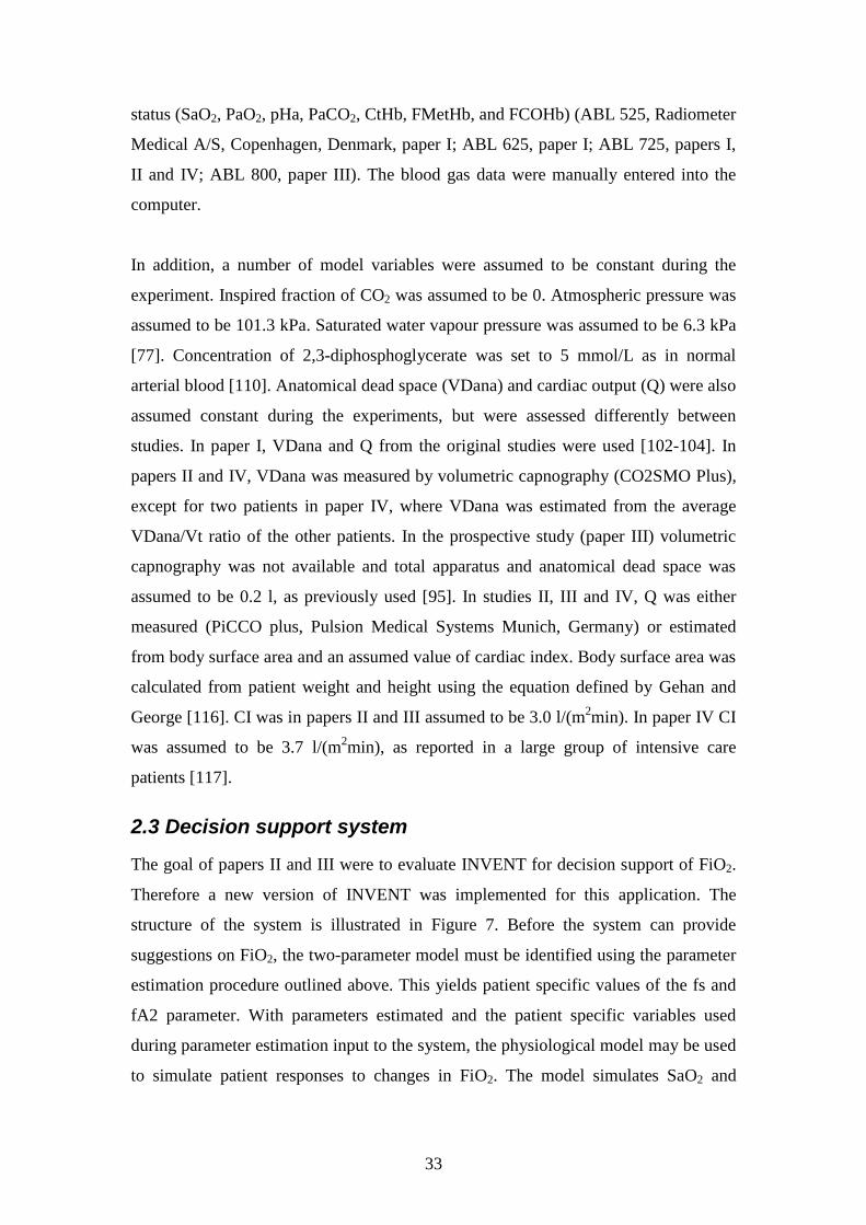
2.3 Decision support system
The goal of papers II and III were to evaluate INVENT for decision support of FiO2.
Therefore a new version of INVENT was implemented for this application. The
structure of the system is illustrated in Figure 7. Before the system can provide
suggestions on FiO2, the two-parameter model must be identified using the parameter
estimation procedure outlined above. This yields patient specific values of the fs and
fA2 parameter. With parameters estimated and the patient specific variables used
during parameter estimation input to the system, the physiological model may be used
to simulate patient responses to changes in FiO2. The model simulates SaO2 and

34
estimates mixed venous O2 saturation (SmvO2) assuming venous pH to be 0.04 less
than pHa, and mixed venous PCO2 to be 0.8 kPa higher than PaCO2.
The INVENT system uses utility theory in the form of penalty functions to model
clinical preferences. Each level of FiO2 and predicted oxygenation values are
associated with a total penalty calculated as the unweighted sum of penalties due to
local and general ischemia quantified as functions of SaO2 and SmvO2, respectively,
and due to the risk of oxygen toxicity quantified as a function of FiO2. The
optimization component of INVENT automatically varies FiO2 and locates the
optimal level, which is that incurring minimal total penalty.
Figure 7: The structure for the INVENT system for decision support on FiO2. Used with
permission from paper II.
Figure 8 illustrates the user interface of INVENT for FiO2 management. The figure is
a screenshot taken with data input for a patient from paper III. The left hand side of
the screen shows the patient specific predicted FiO2-SaO2 curve. On the curve a cross
encircled by a green circle identifies the system suggested level of FiO2. The system
allows the clinician to manually vary the FiO2 and see the resulting total penalty. This
can be done using the button next to FiO2 under the curve in the column “Manual”.
The currently selected manual FiO2 is marked on the FiO2-SaO2 curve by a vertical
and horizontal line. In the column “Optimal” under the curve, the system suggested
level of FiO2 is shown. Under “Manual” and “Optimal” model predicted values of
SaO2, arterial oxygen concentration (CaO2) and oxygen delivery (DO2) are also

35
shown for the manual and optimal levels of FiO2, respectively. The manual and
optimal penalties are summarized in the bar plot next to the FiO2-SaO2 curve. The
manual FiO2 in the screenshot corresponds to the FiO2 selected by the attending
clinician in paper III.
The right hand side shows the three penalty functions in the system, and a summary of
the penalties associated with the manually selected FiO2 level.
Figure 8: A screenshot of the INVENT system taken during study III. Used with permission from
paper III.
2.4 ICARE system and database
The INVENT system and the parameter estimation procedure are implemented in a
system, ICARE, developed at MMDS, Aalborg University [118]. The system includes
software for communication with devices using RS-232 interfacing. It incorporates a
MySQL database for storing all data from devices as well as model simulation data
and INVENT suggestions. The system also includes autonomous agents responding to
changes in specific data and calculating new values of derived variables. E.g. when a
new value of height or weight is available an autonomous agent calculates a new
value of body surface area. All added data are stored with an ID referring to the
patient they describe. All stored data are also associated with a timestamp, a rank, and

36
their origin. The rank can be measured, calculated, estimated or default illustrating the
quality of the data. Origin allows the user to see the device or software system, e.g.
INVENT, which input the data.
37
3. Summary of Papers
3.1 Paper I
Aim
To evaluate the relevance of variation in the PaO2/FiO2 ratio with FiO2. PaO2/FiO2 is
the current dominating hypoxemia index used in intensive care and clinical trials. The
study was also performed to evaluate the ability of a shunt only model to describe this
variation in comparison with a two parameter model describing shunt and
ventilation/perfusion mismatch.
Methods
The study was a retrospective evaluation. Several patient groups were included to
allow the analysis to be performed simulating as many different forms of lung
disorders and severities of gas exchange problems as possible. Experimental data
were included from normal subjects [103], postoperative patients following
gynaecological laparotomy [102,103] and cardiac surgery [103,104], patients
suffering from cardiac incompensation [103], intensive care patients from a
previously published study [103] and previously unpublished experimental data from
a further 8 intensive care patients (see paper II, section 3.2) totaling 93 patients
studied. A total of 36 patients were mechanically ventilated intensive care patients
whereas 57 were spontaneously breathing. Some of the patients were studied on more
than one occasion, e.g. after changes in PEEP, yielding a total of 134 patient cases. 18
patient cases (spontaneous breathing) were excluded as measurement data only
included arterial blood gas measurements at two levels of FiO2.
First the two-parameter model was used to show the theoretical variation in
PaO2/FiO2 upon changing FiO2 under different levels of shunt and A/QV mismatch.
The variation was analysed in a clinically relevant range, which was defined as the
range of FiO2 corresponding to simulated SaO2 values in the range 92-98%. The
variation was then analysed in 116 patient cases, using both the one parameter
„effective‟ shunt model and the twoparameter model to fit patient data and simulate
variation in PaO2/FiO2 ratio. The value of the PaO2/FiO2 ratio as hypoxemia index
was then evaluated by classifying each patient as being normal (PaO2/FiO2 > 47 kPa),
38
having mild hypoxemia (40 kPa ≤ PaO2/FiO2 < 47 kPa), ALI (27 kPa ≤ PaO2/FiO2 <
40 kPa) or ARDS (PaO2/FiO2 < 27 kPa) [1]. This was done at the minimum and
maximum FiO2 of the patient specific clinically relevant ranges, and the number of
patients changing classification from low to high FiO2 were quantified.
Data are reported as means ± SD appearing normally distributed on graphic
evaluation of Q-Q plots [119]. F-tests were used to compare goodness of fit between
the shunt-only model and the two parameter model, taking into account the degrees of
freedom lost with additional complexity. A confusion matrix [120] was used to
illustrate the number of patients classified as normal, with mild hypoxemia, ALI or
ARDS upon changing FiO2. A cut-off value of 0.05 was used for signifying statistical
significant differences in the F-test.
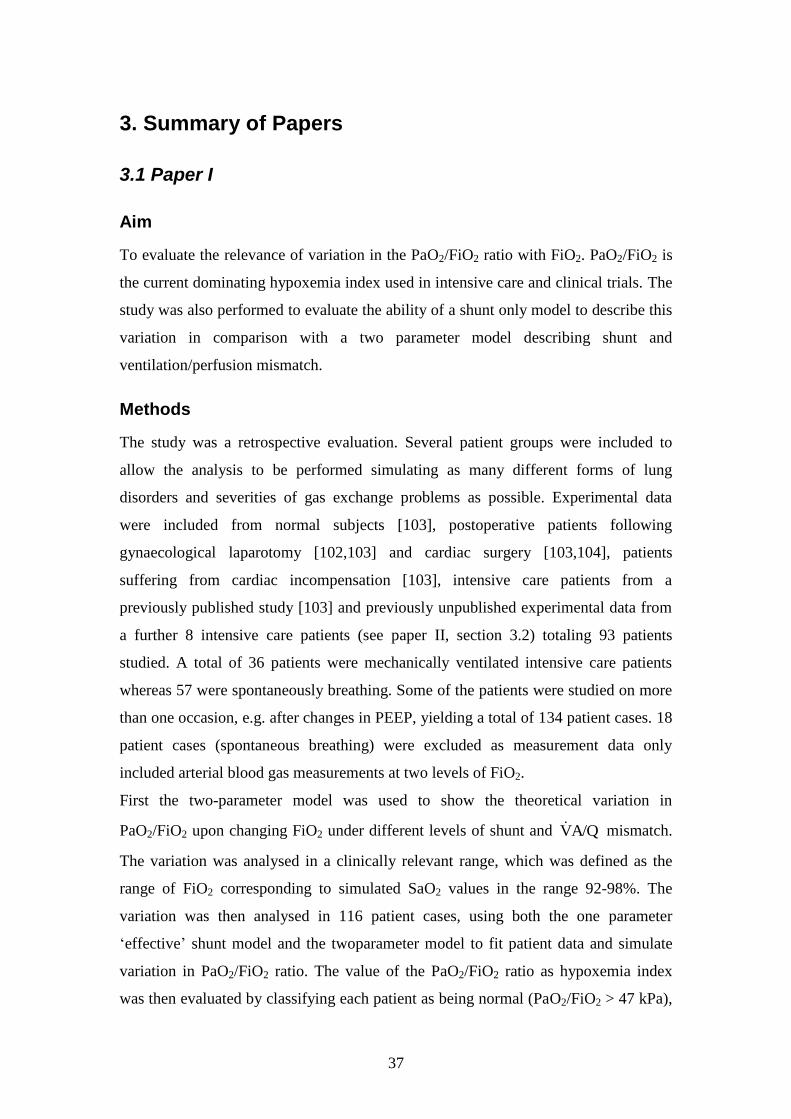
Results
Figure 9 and Figure 10 illustrate the theoretical variation in SaO2 and PaO2/FiO2 ratio
upon changing FiO2 under different levels of shunt and A/QV mismatch,
respectively. Figure 11 illustrates measured and model simulated variation in SaO2
and PaO2/FiO2 in six patients representing typical examples from the studied patient
groups. The two parameter model was shown to give a statistically better fit to data
than the „effective‟ shunt model (P < 0.005).
Figure 9: Simulated variation in SaO2 (A) and PaO2/FiO2 ratio (B) upon changing FiO2 under
varying levels of shunt (fs). Thick solid lines indicate the portion of the curves within the
clinically relevant range of FiO2. a and b in subplot B indicate a variation in FiO2 from 0.19 to
0.57 for fs=20%. Simulations were performed using ΔPO2 = 0 kPa (fA2=0.9), VO2 = 0.26 l/min,
alveolar minute volume = 5.25 l. Used with permission from paper I.
39
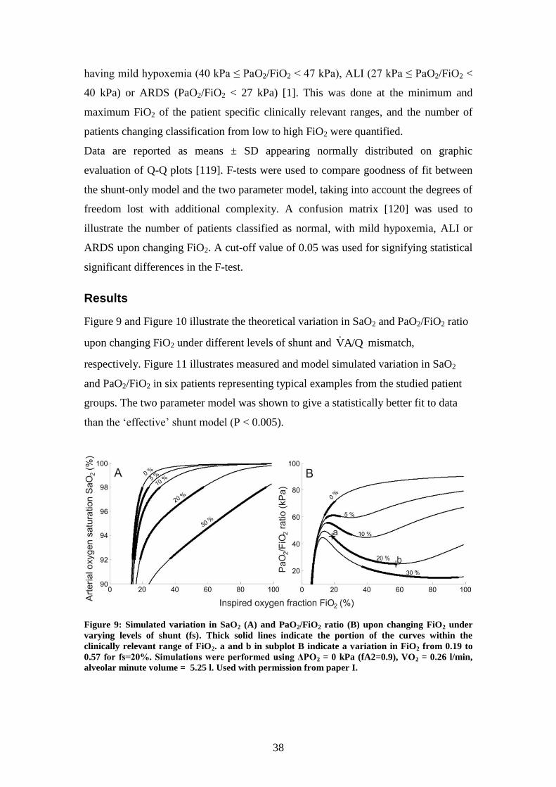
Figure 10: Simulated variation in SaO2 (A) and PaO2/FiO2 ratio (B) upon changing FiO2 under
varying levels of A/QV mismatch (ΔPO2). Thick solid lines indicate the portion of the curves
within the clinically relevant range of FiO2. a and b in subplot B indicate a variation in FiO2 from
0.26 to 0.35 for fs=20%. Simulations were performed using fs = 5 %, VO2 = 0.26 l/min, alveolar
minute volume = 5.25 l. Used with permission from paper I.
Disease classification changed upon varying FiO2 within the clinically relevant range
in 38 of the 116 patient cases (~30%) according to the two-parameter model. The
number of patient cases classified as ALI or ARDS according to the two-parameter
model changed from 23 to 31 (~35% increase) and from 18 to 24 (~33% increase),
respectively.
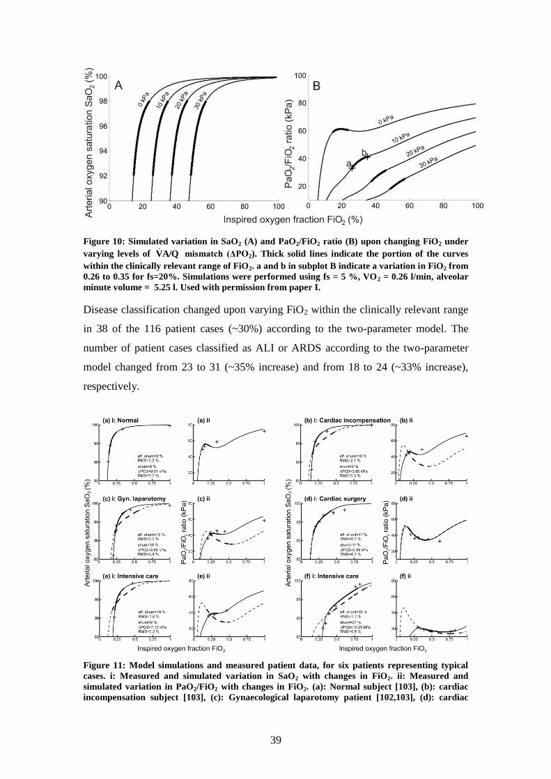
Figure 11: Model simulations and measured patient data, for six patients representing typical
cases. i: Measured and simulated variation in SaO2 with changes in FiO2. ii: Measured and
simulated variation in PaO2/FiO2 with changes in FiO2. (a): Normal subject [103], (b): cardiac
incompensation subject [103], (c): Gynaecological laparotomy patient [102,103], (d): cardiac
40
surgery patient [104], (e): intensive care patient [103], (f): patient from the previously
unpublished study in intensive care patients. Solid lines and dashed lines indicate models
simulations with the two-parameter model, and the ‘effective’ shunt model, respectively, thick
part of curves correspond to the clinically relevant range of FiO2. Model parameters and root
mean square (RMS) error are specified for each model. + represent measured patient data. Used
with permission from paper I.
Conclusions
The PaO2/FiO2 ratio is dependent on both the levels of FiO2 and SaO2. Within the
ranges investigated (SaO2 = 92-98 %) almost a third of patients change disease
classification. Therefore the scientific and clinical utility of the PaO2/FiO2 ratio
appears questionable. If used, then at least the FiO2 level at which the ratio was
measured should be reported. The results indicate that the one-parameter „effective‟
shunt model is not capable of describing this variation correctly, but that the two
parameter model is.
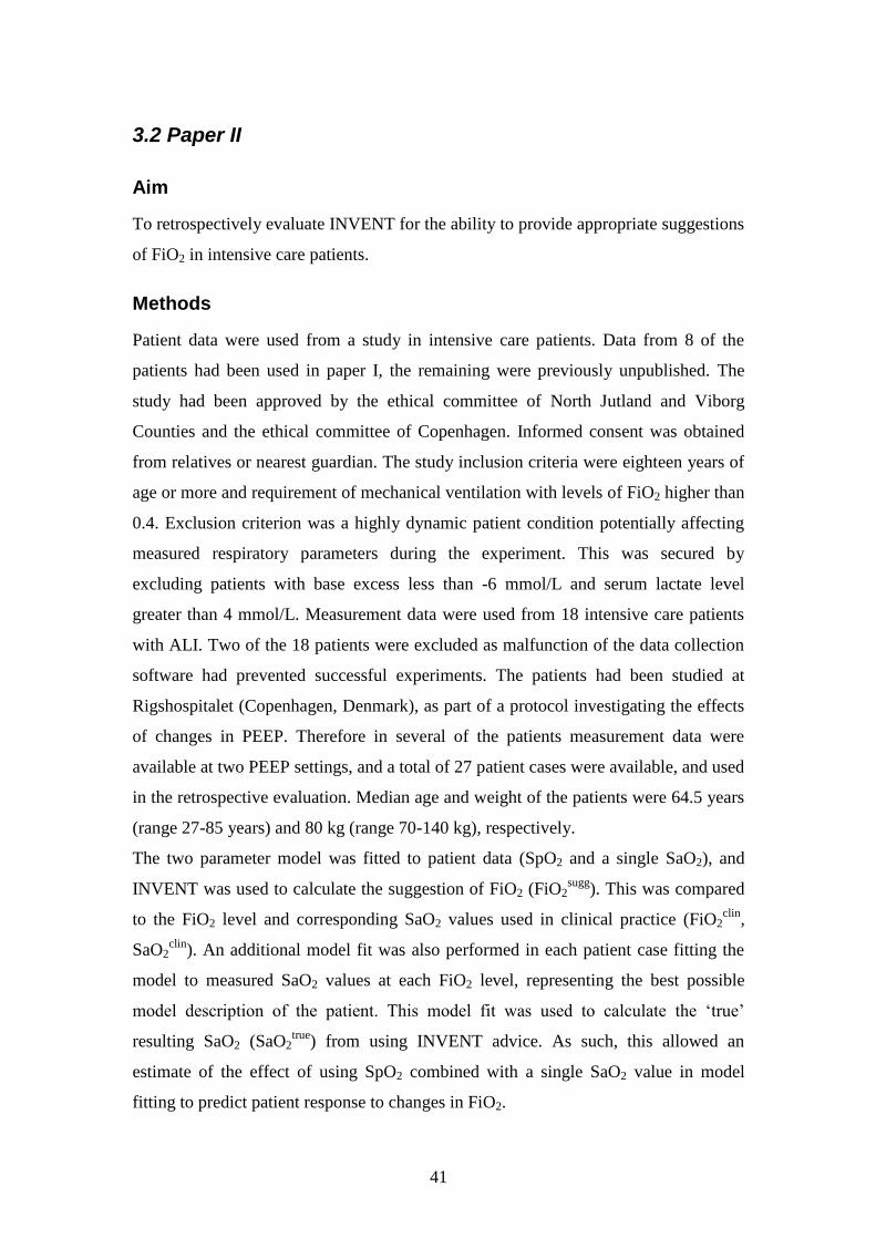
41
3.2 Paper II
Aim
To retrospectively evaluate INVENT for the ability to provide appropriate suggestions
of FiO2 in intensive care patients.
Methods
Patient data were used from a study in intensive care patients. Data from 8 of the
patients had been used in paper I, the remaining were previously unpublished. The
study had been approved by the ethical committee of North Jutland and Viborg
Counties and the ethical committee of Copenhagen. Informed consent was obtained
from relatives or nearest guardian. The study inclusion criteria were eighteen years of
age or more and requirement of mechanical ventilation with levels of FiO2 higher than
0.4. Exclusion criterion was a highly dynamic patient condition potentially affecting
measured respiratory parameters during the experiment. This was secured by
excluding patients with base excess less than -6 mmol/L and serum lactate level
greater than 4 mmol/L. Measurement data were used from 18 intensive care patients
with ALI. Two of the 18 patients were excluded as malfunction of the data collection
software had prevented successful experiments. The patients had been studied at
Rigshospitalet (Copenhagen, Denmark), as part of a protocol investigating the effects
of changes in PEEP. Therefore in several of the patients measurement data were
available at two PEEP settings, and a total of 27 patient cases were available, and used
in the retrospective evaluation. Median age and weight of the patients were 64.5 years
(range 27-85 years) and 80 kg (range 70-140 kg), respectively.
The two parameter model was fitted to patient data (SpO2 and a single SaO2), and
INVENT was used to calculate the suggestion of FiO2 (FiO2sugg
). This was compared
to the FiO2 level and corresponding SaO2 values used in clinical practice (FiO2clin
,
SaO2clin
). An additional model fit was also performed in each patient case fitting the
model to measured SaO2 values at each FiO2 level, representing the best possible
model description of the patient. This model fit was used to calculate the „true‟
resulting SaO2 (SaO2true
) from using INVENT advice. As such, this allowed an
estimate of the effect of using SpO2 combined with a single SaO2 value in model
fitting to predict patient response to changes in FiO2.
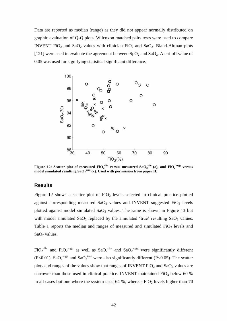
42
Data are reported as median (range) as they did not appear normally distributed on
graphic evaluation of Q-Q plots. Wilcoxon matched pairs tests were used to compare
INVENT FiO2 and SaO2 values with clinician FiO2 and SaO2. Bland-Altman plots
[121] were used to evaluate the agreement between SpO2 and SaO2. A cut-off value of
0.05 was used for signifying statistical significant difference.
Figure 12: Scatter plot of measured FiO2clin
versus measured SaO2clin
(o), and FiO2´sugg
versus
model simulated resulting SaO2sugg
(x). Used with permission from paper II.
Results
Figure 12 shows a scatter plot of FiO2 levels selected in clinical practice plotted
against corresponding measured SaO2 values and INVENT suggested FiO2 levels
plotted against model simulated SaO2 values. The same is shown in Figure 13 but
with model simulated SaO2 replaced by the simulated „true‟ resulting SaO2 values.
Table 1 reports the median and ranges of measured and simulated FiO2 levels and
SaO2 values.
FiO2clin
and FiO2sugg
as well as SaO2clin
and SaO2sugg
were significantly different
(P<0.01). SaO2sugg
and SaO2true
were also significantly different (P<0.05). The scatter
plots and ranges of the values show that ranges of INVENT FiO2 and SaO2 values are
narrower than those used in clinical practice. INVENT maintained FiO2 below 60 %
in all cases but one where the system used 64 %, whereas FiO2 levels higher than 70
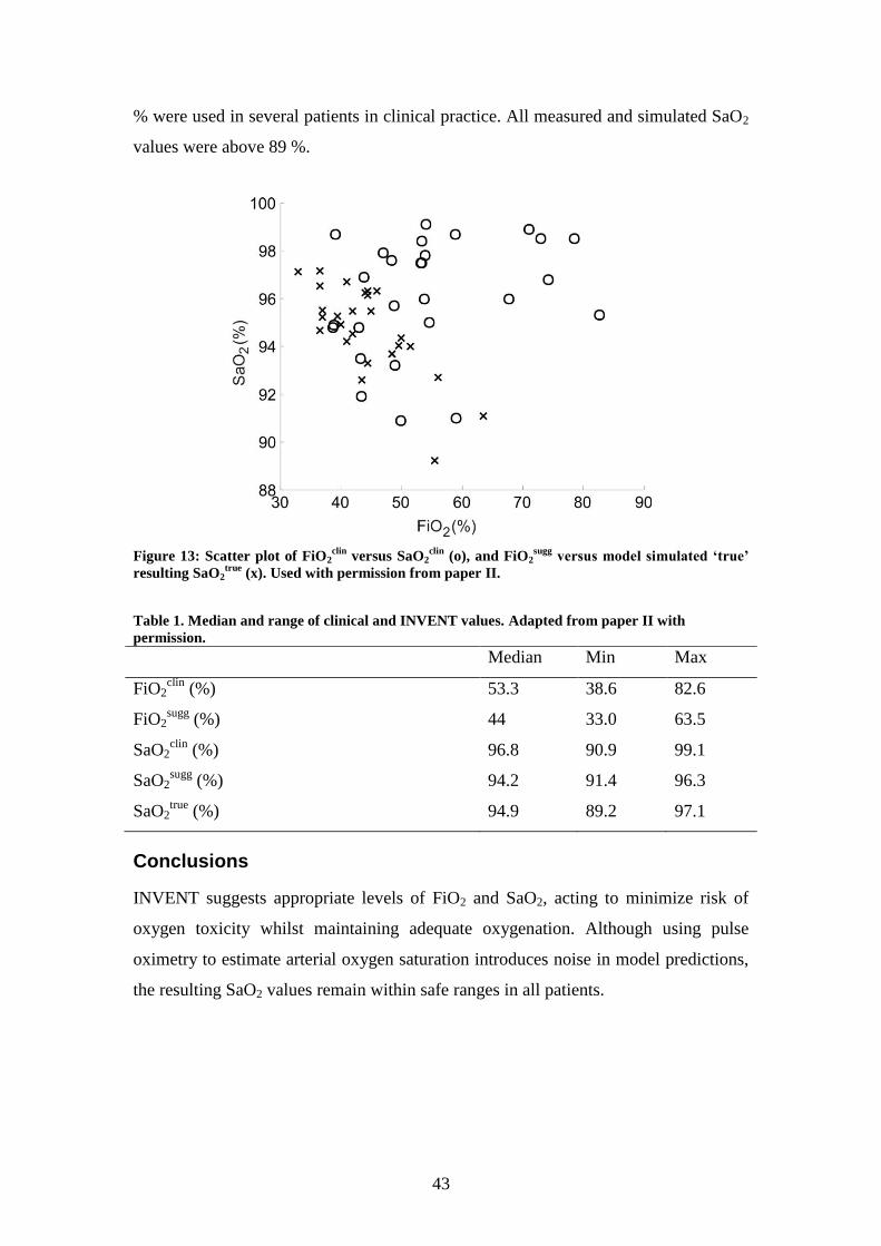
43
% were used in several patients in clinical practice. All measured and simulated SaO2
values were above 89 %.
Figure 13: Scatter plot of FiO2clin
versus SaO2clin
(o), and FiO2sugg
versus model simulated ‘true’
resulting SaO2true
(x). Used with permission from paper II.
Table 1. Median and range of clinical and INVENT values. Adapted from paper II with
permission.
Median Min Max
FiO2clin
(%) 53.3 38.6 82.6
FiO2sugg
(%) 44 33.0 63.5
SaO2clin
(%) 96.8 90.9 99.1
SaO2sugg
(%) 94.2 91.4 96.3
SaO2true
(%) 94.9 89.2 97.1
Conclusions
INVENT suggests appropriate levels of FiO2 and SaO2, acting to minimize risk of
oxygen toxicity whilst maintaining adequate oxygenation. Although using pulse
oximetry to estimate arterial oxygen saturation introduces noise in model predictions,
the resulting SaO2 values remain within safe ranges in all patients.
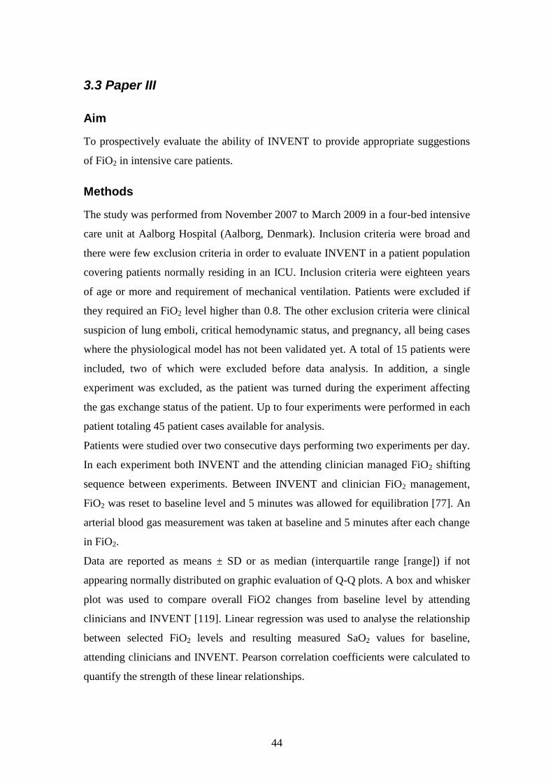
44
3.3 Paper III
Aim
To prospectively evaluate the ability of INVENT to provide appropriate suggestions
of FiO2 in intensive care patients.
Methods
The study was performed from November 2007 to March 2009 in a four-bed intensive
care unit at Aalborg Hospital (Aalborg, Denmark). Inclusion criteria were broad and
there were few exclusion criteria in order to evaluate INVENT in a patient population
covering patients normally residing in an ICU. Inclusion criteria were eighteen years
of age or more and requirement of mechanical ventilation. Patients were excluded if
they required an FiO2 level higher than 0.8. The other exclusion criteria were clinical
suspicion of lung emboli, critical hemodynamic status, and pregnancy, all being cases
where the physiological model has not been validated yet. A total of 15 patients were
included, two of which were excluded before data analysis. In addition, a single
experiment was excluded, as the patient was turned during the experiment affecting
the gas exchange status of the patient. Up to four experiments were performed in each
patient totaling 45 patient cases available for analysis.
Patients were studied over two consecutive days performing two experiments per day.
In each experiment both INVENT and the attending clinician managed FiO2 shifting
sequence between experiments. Between INVENT and clinician FiO2 management,
FiO2 was reset to baseline level and 5 minutes was allowed for equilibration [77]. An
arterial blood gas measurement was taken at baseline and 5 minutes after each change
in FiO2.
Data are reported as means ± SD or as median (interquartile range [range]) if not
appearing normally distributed on graphic evaluation of Q-Q plots. A box and whisker
plot was used to compare overall FiO2 changes from baseline level by attending
clinicians and INVENT [119]. Linear regression was used to analyse the relationship
between selected FiO2 levels and resulting measured SaO2 values for baseline,
attending clinicians and INVENT. Pearson correlation coefficients were calculated to
quantify the strength of these linear relationships.
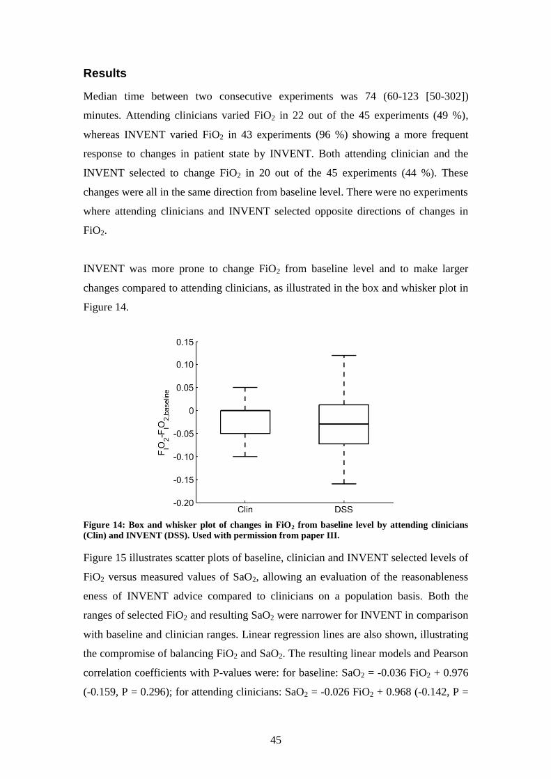
45
Results
Median time between two consecutive experiments was 74 (60-123 [50-302])
minutes. Attending clinicians varied FiO2 in 22 out of the 45 experiments (49 %),
whereas INVENT varied FiO2 in 43 experiments (96 %) showing a more frequent
response to changes in patient state by INVENT. Both attending clinician and the
INVENT selected to change FiO2 in 20 out of the 45 experiments (44 %). These
changes were all in the same direction from baseline level. There were no experiments
where attending clinicians and INVENT selected opposite directions of changes in
FiO2.
INVENT was more prone to change FiO2 from baseline level and to make larger
changes compared to attending clinicians, as illustrated in the box and whisker plot in
Figure 14.
Figure 14: Box and whisker plot of changes in FiO2 from baseline level by attending clinicians
(Clin) and INVENT (DSS). Used with permission from paper III.
Figure 15 illustrates scatter plots of baseline, clinician and INVENT selected levels of
FiO2 versus measured values of SaO2, allowing an evaluation of the reasonableness
eness of INVENT advice compared to clinicians on a population basis. Both the
ranges of selected FiO2 and resulting SaO2 were narrower for INVENT in comparison
with baseline and clinician ranges. Linear regression lines are also shown, illustrating
the compromise of balancing FiO2 and SaO2. The resulting linear models and Pearson
correlation coefficients with P-values were: for baseline: SaO2 = -0.036 FiO2 + 0.976
(-0.159, P = 0.296); for attending clinicians: SaO2 = -0.026 FiO2 + 0.968 (-0.142, P =
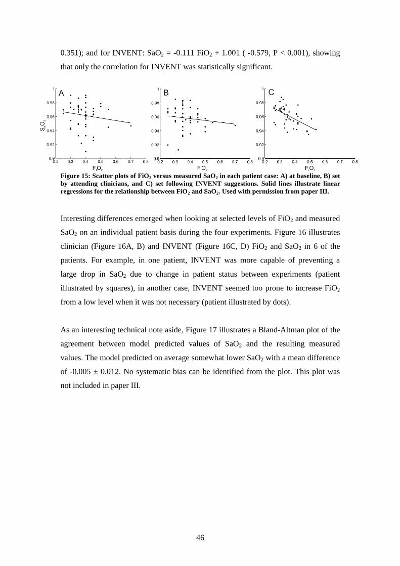
46
0.351); and for INVENT: SaO2 = -0.111 FiO2 + 1.001 ( -0.579, P < 0.001), showing
that only the correlation for INVENT was statistically significant.
Figure 15: Scatter plots of FiO2 versus measured SaO2 in each patient case: A) at baseline, B) set
by attending clinicians, and C) set following INVENT suggestions. Solid lines illustrate linear
regressions for the relationship between FiO2 and SaO2. Used with permission from paper III.
Interesting differences emerged when looking at selected levels of FiO2 and measured
SaO2 on an individual patient basis during the four experiments. Figure 16 illustrates
clinician (Figure 16A, B) and INVENT (Figure 16C, D) FiO2 and SaO2 in 6 of the
patients. For example, in one patient, INVENT was more capable of preventing a
large drop in SaO2 due to change in patient status between experiments (patient
illustrated by squares), in another case, INVENT seemed too prone to increase FiO2
from a low level when it was not necessary (patient illustrated by dots).
As an interesting technical note aside, Figure 17 illustrates a Bland-Altman plot of the
agreement between model predicted values of SaO2 and the resulting measured
values. The model predicted on average somewhat lower SaO2 with a mean difference
of -0.005 ± 0.012. No systematic bias can be identified from the plot. This plot was
not included in paper III.
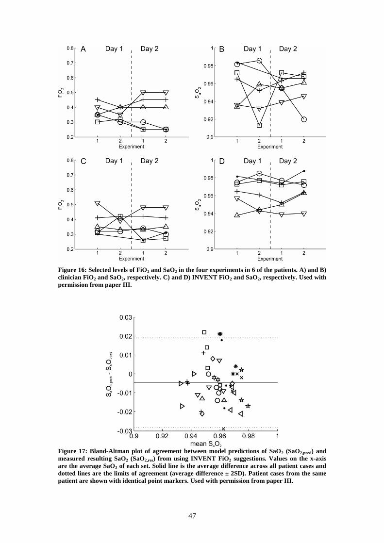
47
Figure 16: Selected levels of FiO2 and SaO2 in the four experiments in 6 of the patients. A) and B)
clinician FiO2 and SaO2, respectively. C) and D) INVENT FiO2 and SaO2, respectively. Used with
permission from paper III.
Figure 17: Bland-Altman plot of agreement between model predictions of SaO2 (SaO2,pred) and
measured resulting SaO2 (SaO2,res) from using INVENT FiO2 suggestions. Values on the x-axis
are the average SaO2 of each set. Solid line is the average difference across all patient cases and
dotted lines are the limits of agreement (average difference ± 2SD). Patient cases from the same
patient are shown with identical point markers. Used with permission from paper III.
48
Conclusions
Results indicate that the INVENT system is safe to use for suggesting FiO2 levels in
intensive care patients. The physiological model accurately predicts SaO2, and all
FiO2 levels suggested by INVENT resulted in appropriate values of SaO2. Both
clinicians and INVENT often changed FiO2 when evaluating patients suggesting that
frequent reevaluation of the patients is valuable. INVENT may help to understand
difficult patients, and in easily managed patients the system may be used to free the
focus of clinicians to concentrate on more challenging therapy.

49
3.4 Paper IV
Aim
To perform a systematic comparison of different minimal models to find the
necessary degree of modeling complexity to describe the pulmonary gas exchange of
both O2 and CO2 and provide an adequate description of gas exchange abnormality in
intensive care patients.
Methods
The study was carried out as a retrospective study. The data used in paper II were
selected, as these patients had severe disorders in pulmonary gas exchange. After
publication of paper II the data from two additional patients were made available
totaling 18 patients for the analysis. As several patients had been studied at two levels
of PEEP a total of 30 patient cases were available.
Three different models were compared: an one parameter „effective‟ shunt model
(Model I); a two parameter model as used in papers I, II, and III describing shunt and
A/QV mismatch, with perfusion locked between two ventilated compartments, and
fraction of ventilation varied to fit patient data (model II); and a three parameter
model, similar to model II but where also the fraction of perfusion going to the two
ventilated compartments is varied to fit patient data. The three models were compared
quantitatively for their ability to fit patient data taking into account the degrees of
freedom lost with increasing complexity, and qualitatively for their ability to describe
the gas exchange abnormality of the individual patients.
Data are reported as means ± SD appearing normally distributed on graphic
evaluation of Q-Q plots. F-tests were used to compare goodness of fit between
models, taking into account the degrees of freedom lost with more complex models. χ2
tests were also used to evaluate the goodness of fit of the models on a patient case
basis. A Bland-Altman plot was used to evaluate the agreement between SpO2 and
SaO2. A cut-off value of 0.05 was used for signifying statistical significant differences
in F-tests. In the χ2 tests P>0.1 was used as cut-off for signifying an adequate fit to
measurement data.

50
Results
Chi-squared tests of quality of fit to individual patient cases indicated adequate fit to
measured data (P>0.1) in 1 patient case for model I (3% of cases), 19 (63%) patient
cses for model II and in 24 (80%) patient cases for model III. Pairwise F-test
comparisons showed model II to give a significantly better fit to measured data than
model I (P<0.001), and indicate model III to give better fit than model II (P<0.1),
however with low statistical significance.
Figure 18 shows an example of model fits to measured O2 and CO2 data in a patient
case where only model III provides an adequate fit to both O2 and CO2. Both models
II and III fit the CO2 data, but model II can not simulate sufficient right shift in the
FetO2-SpO2 curve, i.e. describe a sufficient alveolar to lung capillary drop in PO2.
Of the 6 patient cases where chi-squared tests showed that model III produced an
inadequate fit to data, 1 patient could be described by model I, i.e. shunt was
sufficient to describe the data. In another case, model II was sufficiently complex to
describe measured data. In the last 4 patient cases, there was a significant bias
between SpO2 and SaO2. However, in these cases fitting model III to SaO2 showed
small differences in resulting described degree of lung disorder according to the
difference between alveolar and arterial partial pressures of O2 and CO2.
Figure 18: Fit of the three models to measured patient data, in a patient case, where only model
III provides a good fit to both O2 and CO2 data. Left) Model fitted simulations of oxygenation for
model I (dashed line), model II (dotted line) and model III (solid line), and measured FetO2-SpO2
(+) and FetO2-SaO2 (o) points. Right) Model fitted simulations of FetCO2-PaCO2 for model I
(diamond), model II (triangle) and model III (x) and measured FetCO2-PaCO2 (o) point.

51
Figure 19 shows model fitted ventilations and perfusions of the two ventilated
compartments versus A/QV of the respective compartments for models II and III. For
each patient case there are two points in each sub plot: one for the low A/QV
compartment and one for the high A/QV compartment. The ranges of A/QV ratios
that model III can describe are broader than those of model II. This is in particular
obvious in the middle ranges of A/QV ratios when comparing ranges of perfusion (A
and C), and in the lowest range of A/QV ratios when comparing ventilation (B and
D).
Figure 19: Model fitted perfusion and ventilation of ventilated compartments 1 (upward
triangles) and 2 (downward triangles) versus ventilation/perfusion ratios in the respective
compartments. A: perfusion to ventilated compartments of model II. B: ventilation to ventilated
compartments of model II. C: perfusion to ventilated compartments of model III. D: ventilation
to ventilated compartments of model III. Filled triangles indicate patient cases where Chi-
squared tests of quality of fit resulted in p>0.1 for model III and p<0.1 for model II, or where the
resulting p for model III was at least 0.2 larger than that for model II.
52
Conclusions
The results show that the one parameter model is not able to describe pulmonary gas
exchange of O2 and CO2. The two parameter model is sufficiently complex to
describe gas exchange of one of the two gases, but is not able describe gas exchange
of both gases in all patients. The three parameter model is able to provide adequate
fits to measured O2 and CO2 data, and is robust in the cases where SpO2 provide a
poor estimate of SaO2. The three parameter model is able to provide a more varied
description of A/QV ratios in different patients. As such this minimal model
represents a good compromise between complexity and feasibility, and may be used
in clinical practice to describe lung status in patients with severe lung disorders.
53
4. Discussion
Ventilator management can be considered a process of finding the appropriate
compromise between conflicting goals, it is important to secure gas exchange, but at
the same time excessive levels of pressures, volumes and FiO2 should be avoided to
prevent VILI. This is a complex task requiring a good understanding of the
pathophysiology of the individual patient. Although numerous measurements are
available in the ICU to describe the gas exchange status of the lungs, currently
available measurements are over-simplified and vary with changes in therapy not
affecting lung status. Model-based DSSs constitute a potential solution by offering a
deeper understanding of the patient‟s lung status and by integrating measurement data
and suggesting optimal therapy.
The overall aim of this PhD project was to evaluate the use of minimal models of gas
exchange in decision support of ventilator management. Four questions were
addressed during the project, see section 1.5. In the following the answers to these
four questions are discussed based on the results presented in the four papers. In the
following sections the INVENT system as well as results of the PhD project are
discussed in relation to: rule-based DSSs and other model-based DSSs. In addition,
this chapter will discuss the necessary future work to allow INVENT to provide
decision support of FiO2, Vt and f in intensive care patients, and what is necessary to
also provide suggestions on PEEP. As a final discussion on decision support, this
chapter will discuss the necessary steps to be taken to achieve a successful integration
of INVENT in clinical practice. This chapter will also examine the limitations of the
mathematical models and parameter estimation methods used in all the studies
presented in this thesis. Finally other relevant clinical perspectives related to the
project are discussed.
4.1 The major findings of this thesis
Paper I shows that the currently dominating oxygenation index, the PaO2/FiO2 ratio
varies significantly with changes in FiO2. This has been shown both theoretically,
according to a two parameter gas exchange model describing shunt and A/QV
mismatch, but also in various patient groups including intensive care patients. The
clinical and scientific value of the index therefore appears doubtful. A shunt only

54
model can not describe this variation accurately, but the two parameter model can.
The results presented in paper I illustrate that a two parameter gas exchange model is
necessary to accurately predict changes in oxygenation with FiO2, and is therefore the
necessary complexity for decision support of FiO2 in the ICU.
A two parameter model describing shunt and A/QV mismatch is an integrated part of
the INVENT system originally presented by Rees et al. [73]. As a first step towards
clinical integration INVENT was modified in this PhD to provide decision support of
FiO2. This version of INVENT is based on the two parameter gas exchange model
alone to predict patient response to changes in FiO2. In addition, the parameter
estimation procedure was modified to include arterial oxygen saturation measured
from an arterial blood sample. Paper II describes the retrospective evaluation of
INVENT for decision support of FiO2 in intensive care patients. The results indicate
that INVENT suggests appropriate levels of FiO2 and SaO2. However, the study was
retrospective and resulting oxygenations were predicted by model simulations,
therefore the study is limited to indicate that INVENT is safe to use in an ICU.
Paper III presents the prospective evaluation of INVENT for providing decision
support of FiO2 in 13 intensive care patients in up to four experiments over two
consecutive days. Although the number of patients was limited, the results indicate
that INVENT provides safe and appropriate suggestions of FiO2 in intensive care
patients with varying severities of respiratory failure. The scatter plots illustrating
INVENT FiO2 versus resulting SaO2 (Figure 15C) show that INVENT standardizes
the compromise of achieving sufficient oxygenation versus avoiding the adverse
effects of hyperoxia, these compromises being patient specific as INVENT
suggestions are based on model parameters estimated to describe the individual
patient. When higher FiO2 is required, INVENT accepts lower SaO2. Both in the
retrospective and the prospective study, it appears that INVENT managed this balance
as well as or better than attending clinicians.
INVENT was originally designed to provide decision support of FiO2, Vt and f [73].
To provide appropriate suggestions of Vt and f, it is necessary to also model the
pulmonary gas exchange of CO2 as changes in alveolar ventilation caused by changes
55
in Vt and f modify gas exchange of CO2 and thereby affect the acid-base status of the
blood. Paper IV describes a systematic evaluation of what modeling complexity is
necessary to obtain an accurate minimal model representation of pulmonary gas
exchange of both O2 and CO2. The results presented in paper IV show that a three
parameter model describing shunt and A/QV mismatch can accurately describe O2
and CO2 gas exchange in intensive care patients. The two parameter model describing
shunt and A/QV mismatch was sufficient in the majority of patient cases but failed in
some. In comparison, the three parameter model can describe broader ranges in
A/QV ratios and is able to describe perfusion to a model compartment with very low
A/QV ratios, close to 0.1. This is an interesting quality as perfusion of lung regions
with very low A/QV ratios has been described in ARDS patients in studies using the
MIGET technique [94,95]. The two parameter model could not provide such a
physiological description.
4.2 Model-based or rule-based decision support systems?
The successful prospective evaluation of INVENT for suggestions of FiO2 is an
important indication of the clinical usability of a model-based DSS. However, in
comparison numerous clinical trials have been performed with rule-based systems
including large multicenter studies and often in complex problems involving several
ventilator settings. Rule-based systems have also been successfully implemented in
commercial ventilators. Development of a model-based decision support system is a
complex and time-consuming task involving mathematical description of a complex
physiological system, quantification of clinical preferences and development of
methods for estimating model parameters from clinical data. Indeed, given the current
status of rule-based systems compared to model-based systems one may ask, is it still
worth it?
There is no doubt that under certain conditions use of a rule-based system may result
in improvement in patient care [e.g. 47,53] and in general standardize care, which is a
quality in itself [40]. Multicenter studies have also shown that this can be achieved
with the same system successfully across several institutions [47,53]. It may also be
argued that compared to a model-based DSS, a rule-based DSS in the simplest form is
of relatively low cost to build, for example by implementing a computerized version
56
of a clinical guideline. However, rule-based systems remain blackbox systems, which
do not provide the clinician with a deeper understanding of the patient nor the
provided advice. Implementation of intelligent graphic user interfaces have been
suggested to address this problem [67], however, graphic interfaces do not help
clinicians in understanding the provided advice, and would likely be more valuable
with an underlying physiological interpretation of the patient as in a model-based
DSS.
When estimated, the model parameters for the two parameter gas exchange model
used in INVENT provide a physiological interpretation of the individual patient. The
fs parameter quantifies the degree of intrapulmonary shunt, and the fA2 parameter
describes the degree of A/QV mismatching. This is directly related to the response of
the individual patient to changes in FiO2, as increases in intrapulmonary shunt causes
a vertical depression of the FiO2-SaO2 curve, i.e. changes in FiO2 have less effect on
SaO2. An increase in A/QV mismatching (low fA2) causes a horizontal right shift in
the FiO2-SaO2 curve. This can be translated to a ΔPO2 value, which describes the
extra amount of oxygen necessary at the mouth to counter the oxygenation problem
due to A/QV mismatch.
The requirement of a parameter estimation procedure may be regarded as a limitation
of model-based DSS compared to rule-based DSSs. The parameter estimation
procedure used to identify the two parameter gas exchange model in INVENT
requires variation in FiO2 and measurement of oxygenation using pulse oximetry, a
single arterial blood gas analysis and measurement of oxygen fraction in the expired
air. Variation of FiO2 is a common procedure in an ICU and only measurement of
expired gas fractions can be considered not a part of routine clinical data. However,
these measurements can be obtained from medical equipment, and as shown in paper
I, they are necessary for an accurate description of pulmonary gas exchange. In
addition, the parameter estimation procedure could safely be performed by a nurse,
and Bayesian methods have been developed for supporting the selection of FiO2 steps
and potentially making the procedure computer controlled [122]. Of course, clinicians
or nurses could vary FiO2 without a computer system to better understand patient
responses to changes in FiO2. This would, however, be time-demanding requiring

57
resources not normally available in clinical practice. Without tools as those presented
by Rees et al. [78] to monitor equilibration after changes in FiO2, it is necessary to
allow 5 minutes for equilibration [77] prolonging the process compared to using the
DSS. In addition, although varying FiO2 without a DSS would give clinicians a better
understanding of the gas exchange status of the patient, it would not help to
standardize clinical preferences when managing FiO2.
Another possible advantage of model-based DSS is removal of the need for a trial and
error approach to locating the appropriate ventilator settings [68]. However, this is
under the assumption that the physiological models accurately predict patient
response to changes in therapy. The good agreement between model simulated SaO2
from fit to pulse oximetry with model simulated SaO2 from fit to SaO2 reported in
paper II (Figure 12 and Figure 13) indicate that the two parameter gas exchange
model accurately predicts patient response to changes in FiO2. This was confirmed in
the prospective study as shown in Figure 17.
INVENT uses utility theory in the form of penalty functions to decide therapy. The
penalty functions in INVENT quantify clinical preferences, e.g. preventing ischemia.
The suggestions provided by INVENT are associated with penalties calculated using
the relevant penalty functions. As such, the compromises between conflicting goals
made by INVENT are made explicit to the clinician allowing an understanding of the
provided advice.
4.3 Current status of model-based decision support of
mechanical ventilation
Several model based DSS or hybrid systems combining models with rules have been
developed to assist clinicians in managing mechanical ventilation as outlined in the
introduction. In the following, the various qualities of the systems are discussed
including how the results obtained in the PhD project contributes to the field of
model-based decision support of mechanical ventilation. The different systems are not
directly comparable for their ability to provide appropriate suggestions of changes in
therapy as they provide advice for different ventilator settings, use different
measurements for evaluation or have been tested under different conditions. In the
58
following discussion the systems are instead compared with regards to the potential
clinical benefits they may provide and their clinical feasibility. The VentPlan system
[71] was very similar to INVENT in terms of modeling and use of decision theory.
However, the development of VentPlan has stopped and it will not be discussed
further, but it should be noted that several of the qualities of INVENT discussed in the
following were also qualities of the VentPlan system.
To the best of my knowledge, the prospective evaluation of INVENT presented in
paper III is the first prospective evaluation of a model-based medical decision support
system for ventilator management since the studies with the OPTPROG system by
Rudowski et al. in the early nineties [69,70]. OPTPROG was prospectively evaluated
for suggestions on f, Vt and PEEP, which represent a more complex problem than
management of FiO2. However, OPTPROG had several issues which likely would
prevent routine clinical use. OPTPROG was based on linear models with model
parameters having no physiological interpretation. As such the users would have little
extra to gain from using this system compared to a rule-based system, except the
potential of preventing trial and error when selecting therapy. Despite the simple
model, the parameter estimation procedure took approximately one hour and required
four arterial blood gas measurements [69,70], a frequency of arterial blood sampling
in excess of that normally used in clinical practice. Rudowski et al. argued that when
enough patient data was accumulated, statistical analysis could be used to acquire a
priori knowledge of model parameters potentially simplifying or obviating the
parameter estimation procedure [69]. However, the heterogeneity of ALI/ARDS
patients reported in clinical studies strongly contradicts this [e.g. 36,37].
The SIVA system [72] is based on a two parameter physiological model of gas
exchange to simulate patient response to changes in FiO2, and empirical models to
simulate patient response to changes in respiratory frequency and inspiratory pressure
[72]. The two parameters of the gas exchange model have a physiological
interpretation [75], however, the authors stated that estimation of model parameters
requires a pulmonary artery catheter and that there were convergence problems. As
such, the authors suggested that for prospective use, shunt should be estimated using a
fuzzy inference system [62] and physiological dead space had to be estimated by the
clinician [72]. The parameter estimation is therefore limited to requiring an arterial
59
blood gas sample at a single level of FiO2. The system has at present only been
evaluated using model simulations, preventing any conclusions on the accuracy of
model predictions.
The FLEX system uses several simple models to describe patient response to changes
in FiO2, PEEP, f and I:E-ratio [74]. All the models implemented in FLEX are either
very simple without any model parameters or empirical having parameters without a
physiological interpretation. These empirical model parameters are not tuned to
describe the individual patient. The simple nature of the models used in FLEX allows
the system to be used from measurements readily available at the bedside in the ICU,
however, this also prevents FLEX from providing the clinician with a deeper
physiological understanding of the patient.
FLEX incorporates scalability such that if certain measurements are not available the
system uses a different, i.e. simpler, approach to calculate suggestions. This can be a
valuable quality of the system, especially when used outside the ICU where
measurements of lung status are sparse. The different methods used in this PhD
project (see section 2.2) illustrate that the parameter estimation procedure used for the
gas exchange model in INVENT is to some extent scalable. In addition it has been
shown that in patients with cardiac incompensation the parameters can be estimated
without an arterial blood gas measurement using default values [123]. However, it
would still be necessary to measure variation in oxygenation with varying FiO2 to
separate the effects of shunt and A/QV mismatch.
Both SIVA and FLEX are hybrid systems using models in combination with rules to
calculate suggestions on changes in therapy. As such these systems, like rule-based
systems, do not make it obvious to the clinician what compromises have been made
by the system.
4.4 Future work
The version of INVENT originally presented by Rees et al. in 2006 was intended for
decision support of not only FiO2 but also of Vt and f [73]. In the following, the
relevance of providing decision support on these settings is discussed as well as the
60
future work necessary to enable prospective evaluation of INVENT for suggesting
FiO2, Vt and f in an ICU. Thereafter it is discussed what future work may allow
INVENT to provide suggestions on PEEP, for which the appropriate levels remains
elusive. Finally this section will address the necessary work required to facilitate
future clinical integration of INVENT.
Advice on FiO2, Vt, and f
A recent epidemiological study by Esteban et al. showed that tidal volumes in general
have been decreased in ARDS patients in clinical practice, but also that this has not
reduced mortality more than 5 percent since 1998 [124] and ICU and hospital
mortality in ARDS patients remain above 50% and 60%, respectively [124]. Several
reasons may have contributed to this apparently high mortality in comparison with
those of the multicenter clinical trials reporting mortalities below 30% [e.g. 28,30]:
the studied populations may have been more heterogeneous; the design of the
retrospective study by Esteban et al.; change in composition of patients presenting in
the ICU; or perhaps the difficulty for clinicians to use the guidelines in clinical
practice for the benefit of individual patients? Use of a DSS could potentially help to
standardize care according to the individual patient.
INVENT has been shown retrospectively to provide appropriate suggestions of FiO2,
Vt and f in cardiac surgery patients mechanically ventilated in an ICU [125]. In these
patients the two parameter gas exchange model was successfully used to describe the
pulmonary gas exchange of O2 and CO2. However, the results presented in paper IV
illustrate that a three parameter model is necessary to describe both O2 and CO2 gas
exchange in intensive care patients. As such, this model must be implemented in
INVENT before an eventual retrospective or prospective evaluation in intensive care
patients. This will require a measurement of end-tidal fraction of CO2 in addition to
the measurements used for estimating model parameters to describe O2 gas exchange.
Paper IV was limited to compare minimal models in intensive care patients
representing some of the most complex respiratory failure patients with regards to gas
exchange. The two parameter model may be sufficient to describe gas exchange of
both O2 and CO2 in “simpler” patients, as shown in cardiac surgery patients by
Allerød et al. [125].
61
Currently, INVENT uses a single set of penalty functions to calculate suggestions,
regardless of patient type and clinical circumstances. Although the majority of
changes in FiO2 have been appropriate and all have been safe, results in a few patients
indicate that INVENT may be too prone to increase FiO2 from low levels when SaO2
is sufficient (see patient illustrated with dots in Figure 16). It may be sufficient to
remove this tendency by making ischemia penalties smaller at SaO2 above 0.97 or
toxicity penalty a little higher at low FiO2 (see Figure 7). Alternatively a new set of
penalties could be formulated for example for weaning patients. In all circumstances,
a new set of penalties would be relevant for patients with severe chronic obstructive
pulmonary disease, where the patient‟s normal levels of oxygenation may be reduced.
Some additional modifications of INVENT are necessary before a prospective
evaluation can be performed for the three settings. The current simple model of lung
mechanics implemented in INVENT assumes a constant linear compliance relating
tidal volume to the difference between peak pressure and PEEP. Changes in tidal
volume can, however, affect lung status, e.g. increases in tidal volume may increase
peak pressure potentially recruiting collapsed alveoli reducing shunt thereby leading
to less accurate predictions using the gas exchange model. As described in the
following section on PEEP advice, it is difficult to formulate a mathematical model of
lung mechanics, which is able to describe lung mechanics of patients from clinical
data. Therefore it may be necessary to resort to a trial and error approach as in rule-
based systems. This can be performed by implementing a step to target algorithm in
INVENT such that large changes in tidal volume can be performed in steps and gas
exchange can be evaluated at each step. Similarly increases in f may cause intrinsic
PEEP. As such, the step to target functionality should include f, and end-expiratory
occlusions should be performed allowing quantification of intrinsic PEEP.
Advice on PEEP
The large number of clinical trials which have failed at finding an optimal PEEP
strategy in ALI/ARDS patients [e.g. 29,31,32] illustrate the need for better
understanding of how PEEP should be managed. An important reason may be the lack
of understanding of how changes in PEEP affect lung mechanics as well as gas
exchange. Physiological models have been constructed to describe lung mechanics

62
either addressing specific components of the respiratory system in great detail [e.g.
126] or the complete respiratory system using an empirical approach [127]. Whilst
these models have added to the current understanding of lung mechanics they have
not provided a fundamental understanding of the mechanical effects of changes in
PEEP. In addition, attempts to link lung mechanics and gas exchange have been few
[128], despite the fact that securing gas exchange is one of the goals of changes in
PEEP.
In order to enable model-based decision support of PEEP a novel model must be
constructed, capable of explaining the effects of changes in PEEP. The model should
be able to describe the effect of PEEP on both ventilation and perfusion, thereby
allowing also description of gas exchange.
To describe ventilation, the model should include the contributions of: the hydrostatic
gradient down the lung due to the weight of the lung [4]; the chest wall; the lung
tissue; and pulmonary surfactant. The chest wall and pulmonary surfactant have often
been neglected in models of lung mechanics. The chest wall has been suggested as
important in understanding recruitments maneuvers in ALI/ARDS patients [129].
Pulmonary surfactant is considered vital for mechanical stability during breathing
[130], and inhibition of surfactant due to mechanical ventilation and edematous fluid
entry indicate the importance of understanding surfactant to understand lung
mechanics in respiratory disease [18]. Work has begun describing the different
components in the healthy lungs [131], however future work is required addressing
how the properties of the different components of the respiratory system change in
respiratory disease.
To the best of my knowledge, no mathematical models have been built to describe
how changes in ventilatory pressures affect the distribution of pulmonary perfusion.
We have begun building a model with this aim [132]. The model at its current state,
describes the pulmonary perfusion in the healthy human lungs and is able to describe
experimentally measured total capillary perfusion, volume and surface area [132].
Future work is required for describing pulmonary perfusion in respiratory diseases
including addition of hypoxic pulmonary vasoconstriction, which acts to reduce
pulmonary perfusion in hypoxic lung regions [133].
63
In addition to modeling the different components of the respiratory system, the
heterogeneity of the lungs must be considered. A possible solution is to build
stratified models allowing different layers to have different properties, e.g. due to
different pleural pressures as caused by a hydrostatic gradient [4]. This may enable
such models to describe the „baby-lung‟ of ARDS, that is, a very small volume of
lung being ventilated due to the remaining lung being collapsed, or consolidated [5].
Models of lung mechanics have been built using a stratified structure including the
model by Steimle et al. [127,128,131,134]. The model of perfusion introduced by
Mogensen et al. [133] is based on the same stratified structure as the model by
Steimle et al. [134], illustrating the possible combination of these models in the future
to describe gas exchange and thereby also the link between lung mechanics and gas
exchange.
Alternatively to modeling the effects of changes in PEEP, one may derive simple
algorithms describing compromises considering changes in FiO2 versus PEEP as done
in clinical trials [e.g. 28]. However, changes in PEEP will likely impact the patient‟s
gas exchange status and should as such be followed by a re-estimation of parameters
of the gas exchange model. This may introduce the need for trial and error in some
patients, similar to that necessary in rule-based systems.
Clinical integration
Whilst a DSS may provide sound advice, provide physiological understanding and in
general improve patient care, it is of no value if it is not used at the bedside. An
obvious way to enable successful clinical integration is through commercial
collaboration with companies producing mechanical ventilators. This has been
demonstrated with the GANESH system originally presented by Dojat et al. [49],
which is now implemented as part of the SmartCareTM
system by Dräger Medical
[64]. Besides performing several large studies demonstrating the efficiency of the
system, as performed with GANESH [51-53], INVENT could benefit significantly
from collaboration with the industry. However, as long as data can be retrieved from
the ventilator, a system as INVENT could potentially be developed as a stand-alone
system, albeit this would introduce significant obstacles, the main being that it would
64
be difficult if not impossible to make the system or parts of the system such as
parameter estimation, closed loop.
Morris described in 2000 several barriers to the use of computerized clinical
protocols, i.e. rule-based DSSs [40]. The list presented by Morris included a lot of
barriers concerning clinical culture and fear of losing authority. However, several
barriers addressed qualities of the DSS, which would apply for an eventual integration
of INVENT as well. These barriers can be translated to a few qualities a DSS should
have: The system should not add more burdens to the already stressed critical care
staff either in form of excessive data entry or high complexity; the system should be
usable in a large variation of clinical cases not just the most prevalent; and the system
should be supported by a technological infrastructure such as electronic patient
records.
As discussed previously the parameter estimation procedure used for estimating
parameters of the gas exchange model in INVENT does not add considerable burden
to the clinical staff, and it could potentially be automated. Further automation could
include automatic detection of when new parameter estimation is necessary, e.g. after
changes in settings or posture. When estimated, model parameters act to reduce
complexity integrating data from various devices, and providing a physiological
understanding of the patient directly related to changes in therapy.
Paper III showed that INVENT could provide decision support of FiO2 in patients
with different severities of lung disorder and in controlled as well as spontaneous
ventilator modes. The current version of INVENT for providing suggestions of Vt and
f is limited to volume controlled mode. Inclusion of more complex models of lung
mechanics and respiratory drive would potentially allow INVENT to provide decision
support in patients ventilated in pressure control mode as well as support ventilator
modes. Models of respiratory drive have been built, which potentially could be used
for such application [e.g. 135,136].
INVENT is implemented in the database system ICARE [118], which can facilitate
automatic retrieval and storage of measurement data from several medical devices. In
addition calculated values, estimated model parameters and INVENT advice can be
65
stored in the database, and values not automatically retrieved may be input manually.
As such ICARE provides a technological infrastructure supporting all tasks related to
using INVENT. Integration of ICARE with existing clinical databases would further
strengthen this technological infrastructure, perhaps similar to the HELP database
used in the successful Salt-Lake city protocols [45-48].
4.5 Model limitations
A number of assumptions and simplifications have been made in the project regarding
the use of minimal models to describe pulmonary gas exchange and when estimating
model parameters. These are discussed in the following.
All minimal models used in this PhD project have assumed continuous ventilation and
perfusion, steady state, and that end-tidal gas and mixed alveolar gas fractions are
equal. These assumptions do not represent the true nature of human breathing, which
is tidal [137]. Models of gas exchange have been built describing tidal ventilation
[e.g. 138,139]. However, estimation of model parameters for these models using
clinical data has so far not been demonstrated, and the models have either been
limited to simulations studies [138] or parameters have been estimated using a
combination of MIGET and multiple breath nitrogen washout measurements [139].
As such, minimal models as those presented here, appear currently to represent the
most accurate descriptions of gas exchange from clinical data.
Effects of diffusion limitations have not been included in the models. Diffusion
limitation has a similar effect on pulmonary gas exchange as A/QV causing a ΔPO2
[140]. Studies with MIGET, however, has demonstrated that in the majority of
patients, A/QV mismatch is likely a better description of physiology [93,93,140]
except in cases of pulmonary fibrosis [93], exercise [141] or mild exercise during
hypoxia [141,142].
The parameter estimation procedure depends on measurement of arterial oxygenation
at steady state at varying levels of FiO2. This is done under the assumption, that
varying FiO2 does not alter the physiology of the patient in such a way that
A/QV mismatch and shunt are affected significantly. Variation in FiO2 may affect
66
lung physiology through absorption atelectasis and hypoxic pulmonary
vasoconstriction (HPV). Absorption atelectasis is not likely to have an impact on
parameter estimation, as it has been shown during induction of anaesthesia that
absorption atelectasis mainly occurs at FiO2 levels of 0.8 or more [20], whilst
parameter estimation rarely requires FiO2 levels as high as 0.8. HPV reported effects
on gas exchange have been moderate [143] or considerable [144], but has represented
maximal responses to changes in FiO2, the expected changes with the smaller
variations in FiO2 being less likely to significantly affect pulmonary gas exchange.
Studies with MIGET [145,146] and computer simulations [147,148] have also shown
small changes in model parameters with large variations in FiO2.
VDana and Q were assumed constant during the parameter estimation procedure, see
section 2.2. In paper I, VDana and Q from the original papers were used [102-104]. In
papers II and IV VDana was measured. In paper III, VDana including apparatus dead
space was assumed to be 0.2 l in all patients. As VDana varies with posture, body size
etc. [77], this simplification may have affected the correctness of estimated model
parameters. However, it would have no consequence on INVENT suggestions of
FiO2, as they would be based on model fits to measured FetO2-SpO2 curves and as
such would reflect patient status as long as the model fitted data well.
In papers II-IV, Q was either measured or estimated from body surface area and a
population characteristic CI. This may likely not have reflected the true Q in some
patients. However, a previous study with a two parameter model describing shunt and
diffusion limitation, i.e. a ΔPO2, based on mass conservation and the same
assumptions as models used in this project, showed that parameter estimation was
insensitive to moderate variations in Q [106], with changes in Q of 40% changing fs
by 0.04 and ΔPO2 by 0.5 kPa.
4.6 Clinical perspectives
The results presented in this thesis illustrate that a model-based DSS for mechanical
ventilation may help to standardize therapy according to the individual patient. A
model-based DSS may also help to diminish information overload, which if
unattended affects clinical decision making [38,40]. INVENT uses a gas exchange
model to integrate information from ventilator settings, hemodynamic parameters,
oxygenation, acid base chemistry of blood and metabolism into two model

67
parameters, shunt and fA2 (ΔPO2) describing the gas exchange status of the patient.
This can be valuable in the difficult to manage patients. In the easily managed
patients, a model-based DSS as INVENT may free the focus of the clinicians on more
challenging therapy.
Results in papers I and IV illustrate that several measurements currently used in
clinical practice are insufficient to provide an accurate description of pulmonary gas
exchange in intensive care patients including ALI/ARDS patients. Results in paper I
illustrated how the PaO2/FiO2 ratio varies with changes in FiO2 and SaO2. The
PaO2/FiO2 ratio is the hypoxemia index used in the definition of ALI/ARDS and
constitutes the single value separating ALI and ARDS diagnosis [1]. Paper I showed
several patients changing disease classification due to variation in FiO2, a common
therapeutical intervention normally not affecting pulmonary gas exchange. Other
theoretical studies have come to similar conclusions but without involving clinically
measured variations in the PaO2/FiO2 ratio [149-151].
So how should the refractory hypoxemia evident in ALI/ARDS patients be defined?
As suggested in paper I, at least the FiO2 level should be informed when using the
PaO2/FiO2 ratio, however also SaO2 has an impact and would be appropriate to
inform. However, standardizing FiO2 when including patients is unpractical and
standardizing both FiO2 and SaO2 is even more so due to the varying severities of
lung disorder seen in ALI/ARDS. Alternatively, minimal models of gas exchange
could be used by estimating model parameters and classifying hypoxemia according
to the levels of shunt and degree of A/QV mismatch. The ability of the three
parameter model to describe large fractions of shunt and large perfusions going to
regions with low A/QV ratios as shown in studies using MIGET indicate that the
three parameter model may be suitable for this application.
It has also been argued that a future hypoxemia index used in ALI/ARDS definition
should not vary with changes in PEEP [152]. We disagree with this approach, as
changes in PEEP, unlike most changes in FiO2, affect the pulmonary physiology
[94,95] and may have different effects in different patients [37]. However,
standardization of the level of PEEP used when evaluating patients could be very

68
relevant to potentially separate patients whose alveoli are readily recruited and where
PEEP may keep these alveoli open from patients with consolidated lung regions. This
is in line with several other authors advocating use of standardized ventilator settings
when enrolling ALI/ARDS patients for clinical trials [153-154].

69
5. General Conclusions
1. A two parameter model of pulmonary gas exchange describing intrapulmonary
shunt and ventilation-perfusion mismatch can describe variation in
oxygenation with changes in FiO2. The shunt only model can not describe this
variation accurately.
2. The PaO2/FiO2 ratio varies significantly with FiO2 and its use as a hypoxemia
index is questionable. As a minimum requirement measurements of PaO2/FiO2
should be accompanied by the corresponding FiO2 level.
3. The INVENT system provides appropriate suggestions of FiO2 and SaO2 in
intensive care patients.
4. Compared to attending clinicians INVENT standardizes the FiO2 levels and
values of SaO2 according to the individual patient.
5. All suggestions of FiO2 provided by INVENT results in safe values of SaO2.
6. When identified, the two parameter gas exchange model describing shunt and
ventilation-perfusion mismatch is capable of predicting the oxygenation
response of intensive care patients to changes in inspired oxygen.
7. A three parameter model is necessary for an accurate minimal model
representation of the pulmonary gas exchange of both O2 and CO2 in intensive
care patients.
8. Estimation of model parameters for a three parameter model describing shunt
and ventilation-perfusion mismatch may be performed using routine clinical
data, potentially allowing this model to be used at the bedside in an ICU and
as part of INVENT for providing advice on tidal volume and respiratory
frequency.
70
Acknowledgements
I wish to express my sincere gratitude to:
Steve Rees, my supervisor, for his friendship, always positive attitude and valuable
guidance in all areas of the PhD project.
Steen Andreassen, my second supervisor, for inspiration and sharing his immense
insight and intuition.
Søren Kjærgaard, my third supervisor, for an always positive attitude and discussions
on modeling and clinical perspectives.
Charlotte Allerød, my “comrade-in-arms”, for her friendship, substantial contribution
to data collection and our enlightening discussions of our projects.
Per Thorgaard, for his never ending enthusiasm and support, and for opening my eyes
to the interesting field of critical care.
All staff at intensive therapy section 103, Aalborg hospital, for their understanding
and help. Lotte Frilev and Ann-Maj Carius for their help with finding and including
patients for the study.
Bram Smith, for his friendship and support during my first year at MMDS.
All my other colleagues at MMDS for their support and pleasant collaboration.
And, most of all, my wife Pia and my children Silas and Silke for their understanding
and support, and for always being there.
This work was partially supported by the Programme Commission on Nanoscience,
Biotechnology and IT under the Danish Council for Strategic Research.

71
References
1. Bernard GR, Artigas A, Brigham KL, Carlet J, Falke K, Hudson L, Lamy M,
Legall JR, Morris A, Spragg R. The American-European consensus conference
on ARDS. Am J Respir Crit Care Med 1994; 149:818-24.
2. Antonsen K, Wetterskev J, Bonde J. Incidence, severity and mortality of acute
respiratory failure in Denmark. Ugeskr Laeger 2000; 162:2876-81.
3. Brun-Buisson C, Minelli C, Betolini G, Brazzi L, Pimentel J, Lewandowski K,
Bion J, Romand J, Villar J, Thorsteinsson A, Damas P, Armaganidis A, Lemaire
F. Epidemiology and outcome of acute lung injury in European intensive care
units – results from the ALIVE study. Intensive Care Med 2004; 30:51-61.
4. Gattinoni L, Caironi P, Pelosi P, Goodman LR. What has computed tomography
taught us about the acute respiratory distress syndrome syndrome. Am J Respir
Crit Care Med 2001; 164:1701-11.
5. Gattinoni L, Pesenti A. The concept of “baby lung”. Intensive Care Med 2005;
31:776-84.
6. Baker AB. Artificial respiration, the history of an idea. Med Hist 1971; 15:336-
51.
7. International consensus conferences in intensive care medicine – ventilator-
associated lung injury in ARDS. Intensive Care Med 1999; 25:1444-52.
8. Dreyfuss D, Saumon G. Ventilator-induced lung injury – lessons from
experimental studies. Am J Respir Crit Care Med 1998; 157:294-323.
9. Rouby JJ, Lherm T, Martin de Lassale E, Poéte P, Bodin L, Finet JF, Callard P,
Viars P. Histologic aspects of pulmonary barotrauma in critically ill patients with
acute respiratory failure. Intensive Care Med 1993; 19:383-9.
10. Esteban A, Anzueto A, Frutos F, Alía I, Brochard L, Stewart TE, Benito S,
Epstein SK, Apezteguía C, Nightingale P, Arroliga AC, Tobin MJ.
Characteristics and outcomes in adult patients receiving mechanical ventilation –
a 28-day international study. JAMA 2002; 287:345-55.
11. Slutsky AS, Tremblay LN. Multiple system organ failure – is mechanical
ventilation a contributing factor. Am J Respir Crit Care Med 1998; 157:1721-5.

72
12. Dreyfuss D, Saumon G. From ventilator-induced lung injury to multiple organ
dysfunction. Intensive Care Med 1998; 24:102-4.
13. Nin N, Lorente JA, Fernández-Segoviano P, De Paula M, Ferruelo A, Esteban A.
High-tidal volume ventilation aggravates sepsis-induced multiorgan dysfunction
in a dexamethasone-inhibitable manner. Shock 2009; 31:429-34.
14. Tremblay L, Valenza F, Ribeiro SP, Jingfang L, Slutsky AS. Injurious
ventilatory strategies increase cytokines and c-fos m-RNA expression in an
isolated rat lung model. J Clin Invest 1997; 99:944-52.
15. Ranieri VM, Suter PM, Tortorella C, De Tullio R, Dayer JM, Brienza A, Bruno
F, Slutsky AS. Effect of mechanical ventilation on inflammatory mediators in
patients with acute respiratory distress syndrome – a randomized controlled trial.
JAMA 1999; 281:54-61.
16. Dreyfuss D, Rouby JJ. Mechanical ventilation-induced lung release of cytokines
– a key for the future or Pandora‟s box. Anesthesiology 2004; 101:1-3.
17. Mols G, Priebe HJ, Guttmann J. Alveolar recruitment in acute lung injury. Br J
Anaesth 2006; 96:156-66.
18. Verbrugge SJC, Lachmann B, Kesecioglu J. Lung protective ventilatory
strategies in acute lung injury and acute respiratory distress syndrome: from
experimental findings to clinical applications. Clin Physiol Funct Imaging 2007;
27:67-90.
19. Dantzker DR, Wagner PD, West JB. Instability of lung units with low V/Q ratios
during O2 breathing. J Appl Physiol 1975; 5:886-95.
20. Edmark LK, Kostova-Aherdan K, Enlund M, Hedenstierna G. Optimal oxygen
concentration during induction of general anesthesia. Anesthesiology 2003;
98:28-33.
21. Aboab J, Jonson B, Kouatchet A, Taille S, Niklason L, Brochard L. Effect of
inspired oxygen fraction on alveolar derecruitment in acute respiratory distress
syndrome. Intensive Care Med 2006; 32:1979-86.
22. Nash G, Blennerhassett JB, Pontoppidan H. Pulmonary lesions associated with
oxygen therapy and artificial ventilation. N Engl J Med 1967; 276:368-74.

73
23. Altemeier WA, Sinclair SE. Hyperoxia in the intensive care unit: why more is
not always better. Curr Opin Crit Care 2007; 13:73-8.
24. Amato MB, Barbas CS, Medeiros DM, Magaldi RB, Schettino GP, Lorenzi-
Hilho G, Kairalla RA, Deheinzelin D, Munoz C, Oliveira R, Takagaki TY,
Carvalho CR. Effect of a protective-ventilation strategy on mortality in the acute
respiratory distress syndrome. N Engl J Med 1998; 338:347-54.
25. Brochard L, Roudot-Thoraval F, Roupie E, Declaux C, Chastre J, Fernandez-
Mondejar E, Clementi E, Mancebo J, Factor P, Matamis D, Ranieri M, Blanch L,
Rodi G, Mentec H, Dreyfuss D, Ferrer M, Brun-Buisson C, Tobin M, Lemaire F.
Tidal volume reduction for prevention of ventilator-induced lung injury in acute
respiratory distress syndrome. Am J Respir Crit Care Med 1998; 158:1831-8.
26. Stewart TE, Meade MO, Cook DJ, Granton JT, Hodder RV, Lapinsky SE, Mazer
CD, McLean RF, Rogovein TS, Schouten BD, Todd TR, Slutsky AS. Evaluation
of a ventilation strategy to prevent barotraumas in patients at high risk for acute
respiratory distress syndrome. N Engl J Med 1998; 338:355-61.
27. Brower RG, Shanholtz CB, Fessler HE, Shade DM, White P Jr, Wiener CM,
Teeter JG, Dodd-o JM, Almog Y, Piantadosi S. Prospective, randomized,
controlled clinical trial comparing traditional versus reduced tidal volume
ventilation in acute respiratory distress syndrome patients. Crit Care Med 1999;
27:1492-8.
28. The Acute Respiratory Distress Syndrome (ARDS) Network: Ventilation with
lower tidal volumes as compared with traditional tidal volumes for acute lung
injury and the acute respiratory distress syndrome. N Engl J Med 2000;
342:1301-8.
29. The Acute Respiratory Distress Syndrome (ARDS) Network. Higher versus
lower positive end-expiratory pressures in patients with the acute respiratory
distress syndrome. N Engl J Med 2004; 351:327-36.
30. Villar J, Kacmarek RM, Pérez-Méndez L, Aguirre-Jaime A. A high positive end-
expiratory pressure, low tidal volume ventilatory strategy improves outcome in
persistent acute respiratory distress syndrome: A randomized, controlled trial.
Crit Care Med 2006; 34:1311-8.

74
31. Meade MO, Cook DJ, Guyatt GH, Slutsky AS, Arabi YM, Cooper DJ, Davies
AR, Hand LE, Zhou Q, Thabane L, Austin P, Lapinsky S, Baxter A, Russell J,
Skrobik Y, Ronco JJ, Stewart TE. Ventilation strategy using low tidal volumes,
recruitment maneuvers, and high positive end-expiratory pressure for acute lung
injury and acute respiratory distress syndrome – a randomized controlled trial.
JAMA 2008; 299:637-45.
32. Mercat A, Richard JM, Vielle B, Jaber S, Osman D, Diehl J, Lefrant J, Prat G,
Richecoeur J, Nieszkowska A, Cervais C, Baudot J, Bouadma L, Brochard L.
Positive end-expiratory pressure setting in adults with acute lung injury and acute
respiratory distress syndrome – a randomized controlled trial. JAMA 2008;
299:646-55.
33. Gattinoni L, Vagginelli F, Chiumello D, Taccone P, Carlesso E. Physiologic
rationale for ventilator setting in acute lung injury/acute respiratory distress
syndrome patients. Crit Care Med 2003; 31:S300-4.
34. Hedenstierna G. The hidden pulmonary dysfunction in acute lung injury.
Intensive Care Med 2006; 32:1933-4.
35. Tremblay LN, Slutsky AS. Ventilator-induced lung injury: from the bench to the
bedside. Intensive Care Med 2006; 32:24-33.
36. Gattinoni L, Caironi P, Cressoni M, Chiumello D, Ranieri VM, Quintel M,
Russo S, Patroniti N, Cornejo R, Bugedo G. Lung recruitment in patients with
the acute respiratory distress syndrome. N Eng J Med 2006; 354:1755-86.
37. Chiumello D, Carlesso E, Cadringher P, Caironi P, Valenza F, Polli F, Tallarini
E, Cozzi P, Cressoni M, Colombo A, Marini JJ, Gattinoni L. Lung stress and
strain during mechanical ventilation for acute respiratory distress syndrome. Am
J Respir Crit Care Med 2008; 178:346-55.
38. Miller G. The magical number seven, plus or minus two: some limits on our
capacity for processing information. Psychol Rev 1956; 63:81-97.
39. Donchin Y, Seagull FJ. The hostile environment of the intensive care unit. Curr
Opin Crit Care 2002; 8:316-20.
40. Morris AH. Developing and implementing computerized protocols for
standardization of clinical decision. Ann Intern Med 2000; 132:373-83.
75
41. Hernández-Sande C, Moret-Bonillo V, Alonso-Betanzos A. ESTER: An expert
system for management of respiratory weaning therapy. IEEE Trans Biomed Eng
1989; 36:559-64.
42. Strickland JH, Hasson JH. A computer-controlled ventilator weaning system.
Chest 1991; 100:1096-9.
43. Strickland JH, Hasson JH. A computer-controlled ventilator weaning system – a
clinical trial. Chest 1993; 103:1220-6.
44. Tong DA. Weaning patients from mechanical ventilation – a knowledge-based
system approach. Comput Methods Programs Biomed 1991; 35:267-78.
45. Sittig DF, Pace NL, Gardner RM, Beck E, Morris AH. Implementation of a
computerized patient advice system using the HELP clinical information system.
Comput Biomed Res 1989; 22:474-87.
46. Henderson S Crapo RO, Wallace CJ, East TD, Morris AH. Performance of
computerized protocols for the management of arterial oxygenation in an
intensive care unit. Int J Clin Monit Comput 1992; 8:271-80.
47. East TD, Böhm SH, Wallace J, Clemmer TP, Weaver LK, Orme Jr. JF, Morris
AH. A successful computerized protocol for clinical management of pressure
control inverse ratio ventilation in ARDS patients. Chest 1992; 101:697-710.
48. East TD, Heermann LK, Bradshaw RL, Lugo A, Sailors M, Ershler L, Wallace
CJ, Morris AH, McKinley B, Marquez A, Tonnesen A, Parmley L, Shoemaker
W, Meade P, Thaut P, Hill T, Young M, Baughman J, Olterman M, Gooder V,
Quinn B, Summer W, Valentine V, Carlson J, Bonnell B, deBoisblanc B,
McClarity Z, Cachere J, Kovitz K, Callagher E, Pinsky M, Angus D, Cohen M,
Hudson L, Steinberg K. Efficacy of computerized decision support for
mechanical ventilation: results of a prospective multi-center trial. Proc AMIA
Symp 1999; 251-5.
49. Dojat M, Brochard L, Lemaire F, Harf A. A knowledge-based system for
assisted ventilation of patients in intensive care units. Int J Clin Monit Comput
1992; 9:239-50.

76
50. Dojat M, Pachet F, Guessoum Z, Touchard D, Harf A, Brochard L. NéoGanesh:
a working system for the automated control of assisted ventilation in ICUs. Artif
Intell Med 1997; 11:97-117.
51. Dojat M, Harf A, Touchard D, Lemaire F, Brochard L. Clinical evaluation of a
computer-controlled pressure support mode. Am J Respir Crit Care Med 2000;
161:1161-6.
52. Bouadma L, Lellouche F, Cabello B, Taillé S, Mancebo J, Dojat M, Brochard L.
Computer-driven management of prolonged mechanical ventilation and weaning:
a pilot study. Intensive Care Med 2005; 31:1446-50.
53. Lellouche F, Mancebo J, Jolliet P, Roeseler J, Schortgen F, Dojat M, Cabello B,
Bouadma L, Rodriquez P, Maggiore S, Reynaert M, Mersmann S, Brochard L. A
multicenter randomized trial of computer-driven protocolized weaning from
mechanical ventilation. Am J Respir Crit Care Med 2006; 174:894-900.
54. Fagan LM, Kunz JC, Feigenbaum EA, Osborn JJ. Extensions to the rule-based
formalism for a monitoring task. In: Buchanan BG, Shortliffe EH, editors. Rule-
based expert systems; the MYCIN experiments of the Stanford heuristic
programming project. Reading (MA): Addison-Wesley; 1985. p. 397-423.
55. Miller PL. Goal-directed critiquing by computer: ventilator management.
Comput Biomed Res 1985; 18:422-38.
56. Carlo WA. Pacifico L, Chatburn RL, Fanaroff AA. Efficacy of computer-assisted
management of respiratory failure in neonates. Pediatrics 1986; 78:139-43.
57. Rudowski R, Frostell C, Gill H. A knowledge-based support system for
mechanical ventilation of the lungs - the KUSIVAR concept and prototype.
Comput Methods Programs Biomed 1989; 30:59-70.
58. Arrøe M. A computerized aid in ventilating neonates. Comput Biol Med 1991;
21:15-21.
59. Sharsavar N, Ludwigs U, Blomqvist H, Gill H, Wigertz O, Matell G. Evaluation
of a knowledge-based decision support system for ventilator therapy
management. Artif Intell Med 1995; 7:37-52.
77
60. Miksch S, Horn W, Popow C, Paky F. Utilizing temporal data abstraction for
data validation and therapy planning for artificially ventilated patients. Artif
Intell Med 1996; 8:543-76.
61. Nemoto T, Hatzakis GE, Thorpe CW, Olivenstein R, Dial S, Bates JHT.
Automatic control of pressure support mechanical ventilation using fuzzy logic.
Am J Respir Crit Care Med 1999; 160:550-6.
62. Kwok HF, Linkens DA, Mahfouf M, Mills GH. Rule-base derivation for
intensive care ventilator control using ANFIS. Artif Intell Med 2003; 29:185-
201.
63. Belal SY, Taktak AFG, Nevill A, Spencer A. An intelligent ventilation and
oxygenation management system in neonatal intensive care using fuzzy trend
template fitting. Physiol Meas 2005; 26:555-70.
64. Mersmann S, Kück K. SmartCareTM
– optimizing workflow processes in critical
care through automation. In: European society for computing and technology in
anaesthesia and intensive care (ESCTAIC) – Aalborg (Denmark), September 7-
10, 2005. J Clin Monit Comput 2006; 20:117-44.
65. Brunner JX, Iotti GA. Adaptive support ventilation (ASV). Minerva Anestesiol
2002; 68:365-8.
66. Iotti G, Belliato M, Polito A, Pasero D, Beduneau G, Brochard L, Mancebo J,
Ranieri MV. Safety and effectiveness of adaptive support ventilation (ASV) in
acute respiratory failure. Intensive Care Med 2005; 31:S168.
67. Wysocki M, Brunner JX. Closed-loop ventilation: an emerging standard of care?
Crit Care Clin 2007; 23:223-40.
68. Rudowski R, East TD, Gardner RM. Current status of mechanical ventilation
decision support systems: a review. Int J Clin Monit Comput 1996; 13:157-66.
69. Rudowski R, Bokliden A, Carstensen A, Gill H, Ludwigs U, Matell G.
Multivariable optimization of mechanical ventilation. A linear programming
approach. Int J Clin Monit Comput 1991; 8:107-15.
70. Rudowski R, Kaye W, Garner CV, Greenburg AG. Clinical investigation of a
computerized decision support of mechanical ventilation with coronary artery
bypass graft patients. Biocyb and Biomed Eng 1995; 15:81-92.

78
71. Rutledge GW, Thomsen GE, Farr BR, Tovar MA, Polaschek JX, Beinlich IA,
Sheiner LB, Fagan LM. The design and implementation of a ventilator-
management advisor. Artif Intell Med 1993; 5:67-82.
72. Kwok HF, Linkens DA, Mahfouf M, Mills GH. SIVA: A hybrid knowledge-and-
model-based advisory system for intensive care ventilators. IEEE Trans Inf
Technol Biomed 2004; 8:161-72.
73. Rees SE, Allerød C, Murley D, Zhao Y, Smith BW, Kjærgaard S, Thorgaard P,
Andreassen S. Using physiological models and decision theory for selecting
appropriate ventilator settings. J Clin Monit Comput 2006; 20:421-9.
74. Tehrani FT, Roum JH. FLEX: A new computerized system for mechanical
ventilation. J Clin Monit Comput 2008; 22:121-30.
75. Riley RL, Cournand A. „Ideal‟ alveolar air and the analysis of ventilation-
perfusion relationships in the lungs. J Appl Physiol 1949; 1:825-47.
76. Keeney RL, Raiffa H. Decisions with multiple objectives. Cambridge:
Cambridge University Press, 1993.
77. Lumb AB. Nunn‟s applied respiratory physiology. Edinburgh, London, New
York, Oxford, Philadelphia, St Louis, Sydney, Toronton: Butterworth-
Heinemann, 2000.
78. Rees SE, Kjærgaard S, Thorgaard P, Malczynski J, Toft E, Andreassen S. The
automatic lung parameter estimator (APLE) system: non-invasive estimation of
pulmonary gas exchange parameters in 10-15 minutes. J Clin Monit Comput
2002; 17:43-52.
79. Otis AB, Fenn WO, Rahn H. Mechanics of breathing in man. J Appl Physiol
1950; 2:592-607.
80. Tehrani F, Abbasi S. Evaluation of a computerized system for mechanical
ventilation of infants. J Clin Monit Comput 2009; 23:93-104.
81. Wandrup JH. Qunatifying pulmonary oxygen transfer deficits in critically ill
patients. Acta Anaesthesiol Scand Suppl 1995; 107:37-44.
82. Zetterström H. Assessment of the efficiency of pulmonary oxygenation – the
choice of oxygenation index. Acta Anaesthesiol Scand 1988. 32:579-84.

79
83. Ahrens T. The most important vital signs are not being monitored. Aust Crit Care
2008; 21:3-5.
84. Thompson, Craig N. Monitoring during mechanical ventilation. In: Gravenstein
JS, Jaffe MB, Paulus DA, editors. Capnography – clinical aspects. Cambridge,
New York, Melbourne, Madrid, Cape Town: Cambridge; 2004. p. 59-64.
85. Enghoff H. Volumen inefficax. Bemerkungen zur frage des schädlichen raumes.
Upsala Lakareforen Forh 1938; 44:191-218.
86. Riley RL, Cournand A: Analysis of factors affecting partial pressures of oxygen
and carbon dioxide in gas and blood of lungs; theory. J Appl Physiol 1951; 4:77-
101.
87. Riley RL, Cournand A, Donald KW: Analysis of factors affecting partial
pressures of oxygen and carbon dioxide in gas and blood of lungs; methods. J
Appl Physiol 1951; 4:102-20.
88. King TK, Weber B, Okinaka A, Friedman SA, Smith JP, Briscoe WA: Oxygen
transfer in catastrophic respiratory failure. Chest 1974; 65:Suppl:40S-44S.
89. Shapiro BA, Cane RD, Harrison RA, Steiner MC: Changes in intrapulmonary
shunting with administration of 100 percent oxygen. J Appl Physiol 1980;
77:138-41.
90. Wagner PD, Saltzman HA, West JB: Measurement of continuous distributions of
ventilation-perfusion ratios: theory. J Appl Physiol 1974; 36:588-99.
91. Wagner PD, Laravuso RB, Uhl RR , West JB. Continuous distributions of
ventilation-perfusion ratios in normal subjects breathing air and 100 % O2. J Clin
Invest 1974; 54:54-68.
92. Hedenstierna G. Contribution of multiple inert gas elimination technique to
pulmonary medicine. 6. Ventilation-perfusion relationships during anaesthesia.
Thorax 1995; 50:85-91.
93. Agusti AGN, Barbera JA. Contribution of multiple inert gas elimination
technique to pulmonary medicine. 2. Chronic pulmonary diseases: Chronic
obstructive pulmonary disease and idiopathic pulmonary fibrosis. Thorax 1994;
49:924-32.
80
94. Mélot C. Contribution of multiple inert gas elimination technique to pulmonary
medicine. 5. Ventilation-perfusion relationships in acute respiratory failure.
Thorax 1994; 49:1251-8.
95. Dantzker DR, Brook CJ, DeHart P, Lynch JP, Weg JG. Ventilation-perfusion
distributions in the adult respiratory distress syndrome. Am J Respir Dis 1979;
120:1039-52.
96. Matamis DF, Lemaire F, Harf F, Teisseire B, Brun-Buisson C. Redistribution of
pulmonary blood flow induced by positive end-expiratory pressure and dopamine
infusion in acute respiratory failure. Am J Respir Dis 1984; 129:39-44.
97. Pappert D, Rossaint R, Slama K, Gruning T, Falke KJ. Influence of positioning
on ventilation-perfusion relationships in severe adult respiratory distress
syndrome. Chest 1994; 106:1511-6.
98. Bein T, Reber A, Metz C, Jauch KW, Hedenstierna G. Acute effects of
continuous rotational therapy on ventilation-perfusion inequality in lung injury.
Intensive Care Med 1998; 24:132-7.
99. Sapsford DJ, Jones JG: The PiO2 vs. SaO2 diagram: a non-invasive measure of
pulmonary oxygen exchange. Eur J Anaesthesiol 1995; 12:375-86.
100. de Gray L, Rush EM, Jones JG: A noninvasive method for evaluating the effect
of thoracotomy on shunt and ventilation perfusion inequality. Anaesthesia 1997;
52:630-5.
101. Roe PG, Gadelrab R, Sapsford D, Jones JG: Intra-operative gas exchange and
post-operative hypoxemia. Eur J Anaesthesiol 1997; 14:203-10.
102. Kjærgaard S, Rees SE, Nielsen JA, Freundlich M, Thorgaard P, Andreassen S:
Modelling of hypoxaemia after gynaecological laparotomy. Acta Anaesthesiol
Scand 2001; 45:349-56.
103. Kjaergaard S, Rees S, Malczynski J, Nielsen JA, Thorgaard P, Toft E,
Andreassen S: Non-invasive estimation of shunt and ventilation-perfusion
mismatch. Intensive Care Med 2003; 29:727-34.
104. KjærgaardS, Rees SE, Grønlund J, Lambert P, Nielsen EM, Thorgaard P,
Andreassen S. Hypoxemia after cardiac surgery: clinical application of a model
of pulmonary gas exchange. Eur J Anaesthesiol 2004; 21:296-301.

81
105. Andreassen S, Egeberg J, Schröter MP, Andersen PT: Estimation of pulmonary
diffusion resistance and shunt in an oxygen status model. Comput Methods
Programs Biomed 1996; 51: 95-105.
106. Andreassen S, Rees SE, Kjaergaard S, Thorgaard P, Winter SM, Morgan CJ,
Alstrup P, Toft E: Hypoxemia after coronary bypass surgery modeled by
resistance to oxygen diffusion. Crit Care Med 1999; 27:2445-53.
107. Vidal Melo MF, Loeppky JA, Caprihan A, Luft UC: Alveolar ventilation to
perfusion heterogeneity and diffusion impairment in a mathematical model of
gas exchange. Comput Biomed Res 1993; 26:103-20.
108. Loeppky JA, Caprihan A, Altobelli SA, Icenogle MV, Scotto P, Vidal Melo MF:
Validation of a two-compartment model of ventilation/perfusion distribution.
Respir Physiol Neurobiol 2006; 151:74-92.
109. Rees SE, Kjaergaard S, Andreassen S, Hedenstierna G: Reproduction of MIGET
retention and excretion data using a simple mathematical model of gas exchange
in lung damage caused by oleic acid infusion. J Appl Physiol 2006, 101:826-32.
110. Rees S, Andreassen S. Mathematical models of oxygen and carbon dioxide
storage and transport: the acid-base chemistry of blood. Crit Rev Biomed Eng
2005; 33:209-64.
111. Personal communication. Steen Andreassen, Center for Model-based Medical
Decision support (MMDS), Aalborg University, Denmark
112. Radiometer Medical A/S. Blood gas, oximetry and electrolyte systems.
Reference Manual. 1994.
113. Novametrix Medical Systems Inc. CO2SMO plus respiratory profile monitor.
User‟s manual. 2001.
114. Van de Louw A, Cracco C, Cerf C, Harf A, Duvaldestin P, Lemaire F, Brochard
L. Accuracy of pulse oximetry in the intensive care unit. Intensive Care Med
2001; 27:1606-13.
115. Press WH, Teukolsky SA, Vetterling WT, Flannery BP. Numerical recipes in C
– the art of scientific computing. Cambridge, New York, Port Chester,
Melbourne, Sydney: Cambridge University Press, 1992.
82
116. Gehan EA, George SL. Estimation of human body surface area from height and
weight. Cancer Chemother Rep 1970; 54:225-35.
117. Gattinoni L, Brazzi L, Pelosi P, Latini R, Tognoni G, Pesenti A, Fumugalli R. A
trial of goal-oriented hemodynamic therapy in critically ill patients. N Engl J
Med 1995; 333:1025-32.
118. Smith BW, Rees SE, Christensen TF, Karbing DS, Andreassen S. Getting the
most from clinical data through physiological modeling and medical decision
support. In: European society for computing and technology in anaesthesia and
intensive care (ESCTAIC) – Aalborg (Denmark), September 7-10, 2005. J Clin
Monit Comput 2006; 20:117-44.
119. Bland, JM. An introduction to Medical Statistics. New York; Oxford University
Press, 2000.
120. Kohavi R, Provost F. Glossary of terms. Machine Learning 1998; 30:271-4.
121. Bland JM, Altman DG. Statistical methods for assessing agreement between two
methods of clinical measurement. Lancet 1986; 1:307-10.
122. Murley D, Rees S, Rasmussen B, Andreassen S. Decision support of inspired
oxygen selection based on Bayesian learning of pulmonary gas exchange
parameters. Artif Intell Med 2005; 34:53-63.
123. Smith BW, Rees SE, Karbing DS, Kjærgaard S, Andreassen S. Quantitative
assessment of pulmonary shunt and ventilation-perfusion mismatch without a
blood sample. 29th
Annual International conference of the IEEE Engineering in
Medicine and Biology Society, August 23-25, Lyon, France, 2007. p. 4255-8.
124. Esteban A, Ferguson ND, Meade MO, Frutos-Vivar F, Apezteguia C, Brochard
L, Raymondos K, Nin N, Hurtado J, Tomicic V, González, Elizalde J,
Nigthingale P, Abroug F, Pelosi P, Arabi Y, Moreno R, Jibaja M, D‟Empaire G,
Sandi F, Matamis D, Montañes AM, Anzueto A. Evolution of mechanical
ventilation in response to clinical research. Am J Respir Crit Care Med 2008;
177:170-7.
125. Allerød C, Rees SE, Rasmussen BS, Karbing DS, Kjærgaard S, Thorgaard P,
Andreassen S. A decision support system for suggesting ventilator settings:
83
retrospective evaluation in cardiac surgery patients ventilated in the ICU.
Comput Methods Programs Biomed 2008; 92:205-12.
126. Dale PJ, Matthews FL, Schroter RC. Finite element analysis of lung alveolus. J
Biomech 1980; 13: 865-73.
127. Hickling K. The pressure-volume curve is greatly modified by recruitment. Am J
Respir Crit Care Med 1998; 158:194-202.
128. Smith BW, Rees SE, Tvorup J, Christensen CG, Andreassen S. Modeling the
influence of the pulmonary pressure-volume curve on gas exchange. 27th
Annual
International conference of the IEEE engineering in Medicine and Biology
Society, September 1-4, Shanghai, China, 2005. IEEE. p. 176-9.
129. Pelosi P, D‟Onofrio D, Chiumello D, Paolo S, Chiara G, Capelozzi VL, Barbas
CSV, Chiandra M, Gattinoni L. Pulmonary and extrapulmonary acute respiratory
distress syndrome are different. Eur Respir J Suppl 2003; 42:48s-56s.
130. Schurch S, Green FH, Bachofen H. Formatin and structure of surface films:
captive bubble surfactometry. Biochim Biophys Acta 1998; 148:180-202.
131. Steimle KL, Mogensen ML, Karbing DS, Smith BW, Vacek O, Andreassen S. A
mathematical physiological model of the pulmonary ventilation. 7th IFAC
Symposium on Modelling and Control in Biomedical Systems, August 12-14,
Aalborg, Denmark, 2009. Accepted.
132. Mogensen ML, Steimle KL, Karbing DS, Andreassen S. A mathematical
physiological model of the pulmonary capillary perfusion. 7th IFAC Symposium
on Modelling and Control in Biomedical Systems, August 12-14, Aalborg,
Denmark, 2009. Accepted.
133. Weissmann N, Sommer N, Schermuly RT, Ghofrani HA, Seeger W, Grimminger
F. Oxygen sensors in hypoxic pulmonary vasoconstriction. Cardiovasc Res 2006;
71:620-9.
134. Markhorst DG, van Genderingen HR, van Vught AJ. Static pressure-volume
curve characteristics are moderate estimators of optimal airway pressures in a
mathematical model of (primary/pulmonary) acute respiratory distress syndrome.
Intensive Care Med 2004; 30:2086-93.

84
135. Duffin J, Mohan RM, Vasiliou P, Stephenson R, Mahamed S. A model of the
chemoreflex control of breathing in humans: model parameters measurement.
Respir Physiol 2000; 120:13-26.
136. Duffin J. Role of acid-base balance in the chemoreflex control of breathing. J
Appl Physiol 2005; 99:2255-65.
137. Hahn CEW, Farmery AD. Gas exchange modeling: no more gills, please. Br J
Anaesth 2003; 91:2-15.
138. Whiteley JP, Farmery AD, Cavaghan DJ, Hahn CEW. A tidal ventilation model
for oxygenation in respiratory failure. Respir Physiol Neurobiol 2003; 135:77-88.
139. Yem JS, Turner MJ, Baker AB, Young IH, Crawford ABH. A tidally breathing
model of ventilation, perfusion and volume in normal and diseased lungs. Br J
Anaesth 2006; 97:718-31.
140. Piiper J. Search for diffusion limitation in pulmonary gas exchange. In: Wagner,
W.W. Jr., Weir, E.K. (Eds.). The Pulmonary Circulation and Gas Exchange.
Futura, Armonk, New York. 1994, pp. 125-45.
141. Torre-Bueno JR, Wagner PD, Saltzman HA, Gale GE, Moon RE, Diffusion
limitation in normal humans during exercise at sea level and simulated altitude. J
Appl Physiol 1985; 58:989-95.
142. Wagner PD, Gale GE, Moon RE, Torre-Bueno JR, Stolp BW, Saltzman HA.
Pulmonary gas exchange in humans exercising at sea level and simulated
altitude. J Appl Physiol 1986; 61:260-70.
143. Mélot C, Naeije R, Hallemans R, Lejeune P, Mols P, Hypoxic pulmonary
vasoconstriction and pulmonary gas exchange in normal man. Respir Physiol
1987; 68:11-27.
144. Brimioulle S, Julien V, Gust R, Kolowski JK, Naeije R, Schuster DP. Importance
of hypoxic vasoconstriction in maintaining oxygenation during acute lung injury.
Crit Care Med 2002; 30:874-80.
145. Delcroix M, Mélot C, Vermeulen F, Naeije E. Hypoxic pulmonary
vasoconstriction and gas exchange in acute canine pulmonary embolism. J Appl
Physiol 1996; 80:1240-8.

85
146. Domino KB, Hlastala MP, Esisenstein BL, Cheney FW. Effect of regional
hypoxia on gas exchange in dogs. J Appl Physiol 1989; 67:730-5.
147. Marshall BE, Marshall C, Frasch F, Hanson CW. Role of hypoxic pulmonary
vasoconstriction in pulmonary gas exchange and blood flow distribution: 1.
physiological concepts. Intensive Care Med 1994; 20:291-7.
148. Marshall BE, Hanson CW, Frasch F, Marshall C. Role of hypoxic pulmonary
vasoconstriction in pulmonary gas exchange and blood flow distribution: 2.
pathophysiology. Intensive Care Med 1994; 20:379-89.
149. Aboab J, Louis B, Jonson B, Brochard L. Relation between PaO2/FIO2 ratio and
FIO2: a mathematical description. Intensive Care Med 2006; 32:1494-7.
150. Gowda MS, Klocke RA. Variability of indices of hypoxaemia in adult
respiratory distress syndrome. Crit Care Med 1997; 25:41-5.
151. Whiteley JP, Gavaghan DJ, Hahn CEW. Variation of venous admixture, SF6
shunt, PaO2, and the PaO2/FIO2 ratio with FIO2. Br J Anaesth 2002; 88:771-8.
152. El-Khatib MF, Jamaleddine GW. A new oxygenation index for reflecting
intrapulmonary shunting in patients undergoing open-heart surgery. Chest 2004;
125:592-6.
153. Ferguson ND, Kacmarek RM, Chiche JD, Singh JM, Hallett DC, Mehta S,
Stewart TE. Screening of ARDS patients using standardized ventilator settings:
influence on enrollment in a clinical trial. Intensive Care Med 2004; 30:1111-6.
154. Villar J, Pérez-Méndez L, López J, Belda J, Blanco J, Saralegui I, Suárez-
Sipmann F, López J, Lubillo S, Kacmarek RM. An early PEEP/FiO2 trial
identifies different degrees of lung injury in patients with acute respiratory
distress syndrome. Am J Respir Crit Care Med 2007; 176:795-804.

86
Summary
Mechanical ventilation is one of the key life-sustaining therapies applied in the
intensive care unit. Management of mechanical ventilation is a complex task due to
conflicting clinical goals. Decision support systems provide a tool for clinicians when
selecting therapy by providing patient specific suggestions on therapy. This PhD
thesis addresses the use of minimal models of pulmonary gas exchange, comparing
models of varying complexity and clinically available measurements, and the use of a
minimal model of O2 gas exchange in a decision support system to provide
suggestions on inspired O2 in intensive care patients.
The thesis describes the clinical and technical backgrounds of the project. Acute lung
injury and acute respiratory distress syndrome (ALI/ARDS) are described constituting
some of the most complex diseases with regards to mechanical ventilation, with lungs
being susceptible to ventilator induced lung injury (VILI). Different VILI types are
described as well as major clinical trials with lung protective ventilator strategies. The
review illustrates that controversy exists and the optimal therapy for the individual
patient remains elusive. Whilst volumes and pressures are the focus of current
strategies, levels of inspired O2 are rightfully not ignored and minimized in the
majority of trials. Decision support systems for mechanical ventilation are reviewed
illustrating the predominate role of rule-based systems. Two inherent weaknesses of
these systems are brought forward. Rule-based systems do not provide a deeper
physiological understanding of the patient nor the provided advice, and may require
trial and error to find appropriate therapy. Model-based systems may solve both these
problems using mathematical models with parameters having a physiological
interpretation. Finally pulmonary gas exchange models are reviewed arguing that
currently available measurements and models in clinical practice are oversimplified as
they vary with therapies not affecting physiology. The reference technique for
measuring gas exchange, the multiple inert gas elimination technique, is too complex
for clinical application. Existing minimal models of pulmonary gas exchange
represent compromises between complexity and feasibility.
Four studies were carried out resulting in four corresponding papers forming the basis
for this thesis. Models of different complexities were used, but all being based on
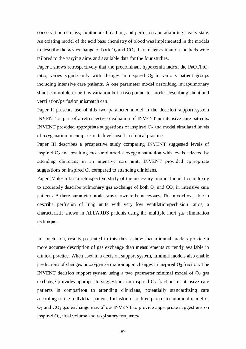
87
conservation of mass, continuous breathing and perfusion and assuming steady state.
An existing model of the acid base chemistry of blood was implemented in the models
to describe the gas exchange of both O2 and CO2. Parameter estimation methods were
tailored to the varying aims and available data for the four studies.
Paper I shows retrospectively that the predominant hypoxemia index, the PaO2/FiO2
ratio, varies significantly with changes in inspired O2 in various patient groups
including intensive care patients. A one parameter model describing intrapulmonary
shunt can not describe this variation but a two parameter model describing shunt and
ventilation/perfusion mismatch can.
Paper II presents use of this two parameter model in the decision support system
INVENT as part of a retrospective evaluation of INVENT in intensive care patients.
INVENT provided appropriate suggestions of inspired O2 and model simulated levels
of oxygenation in comparison to levels used in clinical practice.
Paper III describes a prospective study comparing INVENT suggested levels of
inspired O2 and resulting measured arterial oxygen saturation with levels selected by
attending clinicians in an intensive care unit. INVENT provided appropriate
suggestions on inspired O2 compared to attending clinicians.
Paper IV describes a retrospective study of the necessary minimal model complexity
to accurately describe pulmonary gas exchange of both O2 and CO2 in intensive care
patients. A three parameter model was shown to be necessary. This model was able to
describe perfusion of lung units with very low ventilation/perfusion ratios, a
characteristic shown in ALI/ARDS patients using the multiple inert gas elimination
technique.
In conclusion, results presented in this thesis show that minimal models provide a
more accurate description of gas exchange than measurements currently available in
clinical practice. When used in a decision support system, minimal models also enable
predictions of changes in oxygen saturation upon changes in inspired O2 fraction. The
INVENT decision support system using a two parameter minimal model of O2 gas
exchange provides appropriate suggestions on inspired O2 fraction in intensive care
patients in comparison to attending clinicians, potentially standardizing care
according to the individual patient. Inclusion of a three parameter minimal model of
O2 and CO2 gas exchange may allow INVENT to provide appropriate suggestions on
inspired O2, tidal volume and respiratory frequency.
88
Danish summary
Mekanisk ventilation er en af de primære livreddende terapiformer i anvendelse på en
intensivafdeling. Modstridende kliniske mål komplicerer indstilling af en respirator.
Beslutningsstøttesystemer udgør et muligt værktøj til at hjælpe læger med valg af den
mest hensigtsmæssige terapi. Denne PhD-afhandling omhandler brugen af minimal-
modeller af den pulmonære gasudveksling. Under projektet er minimal-modeller med
forskellige grader af kompleksitet samt klinisk tilgængelige måleteknikker for
evaluering af gasudveksling blevet sammenlignet, og en minimal-model af O2
gasudveksling er blevet anvendt i et beslutningsstøttesystem til rådgivning om den
inspirerede iltfraktion ved patienter på en intensivafdeling.
Som udgangspunkt beskrives projektets kliniske og teknologiske baggrunde. Akut
lungeskade og akut respiratorisk distress syndrome (ALI/ARDS) beskrives, idet de
udgør komplekse former for respiratorisk svigt og patienterne er kendetegnet ved at
være tilbøjelige til at udvikle respiratorinduceret lungeskade (ventilator induced lung
injury - VILI). Der redegøres for forskellige VILI typer samt vigtige kliniske studier i
strategier for mekanisk ventilation. Redegørelsen viser, at der er kontroverser omkring
hvad VILI er, og hvordan strategier for mekanisk ventilation bedst muligt
tilrettelægges for den individuelle patient. De fleste studier har fokus på volumen og
tryk, men den inspirerede iltfraktion er med rette ikke blevet ignoreret og søges
minimeret i størstedelen. En redegørelse for udviklingen indenfor
beslutningsstøttesystemer gør det klart, at regel-baserede systemer dominerer. Der er
dog to grundlæggende svagheder ved regel-baserede systemer. De giver ikke en
forståelse for patientens fysiologi eller rådene de tilbyder, og de kan kræve, at man
prøver sig frem for at finde den mest hensigtsmæssige terapi. Model-baserede
systemer kan potentielt løse begge problemer ved brug af matematiske modeller med
parametre tilknyttet en fysiologisk fortolkning. Til slut redegøres for matematiske
modeller af den pulmonære gasudveksling. Her argumenteres for, at tilgængelige
målinger af gasudveksling i klinisk praksis er for simple, idet de varierer med
terapiændringer, der ikke påvirker patientens fysiologi. Referenceteknikken, the
multiple inert gas elimination technique (MIGET) er for kompleks til klinisk brug.
Minimal-modeller udgør et kompromis imellem kompleksitet og anvendelighed.
89
Afhandlingen er baseret på fire artikler. Minimal-modeller af forskellig kompleksitet
er blevet anvendt, men alle er baseret på massebevarelse, kontinuert vejrtrækning og
perfusion og antager ligevægtstilstand. En eksisterende model af blodets syre-base
kemi blev implementeret i modellerne for at kunne beskrive gasudveksling af både O2
og CO2. Metoder til parameterestimering blev tilpasset til tilgængelige data og de
enkelte artiklers formål.
Artikel I viser retrospektivt, at det dominerende hypoksæmi-index, PaO2/FiO2,
varierer signifikant med ændringer i den inspirerede iltfraktion. En en-parameter
model af intrapulmonær shunt kunne ikke beskrive denne variation i modsætning til
en to-parameter minimal-model af shunt og ventilation/perfusions misforhold.
Artikel II beskriver brug af den samme to-parameter model i
beslutningsstøttesystemet INVENT, som blev retrospektivet evalueret i patienter på
en intensivafdeling. INVENTs råd om inspireret iltfraktion og model-simulerede
arterielle iltmætninger var hensigtsmæssige i sammenligning med klinisk praksis.
Artikel III beskriver et prospektiv studie på en intensivafdeling, som sammenlignede
INVENTs råd om inspireret iltfraktion og de målte resulterende arterielle
iltmætninger med niveauer valgt af vagthavende læger. INVENTs råd var
hensigtsmæssige i sammenligning med lægernes.
Artikel IV beskriver en retrospektiv undersøgelse af den nødvendige minimal-model
kompleksitet for at beskrive den pulmonære gasudveksling af både O2 og CO2 i
patienter på en intensivafdeling. Studiet viste, at en tre-parameter model er nødvendig.
Denne model kan beskrive perfusion af lungeenheder med meget lav
ventilation/perfusions ratio, hvilket tidligere er beskrevet i ALI/ARDS patienter med
MIGET.
Resultaterne præsenteret i denne afhandling viser, at minimal-modeller giver en mere
nøjagtig beskrivelse af gasudveksling end tilgængelige målinger i klinisk praksis. I
beslutningsstøttesystemer kan disse modeller anvendes til at simulere ændring i
arteriel iltmætning ved ændringer i inspireret iltfraktion. INVENT har ved brug af en
minimal-model givet hensigtsmæssige råd om inspireret iltfraktion i forhold til
vagthavende læger på en intensivafdeling. Potentielt kunne systemet standardisere,
hvordan inspireret iltfraktion indstilles i forhold til den specifikke patient. Inklusion af
en tre-parameter minimal-model kan muliggøre at INVENT i fremtiden også kan
rådgive om tidalvolumen og respirationsfrekvens på en intensivafdeling.