Embed Size (px)
Citation preview
Version 3 Last Updated 19 December 2014
ab109202
DRAQ7TM 1ml (0.3mM)
Protocol
Instructions for Use
This product is for research use only and is not intended for in vitro diagnostic use.
1
Table of Contents
1. Introduction 2
2. Reagents Required 3
3. Storage and Handling 3
4. Overview: Use of DRAQ7™ 3
5. General Protocols 5
Protocol I: Apoptosis/ cell death by flow cytometry 5
Protocol II: Real-time cell viability assays 9
Protocol III: Immunofluorescence 13
6. FAQ 16
2
1. Introduction
DRAQ7™ is a cell impermeable far-red fluorescent DNA dye that
only stains the nuclei in dead and permeabilized cells. It can be used
in combination with common labels such as GFP or FITC.
DRAQ7™ is the ideal tool to study dead or membrane-compromised
cells because it does not enter intact, live cells. DRAQ7™ is an ideal
replacement for Propidium Iodide and 7-AAD having far better
spectral properties; it has no UV excitation and no emission overlap
with PE and homologues. DRAQ7™ can be used in flow cytometry,
cell imaging and cell-based assays and is highly compatible with
existing protocols across a wide range of instrumentation platforms.
Key features of DRAQ7™ include:
Rapid staining of dsDNA/ nuclei of dead or permeabilized
cells
Low photobleaching effect
It can be used in most cell types, eukaryotic and prokaryotic:
mammalian, bacterial, parasitic, plant, etc ...
No compensation needed with common FITC/GFP + PE
combinations in flow cytometry
No RNase treatment required
3
2. Reagents Required
ab109202: DRAQ7™ 1ml (0.3mM)
Phosphate Buffered Saline (PBS), without sodium azide, or
other culture medium
3. Storage and Handling
Before handling ab109202 DRAQ7™ please read the MSDS
supplied with the product.
Store product at 4°C in the dark. Do not freeze.
Undiluted product is stable for at least 3 years if kept under the
recommended conditions. Diluted product should be used as soon
as possible.
4. Overview: Use of DRAQ7™
DRAQ7™ is added in the final staining step of a labeling procedure
as there is no further washing step required.
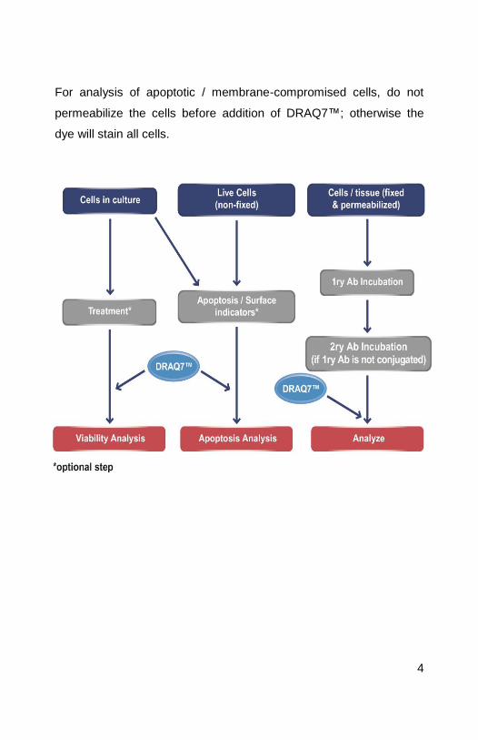
The following chart gives an overview on where DRAQ7™ can be
added depending on the type of experiment.
4
For analysis of apoptotic / membrane-compromised cells, do not
permeabilize the cells before addition of DRAQ7™; otherwise the
dye will stain all cells.
5
5. General Protocols
Protocol I: Cell staining with DRAQ7™ for dead cell / apoptosis
evaluation by flow cytometry.
This general protocol is a guideline, and we recommend adapting it
to each user’s best protocol.
Surface antibodies and apoptosis indicators (such as Annexin V-
FITC) can be use together with DRAQ7™. Simply incubate the cells
with those products prior to the addition of DRAQ7™.
A. Cell Preparation and Fixation
1. Prepare cells for staining with DRAQ7™: centrifuge and
resuspend cells in appropriate buffer such as PBS (or other
culture media) at a concentration of ≤4 x 10⁵ cells/ml in a test
tube.
2. For cells requiring additional fluorescent staining (either
conjugated-primary antibody or primary and secondary
antibody), perform the immunostaining at this step. Wash
thoroughly after antibody incubation.
6
B. Cell Staining
3. Add DRAQ7™ directly as supplied to a final concentration of
3µM to the cells. Alternatively, DRAQ7™ can be added to the
fresh buffer/ media prior to resuspending the cells in it.
4. Gently mix the cells by pipetting and then incubate for 5 – 30
minutes at room temperature.
DRAQ7™ staining is accelerated at 37°C and incubation time
may be reduced but this should be checked by titration and for
each cell type.
Note: Protect cells from light during incubation period if other
fluorescent stains (such secondary antibodies) have been
applied to the cells, as they may otherwise suffer photo-
bleaching.
5. Cells can be analyzed directly without further treatment or
washing, preferably within two hours.
7
C. Data Analysis
It is important to consider the combinations of fluorochromes and
filters for the experiments.
EXCITATION:
In flow cytometry, DRAQ7™ may be excited by wavelengths
from 488 nm and up to 647 nm (Exλmax 646 nm).
Despite low absorbance at 488 nm, this excitation offers optimal
CVs for flow cytometry cell cycle analysis and convenient
combination with FITC and R-PE conjugates and eGFP.
EMISSION:
Emission starts at 655 nm (Emλmax 681 nm/ 697 nm dsDNA-
bound).
Suitable filters include 695LP, 715LP or 780 LP.
For cell cycle analysis it is recommended to choose a filter (such
as 715LP) which excludes a significant proportion of signal from
the small fraction of unbound DRAQ7™.
ANALYSIS BY FLOW CYTOMETRY: what you should expect
to see
To establish the position of DRAQ7™ (+) cells a control
experiment may be performed:
8
1. Analyze untreated, unstained control cells, plotting the results
as an intensity histogram, exciting/detecting in all available
channels to determine the negative event distribution and any
bio-fluorescence.
2. Analyze treated, unstained cells similarly.
3. Based on both samples, adjust the instrument settings to place
the negative population in the first log decade.
4. Add DRAQ7™ to a new aliquot of untreated control cells
according to the protocol.
5. Split the aliquot in half, analyzing one half to establish the
position of the DRAQ7™ (-) cells.
6. Permeabilize the remaining half of the aliquot of untreated
control cells (prepared in Step 4) with the addition of 1% Triton-
X100, vortex to mix and analyze to establish the position of
DRAQ7™ (+) cells (i.e. membrane-compromised cells).
Alternatively, add DRAQ5™ (ab108410) at 20µM and incubate
for 10 minutes at 37°C. The position of DRAQ5™ (+) events is
the upper limit for the DRAQ7™ (+) event signal.
These control experiments should allow setting of DRAQ7 (-) /
DRAQ7 (+) gates.
Remove doublets and clumps by plotting DRAQ7™ peak height/
width versus DRAQ7™ peak area.
For cell cycle/ ploidy analysis, plot DRAQ7™ signals in linear
mode.
9
Protocol II: Use of DRAQ7™ in real-time, dynamic cell viability
assays.
DRAQ7™ has been shown not to have any effect on the proliferation
rate of cells in long-term culture assays.
DRAQ7™ can therefore be used to report, in real-time, the cytotoxic
or apoptotic-effect of a specific treatment on cells.
For example, DRAQ7™ can be used to study the effect of:
A pharmacological agent
RNAi
Virus
Antibody-dependent complement-mediated killing
In vitro toxicology testing
This general protocol is a guideline, and we recommend adapting it
to each user’s best protocol.
A. Cell Preparation and Staining
1. Prepare cells for experiment according to your protocol.
Treat cells as required for your protocol.
2. DRAQ7™ is supplied ready-to-use.
10
Add directly to the cell culture media at a final concentration of
3µM (1/100 dilution).and mix gently.
3. Remove aliquots as required and analyze for far-red (> 665 nm)
fluorescing cells (relative to controls) by flow cytometry.
Normal cells will not need to be washed prior to analysis.
Alternatively, cells on microplates/ slides can be fixed at the
required time points for the experiment and analyzed by cell
imaging microscopy.
B. Data Analysis
It is important to consider the combinations of fluorochromes and
filters for the experiments.
EXCITATION:
In flow cytometry, DRAQ7™ may be excited by wavelengths
from 488 nm and up to 647 nm (Exλmax 646 nm).
Despite low absorbance at 488 nm, this excitation offers optimal
CVs for flow cytometry cell cycle analysis and convenient
combination with FITC and R-PE conjugates and eGFP.
For cell imaging, DRAQ7™ excitation (Exλmax 599/ 644 nm) is
performed with either 633 nm or 647 nm wavelengths.
11
EMISSION:
Emission starts at 655 nm (Emλmax 681 nm/ 697 nm dsDNA-
bound).
Suitable filters include 695LP, 715LP or 780 LP.
For cell cycle analysis in flow cytometry it is recommended to
choose a filter (such as 715LP) which excludes a significant
proportion of signal from the small fraction of unbound
DRAQ7™.
In cell imaging, DRAQ7™ has no spectral emission overlap with
FITC, R-PE, GFP, DyLight® 488 or many other fluoresceing
proteins allowing image acquisition in one scan.
ANALYSIS BY FLOW CYTOMETRY: what you should expect
to see
To establish the position of DRAQ7™ (+) cells a control
experiment may be performed:
1. Analyze untreated, unstained control cells, plotting the results
as an intensity histogram, exciting/detecting in all available
channels to determine the negative event distribution and any
bio-fluorescence.
2. Analyze treated, unstained cells similarly.
12
3. Based on both samples, adjust the instrument settings to place
the negative population in the first log decade.
4. Add DRAQ7™ to a new aliquot of untreated control cells
according to the protocol.
5. Split the aliquot in half, analyzing one half to establish the
position of the DRAQ7™ (-) cells.
6. Permeabilize the remaining half of the aliquot of untreated
control cells (prepared in Step 4) with the addition of 1% Triton-
X100, vortex to mix and analyze to establish the position of
DRAQ7™ (+) cells (i.e. membrane-compromised cells).
Alternatively, add DRAQ5™ (ab108410) at 20µM and incubate
for 10 minutes at 37°C. The position of DRAQ5™ (+) events is
the upper limit for the DRAQ7™ (+) event signal.
These control experiments should allow setting of DRAQ7 (-) /
DRAQ7 (+) gates.
Remove doublets and clumps by plotting DRAQ7™ peak height/
width versus DRAQ7™ peak area.
For cell cycle/ ploidy analysis, plot DRAQ7™ signals in linear
mode.
13
Protocol III: Fixed cell/ tissue staining with DRAQ7™ for nuclear
visualization by epifluorescence microscopy / laser scanning
confocal or HCS imaging platform.
This general protocol is a guideline, and we recommend adapting it
to each user’s best protocol.
DRAQ7™ is an ideal far-red counterstain for IF/IHC assays. It stains
nucleus and cytoplasm differentially and it is spectrally compatible
with all visible range fluors.
DRAQ7™ will stain all fixed and permeabilized cells in the
preparation.
A. Cell Preparation and Fixation
1. Prepare cells for fixation.
Prepare working solutions of:
4% formaldehyde in PBS
5µM DRAQ7™ solution in PBS
2. Fix cells by overlaying the slide (or chamber/ well) with the 4%
formaldehyde solution. Incubate for 15 – 30 minutes at room
temperature / 37°C.
3. Gently aspirate the formaldehyde solution and wash the slide
with PBS.
14
4. For cells expressing fluorescent proteins (e.g. GFP/YFP-tagged
proteins), DRAQ7™ can be mixed with formaldehyde fixative to
provide a one step “fix/permeabilize & stain” protocol. To
prepare, mix equal volumes of 10µM DRAQ7™ and 8%
formaldehyde.
5. For cells requiring indirect immunostaining (primary and / or
secondary antibody), block and permeabilize according to your
favorite protocol. Wash thoroughly after antibody incubation.
B. Cell Staining
6. Overlay the sample with the 5µM DRAQ7™ solution.
Alternatively, DRAQ7™ can be added to the fresh buffer/
medium prior resuspending the cells in it.
7. Gently mix the cells by pipetting and then incubate for 5 – 30
minutes at room temperature.
DRAQ7™ staining is accelerated at 37°C and incubation time
may be reduced but this should be checked by titration and for
each cell type.
Note: Protect cells from light during incubation period if other
fluorescent stains (such secondary antibodies) have been
applied to the cells, as they may otherwise suffer photo-
bleaching.
15
8. Cells can be analyzed directly without further treatment or
washing, preferably within two hours.
C. Data Analysis
It is important to consider the combinations of fluorochromes and
filters for the experiments.
EXCITATION:
For cell imaging, DRAQ7™ excitation (Exλmax 599/ 644 nm) is
performed with either 633 nm or 647 nm wavelengths.
EMISSION:
Emission starts at 655 nm (Emλmax 678 nm/ 694 nm intercalated
to dsDNA).
Suitable filters include 695LP, 715LP or 780 LP.
DRAQ7™ has no spectral emission overlap with FITC, R-PE,
GFP or many other fluorescing proteins allowing image
acquisition in one scan.
16
6. FAQs
1. In which type of cells can I stain with DRAQ7™?
DRAQ7™ is a DNA-specific anthraquinone dye which derives
from DRAQ5™ (ab108410) and will therefore stain all nucleated
cells. DRAQ7™ will not only stain all type of nucleated
eukaryotic cells (primary or cell-line derived), but also bacterial
cells (such as E. coli or Bacillus) and plant cells.
However, DRAQ7™ does not enter live, intact cells. DRAQ7™
will only stain cells whose membrane is compromised, either
naturally (apoptotic cells) or artificially (fixed and permeabilized).
2. In which type of assays can I use DRAQ7™?
DRAQ7™ can be used assays where nuclear DNA staining is
required.
Examples of these assays are:
Cell imaging:
o Apoptosis studies: dead or membrane-compromised
cells
o Fixed & permeabilized cells (primary or cell-line
derived) or tissue sections
o Fixed & permeabilized tissue sections
Fluorescence In Situ Hybridization (FISH)
Flow Cytometry:
o Apoptosis detection
o Dead cell gating
Cell-based HCS assays
17
o Cytotoxicity/ viability assays
3. If DRAQ7™ doesn’t stain live healthy cells, how can I use it
in real-time viability assays?
DRAQ7™ only enters cells with compromised cellular
membranes. This means that, as long as the cell is healthy,
DRAQ7™ will not enter the cell and will not bind the DNA and
therefore won’t have any cytotoxic effect. DRAQ7™ is a very
stable compound and can be used as “viability” dye over several
days. Once the cells start to dye (either naturally or by cytotoxic
treatment), DRAQ7™ will enter the cells and staining will be
visible.
4. Does DRAQ7™ stain mitochondria or RNA?
DRAQ7™ has high specificity for dsDNA and doesn’t seem to
bind to RNA. Although mitochondria contain dsDNA, no
DRAQ7™ signals seem to be detected. This makes DRAQ7™
the ideal tool to study cell cycle profile, as all the signal will come
from nuclear DNA.
5. Why shouldn’t DRAQ7™-stained cells be washed?
There is no need to wash cells for imaging or flow cytometry.
DRAQ7™ binds stoichiometrically to the DNA and reaches an
equilibrium, which could be disrupted by long washes.
DRAQ7™ can be added at the final stage of the protocol prior
analysis, as it will be able to stain the nucleus very quickly (5
minutes incubation is enough for staining). If washes are
essential in protocol, we recommend very brief washes.
6. Can I use DRAQ7™ with anti-fade mountants?
18
Yes. DRAQ7™ is perfectly compatible with anti-fade mountants
such as Fluoroshield (ab104135), BrightMount (ab103746) or
BrightMount Plus (ab103748). Just ensure that the mountant is
the “native” product and does not contain DAPI as the DAPI
quenches the DRAQ7™ staining signal.
7. Which fluorochromes can I use with DRAQ7™?
DRAQ7™ emits in far-red end of the spectra and therefore is
compatible with fluorochromes with emission spectra lower than
600nm.
DRAQ7™ can be used with common labels such as eGFP/YFP,
Cy2, FITC, Cy3, R-PE, and DyLight® 488, 550 and 594.
We do not recommend the use of DRAQ7™ with other far-red
fluorochromes excited by the 488 ot 633 nm laser lines, such as
PE-Cy7, PerCP-Cy5.5, APC, Texas Red or DyLight® 650.
8. How long can I keep my working dilution of DRAQ7™?
We recommend that if you dilute DRAQ7™, you use your
working solution as soon as possible. Do not store diluted
DRAQ7™; the agent is very stable but the dye could be lost from
solution since it is formulated for immediate use.

19
UK, EU and ROW Email: [email protected] | Tel: +44-(0)1223-696000
Austria Email: [email protected] | Tel: 019-288-259
France Email: [email protected] | Tel: 01-46-94-62-96 Germany Email: [email protected] | Tel: 030-896-779-154 Spain Email: [email protected] | Tel: 911-146-554 Switzerland Email: [email protected] Tel (Deutsch): 0435-016-424 | Tel (Français): 0615-000-530 US and Latin America Email: [email protected] | Tel: 888-77-ABCAM (22226)
Canada Email: [email protected] | Tel: 877-749-8807 China and Asia Pacific
Email: [email protected] | Tel: 108008523689 (中國聯通) Japan Email: [email protected] | Tel: +81-(0)3-6231-0940
www.abcam.com | www.abcam.cn | www.abcam.co.jp
Copyright © 2014 Abcam, All Rights Reserved. The Abcam logo is a registered trademark. All information / detail is correct at time of going to print.