Embed Size (px)
Citation preview
137Copyright © The Korean Society of Fisheries and Aquatic Science http://e-fas.org
Abalone Haliotis discus hannai Intestine Digests with Different Molecule Weights Inhibit MMP-2 and MMP-9 Expression in Human Fibrosarcoma CellsVan-Tinh Nguyen, Zhong-Ji Qian, and Won-Kyo Jung*
Department of Marine Life Science and Marine Life Research & Education Center, Chosun University, Gwangju 501-759, Korea
AbstractThe abalone Haliotis discus hannai, is one of the economically important species in the fisheries industry. Abalone intestines are one of the by-products of its processing. To investigate its bioactive potential, abalone intestine was digested using an in vitro gas-trointestinal (GI) digestion system containing pepsin, trypsin, and α-chymotrypsin. The abalone intestine G1 digests (AIGIDs) pro-duced by the GI digestion system were fractionated into AIGID I (> 100 kDa), AIGID II (10-100 kDa), and AIGID III (1-10 kDa) using an ultrafiltration membrane system. Of the three digests, AIGID II and AIGID III exhibited inhibitory effects against matrix metalloproteinase-2 and -9 (MMP-2, MMP-9) in HT1080 human fibrosarcoma cells. Both fractions potently inhibited gelatine digestion by MMP-2 and MMP-9 treated with phorbol 12-myristate 13-acetate (PMA) and migration of HT1080 cells in dose de-pendently. Furthermore, AIGID II and III attenuated expression of p65, a component of nuclear transcription factor kappa B. These results indicate that of the abalone intestine digests inhibit MMP-2 and MMP-9. Thus, the AIGIDs or their active components may have preventive and therapeutic potential for diseases associated with MMP-2 and MMP-9 activation in fibrosarcoma cells.
Key words: Haliotis discus hannai, Matrix metalloproteinases-2, Matrix metalloproteinases-9, Abalone intestine gastrointestinal digests (AIGIDs), Human fibrosarcoma cell line.
Introduction
The matrix metalloproteinases (MMPs) are a family of zinc-dependent endopeptidases that play important roles in the degradation of extracellular matrix (ECM), and in a variety of biological and pathological processes, such as tumor inva-sion and metastasis (Sternlicht and Werb, 2001; Nishida et al., 2008; Hwang et al., 2010; Khan et al., 2010). This family of proteinases has includes at least 28 endopeptidases collective-ly capable of degrading virtually all ECM components. MMPs family members have been classified into eight subgroups ac-cording to their structural and functional characteristics. These are the minimal-domain MMPs, simple hemopexin domain containing MMPs, gelatine-binding MMPs, furin activated secreted MMPs, and vitronectin-like insert MMPs, and mem-
brane bound MMPs include type I trans-membrane MMPs, glycosyl-phosphatidyl inositol-linked MMPs, and type II transmembrane MMPs (Egeblad and Werb, 2002). MMPs reg-ulate the synthesis and secretion of cytokines, growth factors, hormone receptors and cell adhesion molecules. They also contribute to cell growth and development, cell morphogen-esis, tissue remodelling, angiogenesis, cardiovascular, aller-gies, neurodegenerative diseases, some cancers, and a series of physiological and pathological processes (Sang et al., 2006; Hu et al., 2007; Kong et al., 2008).
Gelatinases (MMP-2, MMP-9) play an important role in cancer invasion and metastasis, and have been detected most consistently in malignant tissues. This subgroup of metal-
Received 31 August 2011; Revised 24 February 2012Accepted 10 April 2012
*Corresponding AuthorE-mail: [email protected]
http://dx.doi.org/10.5657/FAS.2012.0137Open Access
This is an Open Access article distributed under the terms of the Creative Commons Attribution Non-Commercial License (http://creativecommons.org/licenses/by-nc/3.0/) which permits unrestricted non-commercial use, distribution, and reproduction in any medium, provided the original work is properly cited. pISSN: 2234-1749 eISSN: 2234-1757
Original ArticleFish Aquat Sci 15(2), 137-143, 2012
Fish Aquat Sci 15(2), 137-143, 2012
http://dx.doi.org/10.5657/FAS.2012.0137 138
Materials
Live adult abalones were collected from Wando Island, Ko-rea. Intestinal organs (guts) were separated from the washed abalone and lyophilized. Dulbecco’s modified Eagle’s medium (DMEM), trypsin-ethylenediaminetetraacetic acid (EDTA), penicillin/streptomycin, and fetal bovine serum (FBS) were obtained from Gibco BRL, Life Technologies (Grand Island, NY, USA). HT1080 cells were obtained from the American Type Culture Collection (Manassas, VA, USA). Primary and secondary antibodies, including MMP-2 (sc-13595), MMP-9 (sc-10737), NF-κB p65 (sc-8008), NF-κB p50 (sc-166588), β-actin (sc-130656), goat anti-rabbit IgG-horseradish peroxi-dase (HRP) (sc-2004) and goat anti-mouse IgG1-HRP (sc-2060), were purchased from Santa Cruz Biotechnology-Inc (Santa Cruz, CA, USA). 3-(4,5-Dimethylthiazol-2-yl)-2,5-di-phenyltetrazolium bromide (MTT), gelatin (type A) and phor-bol 12-myristate 13-acetate (PMA) were from Sigma Chemi-cal Co. (St. Louis, MO, USA). Other chemicals and reagents used were of analytical grade.
Preparation of AIGIDs using ultrafiltration (UF) membrane bioreactor
The method of Kapsokefalou and Miller (1991) was used to prepare AIGIDs (AIGID and AIGID I-III). Abalone intes-tine solution (1 L) was adjusted to pH 2.2 for gastric digestion using 1 M HCl and 10 M NaOH. Pepsin (EC 3.4.23.1) was added at an enzyme:substrate ratio of 1:100 (w/w), then incu-bated at 37°C on a shaker for 2 h. The pH was adjusted to 6.5 to match the conditions of small intestinal digestion. Trypsin (EC 3.4.21.4) and α-chymotrypsin (EC 3.4.21.1) were added at an enzyme to substrate ratio of 1/100 (w/w), then incubated at 37°C for 2.5 h. AIGIDs were centrifuged (10,000 g, 15 min, at 4°C), and the supernatant was lyophilized to obtain AIGID powder. AIGID (100 g) was solublized in 2 L of water and fractionated using UF membranes with a 100 kDa MW cut-off (MWCO). The filtrate was then fractionated further through a 10 kDa MWCO filter. All samples (AIGID and AIGID I-III) were dialyzed through a 1 kDa membrane and lyophilized.
Cell culture
Human fibrosarcoma HT1080 cells were cultured in DMEM containing 10% FBS and antibiotics as monolayers in 10 cm culture dishes at 37°C in a humidified 5% CO2 atmosphere.
MTT assay
Cytotoxic effects of the AIGIDs on cultured cells were measured using the 3-(4,5-dimethylthiazol-2-yl)-2,5-diphe-nyltetrazolium bromide (MTT) assay. To determine the cy-tocompatible effects of AIGIDs, HT1080 cells were seeded in 96-well plates at a 1×10⁴ /well and incubated with various
loproteinases has also been called type IV collagenases, be-cause of their ability to cleave type IV collagen (Salo et al., 1985). One of them, MMP-2 (gelatinase-A, 72 kDa type IV collagenase), was originally purified from a highly progress-ing metastatic murine tumor (Wilhelm et al., 1989). MMP-2 binds to type I collagen through the fibronectin domain, which stabilizes it against autolysis, thereby controlling its activity (Ellerbroek et al., 2001). MMP-2 expression is dependent on extracellular matrix metalloproteinase inducer, growth fac-tors, cytokines, and hormones. Pro-MMP-2 activation needs contributions from MT1-MMP and TIMP metallopeptidase inhibitor 2. A low level has been linked to a favorable progno-sis in patients with hormone receptor-negative tumors, usually associated with poor prognosis. As a zymogen that requires proteolytic activation for catalytic activity, MMP-2 has been implicated in invasion and metastasis in many cancer model systems, including human breast cancer (Jezierska and Mo-tyl, 2009; Fredrich and Illing, 2010). MMP-9 (92 kDa type IV collagenase, gelatinase B) is produced in human macrophages and polymorphonuclear leukocytes. It has also been localized in endothelial cells and synovial fibroblasts in rheumatoid ar-thritis synovium. MMP-9 is expressed by osteoclasts in hu-man normal bone tissues, indicating a role in bone remodel-ling. Intact mature human odontoblasts also express MMP-9, and it has been detected in human dental caries lesions and sa-liva. However, MMP-9 is not expressed by human gingival fi-broblasts. Like MMP-2, MMP-9 may exist in the ECM bound to type I collagen, gelatine, or laminin (Bolcato-Bellemin et al., 2000).
Degradation of denatured collagen I by MMP-2 and MMP-9 is readily demonstrable in biological materials. One tech-nique, known as gelatin zymography, identifies gelatinolytic activity in biological samples using sodium dodecyl sulfate (SDS)-polyacrylamide gels impregnated with gelatine. The human fibrosarcoma cell line HT1080 has been used exten-sively in studies of ECM proteins involved in attachment, in-vasion, and metastasis. Additionally, HT1080 cells are used because they produce both MMP-2 and MMP-9 enzymes (Brooks and Schumacher, 2001).
The abalone Haliotis discus hannai, which is maricultured on the southwestern coast (Wando area) of Korea, is a ma-rine univalve gastropod mollusc, that inhabits temperate and tropical waters in both hemispheres of the globe. Despite its economic importance, few nutraceutical and pharmaceutical studies of abalone have been reported.
In the present study, to investigate the bioactive potential of abalone intestine, abalone intestine G I digests (AIGIDs) were separated into various molecular weight (MW) fractions by ultrafiltration. We focuses on AIGID II and III mediated inhi-bition of both MMP-2 and MMP-9 and cancer cell migration in human fibrosarcoma HT1080 cells.
Materials and Methods
Nguyen et al. (2012) Inhibitory effects of abalone intestine digests on MMPs expression in fibrosarcoma cells
139 http://e-fas.org
dilution) in blocking agent at 4°C overnight. After washing with TBS-T, the membrane was incubated with secondary an-tibody (1:5,000 dilution) for 2 h at room temperature. Bands were developed by enhanced chemiluminescence and visual-ized with LAS-4000 imaging system (Fujifilm, Tokyo, Japan). Basal levels of MMP-2 and MMP-9 protein expression were normalized by assessing the level of β-actin protein.
Amino acid composition
Freeze-dired AIGID II and AIGID III (20 mg) was hydro-lyzed with 6 M HCl at 110°C for 24 h in vacuum. Amino acids derived with phenylisothiocyanate were identified and quanti-fied using an automatic amino acid analyzer (Biochrom 20; Pharmacia Biotech, Cambridge, UK).
Results
Cytocompatible effects of AIGIDs in HT1080 cells
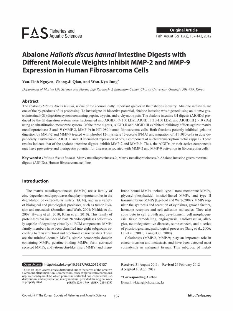
The effects of AIGID and AIGID I-III on proliferation of HT1080 cells were evaluated using an MTT assay. No AIGID had any cytotoxic effect on HT1080 cells up to 400 μg/mL (Fig. 1).
AIGIDs inhibit MMP-2 and MMP-9 activity in HT1080 cells
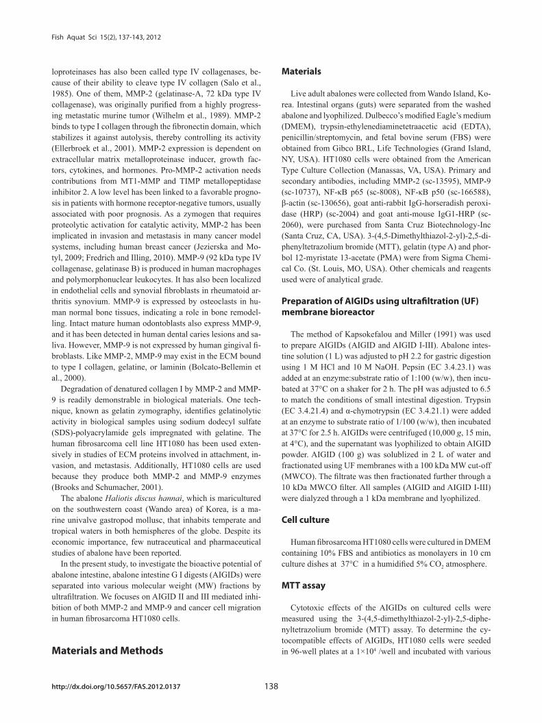
The effects of AIGIDs of differing MWs on MMP-2 and MMP-9 were evaluated. Cells treated with AIGID, AIGID I, AIGID II, or AIGID III at 200 μg/mL were stimulated with PMA. Conditioned media was used to assess MMP-2 and -9 activities. AIGID II and AIGID III showed marked inhibitory effects on MMP-2 and MMP-9 in HT1080 cells compared with
AIGID, AIGID I, AIGID II, and AIGID III concentrations. Af-ter a 24 h incubation, MTT reagent (50 µL) was added to each well and incubation was continued for another 4 h. Finally, DMSO was added to solubilize the formazan salt formed and the absorbance at 540 nm was determined using a microplate reader. Cell viability relative to the non-treated group was cal-culated. Data are expressed as means of at least three inde-pendent experiments and P < 0.05 were considered to indicate statistical significance.
Gelatin zymography
The gelatinase activities of MMP-2 and MMP-9 were de-termined by gelatin zymography (Ta et al., 2006; Kong et al., 2010). Cells were cultured on 24-well plates in serum-free DMEM medium. HT1080 cells were treated with AIGID, AIGID I, AIGID II, and AIGID III for 1 h and then stimulated with 10 ng/mL PMA for 36 h. The conditioned media were collected and centrifuged (3000 g, 10 min). Concentrated medium was electrophoresed under non-reducing conditions through 10% sodium dodecyl sulfate (SDS)-polyacrylamide gels impregnated with 0.15% of gelatin. After electrophoresis, SDS was removed from the gel by washing in 2.5% Triton X-100 solution for 1.5 h and incubated in developing buffer (50 mM Tris-HCl, pH 7.5, 200 mM NaCl, 5 mM CaCl2.2H2O, 0.02% Brif-35) at 37°C. The gels were stained with 1% Coo-massie blue R-250 in 45% methanol and 10% glacial acetic acid. After 30 min, the gels were destained in the same solu-tion with no Coomassie blue dye. The digested area appeared clear on a blue background, indicating the presence of a ge-latinase.
In vitro wound migration assay
HT1080 cells were seeded in six-well plates. Cells were pretreated with mitomycin C (25 μg/mL) for 30 min before an injury line was made with a 2 mm wide tip on cells that were plated in culture dishes at 80% confluence. Cells were then washed with phosphate buffered saline. The cells were allowed to migrate in serum-free medium in the presence of various AIGID II and AIGID III concentrations for 24 h. Re-sults were observed by microscopy.
Western blot analysis
Cultured cells were harvested and then lysed with RIPA buffer (50 mM Tris-HCl, pH 7.5, 150 mM NaCl, 1% Triton X-100, 1% sodium deoxycholate, 2 mM EDTA, and 0.1% SDS). Protein concentration was determined by the BCA method. Proteins were separated by 10% SDS-polyacryl-amide gel electrophoresis and transferred to a nitrocellulose membrane. Membranes were blocked with 5% skim milk in TBS-T buffer (20 mM Tris-HCl, pH 7.6, 136 mM NaCl, 0.1% Tween-20) and then incubated with primary antibody (1:500
Fig. 1. Effects of abalone intestine G I digests (AIGIDs; AIGID, AIGID I, AIGID II and AIGID III) on viability of HT1080 cells. Cells were treated with different concentrations (10-400 μg/mL) of AIGIDs and cell viability was determined by MTT assay after 24 h. Deta are give as means of values ± SD. from three independent experiments.
Fish Aquat Sci 15(2), 137-143, 2012
http://dx.doi.org/10.5657/FAS.2012.0137 140
In vitro effects of AIGID II and III on migration of HT1080 cells
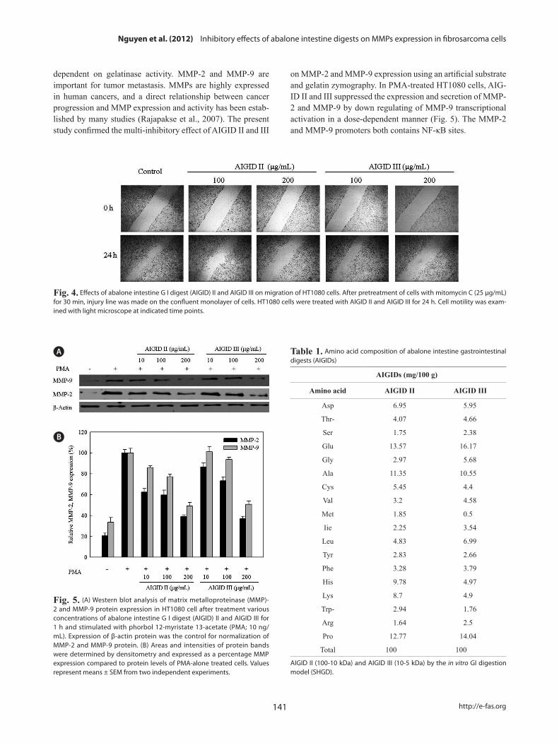
Cell migration is required for cancer cell invasion through the basement membrane. HT 1080 cells were treated with 0-200 μg/mL AIGID II and III. Both AIGID II and III inhib-ited the migration of HT1080 cells at 200 μg/mL (Fig. 4).
EffectS of AIGID II and III on MMP-2 and MMP-9 protein expression and NF-κB activity in HT1080 cells
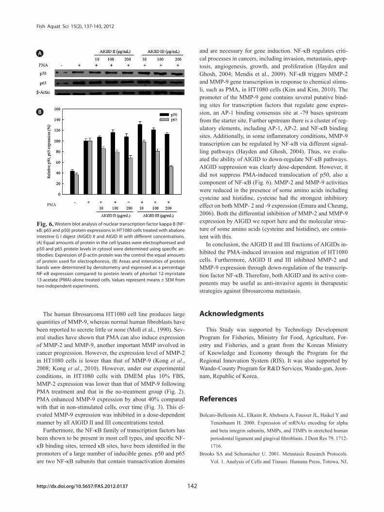
AIGID II and III mediated inhibition of MMP-2 and MMP-9 protein levels were analyzed by Western blotting. AIGID II and III (10, 100, 200 μg/mL) suppressed MMP-2 and MMP-9 protein levels in a concentration-dependent manner, compared with PMA treatment (Fig. 5). Inhibition of MMP-2 expression by both AIGID II and III (200 μg/mL) was more noticeable.
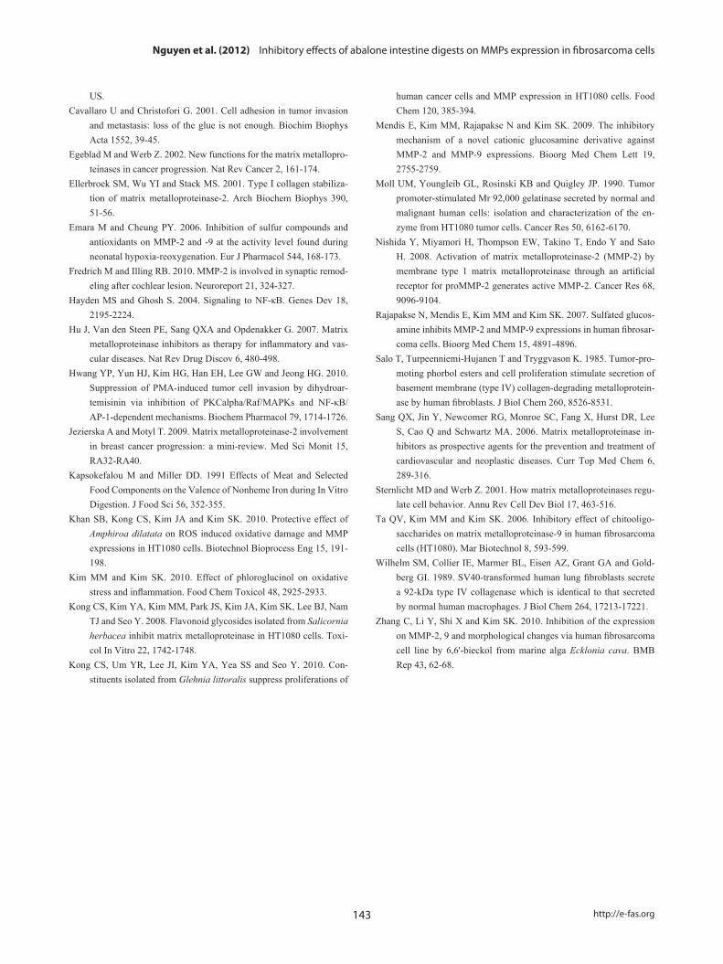
Furthermore, Western blots were used to assess the down-regulation of protein expression and to determine the possible mechanisms for the effects of AIGID II and III on the expres-sion of activator NF-κB. Western blotting was conducted to confirm down-regulation of p50 and p65 protein expression; both are components of NF-кB. AIGID II and III treatment resulted in decreased p65 protein level; this was dose-depen-dent (10, 100, 200 μg/mL) (Fig. 6). Additionally, AIGID III showed a greater inhibition of p65 expression than AIGID II at 200 μg/mL. However, no significant inhibition of p50 expres-sion was detected at the concentrations tested. These results demonstrate that AIGID II and III down-regulated MMP-2 and MMP-9 expression through transcriptional down-regula-tion of NF-κB.
Amino acid composition
The amino acid compositions of AIGID II and III are shown in Table 1. AIGID II and III were rich in acidic amino acids (glutamic acid, aspartic acid), proline (Pro), alanine (Ala), his-tidine (His), and lysine (Lys), which constituted 63.12% of the total amono acid residues in AIGID II and 56.58 % of AIGID III. The amino acid composition of a protein is associated with its biological activities; indeed, Glu, Ala, and His have been reported to be important for the inhibition of MMP-2 and -9 (Emara and Cheung, 2006).
Discussion
In this study, we demonstrated that AIGID II and III mark-edly decreased MMP expression, cell migration, motility and invasiveness (Fig. 4). Tumor cell invasion of the ECM is an important step in tumor metastasis, and involves the attach-ment of tumor cells to ECM (Cavallaro and Christofori, 2001; Zhang et al., 2009). Tumor cell migration and invasion are
the PMA treatment group (Fig. 2). However, neither AIGID nor AIGID I significantly inhibited MMP-2 or MMP-9. Thus, AIGID II and III were used in subsequent experiments.
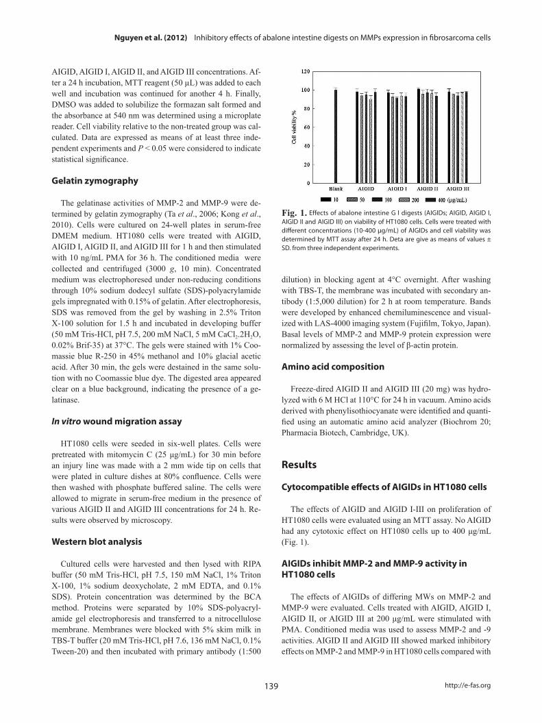
A gelatin zymography assay was performed to assess the dose-dependent inhibitory activities of AIGID II and III on MMP-2 and MMP-9 mRNA expression in HT1080 cells. MMP-2 and MMP-9 expression was suppresed by AIGID II and III in a dose-dependent manner (Fig.3). AIGID III showed greater inhibition of MMP-2 and MMP-9 expression than did AIGID II at 200 μg/mL.
Fig. 2. Gelatin zymography for the determination of matrix metallopro-teinase (MMP)-2 and -9 activities in abalone intestine G I digest (AIGID) treated HT1080 cells. HT1080 cells treated with 200 μg/mL of AIGID, AIGID I, AIGID II and AIGID III for 1 h were stimulated by phorbol 12-myristate 13-acetate (PMA; 10 ng/mL) for 36 h. Gelatinolytic activities of MMP-2 and MMP-9 in conditioned media were detected by electrophoresis of soluble protein on a gelatine containing 10% polyacrylamide gel.
A
B
Fig. 3. Effects of abalone intestine G I digest (AIGID) II and AIGID III on matrix metalloproteinase (MMP)-2 and MMP-9 activities by gelatin zy-mography. (A) Gelatinase activities of MMP-2 and MMP-9 in conditioned media were detected by electrophoresis of soluble protein on a gelatin containing 10% polyacrylamide gel. (B) Areas and relative intensities of gelatin-digested bands by MMP-2 and MMP-9 were quantified by den-sitometry and expressed as relative MMP-9 activity compared to that of phorbol 12-myristate 13-acetate (PMA)-alone treated cells.
Nguyen et al. (2012) Inhibitory effects of abalone intestine digests on MMPs expression in fibrosarcoma cells
141 http://e-fas.org
on MMP-2 and MMP-9 expression using an artificial substrate and gelatin zymography. In PMA-treated HT1080 cells, AIG-ID II and III suppressed the expression and secretion of MMP-2 and MMP-9 by down regulating of MMP-9 transcriptional activation in a dose-dependent manner (Fig. 5). The MMP-2 and MMP-9 promoters both contains NF-κB sites.
dependent on gelatinase activity. MMP-2 and MMP-9 are important for tumor metastasis. MMPs are highly expressed in human cancers, and a direct relationship between cancer progression and MMP expression and activity has been estab-lished by many studies (Rajapakse et al., 2007). The present study confirmed the multi-inhibitory effect of AIGID II and III
Fig. 4. Effects of abalone intestine G I digest (AIGID) II and AIGID III on migration of HT1080 cells. After pretreatment of cells with mitomycin C (25 μg/mL) for 30 min, injury line was made on the confluent monolayer of cells. HT1080 cells were treated with AIGID II and AIGID III for 24 h. Cell motility was exam-ined with light microscope at indicated time points.
Table 1. Amino acid composition of abalone intestine gastrointestinal digests (AIGIDs)
AIGIDs (mg/100 g)
Amino acid AIGID II AIGID III
Asp 6.95 5.95
Thr- 4.07 4.66
Ser 1.75 2.38
Glu 13.57 16.17
Gly 2.97 5.68
Ala 11.35 10.55
Cys 5.45 4.4
Val 3.2 4.58
Met 1.85 0.5
Iie 2.25 3.54
Leu 4.83 6.99
Tyr 2.83 2.66
Phe 3.28 3.79
His 9.78 4.97
Lys 8.7 4.9
Trp- 2.94 1.76
Arg 1.64 2.5
Pro 12.77 14.04
Total 100 100
AIGID II (100-10 kDa) and AIGID III (10-5 kDa) by the in vitro GI digestion model (SHGD).
A
B
Fig. 5. (A) Western blot analysis of matrix metalloproteinase (MMP)-2 and MMP-9 protein expression in HT1080 cell after treatment various concentrations of abalone intestine G I digest (AIGID) II and AIGID III for 1 h and stimulated with phorbol 12-myristate 13-acetate (PMA; 10 ng/mL). Expression of β-actin protein was the control for normalization of MMP-2 and MMP-9 protein. (B) Areas and intensities of protein bands were determined by densitometry and expressed as a percentage MMP expression compared to protein levels of PMA-alone treated cells. Values represent means ± SEM from two independent experiments.
Fish Aquat Sci 15(2), 137-143, 2012
http://dx.doi.org/10.5657/FAS.2012.0137 142
and are necessary for gene induction. NF-κB regulates criti-cal processes in cancers, including invasion, metastasis, apop-tosis, angiogenesis, growth, and proliferation (Hayden and Ghosh, 2004; Mendis et al., 2009). NF-κB triggers MMP-2 and MMP-9 gene transcription in response to chemical stimu-li, such as PMA, in HT1080 cells (Kim and Kim, 2010). The promoter of the MMP-9 gene contains several putative bind-ing sites for transcription factors that regulate gene expres-sion, an AP-1 binding consensus site at -79 bases upstream from the starter site. Further upstream there is a cluster of reg-ulatory elements, including AP-1, AP-2. and NF-κB binding sites. Additionally, in some inflammatory conditions, MMP-9 transcription can be regulated by NF-κB via different signal-ling pathways (Hayden and Ghosh, 2004). Thus, we evalu-ated the ability of AIGID to down-regulate NF-κB pathways. AIGID suppression was clearly dose-dependent. However, it did not suppress PMA-induced translocation of p50, also a component of NF-κB (Fig. 6). MMP-2 and MMP-9 activities were reduced in the presence of some amino acids including cysteine and histidine, cysteine had the strongest inhibitory effect on both MMP- 2 and -9 expression (Emara and Cheung, 2006). Both the differential inhibition of MMP-2 and MMP-9 expression by AIGID we report here and the molecular struc-ture of some amino acids (cysteine and histidine), are consis-tent with this.
In conclusion, the AIGID II and III fractions of AIGIDs in-hibited the PMA-induced invasion and migration of HT1080 cells. Furthermore, AIGID II and III inhibited MMP-2 and MMP-9 expression through down-regulation of the transcrip-tion factor NF-κB. Therefore, both AIGID and its active com-ponents may be useful as anti-invasive agents in therapeutic strategies against fibrosarcoma metastasis.
Acknowledgments
This Study was supported by Technology Development Program for Fisheries, Ministry for Food, Agriculture, For-estry and Fisheries, and a grant from the Korean Ministry of Knowledge and Economy through the Program for the Regional Innovation System (RIS). It was also supported by Wando-County Program for R&D Services, Wando-gun, Jeon-nam, Republic of Korea.
References
Bolcato-Bellemin AL, Elkaim R, Abehsera A, Fausser JL, Haikel Y and Tenenbaum H. 2000. Expression of mRNAs encoding for alpha and beta integrin subunits, MMPs, and TIMPs in stretched human periodontal ligament and gingival fibroblasts. J Dent Res 79, 1712-1716.
Brooks SA and Schumacher U. 2001. Metastasis Research Protocols. Vol. 1. Analysis of Cells and Tissues. Humana Press, Totowa, NJ,
The human fibrosarcoma HT1080 cell line produces large quantities of MMP-9, whereas normal human fibroblasts have been reported to secrete little or none (Moll et al., 1990). Sev-eral studies have shown that PMA can also induce expression of MMP-2 and MMP-9, another important MMP involved in cancer progression. However, the expression level of MMP-2 in HT1080 cells is lower than that of MMP-9 (Kong et al., 2008; Kong et al., 2010). However, under our experimental conditions, in HT1080 cells with DMEM plus 10% FBS, MMP-2 expression was lower than that of MMP-9 following PMA treatment and that in the no-treatment group (Fig. 2). PMA enhanced MMP-9 expression by about 40% compared with that in non-stimulated cells, over time (Fig. 3). This el-evated MMP-9 expression was inhibited in a dose-dependent manner by all AIGID II and III concentrations tested.
Furthermore, the NF-κB family of transcription factors has been shown to be present in most cell types, and specific NF-κB binding sites, termed κB sites, have been identified in the promoters of a large number of inducible genes. p50 and p65 are two NF-κB subunits that contain transactivation domains
A
B
Fig. 6. Western blot analysis of nuclear transcription factor kappa B (NF-κB; p65 and p50) protein expressions in HT1080 cells treated with abalone intestine G I digest (AIGID) II and AIGID III with different concentrations. (A) Equal amounts of protein in the cell lysates were electrophoresed and p50 and p65 protein levels in cytosol were determined using specific an-tibodies. Expression of β-actin protein was the control the equal amounts of protein used for electrophoresis. (B) Areas and intensities of protein bands were determined by densitometry and expressed as a percentage NF-κB expression compared to protein levels of phorbol 12-myristate 13-acetate (PMA)-alone treated cells. Values represent means ± SEM from two independent experiments.
Nguyen et al. (2012) Inhibitory effects of abalone intestine digests on MMPs expression in fibrosarcoma cells
143 http://e-fas.org
human cancer cells and MMP expression in HT1080 cells. Food Chem 120, 385-394.
Mendis E, Kim MM, Rajapakse N and Kim SK. 2009. The inhibitory mechanism of a novel cationic glucosamine derivative against MMP-2 and MMP-9 expressions. Bioorg Med Chem Lett 19, 2755-2759.
Moll UM, Youngleib GL, Rosinski KB and Quigley JP. 1990. Tumor promoter-stimulated Mr 92,000 gelatinase secreted by normal and malignant human cells: isolation and characterization of the en-zyme from HT1080 tumor cells. Cancer Res 50, 6162-6170.
Nishida Y, Miyamori H, Thompson EW, Takino T, Endo Y and Sato H. 2008. Activation of matrix metalloproteinase-2 (MMP-2) by membrane type 1 matrix metalloproteinase through an artificial receptor for proMMP-2 generates active MMP-2. Cancer Res 68, 9096-9104.
Rajapakse N, Mendis E, Kim MM and Kim SK. 2007. Sulfated glucos-amine inhibits MMP-2 and MMP-9 expressions in human fibrosar-coma cells. Bioorg Med Chem 15, 4891-4896.
Salo T, Turpeenniemi-Hujanen T and Tryggvason K. 1985. Tumor-pro-moting phorbol esters and cell proliferation stimulate secretion of basement membrane (type IV) collagen-degrading metalloprotein-ase by human fibroblasts. J Biol Chem 260, 8526-8531.
Sang QX, Jin Y, Newcomer RG, Monroe SC, Fang X, Hurst DR, Lee S, Cao Q and Schwartz MA. 2006. Matrix metalloproteinase in-hibitors as prospective agents for the prevention and treatment of cardiovascular and neoplastic diseases. Curr Top Med Chem 6, 289-316.
Sternlicht MD and Werb Z. 2001. How matrix metalloproteinases regu-late cell behavior. Annu Rev Cell Dev Biol 17, 463-516.
Ta QV, Kim MM and Kim SK. 2006. Inhibitory effect of chitooligo-saccharides on matrix metalloproteinase-9 in human fibrosarcoma cells (HT1080). Mar Biotechnol 8, 593-599.
Wilhelm SM, Collier IE, Marmer BL, Eisen AZ, Grant GA and Gold-berg GI. 1989. SV40-transformed human lung fibroblasts secrete a 92-kDa type IV collagenase which is identical to that secreted by normal human macrophages. J Biol Chem 264, 17213-17221.
Zhang C, Li Y, Shi X and Kim SK. 2010. Inhibition of the expression on MMP-2, 9 and morphological changes via human fibrosarcoma cell line by 6,6'-bieckol from marine alga Ecklonia cava. BMB Rep 43, 62-68.
US.Cavallaro U and Christofori G. 2001. Cell adhesion in tumor invasion
and metastasis: loss of the glue is not enough. Biochim Biophys Acta 1552, 39-45.
Egeblad M and Werb Z. 2002. New functions for the matrix metallopro-teinases in cancer progression. Nat Rev Cancer 2, 161-174.
Ellerbroek SM, Wu YI and Stack MS. 2001. Type I collagen stabiliza-tion of matrix metalloproteinase-2. Arch Biochem Biophys 390, 51-56.
Emara M and Cheung PY. 2006. Inhibition of sulfur compounds and antioxidants on MMP-2 and -9 at the activity level found during neonatal hypoxia-reoxygenation. Eur J Pharmacol 544, 168-173.
Fredrich M and Illing RB. 2010. MMP-2 is involved in synaptic remod-eling after cochlear lesion. Neuroreport 21, 324-327.
Hayden MS and Ghosh S. 2004. Signaling to NF-κB. Genes Dev 18, 2195-2224.
Hu J, Van den Steen PE, Sang QXA and Opdenakker G. 2007. Matrix metalloproteinase inhibitors as therapy for inflammatory and vas-cular diseases. Nat Rev Drug Discov 6, 480-498.
Hwang YP, Yun HJ, Kim HG, Han EH, Lee GW and Jeong HG. 2010. Suppression of PMA-induced tumor cell invasion by dihydroar-temisinin via inhibition of PKCalpha/Raf/MAPKs and NF-κB/AP-1-dependent mechanisms. Biochem Pharmacol 79, 1714-1726.
Jezierska A and Motyl T. 2009. Matrix metalloproteinase-2 involvement in breast cancer progression: a mini-review. Med Sci Monit 15, RA32-RA40.
Kapsokefalou M and Miller DD. 1991 Effects of Meat and Selected Food Components on the Valence of Nonheme Iron during In Vitro Digestion. J Food Sci 56, 352-355.
Khan SB, Kong CS, Kim JA and Kim SK. 2010. Protective effect of Amphiroa dilatata on ROS induced oxidative damage and MMP expressions in HT1080 cells. Biotechnol Bioprocess Eng 15, 191-198.
Kim MM and Kim SK. 2010. Effect of phloroglucinol on oxidative stress and inflammation. Food Chem Toxicol 48, 2925-2933.
Kong CS, Kim YA, Kim MM, Park JS, Kim JA, Kim SK, Lee BJ, Nam TJ and Seo Y. 2008. Flavonoid glycosides isolated from Salicornia herbacea inhibit matrix metalloproteinase in HT1080 cells. Toxi-col In Vitro 22, 1742-1748.
Kong CS, Um YR, Lee JI, Kim YA, Yea SS and Seo Y. 2010. Con-stituents isolated from Glehnia littoralis suppress proliferations of


![Immunohistochemical observations of vitellin synthesis and ......Haliotis discus hannai [4]. Purification of the Vn revealed it to be a large molecule ranging from 700 kDa to 450 kDa](https://img.pdfslide.net/doc/110x75/5f7fa6f7e96c8b63cc0189c4/immunohistochemical-observations-of-vitellin-synthesis-and-haliotis-discus.jpg)