Embed Size (px)
Citation preview
ABCD Arq Bras Cir Dig Letter to the Editor2014;27(3):222-222
EXTRA-PANCREATIC VIPOMAVipoma extra-pancreático
Franz R. APODACA-TORREZ, Marcello TRIVIÑO, Edson José LOBO, Alberto GOLDENBERG, Tarcísio TRIVIÑO
From the Disciplina de Gastroenterologia Cirúrgica, Escola Paulista de Medicina, Universidade Federal de São Paulo (Discipline of Surgical Gastroenterology, Paulista Medical School, Federal University of São Paulo), São Paulo, SP, Brazil
Correspondence: Franz R. Apodaca-TorrezE-mail: [email protected]
INTRODUCTION
Neuroendocrine tumors of the pancreas are rare neoplasms representing approximately 2% of all pancreatic tumors9. Due to the progress of
diagnostic imaging and radioimmunoassay, its diagnosis has become more frequent. Recent epidemiological studies suggest increased frequency12. Among the functioning tumors, vipoma (tumor cells producing vasoactive intestinal polypeptide) is also known as WDHA syndrome (watery diarrhea, hypokalaemia, and achlorhydria), Werner Morrison´s syndrome and pancreatic cholera; it is still rare neoplasia, mainly characterized by profuse diarrhea with hydro-electrolytic disorders. It has an estimated incidence of 0,2 to 0,5 per million inhabitants per year3. Approximately 90% of these tumors originate in the pancreas; however, there are descriptions located in other segments of the gastrointestinal tract, bronchus, adrenal, sympathetic ganglia and liver. There are few cases described in the medical literature of extra-pancreatic location in adults8. Due to its low incidence, it is unknown the true epidemiological data of this unique neoplasm.
The objective of this report is to present another case of extra-pancreatic vipoma.
CASE REPORT
Man of 54 years old had for four months diarrhea, watery stools, often in every three hours lasting 15 days; had 14 kg weight loss during this period, with asthenia, anorexia, malaise, muscle weakness and cramps. Laboratory tests showed VIP (intestinal polypeptide active vessel) 242 pg/ml (<75); Na=138; K=1.6; and creatinine=1.53. Ultrasonography showed hypoechoic image on segment IV of 4.2 cm, confirmed by CT with the presence of several hypervascular images in liver segments IV, III, II (Figure 1). Ultrasonography guided biopsy was performed in one liver nodule that revealed metastatic neuroendocrine carcinoma by immunohistochemistry. Laparotomy confirmed multiple liver metastases (Figure 2). Intraoperative ultrasonography showed nodule in pancreatic body. Bodycaudal pancreatectomy with splenectomy (Figure 3) and left hepatic trisegmentectomy were performed (Figure 4). No tumor was found in the pancreatic parenchyma. Histopathology showed acinar atrophy and hyperplasia of
islets in the caudal region. In peri-pancreatic adipose tissue was confirmed the presence of five nodes with well-differentiated neuroendocrine carcinoma infiltrating the adipose tissue adjacent the neoplastic infiltration beyond perineural and angiolymphatic (Figure 5). Hepatic lesions confirmed the diagnosis of metastatic well-differentiated neuroendocrine carcinoma. Immunohistochemical analysis showed positivity for sinaftofisin, chromogranin and intestinal polypeptide active vessel (VIP). Ki-67 was positive in 10%. Patient had no major complications. There was immediate regression of diarrhea and electrolyte abnormalities. After 12 months, returned again with the same initial clinical picture. Liver CT images showed multiple metastatic nodulation diffusely distributed. Therapy with somatostatin analogue with prolonged action (LAR) and chemotherapy with inhibitors of mTOR (everolimus) was started with regression of liver lesions and clinical symptoms. At the third year postoperatively he was without clinical signs of disease recurrence.
DISCUSSION
Although there is a previous report, this neoplasm was first described by Werner and Morrison in 1958 in two patients with profuse diarrhea and hypokalemia associated with malignancy of non insulin-producing pancreatic islets. Its pathophysiological aspects were more well-known since 1973, the time in which Bloom et al.2 associated WDHA syndrome with increased serum vasoactive intestinal polypeptide5, a fact later confirmed by the studies of Kane et al.7 reproducing this syndrome after intravenous administration of VIP in five volunteers6. It is an aminoacid peptide produced by the delta-2 pancreatic islet cells and is also present in the central and peripheral nervous system and considered as a neurotransmitter. High concentrations are found in the gastrointestinal tract. Among its effects are described: stimulation of the smooth muscles of the gastrointestinal tract; increasing intestinal and pancreatic secretions; vasodilation; inhibition of gastric acid secretion; increased glycogenolysis and hypercalcemia4. Classically, vipomas present profuse diarrhea with consequent electrolyte repercussions, weight loss, and, more rarely, skin lesions, tachycardia and low back pain. Relatively often, these patients are initially investigated by a number of more common diseases whose main symptom is diarrhea. Much of this neoplasm originates in the pancreas and is sporadic; but may also be associated with multiple endocrine neoplasia11. However, these tumors can arise in the ganglia of the sympathetic nervous system, especially in children. Extra-pancreatic vipomas can be classified by their origin in neurogenic and non-neurogenic, the latter very little reported in literature5. From the clinical, laboratory and histopathological findings, it is not possible to differentiate neurogenic extra-pancreatic tumors. The neurogenic appears to have less severe clinical picture, lower VIP levels when compared to gastrointestinal disturbances, lack of production of pancreatic polypeptide and histopathological different characteristics8. The clinical diagnosis is confirmed by the increase in the level of serum VIP and radiological findings, mostly performed by computed tomography or magnetic resonance, localizing the tumor in the pancreas topography in most cases, due to the fact that most of these tumors have more than 3 cm. Likewise, more than 60% of these tumors have liver or lymph node metastasis at the time of diagnosis1. The therapeutic approach is to initially control the electrolyte disturbances, use of somatostatin analogues and subsequently surgical approach. Similar to others gastroenteroneuroendocrine diseases, also to other cancers, surgical resection is the best way to control the clinical symptoms and prolong survival10; it also can be performed cytoreductive operation, resection of liver metastases and even liver transplantation. The completion of adjuvant chemotherapy appears to play an important role in controlling the disease and have been described in addition to biotherapy
Financial source: noneConflicts of interest: none
Received for publication: 19/03/2013Accepted for publication: 11/03/2014
ABCDDV/1042
LETTER TO THE EDITOR
222 ABCD Arq Bras Cir Dig 2014;27(2):222-231
with somatostatin analogues for long-term use to increase survival with good quality of life6,9. In this patient, besides the surgical treatment of liver metastases and the recurrence of the disease, there was considerable gain in life expectancy with the use of adjuvant chemotherapy.
REFERENCES
1. Amir A. Ghaferi & Karen A. Chojnacki & William D. Long & John L. Cameron & Charles J. Yeo. Pancreatic VIPomas: Subject Review and One Institutional Experience. J Gastrointest Surg 2008; 12:382–93.
2. Bloom SR, Polak JM, Pearse AG. Vasoactive intestinal peptide and watery-diarrhoea syndrome. Lancet 1973;2:14-6.
3. Friesen SR. Update on the diagnosis and treatment of rare neuroendocrine tumors. Surg Clin North Am 1987;67:379.
4. Holst JJ, Fahrenkrug J, Knuhtsen S, Jensen SL, Poulsen SS, Nielsen OV. Vasoactive intestinal peptide (VIP) in the pig pancreas: role of VIPergic nerves in the control of fluid and bicarbonate secretion. Regul Pept 1984;8:245.
5. Jo JH, Lim S, Han MS, Cho IR, Kim GJ, Ahn JB, Roh K, Shin SJ. VIPoma that arose from the rectum in a 65-year-old male patient. Int J Colorectal Dis 2012; 27:1385–86.
6. Joyce dl, Hong k, Fishman EK, Wisell J, Pawlik TM. Multi-visceral resection of pancreatic VIPoma in a patient with sinistral portal hypertension. World J Surg Oncol 2008; 6:80.
7. Kane MG, O’Dorisio TM, Krejs GJ. Production of secretory diarrhea by intravenous infusion of vasoactive intestinal polypeptide. N Engl J Med
FIGURE 1 – MRI shows lesions in multiple segments IV, III and II in the liver (arrows)
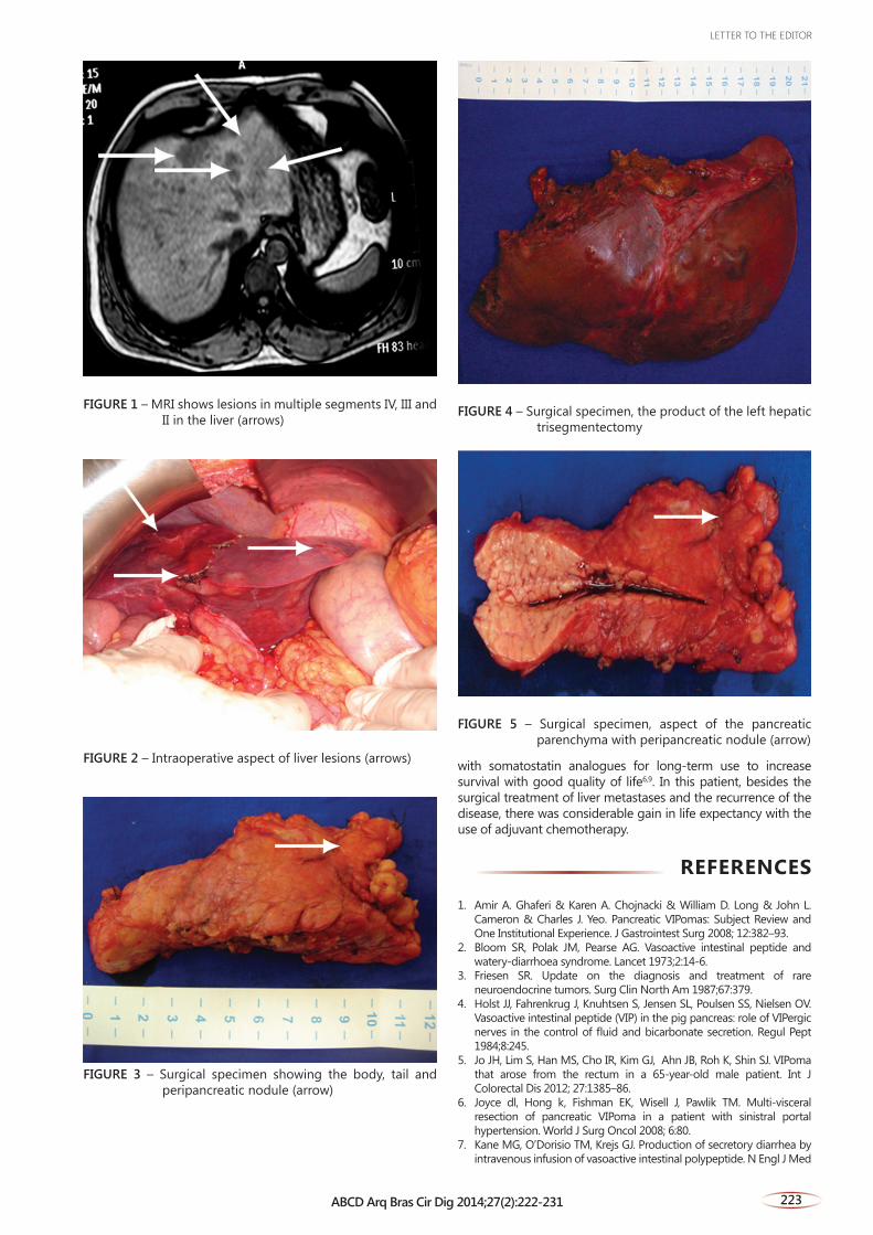
FIGURE 2 – Intraoperative aspect of liver lesions (arrows)
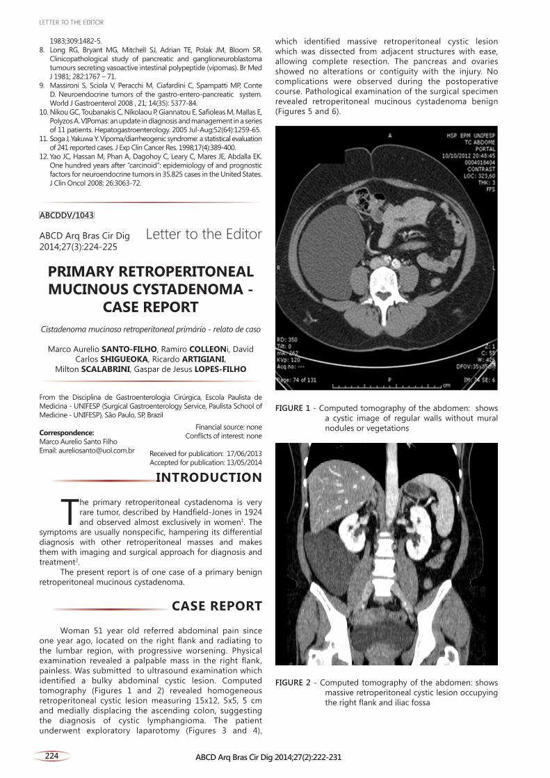
FIGURE 3 – Surgical specimen showing the body, tail and peripancreatic nodule (arrow)
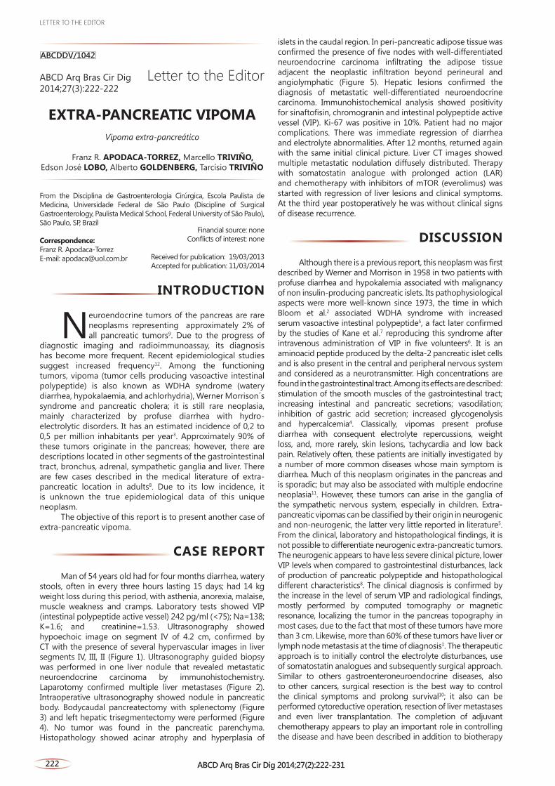
FIGURE 4 – Surgical specimen, the product of the left hepatic trisegmentectomy
FIGURE 5 – Surgical specimen, aspect of the pancreatic parenchyma with peripancreatic nodule (arrow)
LETTER TO THE EDITOR
223ABCD Arq Bras Cir Dig 2014;27(2):222-231
1983;309:1482-5.8. Long RG, Bryant MG, Mitchell SJ, Adrian TE, Polak JM, Bloom SR.
Clinicopathological study of pancreatic and ganglioneuroblastoma tumours secreting vasoactive intestinal polypeptide (vipomas). Br Med J 1981; 282:1767 – 71.
9. Massironi S, Sciola V, Peracchi M, Ciafardini C, Spampatti MP, Conte D. Neuroendocrine tumors of the gastro-entero-pancreatic system. World J Gastroenterol 2008 , 21; 14(35): 5377-84.
10. Nikou GC, Toubanakis C, Nikolaou P, Giannatou E, Safioleas M, Mallas E, Polyzos A. VIPomas: an update in diagnosis and management in a series of 11 patients. Hepatogastroenterology. 2005 Jul-Aug;52(64):1259-65.
11. Soga J, Yakuwa Y. Vipoma/diarrheogenic syndrome: a statistical evaluation of 241 reported cases. J Exp Clin Cancer Res. 1998;17(4):389-400.
12. Yao JC, Hassan M, Phan A, Dagohoy C, Leary C, Mares JE, Abdalla EK. One hundred years after “carcinoid”: epidemiology of and prognostic factors for neuroendocrine tumors in 35.825 cases in the United States. J Clin Oncol 2008; 26:3063-72.
ABCD Arq Bras Cir Dig Letter to the Editor2014;27(3):224-225
PRIMARY RETROPERITONEAL MUCINOUS CYSTADENOMA -
CASE REPORTCistadenoma mucinoso retroperitoneal primário - relato de caso
Marco Aurelio SANTO-FILHO, Ramiro COLLEONi, David Carlos SHIGUEOKA, Ricardo ARTIGIANI,
Milton SCALABRINI, Gaspar de Jesus LOPES-FILHO
From the Disciplina de Gastroenterologia Cirúrgica, Escola Paulista de Medicina - UNIFESP (Surgical Gastroenterology Service, Paulista School of Medicine - UNIFESP), São Paulo, SP, Brazil
Correspondence: Marco Aurelio Santo FilhoEmail: [email protected]
INTRODUCTION
The primary retroperitoneal cystadenoma is very rare tumor, described by Handfield-Jones in 1924 and observed almost exclusively in women1. The
symptoms are usually nonspecific, hampering its differential diagnosis with other retroperitoneal masses and makes them with imaging and surgical approach for diagnosis and treatment2.
The present report is of one case of a primary benign retroperitoneal mucinous cystadenoma.
CASE REPORT
Woman 51 year old referred abdominal pain since one year ago, located on the right flank and radiating to the lumbar region, with progressive worsening. Physical examination revealed a palpable mass in the right flank, painless. Was submitted to ultrasound examination which identified a bulky abdominal cystic lesion. Computed tomography (Figures 1 and 2) revealed homogeneous retroperitoneal cystic lesion measuring 15x12, 5x5, 5 cm and medially displacing the ascending colon, suggesting the diagnosis of cystic lymphangioma. The patient underwent exploratory laparotomy (Figures 3 and 4),
which identified massive retroperitoneal cystic lesion which was dissected from adjacent structures with ease, allowing complete resection. The pancreas and ovaries showed no alterations or contiguity with the injury. No complications were observed during the postoperative course. Pathological examination of the surgical specimen revealed retroperitoneal mucinous cystadenoma benign (Figures 5 and 6).
FIGURE 1 - Computed tomography of the abdomen: shows a cystic image of regular walls without mural nodules or vegetations
FIGURE 2 - Computed tomography of the abdomen: shows massive retroperitoneal cystic lesion occupying the right flank and iliac fossa
Financial source: noneConflicts of interest: none
Received for publication: 17/06/2013Accepted for publication: 13/05/2014
ABCDDV/1043
LETTER TO THE EDITOR
224 ABCD Arq Bras Cir Dig 2014;27(2):222-231