Embed Size (px)
Citation preview
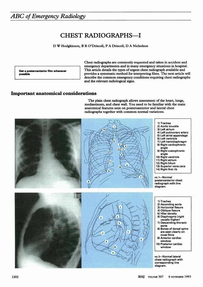
ABC ofEmergency Radiology
CHEST RADIOGRAPHS-ID W Hodgkinson, B R O'Driscoll, P A Driscoll, D A Nicholson
Get a posteroanterior film wheneverpossible
lImportant anatomical considerations
diographs are commonly requested and taken in accident andcy departments and in many emergency situations in hospital.cle details the types ofurgent chest radiograph available anda systematic method for interpreting films. The next article willthe common emergency conditions requiring chest radiographselevant radiological signs.
lain chest radiograph allows assessment of the heart, lungs,cnum, and chest wall. You need to be familiar with the maincal features seen on posteroanterior and lateral chestphs together with common normal variations.
1) Trachea2) Aortic knuckle
-> 3 3)Leftatrium<X7 ::lkJ,4)Leftpulmonary artery
5) Left atrial appendage6) Left ventricle
1 3'. Eg7)Left hemidiaphragm2\14 8) Right cardiophrenic
N ~~~~~~~~~~angle~---~i2A-~ ' ~; |9) Right costophrenic
angle10) Right ventricle1 1) Right atrium12) Right hilum
6tb:t go E /:1Fi i: 13) Superior vena cava14) Right first rib
FIG 1-Normalposteroanterior chestradiograph with linediagram.
1) Trachea2) Ascending aorta3)Horizontalfissure4) Oblique fissure5) Hilar density
2 6) Diaphragms (rightusually higher)
7) Descending thoracicaorta
8) Bones of dorsal spine3 ,>/J// 7 are seen clearly on
most films->(: 8@2 ,6Af 9) Anterior cardiac
4 window10) Posterior cardiac
IC window
FIG 2-Normal lateralchest radiograph withcorresponding linediagram.
BMJ VOLUME 307 6 NOVEMBER 19931202
1) Azygous lobe (mistaken for btolla or- pneumothorax)
4. ... ~~~~~~~~~~~ ~2) Prominent brachiocephalic vessels (mistaken forright upper mediastinal mass or
~~#AI~~~~~~. ~~lymphadenopathy)FIG3) Calcified costal cartilages (mistaken for pleural
or pulmonary lesions)ao4) Pericardial cyst or fat pad (mistaken for
cardiomegaly, tumouth , or consolidation)5) Diaphragmatic hump (mistaken for tumour or
Highdiaphragms cassette andthexposurestakeninfullnspirconsolidation)Rotation~ 6) Unusual cardiac shape or apparent
~~~~3**4~~~~~~ ~ ~ ~ cardiomegaly maybe caused by pectusexcavatum with depressed sternum. (Confirmedby clinical examination and lateral radiograph)Upperlobe 4looddiversion may benormal7) Asymmetrical breast shadows (mistaken for
Smallpneumothorax may be missed (air patients lower zone consolidation)7 ~~~~~8)Prominent nippieshadow (mistaken for
pulmonary nodule)9) Loose folds of skin (especially in anteroposterior
supine film). These may be mistaken for aIgiving pneumothorax but the loose skinfolsd can usually
befollowed outsidethe ribcage
FIG 3-Normal common variants.
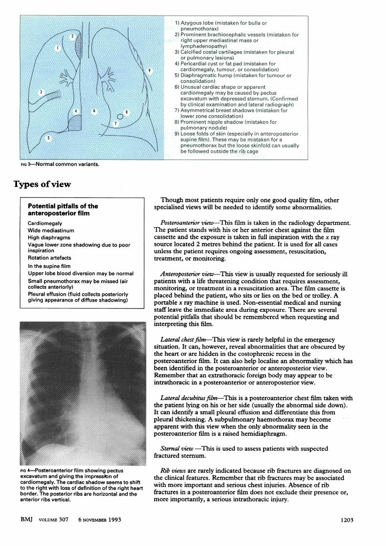
T'ypes ofview
Though most patients require only one good quality film, otherPotential pitfalls of the specialised views will be needed to identify some abnormalities.anteroposterior filmCardiomegaly Posteroanterior view-This film is taken in the radiology department.Wide mediastinum The patient stands with his or her anterior chest against the filmHigh diaphragms cassette and the exposure is taken in full inspiration withi the x rayVague lower zone shadowing due to poor source located 2 metres behind the patient. It is used for all casesinspiration unless the patient requires ongoing assessment, resuscitation,Rotation artefacts treatment, or monritoring.In the supine filmUpper lobe blood diversion may be normal Anteroposterior view-This view is usually requested for seriously illSmall pneumothorax may be missed (air patients with a life threatening condition that requires assessment,collects anteriorly) monitoring, or treatment in a resuscitation area. The film cassette isPleural effusion (fluid collects posteriorly placed behind the patient, who sits or lies on the bed or trolley. Agiving appearance of diffuse shadowing) portable x ray machine is used. Non-essential medical and nursing
staff leave the immediate area during exposure. There are severalpotential piffalls that should be remembered when requesting andinterpreting this film.
Lateral chestfilm-This view is rarely helpful in the emergencysituation. It can, however, reveal abnormalities that are obscured bythe heart or are hidden in the costophrenic recess in theposteroanterior film. It can also help localise an abnormality which hasbeen identified in the posteroanterior or anteroposterior view.Remember that an extrathoracic foreign body may appear to bemntrathoracic in a posteroanterior or anteroposterior view.
Lateral decubitus film-This is a posteroanterior chest film taken with..............the patient lying on his or her side (usually the abnormal side down).
.....Ipleural th iryItapparentwithtif iwwe hnyabsalperlefso noifrmaiysentit tin them
posteroanterior film is a raised hemidiaphragm.
Sternal view -This is used to assess patients with suspectedtractured sternum.~~~~~~~~~~~~~~~~~~~~~~~......... ... ...:
FIG 4-Posteroanterior film showing pectus Rib views are rarely indicated because rib fractures are diagnosed onexcavatum and giving the impressimnof the clinical features. Remember that rib fractures may be associatedcardiomegaly. The cardiac shadow seems to shift wt oeipratadsroscetijre.Asneo ito the right with loss of definition of the right heart with moreimportant and serious chest injuries. Absence of ribborder. The posterior ribs are horizontal and the fractures in a posteroanterior film does not exclude their presence or,anterior ribs vertical. more importantly, a serious intrathoracic injury.
BMJ VOLUME 307 6 NOVEMBER 1993 1 203
The lateral chest radiograph is rarelyhelpful in acute conditions. Howeverit can localise abnormalities seen inthe posteroanterior view
Expiration films may be used to show a small pneumothorax, but it isnot necessary to request this view routinely because mostpneumothoraces will be apparent in the posteroanterior inspirationfilm. Expiration films are occasionally requested to help establish adiagnosis of inhaled foreign body, when "gas trapping" may be seen.Apical lordotic view-This is an oblique view that can show details of
the lung apex which are usually hidden behind the clavicle and upperribs. This technique is seldom indicated in the emergency situation.
System ofradiological assessment
First check the name and age of the patient together with the date onthe radiograph.
.....Check the adequacy and technical quality of the filmNote the following:Projection and exposure-Look at the mid-thoracic intervertebral discs;
they should be clearly visible.Posture-Supine or erect.
.........Rotation-Look at the spinous processes of the upper thoracicEj_ | E : _ ~~~~~~vertebrae. They should be central. Then inspect the medial end of both
_ _ ~~~~~~clavicles to ensure they are equidistant from the central spinous_ ~~~~~~~~~process.
. ~~~~~~~~~~~...;:.::.::.-.
: - ~~~Degree of inspiration-Ths affects the appancoftelwrze_00 < ~~~~~vessels. They appear more prominent with poor inspiration. The right
-- g ~hemidiaphrg shudrahteatroan ftergtsxho,.,::: ::. : ''!!''W' !'"'seventh rib or the ninth rib posteriorly on full inspiration.
g v: ~~~~~~~.
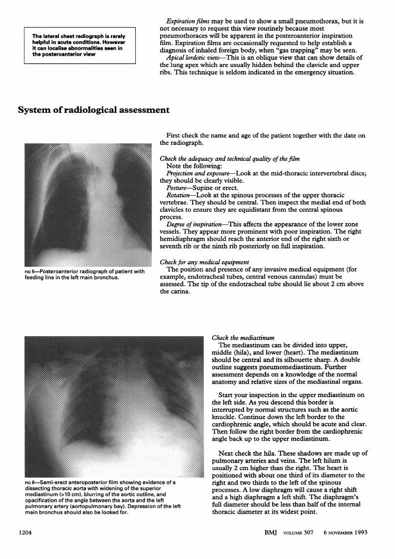
IG-Posteroanterior radiograph of patient with The position and presence of any invasive medical equipment (forfeeding line in the left main bronchus. example, endotracheal tubes, central venous cannulas) must be
assessed. The tip of the endotracheal tube should lie about 2 cm abovethe carina.
FIG 6-Semi-erect anteroposterior film showing evidence of adissecting thoracic aorta with widening of the superiormediastinum (>10 cm), blurring of the aortic outline, andopacification of the angle between the aorta and the leftpulmonary artery (aortopulmonary bay). Depression of the leftmain bronchus should also be looked for.
Check the mediastinumThe mediastinum can be divided into upper,
middle (hila), and lower (heart). The mediastinumshould be central and its silhouette sharp. A doubleoutline suggests pneumomediastinum. Furtherassessment depends on a knowledge of the normalanatomy and relative sizes of the mediastinal organs.
Start your inspection in the upper mediastinum onthe left side. As you descend this border isinterrupted by normal structures such as the aorticknuckle. Continue down the left border to thecardiophrenic angle, which should be acute and clear.Then follow the right border from the cardiophrenicangle back up to the upper mediastinum.
Next check the hila. These shadows are made up ofpulmonary arteries and veins. The left hilum isusually 2 cm higher than the right. The heart ispositioned with about one third of its diameter to theright and two thirds to the left of the spinousprocesses. A low diaphragm will cause a right shiftand a high diaphragm a left shift. The diaphragm'sfull diameter should be less than half of the internalthoracic diameter at its widest point.
BMJ VOLUME 307 6 NOVEMBER 19931204
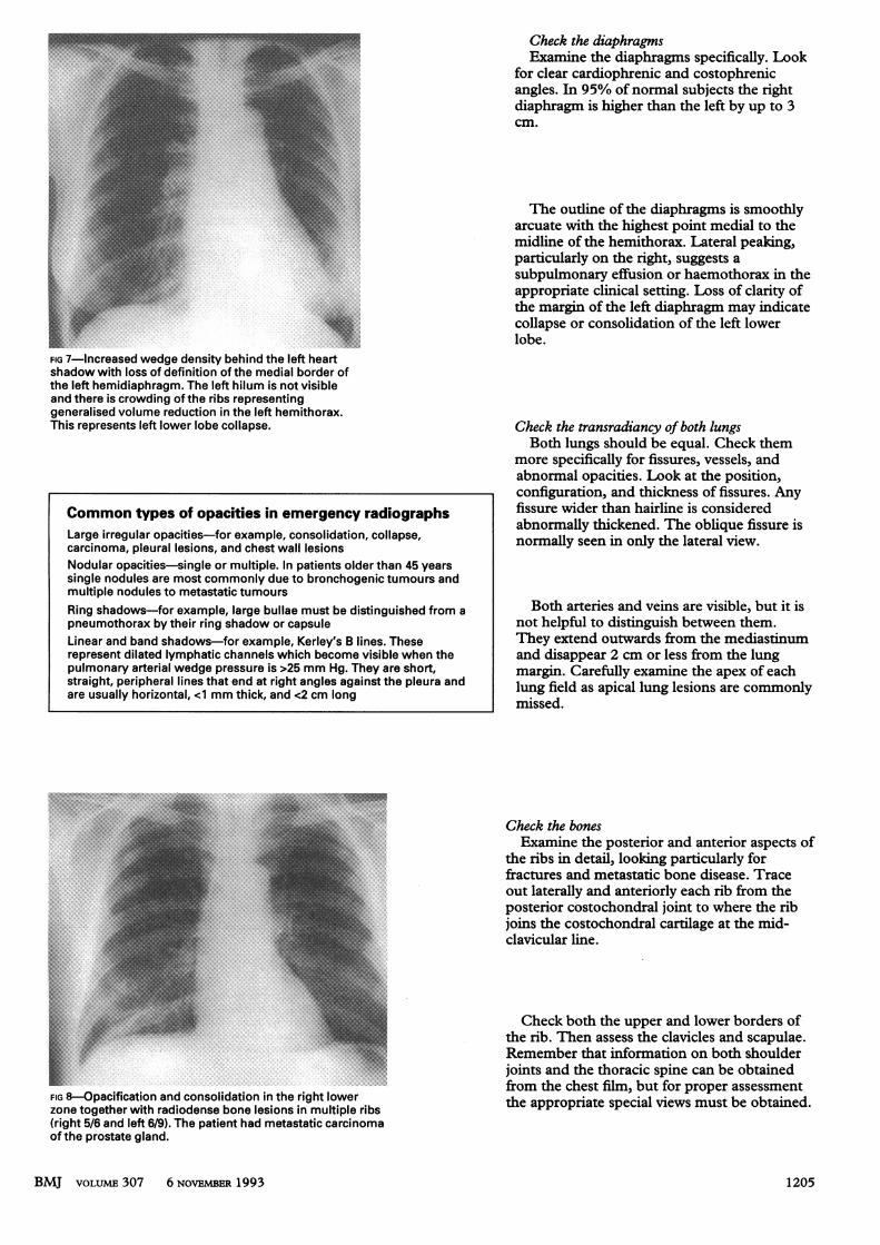
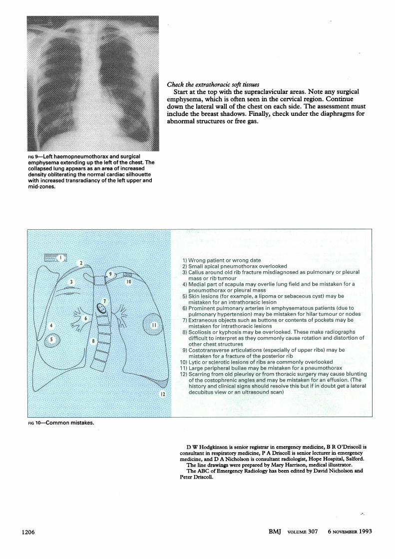
FIG 7-Increased wedge density behind the left heartshadow with loss of definition of the medial border ofthe left hemidiaphragm. The left hilum is not visibleand there is crowding of the ribs representinggeneralised volume reduction in the left hemithorax.This represents left lower lobe collapse.
FIG 8-Opacification and consolidation in the right lowerzone together with radiodense bone lesions in multiple ribs(right 5/6 and left 6/9). The patient had metastatic carcinomaof the prostate gland.
Check the diaphragmsExamine the diaphragms specifically. Look
for clear cardiophrenic and costophrenicangles. In 95% of normal subjects the rightdiaphragm is higher than the left by up to 3cm.
The outline of the diaphragms is smoothlyarcuate with the highest point medial to themidline of the hemithorax. Lateral peaking,particularly on the right, suggests asubpulmonary effusion or haemothorax in theappropriate clinical setting. Loss of clarity ofthe margin of the left diaphragm may indicatecollapse or consolidation of the left lowerlobe.
Check the transradiancy of both lungsBoth lungs should be equal. Check them
more specifically for fissures, vessels, andabnormal opacities. Look at the position,configuration, and thickness of fissures. Anyfissure wider than hairline is consideredabnormally thickened. The oblique fissure isnormally seen in only the lateral view.
Both arteries and veins are visible, but it isnot helpful to distinguish between them.They extend outwards from the mediastinumand disappear 2 cm or less from the lungmargin. Carefully examine the apex of eachlung field as apical lung lesions are commonlymissed.
Check the bonesExamine the posterior and anterior aspects of
the ribs in detail, looking particularly forfractures and metastatic bone disease. Traceout laterally and anteriorly each rib from theposterior costochondral joint to where the ribjoins the costochondral cartilage at the mid-clavicular line.
Check both the upper and lower borders ofthe rib. Then assess the clavicles and scapulae.Remember that information on both shoulderjoints and the thoracic spine can be obtainedfrom the chest film, but for proper assessmentthe appropriate special views must be obtained.
BMJ VOLUME 307 6 NOVEMBER 1993
Common types of opacities in emergency radiographsLarge irregular opacities-for example, consolidation, collapse,carcinoma, pleural lesions, and chest wall lesionsNodular opacities-single or multiple. In patients older than 45 yearssingle nodules are most commonly due to bronchogenic tumours andmultiple nodules to metastatic tumoursRing shadows-for example, large bullae must be distinguished from apneumothorax by their ring shadow or capsuleLinear and band shadows-for example, Kerley's B lines. Theserepresent dilated lymphatic channels which become visible when thepulmonary arterial wedge pressure is >25 mm Hg. They are short,straight, peripheral lines that end at right angles against the pleura andare usually horizontal, <1 mm thick, and <2 cm long
1205
Check the extrathoracic soft tissuesStart at the top with the supraclavicular areas. Note any surgical
emphysema, which is often seen m the cervical region. Continuedown the lateral wall of the chest on each side. The assessment must
~~~~~~~~~~~icuE th bras shaos Fialy chekundertedahgm foabnormal structures or free gas.
FIG 9-Left haemopneumothorax and surgicalemphysema extending up the left of the chest. Thecollapsed lung appears as an area of increaseddensity obliterating the normal cardiac silhouettewith increased transradiancy of the left upper andmid-zones.
1E1- _-1--- N2 1) Wrong patientorwrong date2) Small apical pneumothorax overlooked
3) CallWus around oledrib fracturearisdinmgnosed as puicmonary or pleusralmass or rib tumour
m4) Meiaie part of scapula,may overllerung fieoldoand be Msistaken for apneu othorax orpleural mass
5) Skin lesions (for exam."ple, a lipoma or,sebaceous cyst) may bemistakein for an intrathoracic -lesion
6) Promlnnt pulmonary arterioes 1n ebmpnhysematous patients(diue topulmonary hypertension) may be Mistaken for hilar tumour or nodes
7).Extraneous objects such as buttons or contents of pockets may beII ~~mistaken for intrathoracic lesions
. ~~~~~~~~8)Scoliosis or kyphosis may be overlooked. These make radio'graphsdifficult to interpret as they commonly cause rotation and distortion ofoyther-chest structures
9) Costotransverse articulations (espcially of upper ribs) may bemistaken' for a fr-acurelof the.-posterior. rib
10) Lytic or sclerotic. lesio"ns,of r"ibs are.comnmonlly overlookedLa~~~rge periph-erl bulle "my:be Mistaken .f'or a pneumotoa
12) Scarring frompld alurs ct.r.... thoracic surgery,may -cause bluntingof the costophrenic bnls m ay emistaken -oan effusion. (The
FIG 10-Common mistakes.
D W Hodgkidnson is senior registrar mn emergency medicine, B R O'Driscoll isconsultant in respiratory medicine, P A Driscoll is senior lecturer in emergencymedicine, and D A Nicholson is consultant radiologist, Hope Hospital, Salford.
The line drawings were prepared by Mary Harrison, medical ilustrator.T'he ABC of Emergency Radiology has been edited by David Nicholson and
Peter Driscoll.
BMJ VOLUME 307 6 NOVEMBER 19931206