Embed Size (px)
Citation preview
-39-
Ass. Univ. Bull. Environ. Res. Vol. 12 No. 2, October 2009
AUCES
TERATOGENIC AND GENOTOXIC EFFECTS OF PERFLUOROALKYL
ACIDS ON EMBRYONIC AND NEONATE MICE
Abd El-Nasser, M.A, Manal A. Abdel-mohsen, Shaaban A.A. and Doha Y. Ahmed
Department of Forensic Medicine and Toxicology, Faculty of Veterinary Medicine, Assiut University
ABSTRACT:
Perfluorinated compounds (PFCs) have emerged as a new class of global environmental pollutants. Perfluoroctane sulfonate (PFOS) and perfluorooctanoic acid (PFOA) comprises a class of environmentally persistent chemicals that have a wide range of industrial applications. 160 pregnant dams were divided into two equal groups, PFOS group and PFOA group. Each group was subdivided to four equal groups (n=20), one of them was kept as control group. The first, second and third subgroups of the first main group were treated with 1, 10 and 20 mg PFOS/kg.b.w daily, respectively. While the other three subdivided groups of the second main group were treated with 1, 5 and 10 mg PFOA/kg.b.w daily. Ten dams of each group were treated from gestation day 0 (GD0) till gestation day 17 (GD17). At GD18 dams were euthanized under anesthesia. The gravid uterus were removed and examined for prenatal evaluation of fetuses. The liver of the fetuses were dissected and used immediately for comet assay. Individual live fetuses were prepared for teratological evaluation. While the other ten dams were treated from GD0 till GD18 and then allowed to give birth. The neonates of 5 dams were monitored for 4 days for postnatal survival. Neonates of the remaining 5 dams were kept in the fixative till histopathological examination. Control group were received an equivalent volume of deionized water. Prenatal finding revealed that PFOS treatment reduce the number of live fetuses accompanied with increased fetal resorption. PFOS reduced fetal body weight in a dose dependent manner, while PFOA reduced the fetal body weight at dose of 5 and 10 mg/kg b.w. Gross examination of the fetuses at GD18 showed presence of an abnormal swelling in the back of the neck in all fetuses of dams treated with 20 mg/kg b.w. Teratological evaluation revealed presence of several skeletal abnormalities in PFOS treated groups which were few in PFOA groups. Neonates were borne with reduction in body weight and showed the presence of the bilateral swelling which accompanied by neonatal death, while in PFOA treated group there was only reduction in body weight and survival rate. Results revealed that PFOS caused DNA damage in fetal liver at 10 and 20 mg/kg b.w. Histopathological examination of both, bilateral swelling and lung revealed dilatation of the blood vessels between cranial bone area and brain, and slight to sever atalectasis, respectively. The study concluded that both PFOS and PFOA were toxic to neonates with different degrees although PFOS was recorded the most toxic and the embryo might be died from the lesion formed over the brain.
INTRODUCTION:
Ass. Univ. Bull. Environ. Res. Vol. 12 No. 2, October 2009
-40-
Perfluorinated compounds (PFCs) have
been used since the 1950s in industrial and
commercial applications ranging from water,
soil and stain resistant coating for clothing
fabrics, leather, upholstery and carpets. They
have also been used as oil-resistant coatings for
paper products approved for food contact,
electroplating, electronic etching bath,
surfactants, photographic emulsifiers, aviation
hydraulic fluids, fire-fighting foams, paints,
adhesives, waxes and polishes. Their extensive
use can be attributed to the strength of the
carbon-fluorine bonds, stability at high
temperatures, being nonflammable and not
subject to photolysis or metabolized. PFCs are
composed of a carbon-fluorine chain and
generally have side moieties attached such as
carboxylic acids or sulfonic acids. These
compounds are respectively called
perfluorinated carboxylates or perfluoroalkyl
carboxylates and perfluorinated sulfonates or
perfluoroalkyl sulfonates and they make up two
major classes of PFCs (Giesy and Kannan,
2002). The carbon-fluorine bond in PFCs is very
strong and gives thermal and chemical stability
to many PFCs (So et al., 2004). PFCs are now
ubiquitous global contaminants. They have been
detected in indoor and outdoor air, in rivers,
lakes and groundwater, in wastewater
treatment effluent, in landfills and in the marine
environment. PFCs have also been found in the
body tissues of many different living organisms
throughout the world including humans
(Allsopp et al., 2005). Due to their widespread
use, persistence and bioaccumulative properties
they are taken up by the general population
from different sources (Midasch et al., 2007).
The reproductive toxicity of Perfluoroctane
sulfonate (PFOS) has been examined in several
species as rabbits (Case et al., 2001), rats and
mice (Lau et al., 2003; and Thibodeaux et al.,
2003). Teratological studies have been
conducted in rat, rabbit and mouse with
potassium and lithium salts of PFOS (Lau et al.,
2003 and 2004). Observed developmental effects
include reduction of fetal weight, cleft palate,
edema, delayed ossification of sternum and
phalanges, and cardiac abnormalities in the
highest PFOS dose group. Significant reductions
of weight gain and food consumption were also
observed in the pregnant dams. PFOS also
produced dose-dependent effects on neonatal
survival and retarded the growth and
development of neonates in rats exposed to
doses ranging from 1–10 mg/kg/day during
gestational days (GD) 2–21 and mice exposed to
1–20 mg PFOS/kg/day on GD 1 – 18 (Lau et al.,
2003). These effects were also reported in a two-
generation study in rats exposed to doses
ranging from 0.1 to 3.2 mg PFOS/kg/day
(Luebker et al., 2005).
In the studies by Lau et al. (2003), rat and
mouse neonates exposed to PFOS in utero died
within hours after birth, and cross-fostering of
the exposed neonatal rats did not improve
survival. Further studied in the rat exposed to
25 or 50 mg PFOS/kg/day on GD19 and GD 20,
or to 25 mg/kg/day across various 4-day
gestational intervals, demonstrated that the
neonatal lethality could be produced by
treatment restricted to the late gestational
period and suggested that impaired lung
function could be involved (Grasty et al., 2005).
Developmental toxicity from PFOA in rodents,
including pregnancy loss, reduced fetal weight,
reduced postnatal survival, and delays in
postnatal growth and development in offspring
were reported by Lau et al., (2006). In the rat,
PFOA and PFOS have been detected in
placenta, fetus, amniotic fluid, and milk, and
these chemicals have also been found in human
breast milk (So, et al., 2006).
Liu et al. (2007) found that PFOS and
PFOA are able to produce oxidative stress
Ass. Univ. Bull. Environ. Res. Vol. 12 No. 2, October 2009
-41-
(generation of ROS) and induce apoptosis and
typical DNA fragmentation (DNA laddering) in
primary cultured tilapia hepatocytes. Exposure
to PFOA increases the incidence of liver tumors
in rodents (Biegel et al., 2001). Although the
mechanisms underlying this carcinogenesis have
not been clarified, (Nilsson et al., 1991).
Peroxisome proliferation may result in elevated
levels of hydrogen peroxide, which may in turn
initiate tumorigensis via oxidative DNA damage
(Reddy and Rao, 1989). At the same time
peroxisome proliferators may act as tumor
promoters by stimulating DNA replication
(Kraupp et al., 1991). In addition they may
inhibit the normal process of apoptosis in the
liver (Schulte et al., 1991). PFOA has been
shown to be a strong tumor promoter, showing
a 56% tumor incidence in 12 months of dietary
exposure at 0.02% (w/w). Another long term
feeding study in men showed that PFOA
exposure at 300 ppm in the diet over 2 years
increased cancers of the liver (liver adenomas)
and pancreas (pancreatic acinar cell adenoma)
(Olsen et al., 1998).
Comet assay, known as the single-cell gel
electrophoresis test used to detect DNA strand
breakage (double, single, and alkali-labile sites
expressed as single strand breaks) in virtually
any nucleated cell. Significant advantages of the
comet assay over other genotoxicity tests are its
fairly straight forward technique, sensitivity,
requirement for small numbers of cells and
rapid production of data (Tice et al., 2000).
Genotoxicity of PFOA was assessed by
estimating tail moment of comet in single cell gel
electrophoresis (SCGE) assay (Yao and Zhong,
2005). Damaged cells have an appearance
similar to astronomical comets, with long tails of
DNA migrating from the center of the exposed
nuclei. Damage is generally quantified using
comet tail length and tail moment (i.e.: tail
length multiplied by the percentage of DNA in
the tail) (Tice et al., 2000). Tail length can be
used to indicate initial DNA damage and
confirm exposure to a genotoxin, while tail
moment and percent DNA in the tail can be
used to indicate the intensity of damage
(Knopper et al., 2005).
The aim of this work is to study the genetic
and teratogenic changes in fetuses after
maternal exposure to PFOS and PFOA.
Histopathological examination of neonatal brain
and lung and explain the cause of death was also
one of research goals.
MATERIALS AND METHODS:
Animals:
ICR male and female mice aged 7 weeks
were purchased from CLEA Japan, Inc., Tokyo
were used for the experiment after one week of
acclimatization. Female mice were checked for
estrous cycle stage and each proestrus female
was placed with an individually housed breeder
male overnight, and those females with
spermatozoa in its vaginal smear and/or with a
copulatory plug were considered to be at
gestational day 0 (GD0).
Reagents:
PFOS: Perfluorooctane sulfonate (potassium
salt 98% pure) was purchased from Fluka
Chemie GmbH, Switzerland. PFOS solutions
were prepared with a concentration of 0.1, 1
and 2 mg/ml of 0.5% Tween-20 vehicle and
administered to the pregnant mice by gavage at
a volume of 10 ml/kg/day
PFOA: Perfluoroocatnoic acid (90% pure) was
purchased from Fluka Chemie GmbH,
Switzerland. PFOA solutions were prepared
with a concentration of 0.1, 0.5 and 1 mg/ml of
deionized water and administered to the
Ass. Univ. Bull. Environ. Res. Vol. 12 No. 2, October 2009
-42-
pregnant mice by gavage once daily from GD0
till GD17 at a volume of 10 ml/kg/day.
EXPERIMENTAL DESIGN:
This study protocol was approved by the
Animal Research Committee. A total number of
160 pregnant dams were divided into two main
equal groups. Each group was subdivided into
two groups, treated group (60 dams) and
control group (20 dams). Each group was
subdivided to four equal groups (n=20), one of
them was kept as control group. The first,
second and third subgroups of the first main
group were treated with 1, 10 and 20 mg
PFOS/kg.b.w daily, respectively. While the
other three subdivided groups of the second
main group were treated with 1, 5 and 10 mg
PFOA/kg.b.w daily. Ten dams of each subgroup
were treated from GD0 till GD17. The gravid
uterus was removed and the numbers of the live
or dead fetuses and resorptions were recorded.
Live fetuses were weighed individually and
examined for external abnormalities. Fetuses
were prepared for skeletal evaluation as
described by Narotsky and Rogers (2000). The
skeleton of all the stained fetuses was examined
using Nikon light microscope (Model C-DSD
115, Japan) and the differences between the
control and treated groups were tabulated. At
parturition, newborns were weighted and
observed for clinical signs and survival. The
number of live pups in each litter was tabulated
daily for the first 4 days after birth. Neonates
head and lungs were processed routinely for
paraffin embedding technique. The processed
tissues were cut using tissues microtome. Tissue
sections were stained with the standard
Haematoxylin and Eosin method (H. & E.)
according to Bancroft and Stevens (1982). while
the other ten dams were treated from GD0 till
GD18 and then allowed to give birth. Control
group were received an equivalent volume of
deionized water. At GD18 dams were
euthanized under diethyl ether anesthesia. The
gravid uterus of the same dams was removed
and examined for prenatal evaluation of fetuses.
The liver of the fetuses were dissected and used
immediately for comet assay. Individual live
fetuses were prepared for teratological
evaluation as described by Narotsky and Rogers
(2000). The neonates of 5 dams were monitored
for 4 days for postnatal survival. All the
neonates of the remaining 5 dams were kept in
Bouin’s fixative (300 ml saturated picric acid,
100 ml formaldehyde and 20 ml glacial acetic
acid) for three days then kept in 70% ethanol
till histopathological examination according to
Bancroft and Stevens (1982).
DNA damage in fetal liver was detected
using comet assay (Single Cell Gel
Electrophoresis) according to the method of
Sasaki et al. (1997) and Tsuda et al. (1998). DNA
damage using comet assay is generally
quantified using comet tail length and tail
moment, the later calculated as tail length
multiplied by the percentage of DNA in the tail
(Collins et al. 1997 and Tice et al., 2000). Tail
length can be used to indicate initial DNA
damage and confirm exposure to a genotoxin,
while tail moment and the percentage of DNA in
the tail can be used to indicate the intensity of
damage (Knopper et al., 2005).
Data are presented as means and standard
errors. Statistical significance was determined
by the analysis of variance (ANOVA). Each
treated group was tested for difference from the
control group using Dunnett’s t-test.
Statistically significant differences were
determined at p≤ 0.05. Statistical Package for
the Social Sciences for Windows (SPSS, version
10.0, Chicago, IL, USA) according to Borenstein
et al. (1997).
Ass. Univ. Bull. Environ. Res. Vol. 12 No. 2, October 2009
-43-
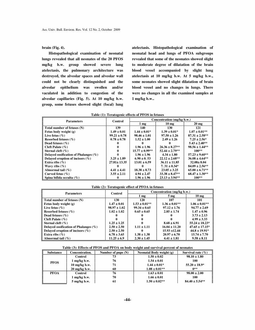
RESULTS:
Reduction in number of live fetuses
accompanied with increased fetal resorption
was recorded only at 20 mg/kg b.w. PFOS
group. Reduced fetal body weight in a dose
dependent manner was found in all used doses
as shown in table (1) while reduced fetal body
weight was shown at 5 and 10 mg/kg b.w. PFOA
groups. There were no significant effects on the
prenatal survival or resorbed fetuses as
presented in table (2).
Teratological examination of the fetuses in
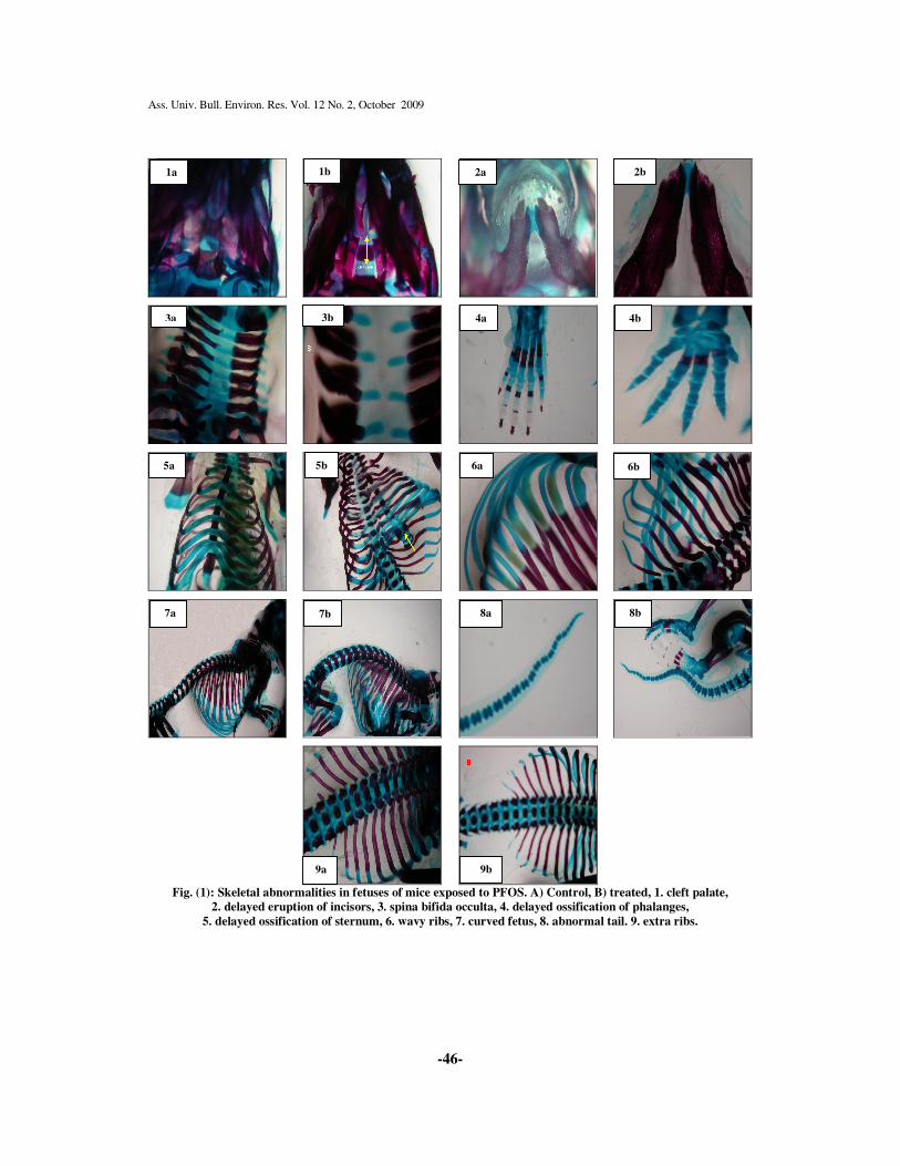
PFOS subgroups revealed, presence of several
skeletal abnormalities such as cleft palate,
delayed eruption of incisors, spina bifida occulta
(delayed closure of the vertebral spin), delayed
ossification of phalanges and sternum, wavy
ribs, curved fetus (curved vertebral column)
and abnormal tail as shown in table (1) and Fig.
(1) mostly at 10 and 20 mg/kg b.w. groups, while
there were few skeletal abnormalities in fetuses
of PFOA group as delayed ossification of the
sternum and phalanges accompanied by delayed
eruption of incisors in the 10 mg/kg b.w. group
(Table 2).
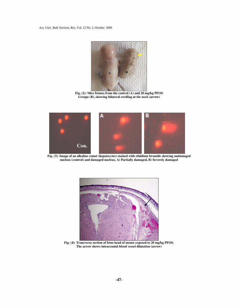
Gross examination of all fetuses of the 20
mg/kg b.w. PFOS subgroup at GD18 showed
presence of abnormal swelling in the back of the
neck region. After peeling of the skin, a bilateral
firm swelling was observed. In addition, some
fetuses of the 10 mg/kg b.w. group showed mild
swelling in the neck region as presented in
Fig. (2).
Examination of neonates born to dams
treated with PFOS revealed that, the neonatal
body weight was significantly reduced at 10 and
20 mg/kg b.w. groups accompanied by neonatal
death (Table 3). At 20 mg/kg b.w. group, the
pups were born weak, inactive and small sized
then all neonates died immediately after birth
(100% neonatal death). At 10 mg/kg, all pups
were born alive, some pups were pale and
inactive and 45% died within 24 hours after
birth, while there was no effect on the survival
rate at 1 mg/kg group (Table 3), PFOA cause
significant reduction in the neonatal body
weight and survival rate at 5 and 10 mg/kg
groups. At 5 mg/kg b.w. group all pups were
born alive and active, and then 16% died during
the first four days after birth. At 10 mg/kg some
pups were still born and others born alive and
active but all died within 6 hours after birth
(100% neonatal death) (Table 3).
Gross examination of the neonates in PFOS
treated subgroups revealed that, the bilateral
firm swelling in the back of the neck was also
observed in all neonates of 20 mg/kg b.w. dose
and some neonates of the 10 mg/kg b.w..
Macroscopically, the area below the bilateral
swelling appeared as a black area. In PFOA
treated group, there are no abnormal structures
in all the treated groups. Some neonates showed
whole body edema at 10 mg/kg b.w.
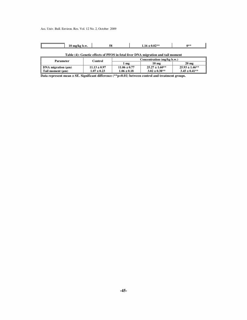
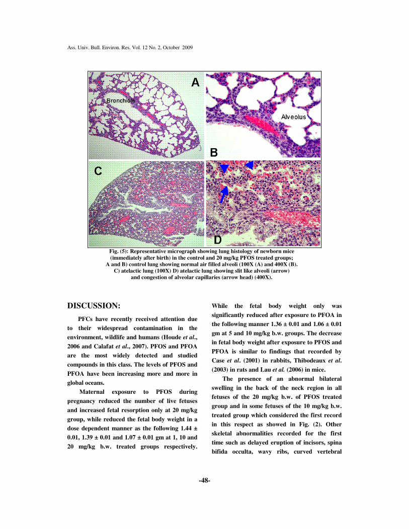
DNA damage was detected in fetal liver of
dams treated with PFOS at 10 and 20 mg/kg
b.w., which appeared in the form of increased
DNA migration represented by tail length and
tail moment as shown in table (4) and Fig. (3).
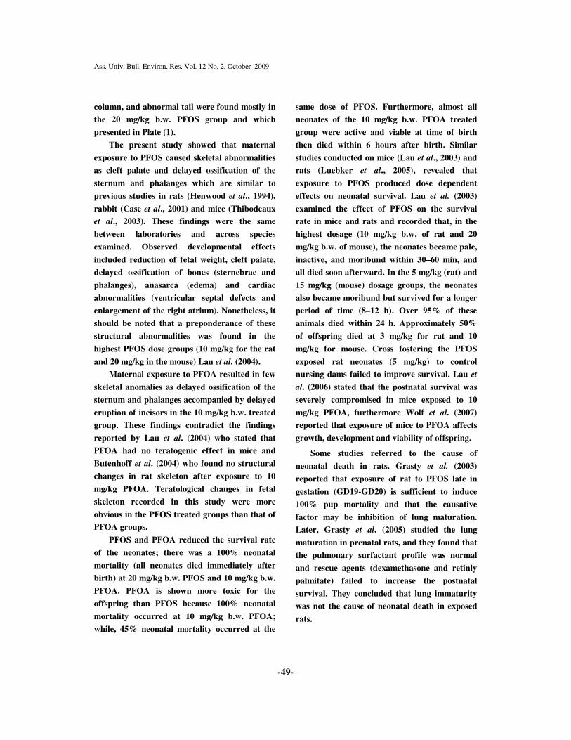
Histopathologically, examination of the
neonates in PFOS group showed a dilatation of
vessels with blood was found between the
cranial bone area and brain relevant to the
black area. At a higher magnification, the
dilatation was located in the Dura matter and
discontinuously trimmed by the endothelial-like
spindle shaped cells. Erythrocytes was found in
the lumen. There were no inflammatory or
hemorrhagic reactions surrounding the dilated
area, indicating that the dilatation area was a
blood vessel, but not hemorrhage or tumor like
lesion. This dilatation of blood vessel was
located either in the central part of the parietal
region, near the center or in one side of the
Ass. Univ. Bull. Environ. Res. Vol. 12 No. 2, October 2009
-44-
brain (Fig. 4).
Histopathological examination of neonatal
lungs revealed that all neonates of the 20 PFOS
mg/kg b.w. group showed severe lung
atelectasis, the pulmonary architecture was
destroyed, the alveolar spaces and alveolar wall
could not be clearly distinguished and the
alveolar epithelium was swollen and/or
vaculated in addition to congestion of the
alveolar capillaries (Fig. 5). At 10 mg/kg b.w.
group, some fetuses showed slight (focal) lung
atelectasis. Histopathological examination of
neonatal head and lungs of PFOA subgroups
revealed that some of the neonates showed slight
to moderate degree of dilatation of the brain
blood vessel accompanied by slight lung
atelectasis at 10 mg/kg b.w. At 5 mg/kg b.w.,
some neonates showed slight dilatation of brain
blood vessel and no changes in lungs. There
were no changes in all the examined samples at
1 mg/kg b.w..
Table (1): Teratogenic effects of PFOS in fetuses
Concentration (mg/kg b.w.) Parameters Control
1 mg 10 mg 20 mg
Total number of fetuses (N) 139 140 130 121
Fetus body weight (g) 1.49 ± 0.01 1.44 ± 0.01* 1.39 ± 0.01* 1.07 ± 0.01**
Live fetus (%) 99.21 ± 0.78 98.46 ± 1.01 97.50 ± 1.26 87.31 ± 2.58** Resorbed fetuses (%) 0.78 ± 0.78 1.52 ± 1.00 2.49 ± 1.26 7.25 ± 2.56*
Dead fetuses (%) 0 0 0 5.43 ± 2.40** Cleft Palate (%) 0 1.96 ± 1.96 26.36 ± 8.27** 98.56 ± 1.44**
Sternal cleft (%) 0 15.77 ± 0.99** 52.44 ± 2.79** 100** Delayed ossification of Phalanges (%) 0 1.96 ± 1.96 4.34 ± 1.80 57.23 ± 9.60**
Delayed eruption of incisors (%) 3.25 ± 1.89 6.90 ± 0. 53 22.12 ± 2.68** 36.08 ± 4.64** Extra ribs (%) 27.81± 13.35 13.01 ± 6.59 36.11 ± 11.85 32.08± 8.04
Wavy ribs (%) 0 0 7. 31 ± 0.34* 84.09 ± 2.56**
Abnormal tail (%) 4.41 ± 4.41 18.38 ± 8.73 23.05 ± 3.25 65.00 ± 6.71**
Curved fetus (%) 3.55 ± 2.11 4.94 ± 2.47 33.38 ± 8.47** 68.47 ± 1.30** Spina bifida occulta (%) 0 1.96 ± 1.96 23.13 ± 3.94** 100**
Table (2): Teratogenic effect of PFOA in fetuses
Concentration (mg/kg b.w.) Parameters Control
1 mg 5 mg 10 mg
Total number of fetuses (N) 130 120 107 101
Fetus body weight (g) 1.47 ± 0.01 1.53 ± 0.01** 1.36 ± 0.01** 1.06 ± 0.01**
Live fetus (%) 98.97 ± 1.02 99.34 ± 0.65 97.12 ± 1.76 94.77 ± 2.69
Resorbed fetuses (%) 1.02 ± 1.02 0.65 ± 0.65 2.85 ± 1.74 1.47 ± 0.96 Dead fetuses (%) 0 0 0 3.73 ± 2.13
Cleft Palate (%) 0 0 0 4.99 ± 3.33 Sternal cleft (%) 1.25 ± 1.25 0 8.68 ± 6.91 55.24 ± 18.23*
Delayed ossification of Phalanges (%) 2.50 ± 2.50 1.11 ± 1.11 16.04 ± 11.20 47.65 ± 17.15* Delayed eruption of incisors (%) 2.50 ± 2.50 0 15.93 ±12.44 44.8 ± 19.91* Extra ribs (%) 6.78 ± 3.65 1.38 ± 1.38 20.97 ± 6.70 15.74 ± 7.70
Abnormal tail (%) 11.25 ± 6.9 2.30 ± 1.45 4.41 ± 1.81 9.58 ± 8.11
Table (3): Effects of PFOS and PFOA on body weight and survival percent of neonates
Substance Concentration. Number of pups (N) Neonatal Body weight (g) Survival rate (%)
Control 73 1.50 ± 0.02 98.10 ± 1.80
1 mg/kg b.w. 76 1.54 ± 0.01 100
10 mg/kg b.w. 71 1.44 ± 0.01* 55.20 ± 18.9* PFOS
20 mg/kg b.w. 60 1.08 ± 0.01** 0**
Control 76 1.63 ± 0.01 98.00 ± 2.00
1 mg/kg b.w. 70 1.66 ± 0.01 100
PFOA
5 mg/kg b.w. 61 1.50 ± 0.02** 84.40 ± 5.54**
Ass. Univ. Bull. Environ. Res. Vol. 12 No. 2, October 2009
-45-
10 mg/kg b.w. 58 1.16 ± 0.02** 0**
Table (4): Genetic effects of PFOS in fetal liver DNA migration and tail moment
Concentration (mg/kg b.w.) Parameter Control
1 mg 10 mg 20 mg
DNA migration (µm) 11.13 ± 0.97 11.06 ± 0.77 25.27 ± 1.60** 25.93 ± 1.46** Tail moment (µm) 1.07 ± 0.23 1.06 ± 0.18 3.02 ± 0.38** 3.45 ± 0.41**
Data represent mean ± SE. Significant difference (**p<0.01) between control and treatment groups.
Ass. Univ. Bull. Environ. Res. Vol. 12 No. 2, October 2009
-46-
Fig. (1): Skeletal abnormalities in fetuses of mice exposed to PFOS. A) Control, B) treated, 1. cleft palate,
2. delayed eruption of incisors, 3. spina bifida occulta, 4. delayed ossification of phalanges,
5. delayed ossification of sternum, 6. wavy ribs, 7. curved fetus, 8. abnormal tail. 9. extra ribs.
1a 1b 2a 2b
3a 3b 4a 4b
5a 5b 6a 6b
7a 7b 8a 8b
9a 9b
Ass. Univ. Bull. Environ. Res. Vol. 12 No. 2, October 2009
-47-
Fig. (2): Mice fetuses from the control (A) and 20 mg/kg PFOS
Groups (B), showing bilateral swelling at the neck (arrow)
Fig. (3): Image of an alkaline comet (hepatocytes) stained with ethidium bromide showing undamaged
nucleus (control) and damaged nucleus, A) Partially damaged, B) Severely damaged
Fig. (4): Transverse section of fetus head of mouse exposed to 20 mg/kg PFOS.
The arrow shows intracranial blood vessel dilatation (arrow)
Con.
Ass. Univ. Bull. Environ. Res. Vol. 12 No. 2, October 2009
-48-
Fig. (5): Representative micrograph showing lung histology of newborn mice
(immediately after birth) in the control and 20 mg/kg PFOS treated groups;
A and B) control lung showing normal air filled alveoli (100X (A) and 400X (B).
C) atelactic lung (100X) D) atelactic lung showing slit like alveoli (arrow)
and congestion of alveolar capillaries (arrow head) (400X).
DISCUSSION:
PFCs have recently received attention due
to their widespread contamination in the
environment, wildlife and humans (Houde et al.,
2006 and Calafat et al., 2007). PFOS and PFOA
are the most widely detected and studied
compounds in this class. The levels of PFOS and
PFOA have been increasing more and more in
global oceans.
Maternal exposure to PFOS during
pregnancy reduced the number of live fetuses
and increased fetal resorption only at 20 mg/kg
group, while reduced the fetal body weight in a
dose dependent manner as the following 1.44 ±
0.01, 1.39 ± 0.01 and 1.07 ± 0.01 gm at 1, 10 and
20 mg/kg b.w. treated groups respectively.
While the fetal body weight only was
significantly reduced after exposure to PFOA in
the following manner 1.36 ± 0.01 and 1.06 ± 0.01
gm at 5 and 10 mg/kg b.w. groups. The decrease
in fetal body weight after exposure to PFOS and
PFOA is similar to findings that recorded by
Case et al. (2001) in rabbits, Thibodeaux et al.
(2003) in rats and Lau et al. (2006) in mice.
The presence of an abnormal bilateral
swelling in the back of the neck region in all
fetuses of the 20 mg/kg b.w. of PFOS treated
group and in some fetuses of the 10 mg/kg b.w.
treated group which considered the first record
in this respect as showed in Fig. (2). Other
skeletal abnormalities recorded for the first
time such as delayed eruption of incisors, spina
bifida occulta, wavy ribs, curved vertebral
Ass. Univ. Bull. Environ. Res. Vol. 12 No. 2, October 2009
-49-
column, and abnormal tail were found mostly in
the 20 mg/kg b.w. PFOS group and which
presented in Plate (1).
The present study showed that maternal
exposure to PFOS caused skeletal abnormalities
as cleft palate and delayed ossification of the
sternum and phalanges which are similar to
previous studies in rats (Henwood et al., 1994),
rabbit (Case et al., 2001) and mice (Thibodeaux
et al., 2003). These findings were the same
between laboratories and across species
examined. Observed developmental effects
included reduction of fetal weight, cleft palate,
delayed ossification of bones (sternebrae and
phalanges), anasarca (edema) and cardiac
abnormalities (ventricular septal defects and
enlargement of the right atrium). Nonetheless, it
should be noted that a preponderance of these
structural abnormalities was found in the
highest PFOS dose groups (10 mg/kg for the rat
and 20 mg/kg in the mouse) Lau et al. (2004).
Maternal exposure to PFOA resulted in few
skeletal anomalies as delayed ossification of the
sternum and phalanges accompanied by delayed
eruption of incisors in the 10 mg/kg b.w. treated
group. These findings contradict the findings
reported by Lau et al. (2004) who stated that
PFOA had no teratogenic effect in mice and
Butenhoff et al. (2004) who found no structural
changes in rat skeleton after exposure to 10
mg/kg PFOA. Teratological changes in fetal
skeleton recorded in this study were more
obvious in the PFOS treated groups than that of
PFOA groups.
PFOS and PFOA reduced the survival rate
of the neonates; there was a 100% neonatal
mortality (all neonates died immediately after
birth) at 20 mg/kg b.w. PFOS and 10 mg/kg b.w.
PFOA. PFOA is shown more toxic for the
offspring than PFOS because 100% neonatal
mortality occurred at 10 mg/kg b.w. PFOA;
while, 45% neonatal mortality occurred at the
same dose of PFOS. Furthermore, almost all
neonates of the 10 mg/kg b.w. PFOA treated
group were active and viable at time of birth
then died within 6 hours after birth. Similar
studies conducted on mice (Lau et al., 2003) and
rats (Luebker et al., 2005), revealed that
exposure to PFOS produced dose dependent
effects on neonatal survival. Lau et al. (2003)
examined the effect of PFOS on the survival
rate in mice and rats and recorded that, in the
highest dosage (10 mg/kg b.w. of rat and 20
mg/kg b.w. of mouse), the neonates became pale,
inactive, and moribund within 30–60 min, and
all died soon afterward. In the 5 mg/kg (rat) and
15 mg/kg (mouse) dosage groups, the neonates
also became moribund but survived for a longer
period of time (8–12 h). Over 95% of these
animals died within 24 h. Approximately 50%
of offspring died at 3 mg/kg for rat and 10
mg/kg for mouse. Cross fostering the PFOS
exposed rat neonates (5 mg/kg) to control
nursing dams failed to improve survival. Lau et
al. (2006) stated that the postnatal survival was
severely compromised in mice exposed to 10
mg/kg PFOA, furthermore Wolf et al. (2007)
reported that exposure of mice to PFOA affects
growth, development and viability of offspring.
Some studies referred to the cause of
neonatal death in rats. Grasty et al. (2003)
reported that exposure of rat to PFOS late in
gestation (GD19-GD20) is sufficient to induce
100% pup mortality and that the causative
factor may be inhibition of lung maturation.
Later, Grasty et al. (2005) studied the lung
maturation in prenatal rats, and they found that
the pulmonary surfactant profile was normal
and rescue agents (dexamethasone and retinly
palmitate) failed to increase the postnatal
survival. They concluded that lung immaturity
was not the cause of neonatal death in exposed
rats.
Ass. Univ. Bull. Environ. Res. Vol. 12 No. 2, October 2009
-50-
Although the mechanisms underlying the
carcinogenesis of PFOS and PFOA have not
been clarified, a number of hypotheses have
been proposed. Peroxisome proliferation may
result in elevated levels of hydrogen peroxide,
which may in turn initiate tumorigensis via
oxidative DNA damage (Reddy and Rao, 1989).
At the same time peroxisome proliferators may
act as tumor promoters by stimulating DNA
replication (Kraupp et al., 1991). In addition
they may inhibit the normal process of
apoptosis in the liver (Schulte et al., 1991).
PFOS and PFOA have been found to cause
hepatic peroxisome proliferation (Berthiaume
and Wallace, 2002).
Our results indicated that PFOS had
genotoxic effects on hepatic cells because these
compounds induced remarkable DNA damage
in hepatic cells. Significant increase in tail
length (DNA migration) and tail moment were
recorded in fetal hepatic cells exposed to PFOS.
The increased tail length in fetal liver were
25.27 ± 1.60 and 25.93 ± 1.46 and tail moment
was 3.02 ± 0.38 and 3.45 ± 0.41 in the 10 and 20
mg/kg PFOS treated subgroups respectively.
Meanwhile maternal exposure to PFOA has no
significant indication in fetal tail length or tail
moment.
In Histopathological examination of 20
mg/kg b.w. PFOS group, all fetuses at GD18
showed intracranial blood vessel dilatation and
severe lung collapse (atelectasis), and after birth
all neonates showed the intracranial blood
vessel dilatation (Fig. 4) and severe lung
atelectasis (Fig. 5). In the 10 mg/kg b.w. dosage
group, some pups showed slight lung atelectasis,
some showed mild to severe dilatation of brain
blood vessel. In addition, all the pups having
slight atelectasis also showed moderate to severe
intracranial blood vessel dilatation. One of the
probable explanations for the neonatal death
might be that the intracranial blood vessel
dilatation press on the respiratory center of the
brain and prevent the lungs to start respiration
after birth which agreement with Doha, et. al.
(2008) who stated that the cause of neonatal
death of mice may be attributed either to the
intracranial dilatation of the blood vessels or to
the sever pulmonary dysfunction, and the
former might be a cause for the latter. In PFOA
group, neonatal brain and lung revealed mild
dilatation of brain blood vessel and slight
collapse of the lung. In spite of the fact that
PFOS and PFOA are similar in causing
neonatal death but the cause of death may be
different. Further studies are required to
explore the cause of neonatal death after
maternal exposure to PFOS and PFOA.
Acknowledgment:
The authors wish to thank Professor Dr.
Shuji Tsuda, Professor of Veterinary Public
Health, School of Veterinary Medicine, Faculty
of Agriculture, Twate University, Japan and
Professor Dr. Midori Yoshida, Professor of
Pathology, Biological Safety Center, National
Institute of Health Science, Japan for their
assistance and advice during this work.
REFERENCES:
Allsopp, M.; Santillo, D.; Walters, A. and
Johnston, P. (2005): Perfluorinated
chemicals: an emerging concern.
Greenpeace Research Laboratories
Technical Note. Department of Biological
Sciences, University of Exeter, Exeter
EX4 4PS, UK, pp. 1-45.
Bancroft, J. D. and Stevens, A. (1982): Theory
and Practice of Histopathological
Techniques. pp. 134. 2nd Ed. Charchill
Living Stone EdinBurgh London
Melbourne and New York.
Ass. Univ. Bull. Environ. Res. Vol. 12 No. 2, October 2009
-51-
Berthiaume, J. and Wallace, K. B. (2002):
Perfluorooctanoate, perflourooctane
sulfonate, and N-ethyl perfluorooctane
sulfonamido ethanol; peroxisome
proliferation and mitochondrial
biogenesis. Toxicol. Lett. 129: 23–32.
Biegel, L. B.; Hurtt, M. E.; Frame, S. R.;
O’Connor, J. C. and Cook, J. C. (2001):
Mechanisms of extrahepatic tumor
induction by peroxisome proliferators in
male CD rats. Toxicol Sci. 60: 44–55.
Borenstein, M.; Rothslein H, and Cohen. J.
(1997): Sample power statisties 1.5. SPSS
I. N. C. Chicago.
Butenhoff, J. L.; Kennedy, G. L.; Frame, S. R.;
O'Connor, J. C. and York, R. G. (2004):
The reproductive toxicology of
ammonium perfluorooctanoate (APFO)
in the rat. Toxicology 196: 95-116.
Calafat, A. M.; Kuklenyik, Z.; Reidy, J. A.;
Caudill, S. P.; Tully, J. S.; Needham, L.
L. (2007): Serum concentrations of 11
polyfluoroalkyl compounds in the U.S.
population: data from the National
Health and Nutrition Examination
Survey (NHANES) 1999–2000. Environ.
Sci. Technol. 41: 2237–2242.
Case, M. T.; York, R. G. and Christian, M. S.
(2001): Rat and rabbit oral
developmental toxicology studies with
two perfluorinated compounds. Int. J.
Toxicol. 20: 101–109.
Collins, A. R.; Dobson, V. L.; Dusinská, M.;
Kennedy, G. and Stĕtina, R. (1997): The
comet assay: what can it really tell us?.
Mutat. Res. 375 (2): 183-193.
Doha, Y. Ahmed, Tsukuba, C., Yoshida, M.,
Sato, I. and Tsuda, S. (2008): Neonatal
death of mice treated with
perfluorooctane sulfonate. The Journal of
toxicological Sciences, vol. 33, No. 2, 219
– 226.
Giesy, J. P. and Kannan, K. (2002):
Perfluorochemical surfactants in the
environment. Environ. Sci. Technol. 36
(7): 146A-152A.
Grasty, R. C.; Grey, B. E.; Lau, C. S. and
Rogers, J. M. (2003): Prenatal window of
susceptibility to perfluorooctane
sulfonate-induced neonatal mortality in
the Sprague Dawley rat. Birth Defects
Res. B Dev. Reprod. Toxicol. 68: 465–
471.
Grasty, R. C.; Bjork, J. A.; Wallace, K. B.; Lau,
C. S. and Rogers, J. M. (2005): Effects of
prenatal perfluorooctane sulfonate
(PFOS) exposure on lung maturation in
the perinatal rat. Birth Defects Res. B
Dev. Reprod. Toxicol. 74: 405–416.
Henwood, S. M.; McKee-Pesick, P.; Costello, A.
C.; and Osmitz, T. G. (1994):
Developmental toxicity study with
lithium perfluorooctane sulfonate in rats.
Teratology 49: 398.
Houde, M.; Martin, J. W.; Letcher, R. J;
Solomon, K. R. and Muir, D. C. (2006):
Biological monitoring of polyfluoroalkyl
substances: a review. Environ. Sci.
Technol. 40: 3463–3473.
Knopper, L. D.; Mineau, P.; McNamee, J. P.
and Lean, D. R. (2005): Use of comet and
micronucleus assays to measure
genotoxicity in meadow voles (Microtus
pennsylvanicus) living in golf course
ecosystems exposed to pesticides.
Ecotoxicology 14 (3): 323-335.
Kraupp, G. B.; Huber, W.; taper, H. and
Schulte, H. R. (1991): Increased
susceptibility of aged rats to
hepatocarcinogenesis by the peroxisome
proliferator nafenopin and the possible
involvement of altered liver foci
occurring spontaneously. Cancer Res. 51:
237-244
Ass. Univ. Bull. Environ. Res. Vol. 12 No. 2, October 2009
-52-
Lau, C.; Thibodeaux, J. R.; Hanson, R. G.;
Rogers, J. M.; Grey, B. E.; Stanton, M.
E.; Butenhoff, J. L. and Stevenson, L. A.
(2003): Exposure to perfluorooctane
sulfonate during pregnancy in rat and
mouse. II: Postnatal evaluation. Toxicol.
Sci. 74: 382–392.
Lau, C.; Butenhoff, J. L. and Rogers, J. M.
(2004): The developmental toxicity of
perfluoroalkyl acids and their
derivatives. Toxicol. Appl. Pharmacol.
198: 231–241.
Lau, C.; Thibodeaux, J. .; Hanson, R. G.;
Narotsky, M. G.; Rogers, J. M.;
Lindstrom, A. B. and Strynar, M. J.
(2006): Effects of perfluorooctanoic acid
exposure during pregnancy in the mouse.
Toxicol. Sci. 90: 510–518.
Liu, C.; Yu, K.; Shi, X.; Wang, J.; Lam, P. K.;
Wu, R. S. and Zhou, B. (2007):Induction
of oxidative stress and apoptosis by PFOS
and PFOA in primary cultured
hepatocytes of freshwater tilapia
(Oreochromis niloticus): Aquati
Toxicology 82: 135–143.
Luebker, D. J.; York, R. G.; Hansen, K. J.,
Moore, J. A. and Butenhoff, J. L. (2005):
Neonatal mortality from in utero
exposure to perfluorooctanesulfonate
(PFOS) in Sprague-Dawley rats:
doseresponse, and biochemical and
pharamacokinetic parameters.
Toxicology 215:149–169.
Midasch, O.; Drexler, H.; Hart, N.; Beckmann,
M. W. and Angerer, J. (2007):
Transplacental exposure of neonates to
perfluorooctanesulfonate and
perfluorooctanoate: a pilot study. Int.
Arch. Occup. Environ. Health 80: 643-
648.
Narotsky, M. G. and Rogers, J. M. (2000):
Examination of the axial skeleton of fetal
rodents. In: Developmental Biology
Protocols Vol. I (R. S. Tuan and C. W.
Lo, Eds.), Humana Press, New Jersey. pp.
139–150
Nilsson, R.; Beije, B.; Preat, V.; Erixon, K. and
Ramel, C. (1991): On the mechanism of
the hepatocarcinogenicity of peroxisome
proliferators. Chem. Biol. Interact. 78:
235-250.
Olsen, G. W., Gilliland, F.D., Burlew, M.M.,
Burris, J.M., Mandel, J.S. and Mandel,
J.H. (1998): An epidemiologic
investigation of reproductive hormones in
men with occupational exposure to
perfluorooctanoic acid. J. Occup.
Environ. Med. 40: 614–622.
Reddy, J. K. and Rao, M. S. (1989): Oxidative
DNA damage caused by persistent
peroxisome proliferation: its role in
hepatocarcinogenesis. Mutat. Res. 214:
63-68.
Sasaki, Y. F.; Izumiyama, F.; Nishidate, E.;
Matsusaka, N. and Tsuda, S. (1997):
Detection of rodent liver carcinogen
genotoxicity by the alkaline single cell gel
electrophoresis (Comet) assay in multiple
mouse organs (liver, lung, spleen, kidney
and bone marrow). Mutat. Res. 391: 201-
214.
Schulte, H. R.; Bursch, W. and Parzefall, W.
(1991): Mitogenesis and programmed cell
death as determinants of carcinogenicity
of nongenotoxic compounds. Prog. Clin.
Biol. Res. 369: 237-244.
So, M. K.; Taniyasu, S.; Yamashita, N.; Giesy,
J. P.; Zheng, J.; Fang, Z.; Im, S. H. and
Lam, P. K. S. (2004): Perfluorinated
compounds in coastal waters of Hong
Kong, South China, and Korea. Environ.
Sci.Technol. 38 (15): 4056-4063.
So, M. K.; Yamashita, N.; Taniyasu, S.; Jiang,
Q.; Giesy, J. P.; Chen, K. So, M. K.;
Ass. Univ. Bull. Environ. Res. Vol. 12 No. 2, October 2009
-53-
Yamashita, N.; Taniyasu, S.; Jiang, Q.;
Giesy, J. P.; Chen, K. and Lam, P. K.
(2006): Health risks in infants associated
with exposure to perfluorinated
compounds in human breast milk from
Zhoushan, China. Environ. Sci. Technol.
40: 2924–2929.
Thibodeaux, J. R.; Hanson, R. G.; Rogers, J.
M.; Grey, B. E.; Barbee, B. D.; Richards,
J.H.; Butenhoff, J.L.; Stevenson, L.A.
and Lau, C. (2003): Exposure to
perfluorooctane sulfonate during
pregnancy in rat and mouse. I: Maternal
and prenatal evaluations. Toxicol. Sci. 74:
369–381.
Tice, R. R.; Agurell, E.; Anderson, D.;
Burlinson, B.; Hartmann, A.; Kobayashi,
H.; Miyamae, Y.; Rojas, E.; Ryu, J. C.
and Sasaki, Y. F. (2000): Single cell
gel/comet assay: guidelines for in vitro
and in vivo genetic toxicology testing.
Environ. Mol. Mutagen. 35 (3): 206-221.
Tsuda, S.; Kosaka, Y.; Matsusaka, N. and
Sasaki, Y. F. (1998): Detection of
pyrimethamine-induced DNA damage in
mouse embryo and maternal organs by
the modified alkaline single cell gel
electrophoresis assay. Mutat. Res. 415:
69-77.
Wolf, C. J.; Fenton, S. E.; Schmid, J. E.;
Calafat, A. M.; Kuklenyik, Z.; Bryant, X.
A.; Thibodeaux, J.; Das, K. P.; White, S.
S.; Lau, C. S. and Abbott, B. D. (2007):
Developmental toxicity of
perfluorooctanoic acid in the CD-1 mouse
after crossfoster and restricted
gestational exposures. Toxicol. Sci. 95:
462-73.
Yao, X. and Zhong, L. (2005): Genotoxic risk
and oxidative DNA damage in HepG2
cells exposed to perfluorooctanoic acid.
Mutat Res. 587 (1-2): 38-44.
Ass. Univ. Bull. Environ. Res. Vol. 12 No. 2, October 2009
-54-
� ��� ا��� وا��ذان ��ی� ا�دة������� أ�#"ض ����روا
�")� ���م&#د �%� ا ،*+&# ، م�"ل �%� ا�-�, �%� ا أ�#�� 2&� ی&�، �%� ا/1ی1 أ�#� 0/%"ن
���� ������� � ���� – ���� ���� ������� –� ��� ����� –���
� �� ������� �� ���!��� "#$%&'� ���(��) *��� . �,���-� ��%�./��� ���� /�� ��� .0��� (�
��,'� %�. /�� ��� : %� 2�#$ ��� ������ � ���� ��� 3�� ��� 0��� %�. �� ��4�� 0�4 ��� 56&
�,����� ����" "$ 7$ /� ��� (�� ��� �� /8 9��� /� ��� 0�' :�� /��� � � ������� )PFOS( 7$
��� ����= � ���� . ��'�&�� � ����� 0��� 3���%�. #$ ��� ������ � ���� ��� �� . 9�� ��4-�� �,
%�. 0� ��� 56& ���� �,����� ���� �� ���� " > "$ /4� ��� (�� ��� �� /8 9��4� /4� ��4�
3� '��� � � �������)PFOA( 7$ ��� ����= � ���� .�� ������ �� /���� �4 ��� (� /� (�� )
���� �� ?����� � ��� %�) (�)�� ( 2� � ��� /� � �� /��&�� � ��� ���4!� ��@ � A��!�4� �)�4��
�'�2� B)� .� ��&�� �� 9����� B)� C���� �'�2� ��� ?���� �. ( 2� � ��� /� D�!2� ����� 0�� ��'��
� /��&�� � ��� %�) 9�@ �� %�) 0��� �& �. �E)6� �� ���� ��� ���� ����� 9��� 0�,�2� F�'� ���!�� �-
0��� �� (���� 9�@ �� . 0��@ �� ?���� �� ��'�� ���!�� �� %� � &���� B):�� � ���� �!2� F�'�. 0�,E�
F�� � � GH��'��� ��&�� �� 9����� %� �'�2� ��� ��6!��� 0=��� %�. PFOS ���'�"$ 7$ (4�� ���
/� ��� �� /8 ���. %� 0���� 3��� �� B-' /������ ��)�� � 9���8 ?� �)��� � 2� �� ��4����� �'� .
/� ����� 9���8 ?� ����� B��'�� �'�2� /8 /�� �� I:' �)=� ��&J��� /�� ��'�� 9��� 4� 9��4� K KKPFOA
���� �'0� > "$ /� ��� (�� ��� �� /8 ��� . B):�� �' �� �E�� �� �'�L� M�:�'� F4�! N=�
������� �'�2� (� ,�L� 0� � �������� 7$ 9��� /� ��� (�� ��� PFOS 0� ���� /� ������ � � 4�
�'�L� %�E��� (��,�� 0� ���� �� 0�� ��'��� �'�2� ��� 9���� 0�� PFOA . B4)� �' /���4���
�&��) 9� � ���0�,�2 0�� � "$ 7$ /� ��� (�� ��� /8 �� /� PFOS �� *�:!'� � 4'�� 4�
'8 �, �,� � � � ?� M�:�'@� F�! ������ . ��'��� � ����� ��� 9���� 0�� PFOA ���4'�> "$
/� ��� (�� ���/8 �� E) �*�:!'� � 3��� ���� ��� /8 �� E) � ��� O :'�� ���' � 4� M�4:�'@�
F�! ������ . �' /� (�� %� � &���� B):��M�:�'@� �� ���� ��� �H��� P���� � /�4� �� �� ���� �� 2�
������� ��E � � ?� Q��� ��H� ,�� 06�� )�� %� O�= ��H��� . /��� ������� /����A��' /4� (� /�
�:��!� 0����� ���� ��� �'�2� %� ��� ��&J� �,� ������� 0)� /�������. 0'�� / 9��� PFOS ���4� �&��
0�,�L�PFOA �L� ���� �&���'.