Embed Size (px)
Citation preview

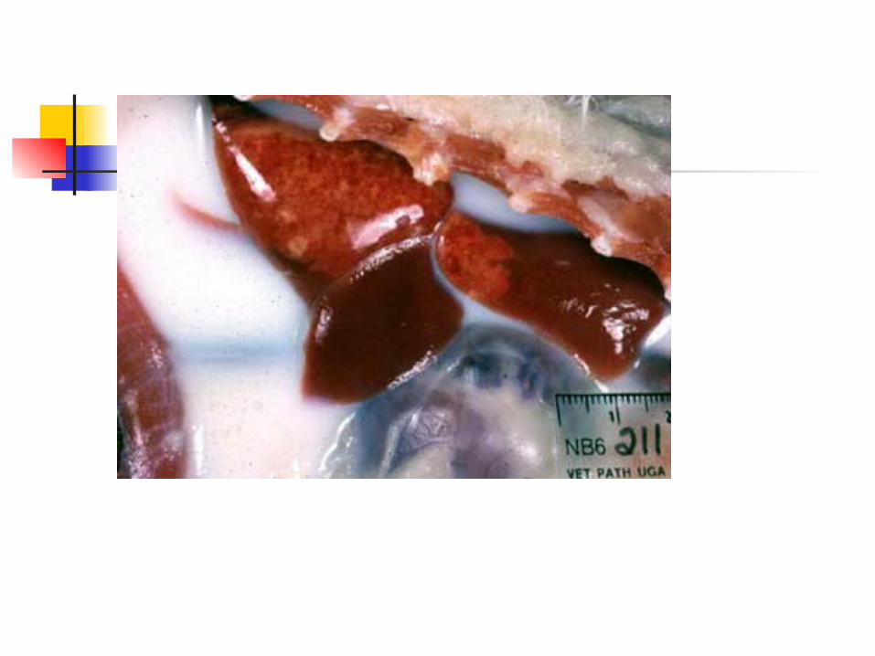
Abdominal and Thoracic Effusions
Clinical Pathology

Abdominal/thoracic fluids Abdominal and thoracic organs are bathed
in and lubricated by a small amount of fluid Fluid increases when the amount entering
the cavity is more than is removed fro it An increased amount of fluid in the
abdominal/thoracic cavity is not a disease in itself, but rather an indication of a pathologic process in the fluid production and/or removal system.

Abdominal/Thoracic Effusion
Fluid analysis and cytology is quick, easy and inexpensive, and relatively safe.
May obtain useful information for diagnosis, prognosis, and treatment.


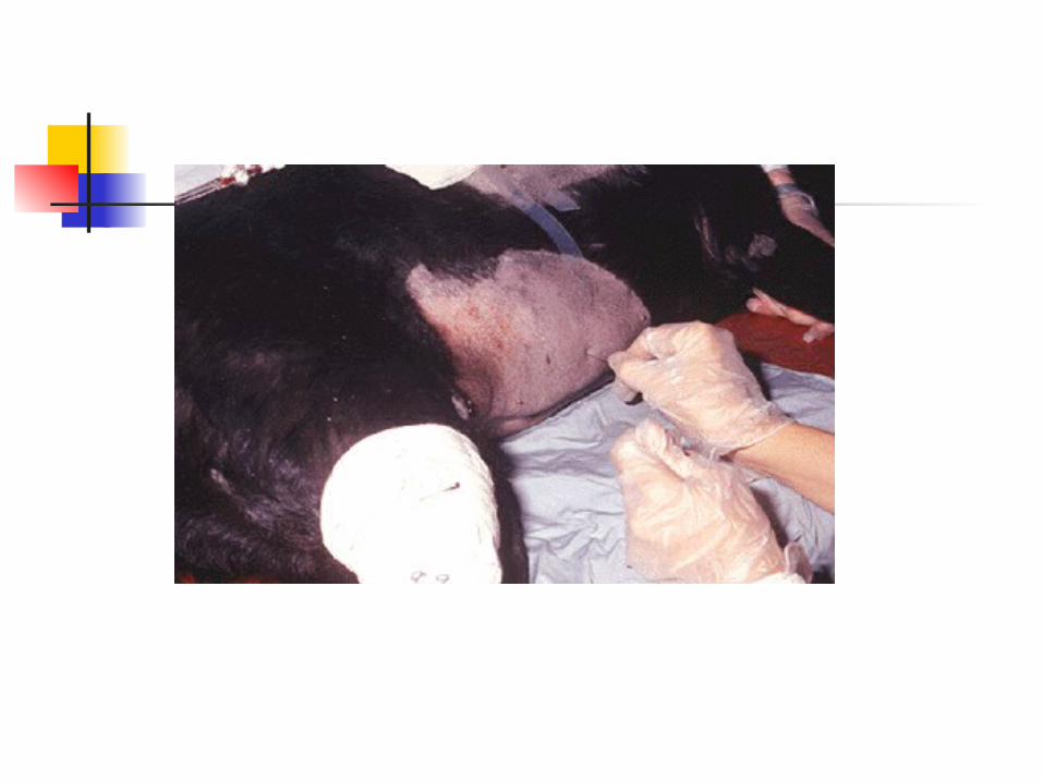
Abdominocentesis Collection
Aseptic prep of the skin Usually done with animal
standing Use a sterile needle or cannula Tap the ventral midline of the
abdomen, 1-2 cm caudal to the umbilicus
Collect fluid and place into an EDTA and red top tube
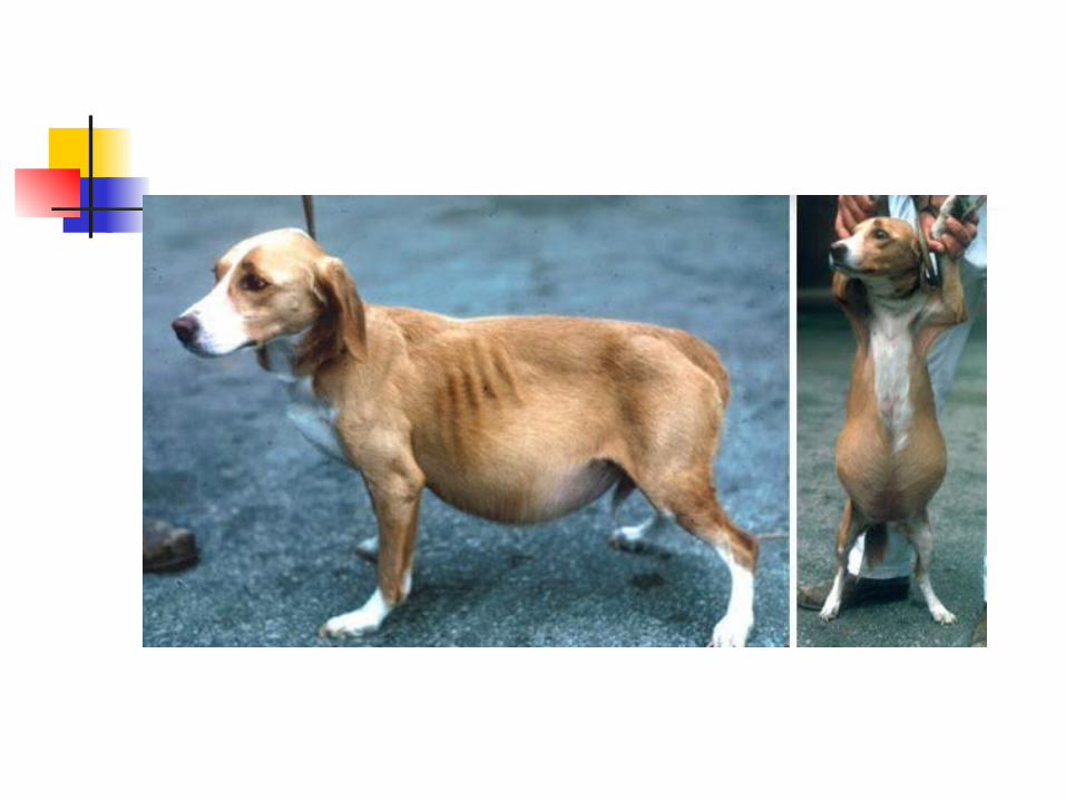
Indications of a peritoneal tap
Ascites: due to cardiac or liver disease or neoplasia, etc.
Peritonitis: Ruptured bowel, ruptured bladder.
In horses-colic
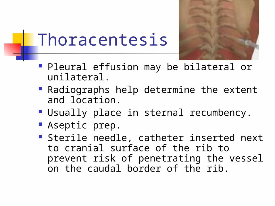
Thoracentesis Pleural effusion may be bilateral or
unilateral. Radiographs help determine the extent
and location. Usually place in sternal recumbency. Aseptic prep. Sterile needle, catheter inserted next to
cranial surface of the rib to prevent risk of penetrating the vessel on the caudal border of the rib.
Indications for Thoracentesis
Hemorrhage Inflammation (FIP) Neoplasia Ruptured thoracic lymphatic duct
(chylothorax)
Place in EDTA and Red top tube

Characteristics of Effusions Transparency/turbidity Color Protein concentration Specific gravity Cells: counts, types, and morphology Fluid should be odorless- if an
abdominal tap yields a malodorous fluid- may indicate a ruptured bowel

Color/turbidity of Effusions
Influenced by protein concentrations and cell numbers.
Normal peritoneal/pleural fluid is colorless and transparent to slightly turbid.
FIP may cause an amber, turbid, thick effusion (straw-yellow color)


Total Protein and Specific Gravity
Centrifuge sample at low speed (1500 rpm for 5 min)
TP can be measured on refractometer Normal is <2.5 g/dl
Specific gravity is measured on refractometer as well Normal is <1.018
Slide preparation Centrifuge fluid at low speed Pour off supernatant, leave about 0.5 ml
at the bottom of tube Resuspend by gentle agitation Place a drop on slide
Routine blood smear type technique Squash prep
Air dry Stain with diff quick

Cell counts on Effusions
Total nucleated cell counts Unopette procedure Automated
Normal peritoneal/pleural fluid has <10,000 nucleated cells/ul
Estimated cell counts can be made on a blood smear

Types of cells found in effusions
Neutrophils Mesothelial/macrophage type cells Lymphocytes Eosinophils Mast cells Neoplastic cells

Classifications of Effusions
Transudates Modified transudates Exudates

Transudates
Clear, colorless effusion <2.5 g/dl protein (low protein) Low total nucleated cell count
Non-degenerative neutrophils and mesothelial cells
Specific gravity < 1.013

Causes of Transudates
Hypoalbunemia: due to renal glomerular disease, hepatic insufficiency, and protein-losing enteropathy.
Ruptured bladder Rarely from blockage of lymph
from lymphatic vessel in the intestines

Modified Transudates Vary in color- amber to white to red Frequently slightly turbid to turbid High protein concentration (2.5-7.5
g/dl) Moderate cellularity: 1000-7000
cells/ul Occur as a result of fluid leakage from
lymphatics carrying high protein lymph or blood vessels.

Modified transudate causes Lease specific, variety of disorders Cardiovascular disease (right sided
heart failure) Neoplastic disease FIP Chylothorax Hemorrhage Hepatic disease- hypoalbunemia and
hypertension

Exudates (infections) Color varies- amber to
white to red Turbid to cloudy High protein > 3.0 g/dl High total nucleated cell
count (>7000 cell/ul) Neutrophils are the
predominant cell type.
Exudates continued Septic exudates: Degenerative
neutrophils and intracellular/extracellular bacteria present Ex: GI perforation, systemic sepsis,
pneumonia Non-septic exudates: non-degenerative
neutrophils, small lymphocytes, and/or neoplastic cells. Ex: Pancreatitis, neoplasia, uroperitoneum
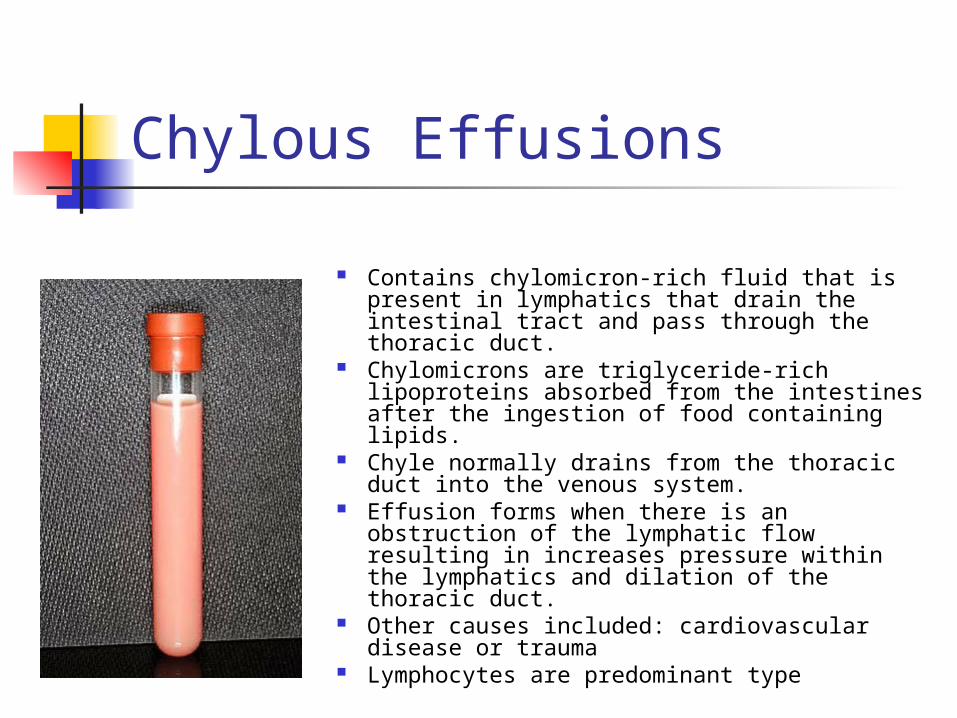
Chylous Effusions
Contains chylomicron-rich fluid that is present in lymphatics that drain the intestinal tract and pass through the thoracic duct.
Chylomicrons are triglyceride-rich lipoproteins absorbed from the intestines after the ingestion of food containing lipids.
Chyle normally drains from the thoracic duct into the venous system.
Effusion forms when there is an obstruction of the lymphatic flow resulting in increases pressure within the lymphatics and dilation of the thoracic duct.
Other causes included: cardiovascular disease or trauma
Lymphocytes are predominant type