Embed Size (px)
Citation preview
Rev. Inst. Med. trop. S. Paulo47(6):359-361, November-December, 2005
(1) Serviço de Pneumologia, Hospital Universitário de Santa Maria, Santa Maria, RS, Brasil.(2) Departamento de Patologia, Hospital Universitário Santa Maria, Santa Maria, RS, Brasil.(3) Departamento de Cirurgia, Hospital Universitário de Santa Maria, Santa Maria, RS, Brasil.(4) Laboratórios de Biologia Parasitária e Parasitologia Molecular, PUCRS, Porto Alegre, RS, Brasil.(5) Faculdade de Biociências e Instituto de Pesquisas Biomédicas, PUCRS, Porto Alegre, RS, Brasil.Correspondence to: José Wellington Alves dos Santos, Rua Venâncio Aires 2020/403, 97010-004 Santa Maria, RS, Brazil. Phone: +55 55 3225-3018; Fax: +55 55 3220-8005; e-mail:
ABDOMINAL ANGIOSTRONGYLIASIS: A CASE WITH SEVERE EVOLUTION
José Wellington Alves dos SANTOS(1), Ricardo Morgental ZAMBENEDETTI(1), Keli Cristina MANN(1), Marta Pires da ROCHA(2),Ewerton Nunes MORAIS(3) & Carlos GRAEFF-TEIXEIRA(4,5)
SUMMARY
A case of acute abdomen disease caused by abdominal angiostrongyliasis is reported. A 42-year-old otherwise healthy patientpresented with a complaint of nine days of abdominal pain, constipation, disury, fever and right iliac fossa palpable mass. Exploratorylaparotomy was performed. After surgical treatment the patient presented serious complications.
KEYWORDS: Abdominal angiostrongyliasis; Angiostrongylus costaricensis; Acute abdomen.
INTRODUCTION
Abdominal angiostrongyliasis (AA) is a parasitosis caused by theworm Angiostrongylus costaricensis. Since 1967, when the first humaninfection case was published3,17, researches have described theepidemiology, life cycle of the parasite, and the clinical manifestationsof this disease7,8,9,18,19,20.
A case of acute abdomen caused by AA is reported.
CASE REPORT
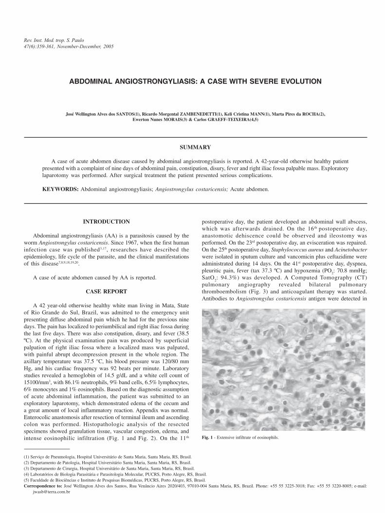
A 42 year-old otherwise healthy white man living in Mata, Stateof Rio Grande do Sul, Brazil, was admitted to the emergency unitpresenting diffuse abdominal pain which he had for the previous ninedays. The pain has localized to periumbilical and right iliac fossa duringthe last five days. There was also constipation, disury, and fever (38.5ºC). At the physical examination pain was produced by superficialpalpation of right iliac fossa where a localized mass was palpated,with painful abrupt decompression present in the whole region. Theaxillary temperature was 37.5 °C, his blood pressure was 120/80 mmHg, and his cardiac frequency was 92 beats per minute. Laboratorystudies revealed a hemoglobin of 14.5 g/dL and a white cell count of15100/mm3, with 86.1% neutrophils, 9% band cells, 6.5% lymphocytes,6% monocytes and 1% eosinophils. Based on the diagnostic assumptionof acute abdominal inflammation, the patient was submitted to anexploratory laparotomy, which demonstrated edema of the cecum anda great amount of local inflammatory reaction. Appendix was normal.Enterocolic anastomosis after resection of terminal ileum and ascendingcolon was performed. Histopathologic analysis of the resectedspecimens showed granulation tissue, vascular congestion, edema, andintense eosinophilic infiltration (Fig. 1 and Fig. 2). On the 11th
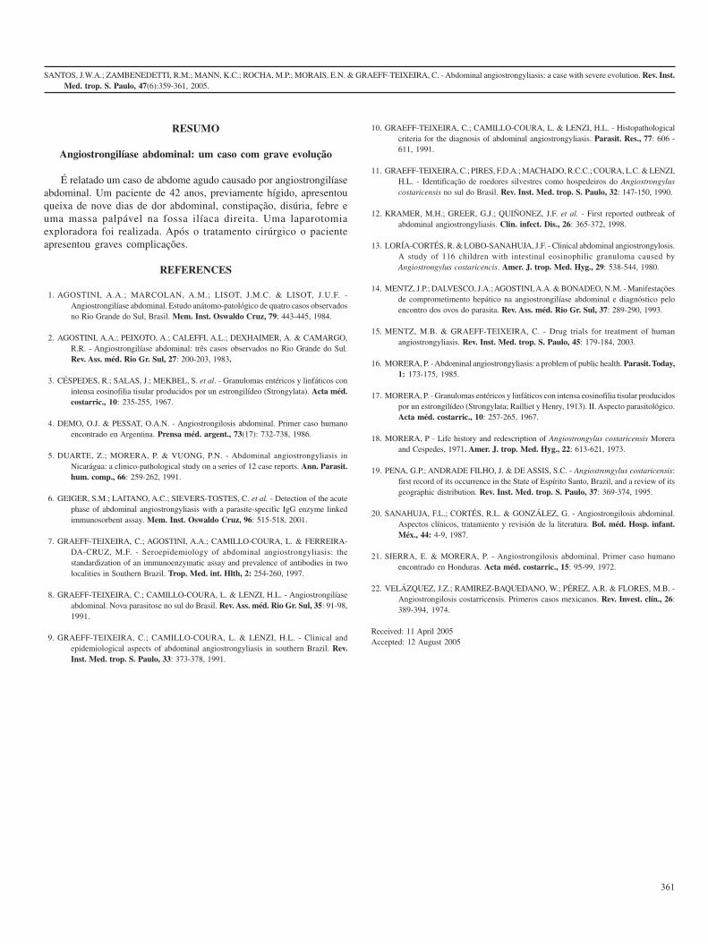
postoperative day, the patient developed an abdominal wall abscess,which was afterwards drained. On the 16th postoperative day,anastomotic dehiscence could be observed and ileostomy wasperformed. On the 23rd postoperative day, an evisceration was repaired.On the 25th postoperative day, Staphylococcus aureus and Acinetobacterwere isolated in sputum culture and vancomicin plus ceftazidime wereadministrated during 14 days. On the 41st postoperative day, dyspnea,pleuritic pain, fever (tax 37.3 ºC) and hypoxemia (PO
2: 70.8 mmHg;
SatO2: 94.3%) was developed. A Computed Tomography (CT)
pulmonary angiography revealed bilateral pulmonarythromboembolism (Fig. 3) and anticoagulant therapy was started.Antibodies to Angiostrongylus costaricensis antigen were detected in
Fig. 1 - Extensive infiltrate of eosinophils.

360
SANTOS, J.W.A.; ZAMBENEDETTI, R.M.; MANN, K.C.; ROCHA, M.P.; MORAIS, E.N. & GRAEFF-TEIXEIRA, C. - Abdominal angiostrongyliasis: a case with severe evolution. Rev. Inst.Med. trop. S. Paulo, 47(6):359-361, 2005.
serum by ELISA at 50th postoperative day. On the 53rd postoperativeday the patient was discharged. After six months follow-up the patienthas been well and no complaints are observed.
DISCUSSION
Angiostrongylus costaricensis is an intra-arterial nematode parasiteof wild rodents11. Man is not its definitive host, but can be accidentally
infected by ingestion of water or food contaminated with third stagelarvae (L3) produced in the intermediate host, generally slugs ofVeronicellidae family8. AA is found in the American continent, mainlyin Central America4,5,13,21,22, having been considered a problem of publichealth in Costa Rica16. Brazil, the country with the second largestnumber of reported diagnosed cases, has most of the reported diagnosisin patients from Rio Grande do Sul (RGS). This zoonosis presentspeculiar geographic distribution. In RGS, the majority of cases hasbeen found in the north of state and with rare occurrences in its centralregion, where the patient of the reported case lives1,2,9,14,19.
The most affected individuals are asymptomatic oroligosymptomatic. Abdominal pain, fever, anorexia, malaise, nausea,vomits and intestinal habit alterations are the most frequent symptoms.Presentation of palpable mass in the right iliac fossa also can occur.Usually, these clinical manifestations may end abruptly and reoccur atrelapsing episodes for many months8,12.
The ileocecal region is the most affected by parasite3,10,17. Areas ofnecrosis and congestion or intestinal wall thickening are found due tothe presence of adult worms and eggs in terminal branches of mesentericvessels. Thrombotic phenomena in the injured vessels endotheliumalso can occur and frequently are associated with degenerated worms.Microscopically, massive eosinophilic infiltration compromising alllayers of the intestinal wall, eosinophilic granulomatous reaction andeosinophilic vasculitis affecting arteries, veins and lymphatics may beobserved8,11.
The described case presented with an acute abdomen disease withsurgical indication. Exploratory laparotomy evidenced the presenceof normal appendix and intense inflammatory reaction in the cecumsuggestive of ileocecal valve neoplasia. AA was suspected after thehistopathologic analysis of the surgical specimen, which presentedintense edema, local inflammatory process, vascular congestion,granulomatous reaction and massive transmural eosinophilic infiltrationin the ileocecal region. The diagnostic hypothesis was reinforced byrevision of clinic aspects and a positive ELISA IgG, a 76.2% sensitiveand 91.1% specific immunoenzymatic test that employs crude antigensof female worms6.
During postoperative evolution the patient presented, initially, localcomplications due to the surgical process. Nosocomial pneumonia wastreated with antibiotic therapy by 14 days. On the 41st postoperativeday massive bilateral pulmonary embolism was diagnosed evidencingthe severity of this case, probably associated with vascularcomplications. The usual complications of this parasitosis areinflammatory thickening of the intestinal wall leading to intestinalobstruction or perforation, both requiring surgical intervention. Theevolution of these patients is satisfactory, with a death rate by peritonitisand sepsis ranging from 1.3%13 to 7.4%9.
In conclusion, AA is a disease with unspecified clinicalmanifestations, often misdiagnosed due to being unknown and that iscurrently clinically untreatable15. Because it is potentially fatal andpresents an undefined natural course, should be considered differentialdiagnostic in patients with acute abdominal disease.
Fig. 2 - Section showing edema, granulomatous reaction, extensive inflammatory infiltrate,
vascular congestion.
Fig. 3 - CT pulmonary angiography: bilateral pulmonary embolism (arrows).
SANTOS, J.W.A.; ZAMBENEDETTI, R.M.; MANN, K.C.; ROCHA, M.P.; MORAIS, E.N. & GRAEFF-TEIXEIRA, C. - Abdominal angiostrongyliasis: a case with severe evolution. Rev. Inst.Med. trop. S. Paulo, 47(6):359-361, 2005.
361
RESUMO
Angiostrongilíase abdominal: um caso com grave evolução
É relatado um caso de abdome agudo causado por angiostrongilíaseabdominal. Um paciente de 42 anos, previamente hígido, apresentouqueixa de nove dias de dor abdominal, constipação, disúria, febre euma massa palpável na fossa ilíaca direita. Uma laparotomiaexploradora foi realizada. Após o tratamento cirúrgico o pacienteapresentou graves complicações.
REFERENCES
1. AGOSTINI, A.A.; MARCOLAN, A.M.; LISOT, J.M.C. & LISOT, J.U.F. -Angiostrongilíase abdominal. Estudo anátomo-patológico de quatro casos observadosno Rio Grande do Sul, Brasil. Mem. Inst. Oswaldo Cruz, 79: 443-445, 1984.
2. AGOSTINI, A.A.; PEIXOTO, A.; CALEFFI, A.L.; DEXHAIMER, A. & CAMARGO,R.R. - Angiostrongilíase abdominal: três casos observados no Rio Grande do Sul.Rev. Ass. méd. Rio Gr. Sul, 27: 200-203, 1983.
3. CÉSPEDES, R.; SALAS, J.; MEKBEL, S. et al. - Granulomas entéricos y linfáticos conintensa eosinofilia tisular producidos por un estrongilídeo (Strongylata). Acta méd.costarric., 10: 235-255, 1967.
4. DEMO, O.J. & PESSAT, O.A.N. - Angiostrongilosis abdominal. Primer caso humanoencontrado en Argentina. Prensa méd. argent., 73(17): 732-738, 1986.
5. DUARTE, Z.; MORERA, P. & VUONG, P.N. - Abdominal angiostrongyliasis inNicarágua: a clinico-pathological study on a series of 12 case reports. Ann. Parasit.hum. comp., 66: 259-262, 1991.
6. GEIGER, S.M.; LAITANO, A.C.; SIEVERS-TOSTES, C. et al. - Detection of the acutephase of abdominal angiostrongyliasis with a parasite-specific IgG enzyme linkedimmunosorbent assay. Mem. Inst. Oswaldo Cruz, 96: 515-518, 2001.
7. GRAEFF-TEIXEIRA, C.; AGOSTINI, A.A.; CAMILLO-COURA, L. & FERREIRA-DA-CRUZ, M.F. - Seroepidemiology of abdominal angiostrongyliasis: thestandardization of an immunoenzymatic assay and prevalence of antibodies in twolocalities in Southern Brazil. Trop. Med. int. Hlth, 2: 254-260, 1997.
8. GRAEFF-TEIXEIRA, C.; CAMILLO-COURA, L. & LENZI, H.L. - Angiostrongilíaseabdominal. Nova parasitose no sul do Brasil. Rev. Ass. méd. Rio Gr. Sul, 35: 91-98,1991.
9. GRAEFF-TEIXEIRA, C.; CAMILLO-COURA, L. & LENZI, H.L. - Clinical andepidemiological aspects of abdominal angiostrongyliasis in southern Brazil. Rev.Inst. Med. trop. S. Paulo, 33: 373-378, 1991.
10. GRAEFF-TEIXEIRA, C.; CAMILLO-COURA, L. & LENZI, H.L. - Histopathologicalcriteria for the diagnosis of abdominal angiostrongyliasis. Parasit. Res., 77: 606 -611, 1991.
11. GRAEFF-TEIXEIRA, C.; PIRES, F.D.A.; MACHADO, R.C.C.; COURA, L.C. & LENZI,H.L. - Identificação de roedores silvestres como hospedeiros do Angiostrongyluscostaricensis no sul do Brasil. Rev. Inst. Med. trop. S. Paulo, 32: 147-150, 1990.
12. KRAMER, M.H.; GREER, G.J.; QUIÑONEZ, J.F. et al. - First reported outbreak ofabdominal angiostrongyliasis. Clin. infect. Dis., 26: 365-372, 1998.
13. LORÍA-CORTÉS, R. & LOBO-SANAHUJA, J.F. - Clinical abdominal angiostrongylosis.A study of 116 children with intestinal eosinophilic granuloma caused byAngiostrongylus costaricencis. Amer. J. trop. Med. Hyg., 29: 538-544, 1980.
14. MENTZ, J.P.; DALVESCO, J.A.; AGOSTINI, A.A. & BONADEO, N.M. - Manifestaçõesde comprometimento hepático na angiostrongilíase abdominal e diagnóstico peloencontro dos ovos do parasita. Rev. Ass. méd. Rio Gr. Sul, 37: 289-290, 1993.
15. MENTZ, M.B. & GRAEFF-TEIXEIRA, C. - Drug trials for treatment of humanangiostrongyliasis. Rev. Inst. Med. trop. S. Paulo, 45: 179-184, 2003.
16. MORERA, P. - Abdominal angiostrongyliasis: a problem of public health. Parasit. Today,1: 173-175, 1985.
17. MORERA, P. - Granulomas entéricos y linfáticos con intensa eosinofilia tisular producidospor un estrongilídeo (Strongylata; Railliet y Henry, 1913). II. Aspecto parasitológico.Acta méd. costarric., 10: 257-265, 1967.
18. MORERA, P - Life history and redescription of Angiostrongylus costaricensis Moreraand Cespedes, 1971. Amer. J. trop. Med. Hyg., 22: 613-621, 1973.
19. PENA, G.P.; ANDRADE FILHO, J. & DE ASSIS, S.C. - Angiostrongylus costaricensis:first record of its occurrence in the State of Espírito Santo, Brazil, and a review of itsgeographic distribution. Rev. Inst. Med. trop. S. Paulo, 37: 369-374, 1995.
20. SANAHUJA, F.L.; CORTÉS, R.L. & GONZÁLEZ, G. - Angiostrongilosis abdominal.Aspectos clínicos, tratamiento y revisión de la literatura. Bol. méd. Hosp. infant.Méx., 44: 4-9, 1987.
21. SIERRA, E. & MORERA, P. - Angiostrongilosis abdominal. Primer caso humanoencontrado en Honduras. Acta méd. costarric., 15: 95-99, 1972.
22. VELÁZQUEZ, J.Z.; RAMIREZ-BAQUEDANO, W.; PÉREZ, A.R. & FLORES, M.B. -Angiostrongilosis costarricensis. Primeros casos mexicanos. Rev. Invest. clín., 26:389-394, 1974.
Received: 11 April 2005Accepted: 12 August 2005


![The Abdominal Sepsis Study: Epidemiology of Etiology and ... · abdominal infection [1]. Abdominal sepsis is a severe infectious complication associated with considerable mortality](https://img.pdfslide.net/doc/110x75/6049ed16a497f05e454be51a/the-abdominal-sepsis-study-epidemiology-of-etiology-and-abdominal-infection.jpg)