Embed Size (px)
Citation preview
Abdominal Wall Hernia Mesh RepairSonography of Mesh and Common Complications
David A. Jamadar, MBBS, Jon A. Jacobson, MD,Gandikota Girish, MBBS, Jefferson Balin, MD,Catherine J. Brandon, MD, Elaine M. Caoili, MD, Yoav Morag, MD, Michael G. Franz, MD
Objective. The purposes of this study were (1) to review the sonographic in vitro and in vivo appear-ances of mesh for surgical repair of abdominal wall hernias, (2) to describe sonographic techniquesand discuss the limitations of sonography in evaluation of mesh hernia repair, and (3) to illustrate com-mon complications after mesh repair shown with sonography. Methods. We identified interestingcases from the musculoskeletal sonographic database as well as from the teaching files of the authors,with surgical or other cross-sectional imaging corroboration. Results. A compilation of the sono-graphic appearances of mesh used for anterior abdominal wall and inguinal hernia repair and compli-cations diagnosable by sonography is presented. Conclusions. Sonography can be effective forevaluation of mesh and complications after mesh repair of anterior abdominal wall and inguinal her-nias. Key words: abdominal wall hernia; mesh repair; sonography.
Received December 27, 2007, from the Departmentsof Radiology (D.A.J., J.A.J., G.G., J.B., C.J.B., E.M.C.,Y.M.) and Surgery (M.G.F.), University of MichiganHospitals, Ann Arbor, Michigan USA. Revisionrequested December 30, 2007. Revised manuscriptaccepted for publication January 29, 2008.
We thank Brian Robertson, Stephanie Creel,Tracy Boon, Heidi Taraskiewicz, and Wenzhen Liangfor help, ideas, and suggestions.
Address correspondence to David A. JamadarMBBS, Department of Radiology, University ofMichigan Hospitals, 1500 E Medical Center Dr, AnnArbor, MI 48109 USA.
E-mail: [email protected]
AbbreviationsCT, computed tomography
epair of abdominal wall hernias with syntheticpatches was first described in 1962.1 Since thattime, these materials have been used widely, andthe various procedures using mesh in abdominal
wall repair have become commonplace. Several reportshave shown that compared with simple sutures, mesh issuperior, with significantly reduced recurrence rates.2
Materials from which mesh is manufactured are usuallyderived from polypropylene or polytetrafluoroethylene3
and typically function by providing a bridge across defi-cient tissue. The mesh is incorporated into the adjacent tis-sues and should restore the structure and function of theabdominal wall. Implanted mesh is a foreign body andtherefore causes an inflammatory reaction. Potential com-plications may relate to the mesh as a foreign body or mayrelate to the surgical repair of abdominal wall hernias.
We present the sonographic in vitro and in vivo appear-ances of mesh and sonographic techniques for identify-ing mesh in the anterior abdominal wall. We also describecomplications of mesh implants and discuss potentiallimitations of sonography.
© 2008 by the American Institute of Ultrasound in Medicine • J Ultrasound Med 2008; 27:907–917 • 0278-4297/08/$3.50
R
Image Presentation
276jumonline.qxp:Layout 1 5/15/08 9:14 AM Page 907
In Vitro Appearances
Sonography is a useful imaging tool that caneffectively evaluate the anterior abdominalwall, identifying mesh and many of the compli-cations associated with its surgical placement.4
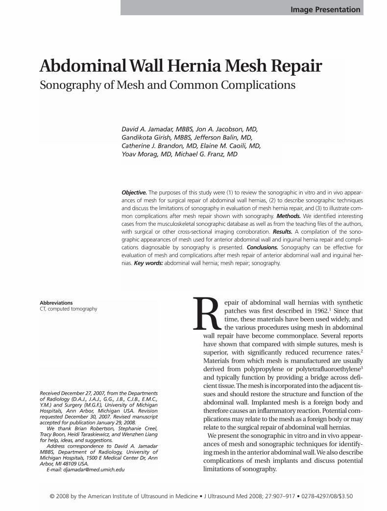
Mesh may be placed in a variety of locations inrelation to the structures of the anterior abdom-inal wall and inguinal region (Figure 1), all ofwhich may be evaluated by sonography.
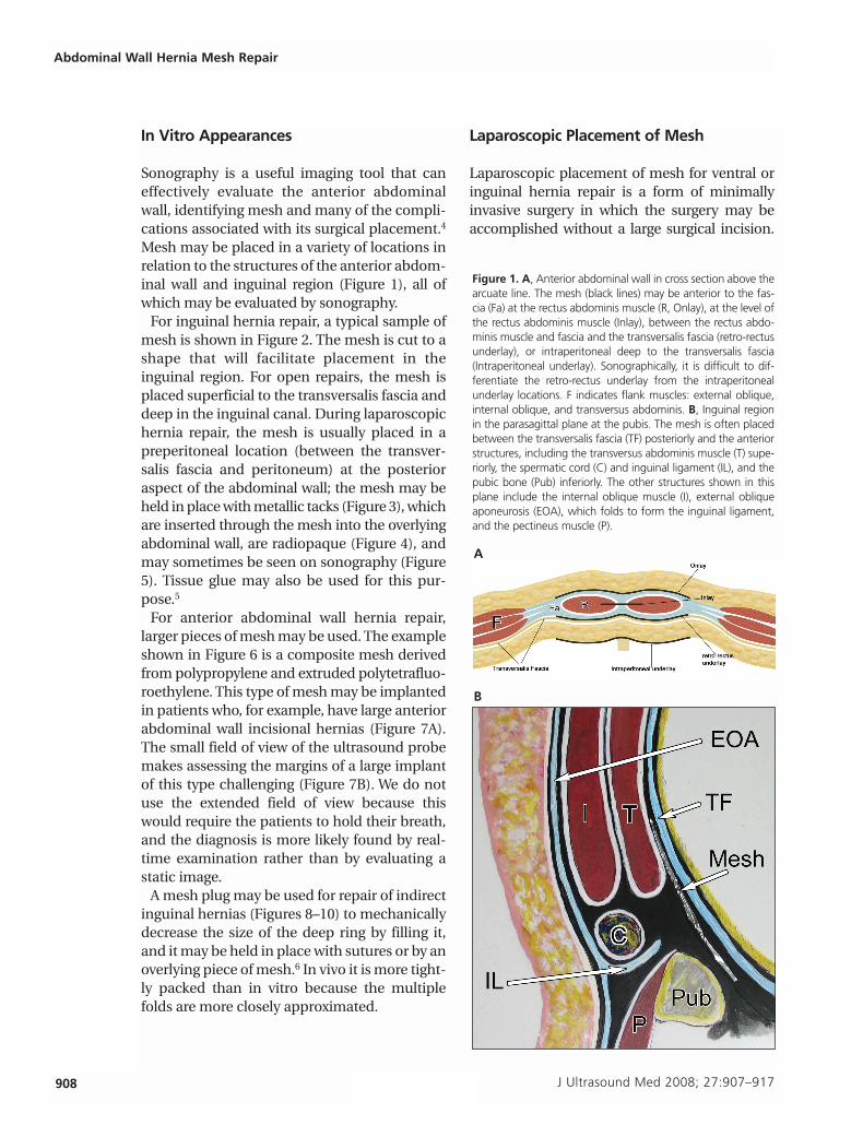
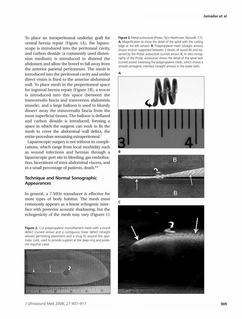
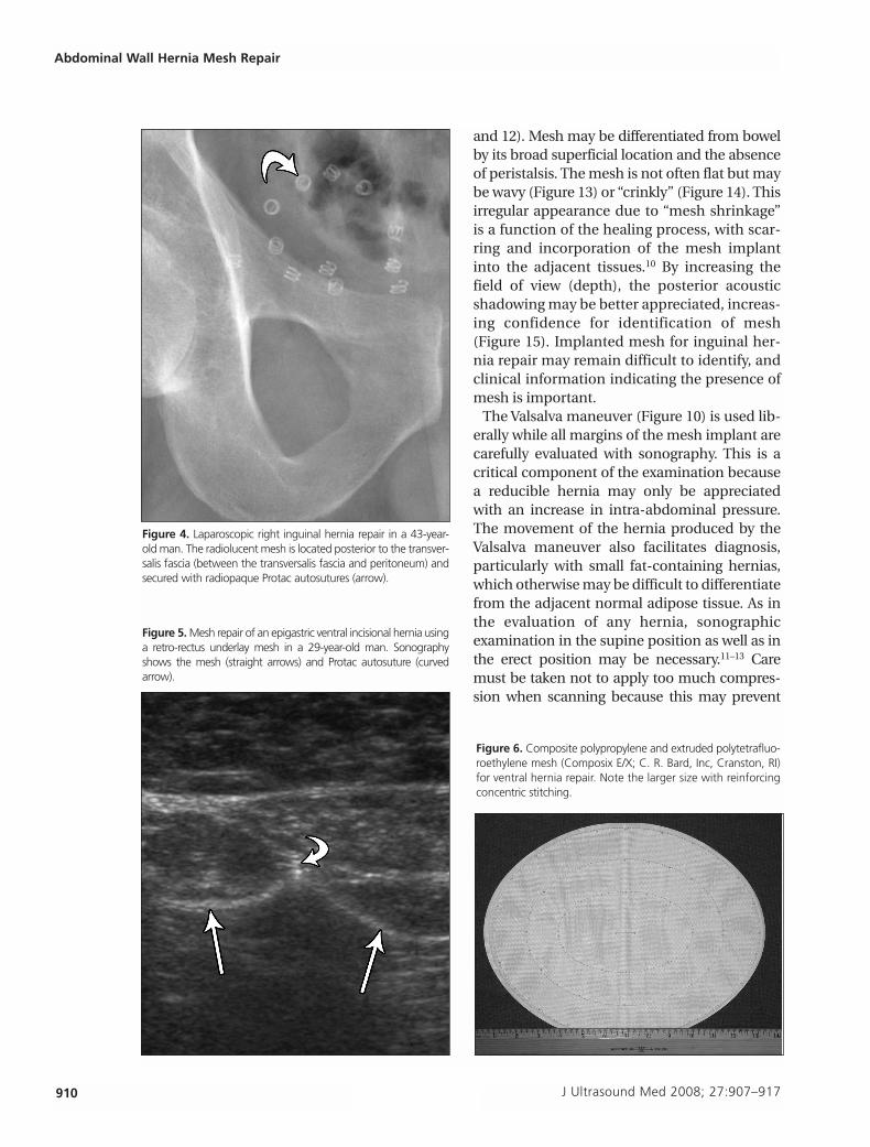
For inguinal hernia repair, a typical sample ofmesh is shown in Figure 2. The mesh is cut to ashape that will facilitate placement in theinguinal region. For open repairs, the mesh isplaced superficial to the transversalis fascia anddeep in the inguinal canal. During laparoscopichernia repair, the mesh is usually placed in apreperitoneal location (between the transver-salis fascia and peritoneum) at the posterioraspect of the abdominal wall; the mesh may beheld in place with metallic tacks (Figure 3), whichare inserted through the mesh into the overlyingabdominal wall, are radiopaque (Figure 4), andmay sometimes be seen on sonography (Figure5). Tissue glue may also be used for this pur-pose.5
For anterior abdominal wall hernia repair,larger pieces of mesh may be used. The exampleshown in Figure 6 is a composite mesh derivedfrom polypropylene and extruded polytetrafluo-roethylene. This type of mesh may be implantedin patients who, for example, have large anteriorabdominal wall incisional hernias (Figure 7A).The small field of view of the ultrasound probemakes assessing the margins of a large implantof this type challenging (Figure 7B). We do notuse the extended field of view because thiswould require the patients to hold their breath,and the diagnosis is more likely found by real-time examination rather than by evaluating astatic image.
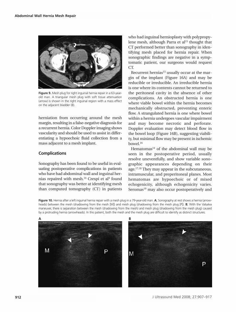
A mesh plug may be used for repair of indirectinguinal hernias (Figures 8–10) to mechanicallydecrease the size of the deep ring by filling it,and it may be held in place with sutures or by anoverlying piece of mesh.6 In vivo it is more tight-ly packed than in vitro because the multiplefolds are more closely approximated.
Laparoscopic Placement of Mesh
Laparoscopic placement of mesh for ventral oringuinal hernia repair is a form of minimallyinvasive surgery in which the surgery may beaccomplished without a large surgical incision.
908 J Ultrasound Med 2008; 27:907–917
Abdominal Wall Hernia Mesh Repair
Figure 1. A, Anterior abdominal wall in cross section above thearcuate line. The mesh (black lines) may be anterior to the fas-cia (Fa) at the rectus abdominis muscle (R, Onlay), at the level ofthe rectus abdominis muscle (Inlay), between the rectus abdo-minis muscle and fascia and the transversalis fascia (retro-rectusunderlay), or intraperitoneal deep to the transversalis fascia(Intraperitoneal underlay). Sonographically, it is difficult to dif-ferentiate the retro-rectus underlay from the intraperitonealunderlay locations. F indicates flank muscles: external oblique,internal oblique, and transversus abdominis. B, Inguinal regionin the parasagittal plane at the pubis. The mesh is often placedbetween the transversalis fascia (TF) posteriorly and the anteriorstructures, including the transversus abdominis muscle (T) supe-riorly, the spermatic cord (C) and inguinal ligament (IL), and thepubic bone (Pub) inferiorly. The other structures shown in thisplane include the internal oblique muscle (I), external obliqueaponeurosis (EOA), which folds to form the inguinal ligament,and the pectineus muscle (P).
A
B
276jumonline.qxp:Layout 1 5/15/08 9:14 AM Page 908
To place an intraperitoneal underlay graft forventral hernia repair (Figure 1A), the laparo-scope is introduced into the peritoneal cavity,and carbon dioxide (a commonly used disten-sion medium) is introduced to distend theabdomen and allow the bowel to fall away fromthe anterior parietal peritoneum. The mesh isintroduced into the peritoneal cavity and underdirect vision is fixed to the anterior abdominalwall. To place mesh in the preperitoneal spacefor inguinal hernia repair (Figure 1B), a trocaris introduced into this space (between thetransversalis fascia and transversus abdominismuscle), and a large balloon is used to bluntlydissect away the transversalis fascia from themore superficial tissues. The balloon is deflatedand carbon dioxide is introduced, forming aspace in which the surgeon can work to fix themesh to cover the abdominal wall defect, theentire procedure remaining extraperitoneal.7
Laparoscopic surgery is not without its compli-cations, which range from local morbidity suchas wound infections and hernias through alaparoscopic port site to bleeding, gas emboliza-tion, lacerations of intra-abdominal viscera, andin a small percentage of patients, death.8,9
Technique and Normal SonographicAppearances
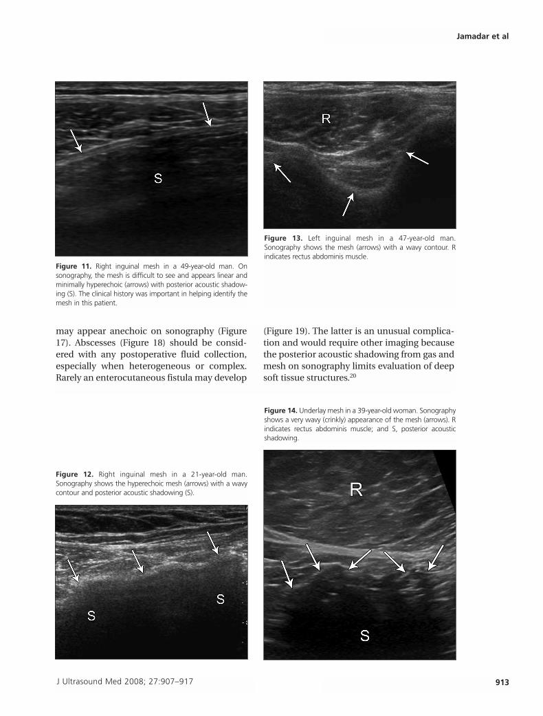
In general, a 7-MHz transducer is effective formost types of body habitus. The mesh mostcommonly appears as a linear echogenic inter-face with posterior acoustic shadowing, but theechogenicity of the mesh may vary (Figures 11
J Ultrasound Med 2008; 27:907–917 909
Jamadar et al
Figure 2. Cut polypropylene monofilament mesh with a rounddefect (curved arrow) and a contiguous linear defect (straightarrows) permitting placement and a snug fit around the sper-matic cord, used to provide support at the deep ring and poste-rior inguinal canal.
Figure 3. Metal autosuture (Protac; Tyco Healthcare, Norwalk, CT).A, Magnification to show the detail of the spiral with the cuttingedge at the left (arrow). B, Polypropylene mesh (straight arrows)shown end-on supported between 2 blocks of wood (B) and tra-versed by the Protac autosuture (curved arrow). C, In vitro sonog-raphy of the Protac autosuture shows the detail of the spiral tack(curved arrow) traversing the polypropylene mesh, which shows asmooth echogenic interface (straight arrows) in the water bath.
A
B
C
276jumonline.qxp:Layout 1 5/15/08 9:14 AM Page 909
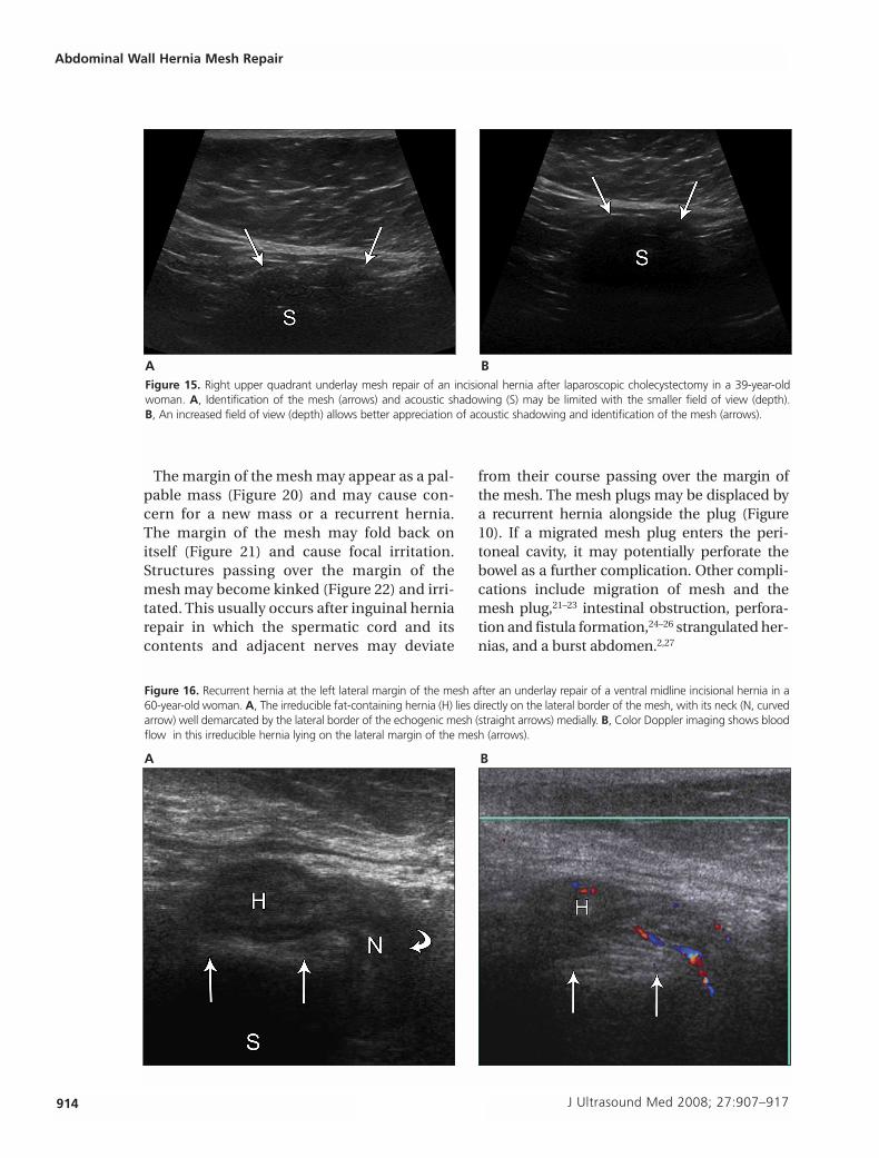
and 12). Mesh may be differentiated from bowelby its broad superficial location and the absenceof peristalsis. The mesh is not often flat but maybe wavy (Figure 13) or “crinkly” (Figure 14). Thisirregular appearance due to “mesh shrinkage”is a function of the healing process, with scar-ring and incorporation of the mesh implantinto the adjacent tissues.10 By increasing thefield of view (depth), the posterior acousticshadowing may be better appreciated, increas-ing confidence for identification of mesh(Figure 15). Implanted mesh for inguinal her-nia repair may remain difficult to identify, andclinical information indicating the presence ofmesh is important.
The Valsalva maneuver (Figure 10) is used lib-erally while all margins of the mesh implant arecarefully evaluated with sonography. This is acritical component of the examination becausea reducible hernia may only be appreciatedwith an increase in intra-abdominal pressure.The movement of the hernia produced by theValsalva maneuver also facilitates diagnosis,particularly with small fat-containing hernias,which otherwise may be difficult to differentiatefrom the adjacent normal adipose tissue. As inthe evaluation of any hernia, sonographicexamination in the supine position as well as inthe erect position may be necessary.11–13 Caremust be taken not to apply too much compres-sion when scanning because this may prevent
910 J Ultrasound Med 2008; 27:907–917
Abdominal Wall Hernia Mesh Repair
Figure 4. Laparoscopic right inguinal hernia repair in a 43-year-old man. The radiolucent mesh is located posterior to the transver-salis fascia (between the transversalis fascia and peritoneum) andsecured with radiopaque Protac autosutures (arrow).
Figure 5. Mesh repair of an epigastric ventral incisional hernia usinga retro-rectus underlay mesh in a 29-year-old man. Sonographyshows the mesh (straight arrows) and Protac autosuture (curvedarrow).
Figure 6. Composite polypropylene and extruded polytetrafluo-roethylene mesh (Composix E/X; C. R. Bard, Inc, Cranston, RI)for ventral hernia repair. Note the larger size with reinforcingconcentric stitching.
276jumonline.qxp:Layout 1 5/15/08 9:14 AM Page 910
J Ultrasound Med 2008; 27:907–917 911
Jamadar et al
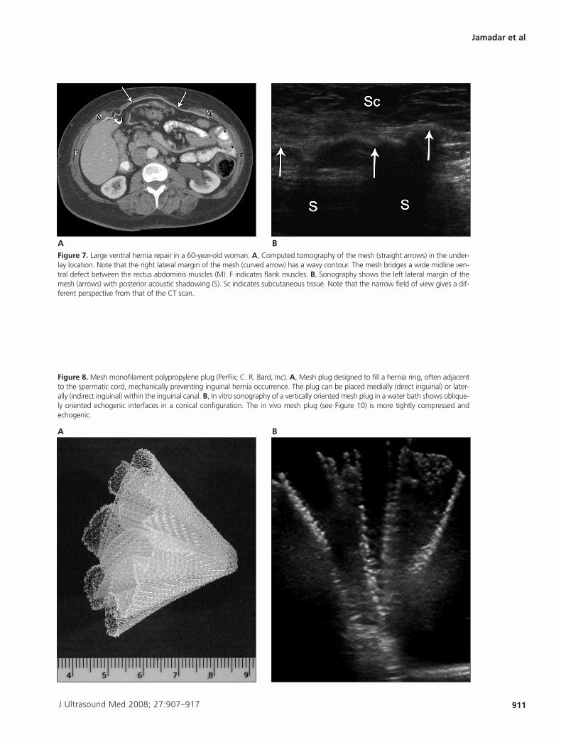
Figure 7. Large ventral hernia repair in a 60-year-old woman. A, Computed tomography of the mesh (straight arrows) in the under-lay location. Note that the right lateral margin of the mesh (curved arrow) has a wavy contour. The mesh bridges a wide midline ven-tral defect between the rectus abdominis muscles (M). F indicates flank muscles. B, Sonography shows the left lateral margin of themesh (arrows) with posterior acoustic shadowing (S). Sc indicates subcutaneous tissue. Note that the narrow field of view gives a dif-ferent perspective from that of the CT scan.
A B
Figure 8. Mesh monofilament polypropylene plug (PerFix; C. R. Bard, Inc). A, Mesh plug designed to fill a hernia ring, often adjacentto the spermatic cord, mechanically preventing inguinal hernia occurrence. The plug can be placed medially (direct inguinal) or later-ally (indirect inguinal) within the inguinal canal. B, In vitro sonography of a vertically oriented mesh plug in a water bath shows oblique-ly oriented echogenic interfaces in a conical configuration. The in vivo mesh plug (see Figure 10) is more tightly compressed andechogenic.
A B
276jumonline.qxp:Layout 1 5/15/08 9:14 AM Page 911
herniation from occurring around the meshmargin, resulting in a false-negative diagnosis fora recurrent hernia. Color Doppler imaging showsvascularity and should be used to assist in differ-entiating a hypoechoic fluid collection from amass adjacent to a mesh implant.
Complications
Sonography has been found to be useful in eval-uating postoperative complications in patientswho have had abdominal wall and inguinal her-nias repaired with mesh.14 Crespi et al4 foundthat sonography was better at identifying meshthan computed tomography (CT) in patients
who had inguinal hernioplasty with polypropy-lene mesh, although Parra et al15 thought thatCT performed better than sonography in iden-tifying mesh placed for hernia repair. Whensonographic findings are negative in a symp-tomatic patient, our surgeons would requestCT.
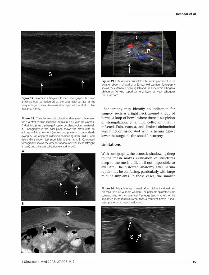
Recurrent hernias15 usually occur at the mar-gin of the implant (Figure 16A) and may bereducible or irreducible. An irreducible herniais one where its contents cannot be returned tothe peritoneal cavity in the absence of othercomplications. An obstructed hernia is onewhere viable bowel within the hernia becomesmechanically obstructed, preventing entericflow. A strangulated hernia is one where bowelwithin a hernia undergoes vascular impairmentand may become necrotic and perforate.Doppler evaluation may detect blood flow inthe bowel loop (Figure 16B), suggesting viabili-ty, but minimal flow may be present in ischemicbowel.16
Hematomas14 of the abdominal wall may beseen in the postoperative period, usuallyresolve uneventfully, and show variable sono-graphic appearances depending on theirage.17,18 They may appear in the subcutaneous,intramuscular, and preperitoneal planes. Mosthematomas are hypoechoic or of mixedechogenicity, although echogenicity varies.Seromas19 may also occur postoperatively and
912 J Ultrasound Med 2008; 27:907–917
Abdominal Wall Hernia Mesh Repair
Figure 9. Mesh plug for right inguinal hernia repair in a 63-year-old man. A triangular mesh plug with soft tissue attenuation(arrow) is shown in the right inguinal region with a mass effecton the adjacent bladder (B).
Figure 10. Hernia after a left inguinal hernia repair with a mesh plug in a 79-year-old man. A, Sonography at rest shows a hernia (arrow-heads) between the mesh (shadowing from the mesh [M]) and mesh plug (shadowing from the mesh plug [P]). B, With the Valsalvamaneuver, there is separation between the mesh (shadowing from the mesh) and mesh plug (shadowing from the mesh plug) causedby a protruding hernia (arrowheads). In this patient, both the mesh and the mesh plug are difficult to identify as distinct structures.
A B
276jumonline.qxp:Layout 1 5/15/08 9:14 AM Page 912
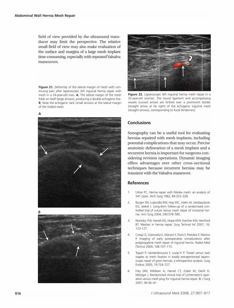
may appear anechoic on sonography (Figure17). Abscesses (Figure 18) should be consid-ered with any postoperative fluid collection,especially when heterogeneous or complex.Rarely an enterocutaneous fistula may develop
(Figure 19). The latter is an unusual complica-tion and would require other imaging becausethe posterior acoustic shadowing from gas andmesh on sonography limits evaluation of deepsoft tissue structures.20
J Ultrasound Med 2008; 27:907–917 913
Jamadar et al
Figure 11. Right inguinal mesh in a 49-year-old man. Onsonography, the mesh is difficult to see and appears linear andminimally hyperechoic (arrows) with posterior acoustic shadow-ing (S). The clinical history was important in helping identify themesh in this patient.
Figure 13. Left inguinal mesh in a 47-year-old man.Sonography shows the mesh (arrows) with a wavy contour. Rindicates rectus abdominis muscle.
Figure 12. Right inguinal mesh in a 21-year-old man.Sonography shows the hyperechoic mesh (arrows) with a wavycontour and posterior acoustic shadowing (S).
Figure 14. Underlay mesh in a 39-year-old woman. Sonographyshows a very wavy (crinkly) appearance of the mesh (arrows). Rindicates rectus abdominis muscle; and S, posterior acousticshadowing.
276jumonline.qxp:Layout 1 5/15/08 9:14 AM Page 913
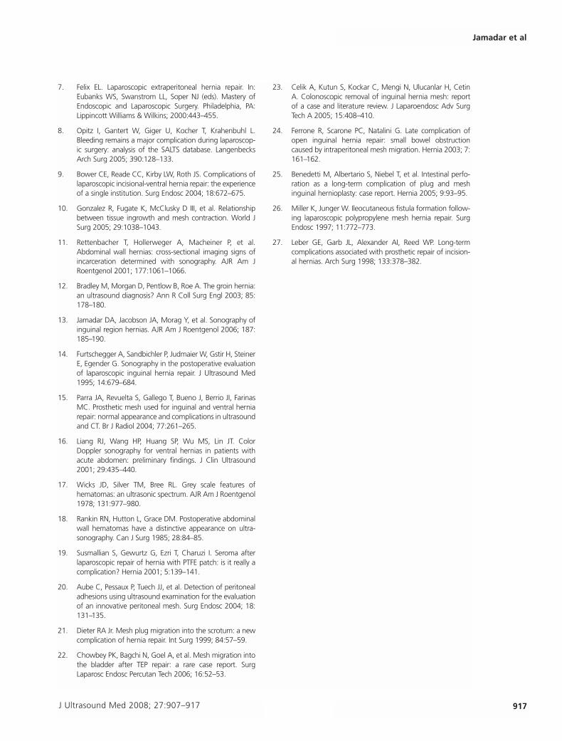
The margin of the mesh may appear as a pal-pable mass (Figure 20) and may cause con-cern for a new mass or a recurrent hernia.The margin of the mesh may fold back onitself (Figure 21) and cause focal irritation.Structures passing over the margin of themesh may become kinked (Figure 22) and irri-tated. This usually occurs after inguinal herniarepair in which the spermatic cord and itscontents and adjacent nerves may deviate
from their course passing over the margin ofthe mesh. The mesh plugs may be displaced bya recurrent hernia alongside the plug (Figure10). If a migrated mesh plug enters the peri-toneal cavity, it may potentially perforate thebowel as a further complication. Other compli-cations include migration of mesh and themesh plug,21–23 intestinal obstruction, perfora-tion and fistula formation,24–26 strangulated her-nias, and a burst abdomen.2,27
914 J Ultrasound Med 2008; 27:907–917
Abdominal Wall Hernia Mesh Repair
Figure 15. Right upper quadrant underlay mesh repair of an incisional hernia after laparoscopic cholecystectomy in a 39-year-oldwoman. A, Identification of the mesh (arrows) and acoustic shadowing (S) may be limited with the smaller field of view (depth). B, An increased field of view (depth) allows better appreciation of acoustic shadowing and identification of the mesh (arrows).
A B
Figure 16. Recurrent hernia at the left lateral margin of the mesh after an underlay repair of a ventral midline incisional hernia in a60-year-old woman. A, The irreducible fat-containing hernia (H) lies directly on the lateral border of the mesh, with its neck (N, curvedarrow) well demarcated by the lateral border of the echogenic mesh (straight arrows) medially. B, Color Doppler imaging shows bloodflow in this irreducible hernia lying on the lateral margin of the mesh (arrows).
A B
276jumonline.qxp:Layout 1 5/15/08 9:14 AM Page 914
Sonography may identify an indication forsurgery, such as a tight neck around a loop ofbowel, a loop of bowel where there is suspicionof strangulation, or a fluid collection that isinfected. Pain, nausea, and limited abdominalwall function associated with a hernia defectlower the surgeon’s threshold for surgery.
Limitations
With sonography, the acoustic shadowing deepto the mesh makes evaluation of structuresdeep to the mesh difficult if not impossible toevaluate. The distorted anatomy after herniarepair may be confusing, particularly with largemidline implants. In these cases, the smaller
J Ultrasound Med 2008; 27:907–917 915
Jamadar et al
B
Figure 17. Seroma in a 68-year-old man. Sonography shows ananechoic fluid collection (S) at the superficial surface of thewavy echogenic mesh (arrows) after repair of a ventral midlineincisional hernia.
Figure 19. Enterocutaneous fistula after mesh placement in theanterior abdominal wall of a 53-year-old woman. Sonographyshows the cutaneous opening (O) and the hyperemic echogenicphlegmon (P) lying superficial to 2 layers of wavy echogenicmesh (arrows).
Figure 18. Complex wound collection after mesh placementfor a ventral midline incisional hernia in a 50-year-old woman.A draining sinus discharged sterile purulent-looking material.A, Sonography in the axial plane shows the mesh with anechogenic folded contour (arrows) and posterior acoustic shad-owing (S). An adjacent collection comprising both fluid (F) anddebris (D) is shown just superficial to the mesh. B, Computedtomography shows the anterior abdominal wall mesh (straightarrows) and adjacent collection (curved arrow).
A
Figure 20. Palpable edge of mesh after midline incisional her-nia repair in a 46-year-old woman. The palpable epigastric lumpcorresponded to the superficial free edge (arrow at left) of theimplanted mesh (arrows) rather than a recurrent hernia. S indi-cates posterior acoustic shadowing.
276jumonline.qxp:Layout 1 5/15/08 9:14 AM Page 915
field of view provided by the ultrasound trans-ducer may limit the perspective. The relativesmall field of view may also make evaluation ofthe surface and margins of a large mesh implanttime-consuming, especially with repeated Valsalvamaneuvers.
Conclusions
Sonography can be a useful tool for evaluatinghernias repaired with mesh implants, includingpotential complications that may occur. Preciseanatomic delineation of a mesh implant and arecurrent hernia is important for surgeons con-sidering revision operations. Dynamic imagingoffers advantages over other cross-sectionaltechniques because recurrent hernias may betransient with the Valsalva maneuver.
References
1. Usher FC, Hernia repair with Marlex mesh: an analysis of541 cases. Arch Surg 1962; 84:325–328.
2. Burger JW, Luijendikj RW, Hop WC, Halm JA, VerdaasdonkEG, Jeekel J. Long-term follow-up of a randomized con-trolled trial of suture versus mesh repair of incisional her-nia. Ann Surg 2004; 240:578–585.
3. Novitsky YW, Harrell AG, Hope WW, Kercher KW, HenifordBT. Meshes in hernia repair. Surg Technol Int 2007; 16:123–127.
4. Crespi G, Giannetta E, Mariani F, Floris F, Pretolesi F, MarinoP. Imaging of early postoperative complications afterpolypropylene mesh repair of inguinal hernia. Radiol Med(Torino) 2004; 108:107–115.
5. Topart P, Vandenbroucke F, Lozac’h P. Tisseel versus tackstaples as mesh fixation in totally extraperitoneal laparo-scopic repair of groin hernias: a retrospective analysis. SurgEndosc 2005; 19:724–727.
6. Frey DM, Wildisen A, Hamel CT, Zuber M, Oertli D,Metzger J. Randomized clinical trial of Lichtenstein’s oper-ation versus mesh plug for inguinal hernia repair. Br J Surg2007; 94:36–41.
916 J Ultrasound Med 2008; 27:907–917
Abdominal Wall Hernia Mesh Repair
Figure 22. Laparoscopic left inguinal hernia mesh repair in a 32-year-old woman. The round ligament and accompanyingvessels (curved arrow) are kinked over a prominent border(straight arrow at far right) of the echogenic inguinal mesh(straight arrows), corresponding to focal tenderness.
Figure 21. Deformity of the lateral margin of mesh with con-tinuing pain after laparoscopic left inguinal hernia repair withmesh in a 24-year-old man. A, The lateral margin of the meshfolds on itself (large arrows), producing a double echogenic line.B, Note the echogenic tack (small arrows) at the lateral marginof the folded mesh.
A
B
276jumonline.qxp:Layout 1 5/15/08 9:14 AM Page 916
7. Felix EL. Laparoscopic extraperitoneal hernia repair. In:Eubanks WS, Swanstrom LL, Soper NJ (eds). Mastery ofEndoscopic and Laparoscopic Surgery. Philadelphia, PA:Lippincott Williams & Wilkins; 2000:443–455.
8. Opitz I, Gantert W, Giger U, Kocher T, Krahenbuhl L.Bleeding remains a major complication during laparoscop-ic surgery: analysis of the SALTS database. LangenbecksArch Surg 2005; 390:128–133.
9. Bower CE, Reade CC, Kirby LW, Roth JS. Complications oflaparoscopic incisional-ventral hernia repair: the experienceof a single institution. Surg Endosc 2004; 18:672–675.
10. Gonzalez R, Fugate K, McClusky D III, et al. Relationshipbetween tissue ingrowth and mesh contraction. World JSurg 2005; 29:1038–1043.
11. Rettenbacher T, Hollerweger A, Macheiner P, et al.Abdominal wall hernias: cross-sectional imaging signs ofincarceration determined with sonography. AJR Am JRoentgenol 2001; 177:1061–1066.
12. Bradley M, Morgan D, Pentlow B, Roe A. The groin hernia:an ultrasound diagnosis? Ann R Coll Surg Engl 2003; 85:178–180.
13. Jamadar DA, Jacobson JA, Morag Y, et al. Sonography ofinguinal region hernias. AJR Am J Roentgenol 2006; 187:185–190.
14. Furtschegger A, Sandbichler P, Judmaier W, Gstir H, SteinerE, Egender G. Sonography in the postoperative evaluationof laparoscopic inguinal hernia repair. J Ultrasound Med1995; 14:679–684.
15. Parra JA, Revuelta S, Gallego T, Bueno J, Berrio JI, FarinasMC. Prosthetic mesh used for inguinal and ventral herniarepair: normal appearance and complications in ultrasoundand CT. Br J Radiol 2004; 77:261–265.
16. Liang RJ, Wang HP, Huang SP, Wu MS, Lin JT. ColorDoppler sonography for ventral hernias in patients withacute abdomen: preliminary findings. J Clin Ultrasound2001; 29:435–440.
17. Wicks JD, Silver TM, Bree RL. Grey scale features ofhematomas: an ultrasonic spectrum. AJR Am J Roentgenol1978; 131:977–980.
18. Rankin RN, Hutton L, Grace DM. Postoperative abdominalwall hematomas have a distinctive appearance on ultra-sonography. Can J Surg 1985; 28:84–85.
19. Susmallian S, Gewurtz G, Ezri T, Charuzi I. Seroma afterlaparoscopic repair of hernia with PTFE patch: is it really acomplication? Hernia 2001; 5:139–141.
20. Aube C, Pessaux P, Tuech JJ, et al. Detection of peritonealadhesions using ultrasound examination for the evaluationof an innovative peritoneal mesh. Surg Endosc 2004; 18:131–135.
21. Dieter RA Jr. Mesh plug migration into the scrotum: a newcomplication of hernia repair. Int Surg 1999; 84:57–59.
22. Chowbey PK, Bagchi N, Goel A, et al. Mesh migration intothe bladder after TEP repair: a rare case report. SurgLaparosc Endosc Percutan Tech 2006; 16:52–53.
23. Celik A, Kutun S, Kockar C, Mengi N, Ulucanlar H, CetinA. Colonoscopic removal of inguinal hernia mesh: reportof a case and literature review. J Laparoendosc Adv SurgTech A 2005; 15:408–410.
24. Ferrone R, Scarone PC, Natalini G. Late complication ofopen inguinal hernia repair: small bowel obstructioncaused by intraperitoneal mesh migration. Hernia 2003; 7:161–162.
25. Benedetti M, Albertario S, Niebel T, et al. Intestinal perfo-ration as a long-term complication of plug and meshinguinal hernioplasty: case report. Hernia 2005; 9:93–95.
26. Miller K, Junger W. Ileocutaneous fistula formation follow-ing laparoscopic polypropylene mesh hernia repair. SurgEndosc 1997; 11:772–773.
27. Leber GE, Garb JL, Alexander AI, Reed WP. Long-termcomplications associated with prosthetic repair of incision-al hernias. Arch Surg 1998; 133:378–382.
J Ultrasound Med 2008; 27:907–917 917
Jamadar et al
276jumonline.qxp:Layout 1 5/15/08 9:14 AM Page 917