Embed Size (px)
Citation preview
REVIEW Open Access
Aberrant structural and functionalconnectivity and neurodevelopmentalimpairment in preterm childrenCynthia E. Rogers1* , Rachel E. Lean2, Muriah D. Wheelock2 and Christopher D. Smyser3
Abstract
Background: Despite advances in antenatal and neonatal care, preterm birth remains a leading cause ofneurological disabilities in children. Infants born prematurely, particularly those delivered at the earliest gestationalages, commonly demonstrate increased rates of impairment across multiple neurodevelopmental domains. Indeed,the current literature establishes that preterm birth is a leading risk factor for cerebral palsy, is associated withexecutive function deficits, increases risk for impaired receptive and expressive language skills, and is linked withhigher rates of co-occurring attention deficit hyperactivity disorder, anxiety, and autism spectrum disorders. Thesesame infants also demonstrate elevated rates of aberrant cerebral structural and functional connectivity, withpersistent changes evident across advanced magnetic resonance imaging modalities as early as the neonatalperiod. Emerging findings from cross-sectional and longitudinal investigations increasingly suggest that aberrantconnectivity within key functional networks and white matter tracts may underlie the neurodevelopmentalimpairments common in this population.
Main body: This review begins by highlighting the elevated rates of neurodevelopmental disorders across domainsin this clinical population, describes the patterns of aberrant structural and functional connectivity common inprematurely-born infants and children, and then reviews the increasingly established body of literature delineatingthe relationship between these brain abnormalities and adverse neurodevelopmental outcomes. We also detailimportant, typically understudied, clinical, and social variables that may influence these relationships amongpreterm children, including heritability and psychosocial risks.
Conclusion: Future work in this domain should continue to leverage longitudinal evaluations of preterm infantswhich include both neuroimaging and detailed serial neurodevelopmental assessments to further characterizerelationships between imaging measures and impairment, information necessary for advancing our understandingof modifiable risk factors underlying these disorders and best practices for improving neurodevelopmentaltrajectories in this high-risk clinical population.
Keywords: Prematurity, Neurodevelopmental disorders, Functional connectivity, Structural connectivity, Magneticresonance imaging
* Correspondence: [email protected] of Psychiatry and Pediatrics, Washington University School ofMedicine, 660 South Euclid Avenue, Campus Box 8504, St. Louis, MO 63110,USAFull list of author information is available at the end of the article
© The Author(s). 2018 Open Access This article is distributed under the terms of the Creative Commons Attribution 4.0International License (http://creativecommons.org/licenses/by/4.0/), which permits unrestricted use, distribution, andreproduction in any medium, provided you give appropriate credit to the original author(s) and the source, provide a link tothe Creative Commons license, and indicate if changes were made. The Creative Commons Public Domain Dedication waiver(http://creativecommons.org/publicdomain/zero/1.0/) applies to the data made available in this article, unless otherwise stated.
Rogers et al. Journal of Neurodevelopmental Disorders (2018) 10:38 https://doi.org/10.1186/s11689-018-9253-x
Preterm birth remains a major public health issue due toits high incidence combined with the frequency of neu-rodevelopmental impairments among surviving infants.In this review, we begin by highlighting the adverse ef-fects of prematurity on trajectories across neurodevelop-mental domains. Next, we discuss the increasinglyestablished relationship between aberrant brain develop-ment and preterm birth, with particular focus on the ad-vanced magnetic resonance imaging (MRI) techniquesincreasingly utilized to delineate the changes in cerebralstructural and functional connectivity related to prema-turity. We then review selected studies from the extantliterature which suggest that prematurity-associatedchanges in cerebral structural and functional connectiv-ity may underlie the neurodevelopmental impairmentscommon among prematurely born children and adults.Finally, we conclude by detailing relevant clinical and so-cial variables that may influence these relationships inthis high-risk clinical population.
Prematurity and neurodevelopmental disordersPremature birth affects more than 500,000 newborns inthe USA each year, occurring in approximately 10% ofall births in 2016 [1]. Survival rates for these infants haveimproved dramatically due to advances in perinatal andneonatal care. In contrast to this improvement in mor-tality, long-term neurodevelopmental outcomes have notimproved, with preterm birth remaining a leading causeof neurological disabilities in children [2]. These surviv-ing preterm children face a range of neurodevelopmentaland neurobehavioral challenges [3–7], with more than30% experiencing impairments across multiple neurode-velopmental domains [8]. Children delivered very pre-term (VPT; born at ≤ 32 weeks’ gestation) typically facedisproportionate risk, with infants born earliest facing thehighest rates of developmental disability [9]. However,these adverse effects are not universal, with widely variedoutcomes among preterm children with similar neonatalclinical phenotypes. Critically, the associated costs in car-ing for these children are enormous, amounting to morethan $25 billion annually in the USA alone [10].Among preterm children, prominent neurodevelop-
mental difficulties are seen across motor, cognitive,language, and social-emotional domains [11–14]. Theseareas warrant particular focus due not only to their crit-ical functional importance, but also to their significantimpact on quality of life, including poor peer relation-ships [15] and academic underachievement [16–18].Over 50% of children diagnosed with cerebral palsy areborn preterm, with the greatest likelihood among thoseborn at the earliest gestational ages [19]. An even largerproportion of preterm children experience other moresubtle fine and gross motor problems, with approxi-mately 40% displaying mild to moderate motor
impairment [12]. Similarly, 15–20% of intellectual dis-abilities and 10–15% of other learning disorders are at-tributable to preterm birth. VPT children obtain FullScale Intelligence Quotient (IQ) scores up to 10 pointslower than term children [20, 21]. Furthermore, VPTchildren consistently perform worse than term-bornpeers on executive function tasks assessing planning, flu-ency, working memory, and response inhibition [22–24].Preterm children also demonstrate problems in selective,sustained, and executive attention, with up to 41% ofVPT and 62% of extremely preterm (born at < 28 weeks’gestation) children in the impaired range [25–28]. Fur-ther, large effect sizes have been reported for executiveshifting and divided attention [25, 26, 29], suggestingVPT children particularly struggle with top-down con-trol of attention processes. In addition, approximately35% of children born between 31 and 34 weeks’ gestationdemonstrate language impairments at preschool-age,with rates as high as 48% for children born at less than30 weeks gestation [30]. Deficits in both receptive andexpressive language domains persist into school age, af-fecting skills such as word finding, perception, grammar,dialog, and linguistics [30–34]. Critically, across each ofthese neurodevelopmental domains, preterm birth re-mains a strong risk factor for impairment even after ac-counting for sociodemographic risk [19, 35].More recently, elevated rates of social-emotional deficits
and psychiatric disorders have been recognized amongchildren born preterm, with increasing numbers of reportsdetailing the “preterm behavioral phenotype” [36], com-prised of inattention, anxiety, and social-communicationdeficits [37]. These comorbid symptoms and the relateddisorders of Attention-deficit hyperactivity disorder(ADHD), anxiety, and autism spectrum disorder (ASD)are two to four times more common among preterm chil-dren [5, 38–43]. As with other neurodevelopmental im-pairments, children born VPT are at greatest risk for thesesocial-emotional impairments and psychiatric diagnoses[36]. Further, studies examining the trajectory of thesesymptoms demonstrate their persistence into adolescence[5, 44–49]. Importantly, rates of these disorders remain el-evated even after accounting for the increased frequencyof other neurodevelopmental disabilities, including motorand intellectual impairments [36].
Assessment of functional and structuralconnectivity in preterm children using MRIThe timing of key interrelated neurobiological processesunderlying development of early cerebral functional andstructural connectivity make the preterm brain uniquelyvulnerable to the perturbations that have been associatedwith common neurodevelopmental disorders. Most notably,this includes processes such as neuronal migration, synap-togenesis, cortical folding, emergence of thalamo-cortical
Rogers et al. Journal of Neurodevelopmental Disorders (2018) 10:38 Page 2 of 13
connections, and myelination [50]. Early investigations ofchildren born preterm employed conventional MRI tocharacterize the alterations in cerebral structural develop-ment associated with preterm birth [51–55]. These pre-dominantly cross-sectional investigations focused onmetrics of brain growth, regional brain volumes, and cor-tical folding, demonstrating atypical patterns of maturationthroughout the brain across techniques in preterm children[56–58]. However, these modalities provided only limitedability to elucidate the alterations in cerebral developmentthat lead to neurodevelopmental deficits; information crit-ical for understanding the pathway to disability.Advanced MRI techniques, including resting state-
functional MRI (rs-fMRI) and diffusion MRI (dMRI),provide powerful, non-invasive tools with high sensitivityfor delineating alterations in the developing brain.rs-fMRI is used to detect temporal correlations in spon-taneous, low-frequency fluctuations in blood oxygenlevel-dependent signal, thereby identifying functionalconnectivity networks from data acquired without re-quiring subjects to perform tasks during acquisition[59–61]. These resting state networks incorporate graymatter regions known to be anatomically connected andco-activated by task performance [60, 62, 63]. dMRIcharacterizes cerebral structural connectivity throughquantification of water displacement within the whitematter microstructural architecture [64–66]. In manyways, these modalities are well-suited for investigationsof infants and pediatric populations; from a study lastingminutes in duration on an infant at rest, robust mea-sures of global functional and structural connectivity canbe obtained. Further, both modalities have been usedsuccessfully to investigate cerebral connectivity in VPTadults and older pediatric populations, demonstratingatypical connectivity patterns which correlate with neu-rodevelopmental disability [57, 67–71].Unique methodological challenges have now been over-
come to successfully study neonates and young childrenusing rs-fMRI and dMRI, including scan sequence specifi-cation, scanning of non-sedated subjects, effects of smallbrain sizes on atlas registration, and development of dataprocessing streams [72–75]. Our group and others havesubsequently used these techniques to identify immatureforms of multiple canonical resting state networks andwhite matter tracts throughout the brain as early as 26weeks postmenstrual age (PMA). These systems reflectthe functional and structural topography of the developingbrain, gradually maturing with advancing age [74]. Ourrecent applications of rs-fMRI and dMRI demonstrateinfants possess a functional and structural network archi-tecture similar to that described in adults, with maturationrates emulating known histological evidence regardingbrain development [74, 76, 77]. For example, networks(e.g., somatomotor, auditory, visual networks) and tracts
(e.g., corticospinal tracts, optic radiations) in areas of thebrain known to develop early demonstrate mature top-ology by term equivalent PMA. In contrast, networks (e.g.,default mode [DMN], frontoparietal [FPN], cingulo-oper-cular [CO] networks), and tracts (e.g., cingulum bundle,uncinate) located in higher-order association cortices in-volved in top-down control of emotion regulation, atten-tion, and cognition do not demonstrate adult-liketopology until later in life.Further, these methods are sensitive to the changes in
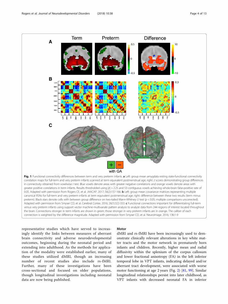
functional and structural connectivity associated withpremature birth (Fig. 1). Across rs-fMRI investigations,infants born prematurely demonstrate similar overallresting state network topography to term-born infantsscanned at comparable PMA, though with weaker in-trinsic brain activity. The magnitude of these differencesin network amplitude and dimensionality differ by net-work and are typically most prominent in those locatedin higher-order association cortices [74, 77, 78]. Infantswith forms of white matter injury common in pretermpopulations (e.g., intraventricular hemorrhage, cysticperiventricular leukomalacia) demonstrate aberrant net-work development, dependent upon severity and prox-imity to the injury site [79]. Interrelated investigations ofstructural connectivity using dMRI also demonstratecomparable regionally specific differences in gray andwhite matter microstructural development between pre-term and term-born infants [80–86]. Across these stud-ies, prematurely born infants demonstrate delayed whitematter tract development, with susceptibility to specificclinical factors (e.g., antenatal steroids, white matter in-jury) also reported. Further, these neuroimaging data areconducive to technically sophisticated analysis ap-proaches designed to investigate complex patterns inneuroimaging data, such as graph theory and machinelearning [76, 87–91]. Use of these methods in neonatesand older pediatric populations have demonstrated theimportance of connectivity within and between networksfor differentiation of term- and prematurely born infantsand continuous measure (i.e., birth gestational age) pre-diction [92–94]. These studies provide converging linesof evidence suggesting neurodevelopmental impairmentmay directly correlate with disruptions in specific struc-tural and functional systems.
Prematurity-related changes in functional andstructural connectivity and developmentalimpairmentThere is a small, but burgeoning literature investigat-ing the relationship between cerebral functional andstructural connectivity changes and motor, cognitive,language, and social-emotional outcomes in prema-turely born children [54, 58, 81, 95–103]. For brevity,across each of these domains, we highlight
Rogers et al. Journal of Neurodevelopmental Disorders (2018) 10:38 Page 3 of 13
representative studies which have served to increas-ingly identify the links between measures of aberrantbrain connectivity and adverse neurodevelopmentaloutcomes, beginning during the neonatal period andextending into adulthood. As the methods for applica-tion of the modality were established earlier, many ofthese studies utilized dMRI, though an increasingnumber of recent studies also include rs-fMRI.Further, many of these investigations have beencross-sectional and focused on older populations,though longitudinal investigations including neonataldata are now being published.
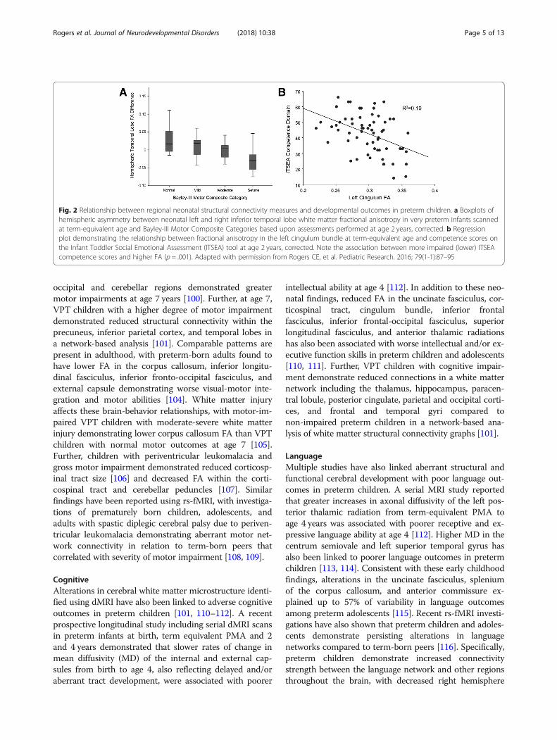
MotordMRI and rs-fMRI have been increasingly used to dem-onstrate clinically relevant alterations in key white mat-ter tracts and the motor network in prematurely borninfants and children. Recently, higher mean and radialdiffusivity within the splenium of the corpus callosumand lower fractional anisotropy (FA) in the left inferiortemporal lobe in VPT infants, indicating delayed and/oraberrant tract development, were associated with worsemotor functioning at age 2 years (Fig. 2) [81, 99]. Similarlongitudinal relationships persist into later childhood, asVPT infants with decreased neonatal FA in inferior
Fig. 1 Functional connectivity differences between term and very preterm infants. a Left: group mean amygdala resting state-functional connectivitycorrelation maps for full-term and very preterm infants scanned at term equivalent postmenstrual age; right: z scores demonstrating group differencesin connectivity obtained from voxelwise t test. Blue voxels denote areas with greater negative correlations and orange voxels denote areas withgreater positive correlations in term infants. Results thresholded using |z| > 2.25 and 53 contiguous voxels achieving whole-brain false-positive rate of0.05. Adapted with permission from Rogers CE, et al. JAACAP. 2017; 56(2):157-166. b Left: group mean covariance matrices representing multiplecanonical RSNs for full-term and very preterm infants at term equivalent postmenstrual age; right: difference between these two results (term minuspreterm). Black stars denote cells with between group difference on two-tailed Mann-Whitney U test (p < 0.05; multiple comparisons uncorrected).Adapted with permission from Smyser CD, et al. Cerebral Cortex. 2016; 26(1):322-333. c Functional connections important for differentiating full-termversus very preterm infants using support vector machine-multivariate pattern analysis to analyze data from 244 regions of interest located throughoutthe brain. Connections stronger in term infants are shown in green; those stronger in very preterm infants are in orange. The caliber of eachconnection is weighted by the difference magnitude. Adapted with permission from Smyser CD, et al. NeuroImage. 2016; 136:1-9
Rogers et al. Journal of Neurodevelopmental Disorders (2018) 10:38 Page 4 of 13
occipital and cerebellar regions demonstrated greatermotor impairments at age 7 years [100]. Further, at age 7,VPT children with a higher degree of motor impairmentdemonstrated reduced structural connectivity within theprecuneus, inferior parietal cortex, and temporal lobes ina network-based analysis [101]. Comparable patterns arepresent in adulthood, with preterm-born adults found tohave lower FA in the corpus callosum, inferior longitu-dinal fasciculus, inferior fronto-occipital fasciculus, andexternal capsule demonstrating worse visual-motor inte-gration and motor abilities [104]. White matter injuryaffects these brain-behavior relationships, with motor-im-paired VPT children with moderate-severe white matterinjury demonstrating lower corpus callosum FA than VPTchildren with normal motor outcomes at age 7 [105].Further, children with periventricular leukomalacia andgross motor impairment demonstrated reduced corticosp-inal tract size [106] and decreased FA within the corti-cospinal tract and cerebellar peduncles [107]. Similarfindings have been reported using rs-fMRI, with investiga-tions of prematurely born children, adolescents, andadults with spastic diplegic cerebral palsy due to periven-tricular leukomalacia demonstrating aberrant motor net-work connectivity in relation to term-born peers thatcorrelated with severity of motor impairment [108, 109].
CognitiveAlterations in cerebral white matter microstructure identi-fied using dMRI have also been linked to adverse cognitiveoutcomes in preterm children [101, 110–112]. A recentprospective longitudinal study including serial dMRI scansin preterm infants at birth, term equivalent PMA and 2and 4 years demonstrated that slower rates of change inmean diffusivity (MD) of the internal and external cap-sules from birth to age 4, also reflecting delayed and/oraberrant tract development, were associated with poorer
intellectual ability at age 4 [112]. In addition to these neo-natal findings, reduced FA in the uncinate fasciculus, cor-ticospinal tract, cingulum bundle, inferior frontalfasciculus, inferior frontal-occipital fasciculus, superiorlongitudinal fasciculus, and anterior thalamic radiationshas also been associated with worse intellectual and/or ex-ecutive function skills in preterm children and adolescents[110, 111]. Further, VPT children with cognitive impair-ment demonstrate reduced connections in a white matternetwork including the thalamus, hippocampus, paracen-tral lobule, posterior cingulate, parietal and occipital corti-ces, and frontal and temporal gyri compared tonon-impaired preterm children in a network-based ana-lysis of white matter structural connectivity graphs [101].
LanguageMultiple studies have also linked aberrant structural andfunctional cerebral development with poor language out-comes in preterm children. A serial MRI study reportedthat greater increases in axonal diffusivity of the left pos-terior thalamic radiation from term-equivalent PMA toage 4 years was associated with poorer receptive and ex-pressive language ability at age 4 [112]. Higher MD in thecentrum semiovale and left superior temporal gyrus hasalso been linked to poorer language outcomes in pretermchildren [113, 114]. Consistent with these early childhoodfindings, alterations in the uncinate fasciculus, spleniumof the corpus callosum, and anterior commissure ex-plained up to 57% of variability in language outcomesamong preterm adolescents [115]. Recent rs-fMRI investi-gations have also shown that preterm children and adoles-cents demonstrate persisting alterations in languagenetworks compared to term-born peers [116]. Specifically,preterm children demonstrate increased connectivitystrength between the language network and other regionsthroughout the brain, with decreased right hemisphere
Fig. 2 Relationship between regional neonatal structural connectivity measures and developmental outcomes in preterm children. a Boxplots ofhemispheric asymmetry between neonatal left and right inferior temporal lobe white matter fractional anisotropy in very preterm infants scannedat term-equivalent age and Bayley-III Motor Composite Categories based upon assessments performed at age 2 years, corrected. b Regressionplot demonstrating the relationship between fractional anisotropy in the left cingulum bundle at term-equivalent age and competence scores onthe Infant Toddler Social Emotional Assessment (ITSEA) tool at age 2 years, corrected. Note the association between more impaired (lower) ITSEAcompetence scores and higher FA (p = .001). Adapted with permission from Rogers CE, et al. Pediatric Research. 2016; 79(1-1):87–95
Rogers et al. Journal of Neurodevelopmental Disorders (2018) 10:38 Page 5 of 13
lateralization [117, 118]. These differences have beenrelated to language performance, with preterm adoles-cents demonstrating weaker bilateral connectivity be-tween left and right superior temporal regions alsodemonstrating poorer language ability at age 14–15years [115, 116], with other regionally specific rela-tionships also reported [68, 119, 120].
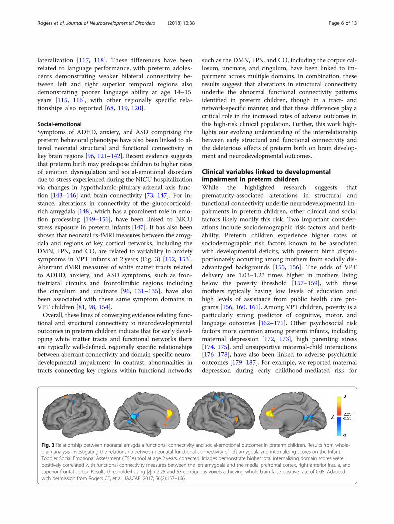
Social-emotionalSymptoms of ADHD, anxiety, and ASD comprising thepreterm behavioral phenotype have also been linked to al-tered neonatal structural and functional connectivity inkey brain regions [96, 121–142]. Recent evidence suggeststhat preterm birth may predispose children to higher ratesof emotion dysregulation and social-emotional disordersdue to stress experienced during the NICU hospitalizationvia changes in hypothalamic-pituitary-adrenal axis func-tion [143–146] and brain connectivity [73, 147]. For in-stance, alterations in connectivity of the glucocorticoid-rich amygdala [148], which has a prominent role in emo-tion processing [149–151], have been linked to NICUstress exposure in preterm infants [147]. It has also beenshown that neonatal rs-fMRI measures between the amyg-dala and regions of key cortical networks, including theDMN, FPN, and CO, are related to variability in anxietysymptoms in VPT infants at 2 years (Fig. 3) [152, 153].Aberrant dMRI measures of white matter tracts relatedto ADHD, anxiety, and ASD symptoms, such as fron-tostriatal circuits and frontolimibic regions includingthe cingulum and uncinate [96, 131–135], have alsobeen associated with these same symptom domains inVPT children [81, 98, 154].Overall, these lines of converging evidence relating func-
tional and structural connectivity to neurodevelopmentaloutcomes in preterm children indicate that for early devel-oping white matter tracts and functional networks thereare typically well-defined, regionally specific relationshipsbetween aberrant connectivity and domain-specific neuro-developmental impairment. In contrast, abnormalities intracts connecting key regions within functional networks
such as the DMN, FPN, and CO, including the corpus cal-losum, uncinate, and cingulum, have been linked to im-pairment across multiple domains. In combination, theseresults suggest that alterations in structural connectivityunderlie the abnormal functional connectivity patternsidentified in preterm children, though in a tract- andnetwork-specific manner, and that these differences play acritical role in the increased rates of adverse outcomes inthis high-risk clinical population. Further, this work high-lights our evolving understanding of the interrelationshipbetween early structural and functional connectivity andthe deleterious effects of preterm birth on brain develop-ment and neurodevelopmental outcomes.
Clinical variables linked to developmentalimpairment in preterm childrenWhile the highlighted research suggests thatprematurity-associated alterations in structural andfunctional connectivity underlie neurodevelopmental im-pairments in preterm children, other clinical and socialfactors likely modify this risk. Two important consider-ations include sociodemographic risk factors and herit-ability. Preterm children experience higher rates ofsociodemographic risk factors known to be associatedwith developmental deficits, with preterm birth dispro-portionately occurring among mothers from socially dis-advantaged backgrounds [155, 156]. The odds of VPTdelivery are 1.03–1.27 times higher in mothers livingbelow the poverty threshold [157–159], with thesemothers typically having low levels of education andhigh levels of assistance from public health care pro-grams [156, 160, 161]. Among VPT children, poverty is aparticularly strong predictor of cognitive, motor, andlanguage outcomes [162–171]. Other psychosocial riskfactors more common among preterm infants, includingmaternal depression [172, 173], high parenting stress[174, 175], and unsupportive maternal-child interactions[176–178], have also been linked to adverse psychiatricoutcomes [179–187]. For example, we reported maternaldepression during early childhood-mediated risk for
Fig. 3 Relationship between neonatal amygdala functional connectivity and social-emotional outcomes in preterm children. Results from whole-brain analysis investigating the relationship between neonatal functional connectivity of left amygdala and internalizing scores on the InfantToddler Social Emotional Assessment (ITSEA) tool at age 2 years, corrected. Images demonstrate higher total internalizing domain scores werepositively correlated with functional connectivity measures between the left amygdala and the medial prefrontal cortex, right anterior insula, andsuperior frontal cortex. Results thresholded using |z| > 2.25 and 53 contiguous voxels achieving whole-brain false-positive rate of 0.05. Adaptedwith permission from Rogers CE, et al. JAACAP. 2017; 56(2):157–166
Rogers et al. Journal of Neurodevelopmental Disorders (2018) 10:38 Page 6 of 13
anxiety disorders associated with preterm birth [188]. Inaddition, these same risk factors have been linked tochanges in brain development, with exposure to povertyand unsupportive caregiving impacting functional andstructural brain development in offspring [186, 189–191].Thus, preterm birth both increases the likelihood of ex-periencing early psychosocial adversity and alters func-tional and structural development of the neonatalbrain. Further, the developing brain may remain highlyvulnerable to continued alterations from repeated expo-sures to psychosocial adversity extending beyond theneonatal period.Another key and understudied risk factor among pre-
term children is heritability. Studies investigating herit-ability suggest that family background determines thelower and upper limits of the range in which a heritableand continuously distributed trait may be expressed, butthat neurodevelopmental disorders increase the pheno-typic variability of trait expression during childhood[192, 193]. For instance, maternal intellectual ability hasa direct influence on her children’s intellectual develop-ment because it is a genetically based and heritable trait[194]. Preterm children born to mothers with low levelsof intellectual ability may therefore be at higher risk ofpoor outcomes. Indeed, our analysis of maternal intellec-tual ability demonstrated that maternal IQ scores wereassociated with both preterm and term child IQ and lan-guage scores at age 5 years [195]. However, the associ-ation between maternal IQ and child IQ and languageoutcomes was weaker for preterm children, indicatingpreterm birth itself was an important factor explainingintellectual and language development. Further, herit-ability is an important variable for social-emotional de-velopment and psychiatric symptoms underlying thepreterm behavioral phenotype, as ADHD, ASD, and anx-iety symptoms are all highly heritable [196–199]. Insome cases, the heritability of social-emotional symp-toms may confound the relationship between prematur-ity and social-emotional development. For instance,substance-abusing mothers are more likely to both haveADHD [200, 201] and anxiety [202] and to deliver pre-term [203, 204]. A similar relationship could exist be-tween the highly related variables of maternal depressionand both preterm delivery [205] and childhood anxiety[206]. These findings highlight the need to assess psy-chosocial risk factors and heritability among families inall research investigating links between preterm birthand neurodevelopmental outcomes.
Future directions and conclusionsContinued research remains necessary to both furtherdelineate the relationships between imaging measuresand neurodevelopmental impairment in prematurelyborn children and better characterize the role of
modifiable risk factors such as psychosocial adversity inthis trajectory. While MRI affords several advantages forstudying these associations, including improved spatialresolution and anatomic specificity, future investigationsmay utilize other complementary modalities for asses-sing brain development and function. These includefunctional near infrared spectroscopy (fNIRS), whichmeasures hemodynamic contrasts [207–209] and elec-troencephalography (EEG), which assesses the coherenceof cortical electrical activity and has been used to suc-cessfully model brain connectivity-behavior associations[210]. In addition, diffuse optical tomography (DOT) en-ables measurements of functional connectivity whichalign with rs-fMRI, though with a more limited field ofview [211, 212]. Limitations notwithstanding, these port-able methods can be readily employed to perform serialstudies at the bedside, providing avenues for novel inves-tigation by enabling the study of clinical populations ofinterest unable to undergo MRI.Future work should also focus on extending longitu-
dinal evaluations of preterm children across early child-hood, leveraging recent advances in MRI acquisition andanalysis methods and incorporating advances developedand implemented among other clinical populations. Forexample, the Infant Brain Imaging Study has performedlongitudinal MRI scanning of infants at risk for autismbeginning at 6 months of age with repeat MRI scans at12 and 24 months, reporting changes in both structuraland functional connectivity parameters utilizing longitu-dinal analyses of brain development and innovativebrain-behavior analyses [127, 213]. More recently, theUNC/UMN Baby Connectome Project (BCP), buildingon sequence development from the Human ConnectomeProject, is studying longitudinal brain developmentacross the first 5 years of life, including imagingpreschool-age children in an awake state [214]. The BCPaims to provide innovative data regarding early typicalstructural and functional brain development through im-proved acquisition resolution, optimized diffusion se-quences, and frequent longitudinal sampling across earlychildhood. While substantive technical challenges re-main, including best practices for studying children inthe setting of evolving tissue contrast and registration ofindividual imaging data sets across multiple time points,these methods are being increasingly established and canbe employed at most institutions.Collectively, the studies reviewed here and elsewhere
[215] provide converging evidence suggesting neurode-velopmental disabilities common in prematurely bornchildren directly relate to early disruptions and/or re-modeling of specific functional and structural networks[102]. Continued use of advanced neuroimaging tech-niques in combination with detailed serial neurodevelop-mental assessments as part of longitudinal studies of
Rogers et al. Journal of Neurodevelopmental Disorders (2018) 10:38 Page 7 of 13
preterm brain development has great potential to advancethe field of developmental neuroimaging. Critically, thesestudies will provide improved understanding of the aber-rant trajectories of structural and functional connectivityin prematurely born children and the role of these differ-ences in adverse outcomes. Further, these investigationswill provide valuable insights into how psychosocial andfamilial factors impact not only neonatal brain develop-ment, but also the nature and evolution of subsequentalterations during early childhood. Ultimately, this infor-mation will prove valuable for both advancing our under-standing of modifiable factors underlying these disordersand defining best practices for improving neurodevelop-mental trajectories in this high-risk population.
AbbreviationsADHD: Attention-deficit hyperactivity disorder; ASD: Autism spectrumdisorder; CO: Cingulo-opercular network; DMN: Default mode network;dMRI: Diffusion magnetic resonance imaging; FA: Fractional anisotropy;FPN: Frontoparietal network; IQ: Intelligence quotient; MD: Mean diffusivity;MRI: Magnetic resonance imaging; PMA: Postmenstrual age; rs-fMRI: Restingstate-functional magnetic resonance imaging; VPT: Very preterm
AcknowledgementsNot applicable
FundingThis work was supported by the National Institutes of Health (grant numbersR01 MH113570, K23 MH105179, K02 NS089852, T32 MH100019-02, U54HD087011, and UL1 TR000448) and the Doris Duke Charitable Foundation.Child Neurology Foundation.Cerebral Palsy International Research Foundation.The Dana Foundation.
Availability of data and materialsNot applicable.
Authors’ contributionsCER and CDS made substantial contributions to the conception and designof the review, were involved in drafting the review and critically revised thereview for important intellectual content. REL and MDW were involved indrafting the review and critically revised the review for important intellectualcontent. All authors read and approved the final submitted review.
Ethics approval and consent to participateNot applicable.
Consent for publicationNot applicable.
Competing interestsThe authors declare that they have no competing interests.
Publisher’s NoteSpringer Nature remains neutral with regard to jurisdictional claims inpublished maps and institutional affiliations.
Author details1Departments of Psychiatry and Pediatrics, Washington University School ofMedicine, 660 South Euclid Avenue, Campus Box 8504, St. Louis, MO 63110,USA. 2Departments of Psychiatry, Washington University School of Medicine,660 South Euclid Avenue, Campus Box 8504, St. Louis, MO 63110, USA.3Departments of Neurology, Pediatrics and Mallinckrodt Institute ofRadiology, Washington University School of Medicine, 660 South EuclidAvenue, Campus Box 8111, St. Louis, MO 63110, USA.
Received: 27 December 2017 Accepted: 14 November 2018
References1. Martin J, Hamilton B, Osterman MJK, et al. Births in the United States, 2016. In:
NCHS data brief, no 287. Hyattsville: National Center for Health Statistics; 2017.2. Mwaniki MK, Atieno M, Lawn JE, Newton CRJC. Long-term
neurodevelopmental outcomes after intrauterine and neonatal insults: asystematic review. Lancet Lond Engl. 2012;379:445–52.
3. Anderson P, Doyle LW. Neurobehavioral outcomes of school-age childrenborn extremely low birth weight or very preterm in the 1990s. JAMA. 2003;289:3264–72.
4. Aylward GP. Cognitive and neuropsychological outcomes: more than IQscores. Ment Retard Dev Disabil Res Rev. 2002;8:234–40.
5. Johnson S, Hollis C, Kochhar P, Hennessy E, Wolke D, Marlow N. Psychiatricdisorders in extremely preterm children: longitudinal finding at age 11 years inthe EPICure study. J Am Acad Child Adolesc Psychiatry. 2010;49:453–463.e1.
6. Marlow N, Wolke D, Bracewell MA, Samara M, EPICure Study Group.Neurologic and developmental disability at six years of age after extremelypreterm birth. N Engl J Med. 2005;352:9–19.
7. Taylor GH, Klein NM, Minich NM, Hack M. Verbal memory deficits in childrenwith less than 750 g birth weight. Child Neuropsychol J Norm Abnorm DevChild Adolesc. 2000;6:49–63.
8. Woodward LJ, Moor S, Hood KM, Champion PR, Foster-Cohen S, Inder TE,et al. Very preterm children show impairments across multipleneurodevelopmental domains by age 4 years. Arch Dis Child Fetal NeonatalEd. 2009;94:F339–44.
9. Saigal S, Doyle LW. An overview of mortality and sequelae of preterm birthfrom infancy to adulthood. Lancet. 2008;371:261–9.
10. Behrman RE, Butler AS. Preterm birth: causes, consequences, andprevention. Washington: National Academies Press (US); 2007. http://www.ncbi.nlm.nih.gov/books/NBK11362/. Accessed 27 Nov 2018.
11. Davis NM, Ford GW, Anderson PJ, Doyle LW, Victorian Infant CollaborativeStudy Group. Developmental coordination disorder at 8 years of age in aregional cohort of extremely-low-birthweight or very preterm infants. DevMed Child Neurol. 2007;49:325–30.
12. Williams J, Lee KJ, Anderson PJ. Prevalence of motor-skill impairment inpreterm children who do not develop cerebral palsy: a systematic review.Dev Med Child Neurol. 2010;52:232–7.
13. Johnson S, Matthews R, Draper ES, Field DJ, Manktelow BN, Marlow N, et al.Early emergence of delayed social competence in infants born late andmoderately preterm. J Dev Behav Pediatr. 2015;36:690–9.
14. Barre N, Morgan A, Doyle LW, Anderson PJ. Language abilities in childrenwho were very preterm and/or very low birth weight: a meta-analysis. JPediatr. 2011;158:766–774.e1.
15. Durkin K, Conti-Ramsden G. Language, social behavior, and the quality offriendships in adolescents with and without a history of specific languageimpairment. Child Dev. 2007;78:1441–57.
16. Conti-Ramsden G, Durkin K, Simkin Z, Knox E. Specific language impairmentand school outcomes. I: identifying and explaining variability at the end ofcompulsory education. Int J Lang Commun Disord. 2009;44:15–35.
17. Horwood L, Mogridge N, Darlow B. Cognitive, educational, and behaviouraloutcomes at 7 to 8 years in a national very low birthweight cohort. ArchDis Child Fetal Neonatal Ed. 1998;79:F12–20.
18. Litt J, Taylor HG, Klein N, Hack M. Learning disabilities in children with verylow birthweight: prevalence, neuropsychological correlates, and educationalinterventions. J Learn Disabil. 2005;38:130–41.
19. Schieve LA, Tian LH, Rankin K, Kogan MD, Yeargin-Allsopp M, Visser S, et al.Population impact of preterm birth and low birth weight on developmentaldisabilities in US children. Ann Epidemiol. 2016;26:267–74.
20. Kerr-Wilson CO, Mackay DF, Smith GCS, Pell JP. Meta-analysis of theassociation between preterm delivery and intelligence. J Public Health OxfEngl. 2012;34:209–16.
21. Mangin KS, Horwood LJ, Woodward LJ. Cognitive development trajectories ofvery preterm and typically developing children. Child Dev. 2017;18(1):282–98.
22. Aarnoudse-Moens CSH, Duivenvoorden HJ, Weisglas-Kuperus N, VanGoudoever JB, Oosterlaan J. The profile of executive function in verypreterm children at 4 to 12 years. Dev Med Child Neurol. 2012;54:247–53.
23. Anderson PJ, Doyle LW. Executive functioning in school-aged children whowere born very preterm or with extremely low birth weight in the 1990s.Pediatrics. 2004;114:50–7.
Rogers et al. Journal of Neurodevelopmental Disorders (2018) 10:38 Page 8 of 13
24. Woodward LJ, Clark CAC, Pritchard VE, Anderson PJ, Inder TE. Neonatalwhite matter abnormalities predict global executive function impairment inchildren born very preterm. Dev Neuropsychol. 2011;36:22–41.
25. Anderson PJ, De Luca CR, Hutchinson E, Spencer-Smith MM, Roberts G,Doyle LW, et al. Attention problems in a representative sample of extremelypreterm/extremely low birth weight children. Dev Neuropsychol. 2011;36:57–73.
26. Lean R, Melzer T, Bora S, Watts R, Woodward L. Attention and regional graymatter development in very preterm children at age 12 years. J IntNeuropsychol Soc. 2017;23(7):539–50.
27. Murray AL, Scratch SE, Thompson DK, Inder TE, Doyle LW, Anderson JFI,et al. Neonatal brain pathology predicts adverse attention and processingspeed outcomes in very preterm and/or very low birth weight children.Neuropsychology. 2014;28:552–62.
28. Delane L, Campbell C, Bayliss DM, Reid C, Stephens A, French N, et al.Poorer divided attention in children born very preterm can be explained bydifficulty with each component task, not the executive requirement to dual-task. Child Neuropsychol. 2017;23:1–13.
29. Bayless S, Stevenson J. Executive functions in school-age children born veryprematurely. Early Hum Dev. 2007;83:247–54.
30. Vieira MEB, Linhares MBM. Developmental outcomes and quality of life inchildren born preterm at preschool- and school-age. J Pediatr. 2011;87:281–91.
31. Foster-Cohen SH, Friesen MD, Champion PR, Woodward LJ. Highprevalence/low severity language delay in preschool children born verypreterm. J Dev Behav Pediatr JDBP. 2010;31:658–67.
32. Reidy N, Morgan A, Thompson DK, Inder TE, Doyle LW, Anderson PJ.Impaired language abilities and white matter abnormalities in children bornvery preterm and/or very low birth weight. J Pediatr. 2013;162:719–24.
33. Wolke D, Samara M, Bracewell M, Marlow N, EPICure Study Group. Specificlanguage difficulties and school achievement in children born at 25 weeksof gestation or less. J Pediatr. 2008;152:256–62.
34. Soleimani F, Zaheri F, Abdi F. Long-term neurodevelopmental outcome safter preterm birth. Iran Red Crescent Med J. 2014;16. https://doi.org/10.5812/ircmj.17965.
35. Pritchard VE, Bora S, Austin NC, Levin KJ, Woodward LJ. Identifying verypreterm children at educational risk using a school readiness framework.Pediatrics. 2014;134:e825–32.
36. Johnson S, Marlow N. Preterm birth and childhood psychiatric disorders.Pediatr Res. 2011;69(5 Pt 2):11R–8R.
37. Montagna A, Nosarti C. Socio-emotional development following verypreterm birth: pathways to psychopathology. Front Psychol. 2016;7:80.
38. Spittle AJ, Treyvaud K, Doyle LW, Roberts G, Lee KJ, Inder TE, et al. Earlyemergence of behavior and social-emotional problems in very preterminfants. J Am Acad Child Adolesc Psychiatry. 2009;48:909–18.
39. Burnett AC, Anderson PJ, Cheong J, Doyle LW, Davey CG, Wood SJ. Prevalenceof psychiatric diagnoses in preterm and full-term children, adolescents andyoung adults: a meta-analysis. Psychol Med. 2011;41:2463–74.
40. Shum D, Neulinger K, O’Callaghan M, Mohay H. Attentional problems inchildren born very preterm or with extremely low birth weight at 7-9 years.Arch Clin Neuropsychol Off J Natl Acad Neuropsychol. 2008;23:103–12.
41. Indredavik MS, Vik T, Heyerdahl S, Kulseng S, Brubakk A-M. Psychiatricsymptoms in low birth weight adolescents, assessed by screeningquestionnaires. Eur Child Adolesc Psychiatry. 2005;14:226–36.
42. Hack M, Taylor HG, Schluchter M, Andreias L, Drotar D, Klein N. Behavioraloutcomes of extremely low birth weight children at age 8 years. J DevBehav Pediatr JDBP. 2009;30:122–30.
43. Elgen I, Sommerfelt K, Markestad T. Population based, controlled study ofbehavioural problems and psychiatric disorders in low birthweight childrenat 11 years of age. Arch Dis Child Fetal Neonatal Ed. 2002;87:F128–32.
44. Breeman LD, Jaekel J, Baumann N, Bartmann P, Wolke D. Attentionproblems in very preterm children from childhood to adulthood: theBavarian Longitudinal Study. J Child Psychol Psychiatry. 2016;57:132–40.
45. Treyvaud K, Ure A, Doyle LW, Lee KJ, Rogers CE, Kidokoro H, et al.Psychiatric outcomes at age seven for very preterm children: rates andpredictors. J Child Psychol Psychiatry. 2013;54:772–9.
46. Taylor HG, Margevicius S, Schluchter M, Andreias L, Hack M. Persistingbehavior problems in extremely low birth weight adolescents. J Dev BehavPediatr JDBP. 2015;36:178–87.
47. Bora S, Pritchard VE, Chen Z, Inder TE, Woodward LJ. Neonatal cerebralmorphometry and later risk of persistent inattention/hyperactivity inchildren born very preterm. J Child Psychol Psychiatry. 2014;55:828–38.
48. Hall J, Wolke D. A comparison of prematurity and small for gestational ageas risk factors for age 6-13 year emotional problems. Early Hum Dev. 2012;88:797–804.
49. Bohnert KM, Breslau N. Stability of psychiatric outcomes of low birth weight:a longitudinal investigation. Arch Gen Psychiatry. 2008;65:1080–6.
50. Bystron I, Blakemore C, Rakic P. Development of the human cerebral cortex:Boulder Committee revisited. Nat Rev Neurosci. 2008;9:110–22.
51. Eikenes L, Løhaugen GC, Brubakk A-M, Skranes J, Håberg AK. Young adultsborn preterm with very low birth weight demonstrate widespread whitematter alterations on brain DTI. NeuroImage. 2011;54:1774–85.
52. Mullen KM, Vohr BR, Katz KH, Schneider KC, Lacadie C, Hampson M, et al.Preterm birth results in alterations in neural connectivity at age 16 years.NeuroImage. 2011;54:2563–70.
53. Nagae L, Hoon AH, Stashinko E, Lin D, Zhang W, Levey E, et al. Diffusiontensor imaging in children with periventricular leukomalacia: variability ofinjuries to white matter tracts. Am J Neuroradiol. 2007;28:1213–22.
54. Schafer RJ, Lacadie C, Vohr B, Kesler SR, Katz KH, Schneider KC, et al.Alterations in functional connectivity for language in prematurely bornadolescents. Brain J Neurol. 2009;132(Pt 3):661–70.
55. Skranes J, Vangberg TR, Kulseng S, Indredavik MS, Evensen KA, MartinussenM, et al. Clinical findings and white matter abnormalities seen on diffusiontensor imaging in adolescents with very low birth weight. Brain J Neurol.2007;130(Pt 3):654–66.
56. Gimenez M, Soria-Pastor S, Junque C, Caldu X, Narberhaus A, Botet F, et al.Proton magnetic resonance spectroscopy reveals medial temporalmetabolic abnormalities in adolescents with history of preterm birth. PediatrRes. 2008;64:572–7.
57. Gozzo Y, Vohr B, Lacadie C, Hampson M, Katz KH, Maller-Kesselman J, et al.Alterations in neural connectivity in preterm children at school age.NeuroImage. 2009;48:458–63.
58. Nosarti C, Shergill SS, Allin MP, Walshe M, Rifkin L, Murray RM, et al. Neuralsubstrates of letter fluency processing in young adults who were born verypreterm: alterations in frontal and striatal regions. NeuroImage. 2009;47:1904–13.
59. Biswal B, Yetkin FZ, Haughton VM, Hyde JS. Functional connectivity in themotor cortex of resting human brain using echo-planar MRI. Magn ResonMed. 1995;34:537–41.
60. Fox MD, Snyder AZ, Vincent JL, Corbetta M, Van Essen DC, Raichle ME. Thehuman brain is intrinsically organized into dynamic, anticorrelatedfunctional networks. Proc Natl Acad Sci U S A. 2005;102:9673–8.
61. Lowe MJ, Mock BJ, Sorenson JA. Functional connectivity in single andmultislice echoplanar imaging using resting-state fluctuations. NeuroImage.1998;7:119–32.
62. Smith SM, Fox PT, Miller KL, Glahn DC, Fox PM, Mackay CE, et al.Correspondence of the brain’s functional architecture during activation andrest. Proc Natl Acad Sci. 2009;106:13040–5.
63. Zhang D, Raichle ME. Disease and the brain’s dark energy. Nat Rev Neurol.2010;6:15–28.
64. McKinstry RC, Mathur A, Miller JH, Ozcan A, Snyder AZ, Schefft GL, et al.Radial Organization of Developing Preterm Human Cerebral CortexRevealed by non-invasive water diffusion anisotropy MRI. Cereb Cortex.2002;12:1237–43.
65. Neil JJ, Shiran SI, McKinstry RC, Schefft GL, Snyder AZ, Almli CR, et al. Normalbrain in human newborns: apparent diffusion coefficient and diffusionanisotropy measured by using diffusion tensor MR imaging. Radiology.1998;209:57–66.
66. Neil J, Miller J, Mukherjee P, Hüppi PS. Diffusion tensor imaging of normal andinjured developing human brain - a technical review. NMR Biomed. 2002;15:543–52.
67. Wheelock MD, Austin NC, Bora S, Eggebrecht AT, Melzer TR, Woodward LJ,Smyser CD. Altered functional network connectivity relates to motordevelopment in children born very preterm. NeuroImage. 2018;183:574–83.
68. Constable RT, Vohr BR, Scheinost D, Benjamin JR, Fulbright RK, Lacadie C,et al. A left cerebellar pathway mediates language in prematurely-bornyoung adults. NeuroImage. 2013;64:371–8.
69. Caldinelli C, Froudist-Walsh S, Karolis V, Tseng C-E, Allin MP, Walshe M,Cuddy M, Murray RM, Nosarti C. White matter alterations to cingulum andfornix following very preterm birth and their relationship with cognitivefunction. NeuroImage. 2017;150:373–82.
70. Myers EH, Hampson M, Vohr B, Lacadie C, Frost SJ, Pugh KR, et al.Functional connectivity to a right hemisphere language center inprematurely born adolescents. NeuroImage. 2010;51:1445–52.
Rogers et al. Journal of Neurodevelopmental Disorders (2018) 10:38 Page 9 of 13
71. Wingert JR, Sinclair RJ, Dixit S, Damiano DL, Burton H. Somatosensory-evoked cortical activity in spastic diplegic cerebral palsy. Hum Brain Mapp.2010;31:1772–85.
72. Pineda RG, Neil J, Dierker D, Smyser CD, Wallendorf M, Kidokoro H, et al.Alterations in brain structure and neurodevelopmental outcome in preterminfants hospitalized in different neonatal intensive care unit environments. JPediatr. 2014;164:52–60.e2.
73. Smith GC, Gutovich J, Smyser C, Pineda R, Newnham C, Tjoeng TH, et al.Neonatal intensive care unit stress is associated with brain development inpreterm infants. Ann Neurol. 2011;70:541–9.
74. Smyser CD, Inder TE, Shimony JS, Hill JE, Degnan AJ, Snyder AZ, et al.Longitudinal analysis of neural network development in preterm infants.Cereb Cortex. 2010;20:2852–62.
75. Smyser CD, Snyder AZ, Neil JJ. Functional connectivity MRI in infants:exploration of the functional organization of the developing brain.NeuroImage. 2011;56:1437–52.
76. Smyser CD, Dosenbach NUF, Smyser TA, Snyder AZ, Rogers CE, Inder TE,et al. Prediction of brain maturity in infants using machine-learningalgorithms. NeuroImage. 2016;136:1–9.
77. Smyser CD, Snyder AZ, Shimony JS, Mitra A, Inder TE, Neil JJ. Resting-statenetwork complexity and magnitude are reduced in prematurely borninfants. Cereb Cortex N Y N 1991. 2016;26:322–33.
78. Toulmin H, Beckmann CF, O’Muircheartaigh J, Ball G, Nongena P,Makropoulos A, et al. Specialization and integration of functionalthalamocortical connectivity in the human infant. Proc Natl Acad Sci. 2015;112:6485–90.
79. Smyser CD, Snyder AZ, Shimony JS, Blazey TM, Inder TE, Neil JJ. Effects ofwhite matter injury on resting state fMRI measures in prematurely borninfants. PLoS One. 2013;8:e68098.
80. Smyser TA, Smyser CD, Rogers CE, Gillespie SK, Inder TE, Neil JJ. Cortical grayand adjacent white matter demonstrate synchronous maturation in verypreterm infants. Cereb Cortex N Y N 1991. 2016;26:3370–8.
81. Rogers CE, Smyser T, Smyser CD, Shimony J, Inder TE, Neil JJ. Regional whitematter development in very preterm infants: perinatal predictors and earlydevelopmental outcomes. Pediatr Res. 2015. https://doi.org/10.1038/pr.2015.172.
82. Duerden EG, Card D, Lax ID, Donner EJ, Taylor MJ. Alterations infrontostriatal pathways in children born very preterm. Dev Med ChildNeurol. 2013;55(10):952–8.
83. Pannek K, Hatzigeorgiou X, Colditz PB, Rose S. Assessment of structuralconnectivity in the preterm brain at term equivalent age using diffusionMRI and t2 relaxometry: a network-based analysis. PLoS One. 2013;8:e68593.
84. Thompson DK, Inder TE, Faggian N, Johnston L, Warfield SK, Anderson PJ,et al. Characterization of the corpus callosum in very preterm and full-terminfants utilizing MRI. NeuroImage. 2011;55:479–90.
85. Pavaine J, Young JM, Morgan BR, Shroff M, Raybaud C, Taylor MJ. Diffusiontensor imaging-based assessment of white matter tracts and visual-motoroutcomes in very preterm neonates. Neuroradiology. 2016;58(3):301–10.
86. Skiöld B, Horsch S, Hallberg B, Engström M, Nagy Z, Mosskin M, et al. Whitematter changes in extremely preterm infants, a population-based diffusiontensor imaging study. Acta Paediatr Oslo Nor 1992. 2010;99:842–9.
87. Dosenbach NUF, Nardos B, Cohen AL, Fair DA, Power JD, Church JA, et al.Prediction of individual brain maturity using fMRI. Science. 2010;329:1358–61.
88. Erus G, Battapady H, Satterthwaite TD, Hakonarson H, Gur RE, Davatzikos C,et al. Imaging patterns of brain development and their relationship tocognition. Cereb Cortex N Y N 1991. 2015;25:1676–84.
89. Greene DJ, Church JA, Dosenbach NUF, Nielsen AN, Adeyemo B, Nardos B,et al. Multivariate pattern classification of pediatric Tourette syndrome usingfunctional connectivity MRI. Dev Sci. 2016;19:581–98.
90. Magnin B, Mesrob L, Kinkingnéhun S, Pélégrini-Issac M, Colliot O, Sarazin M,et al. Support vector machine-based classification of Alzheimer’s diseasefrom whole-brain anatomical MRI. Neuroradiology. 2009;51:73–83.
91. Pruett JR, Kandala S, Hoertel S, Snyder AZ, Elison JT, Nishino T, et al.Accurate age classification of 6 and 12 month-old infants based on resting-state functional connectivity magnetic resonance imaging data. Dev CognNeurosci. 2015;12:123–33.
92. Ben-Hur A, Ong CS, Sonnenburg S, Schölkopf B, Rätsch G. Support vectormachines and kernels for computational biology. PLoS Comput Biol. 2008;4:e1000173.
93. Ecker C, Rocha-Rego V, Johnston P, Mourao-Miranda J, Marquand A, Daly EM,et al. Investigating the predictive value of whole-brain structural MR scans inautism: a pattern classification approach. NeuroImage. 2010;49:44–56.
94. Pereira F, Mitchell T, Botvinick M. Machine learning classifiers and fMRI: atutorial overview. NeuroImage. 2009;45(1 Suppl):S199–209.
95. Bäuml JG, Meng C, Daamen M, Baumann N, Busch B, Bartmann P, et al. Theassociation of children’s mathematic abilities with both adults’ cognitiveabilities and intrinsic fronto-parietal networks is altered in preterm-bornindividuals. Brain Struct Funct. 2017;222(2):799–812.
96. Counsell SJ, Edwards AD, Chew ATM, Anjari M, Dyet LE, Srinivasan L, et al.Specific relations between neurodevelopmental abilities and white mattermicrostructure in children born preterm. Brain J Neurol. 2008;131(Pt 12):3201–8.
97. De Bruïne FT, Van Wezel-Meijler G, Leijser LM, Steggerda SJ, Van Den Berg-Huysmans AA, Rijken M, et al. Tractography of white-matter tracts in very preterminfants: a 2-year follow-up study. Dev Med Child Neurol. 2013;55(5):427–33.
98. Rogers CE, Anderson PJ, Thompson DK, Kidokoro H, Wallendorf M, TreyvaudK, et al. Regional cerebral development at term relates to school-age social-emotional development in very preterm children. J Am Acad Child AdolescPsychiatry. 2012;51:181–91.
99. Thompson DK, Inder TE, Faggian N, Warfield SK, Anderson PJ, Doyle LW,et al. Corpus callosum alterations in very preterm infants: perinatalcorrelates and 2 year neurodevelopmental outcomes. NeuroImage. 2012;59:3571–81.
100. Thompson DK, Lee KJ, Egan GF, Warfield SK, Doyle LW, Anderson PJ, et al.Regional white matter microstructure in very preterm infants: predictors and 7year outcomes. Cortex J Devoted Study Nerv Syst Behav. 2014;52:60–74.
101. Thompson DK, Chen J, Beare R, Adamson CL, Ellis R, Ahmadzai ZM, et al.Structural connectivity relates to perinatal factors and functional impairmentat 7years in children born very preterm. NeuroImage. 2016;134:328–37.
102. Ure AM, Treyvaud K, Thompson DK, Pascoe L, Roberts G, Lee KJ, et al. Neonatalbrain abnormalities associated with autism spectrum disorder in children bornvery preterm. Autism Res Off J Int Soc Autism Res. 2016;9(5):543–52.
103. van Kooij BJM, de Vries LS, Ball G, van Haastert IC, Benders MJNL,Groenendaal F, et al. Neonatal tract-based spatial statistics findings andoutcome in preterm infants. AJNR Am J Neuroradiol. 2012;33:188–94.
104. Sripada K, Løhaugen GC, Eikenes L, Bjørlykke KM, Håberg AK, Skranes J, et al.Visual-motor deficits relate to altered gray and white matter in young adultsborn preterm with very low birth weight. NeuroImage. 2015;109:493–504.
105. Estep ME, Smyser CD, Anderson PJ, Ortinau CM, Wallendorf M, Katzman CS,et al. Diffusion tractography and neuromotor outcome in very pretermchildren with white matter abnormalities. Pediatr Res. 2014;76:86–92.
106. Rha D, Chang WH, Kim J, Sim EG, Park ES. Comparing quantitativetractography metrics of motor and sensory pathways in children withperiventricular leukomalacia and different levels of gross motor function.Neuroradiology. 2012;54:615–21.
107. Wang S, Fan GG, Xu K, Wang C. Altered microstructural connectivity of thesuperior and middle cerebellar peduncles are related to motor dysfunctionin children with diffuse periventricular leucomalacia born preterm: a DTItractography study. Eur J Radiol. 2014;83:997–1004.
108. Lee JD, Park H-J, Park ES, Oh M-K, Park B, Rha D-W, et al. Motor pathwayinjury in patients with periventricular leucomalacia and spastic diplegia.Brain. 2011;134:1199–210.
109. Burton H, Dixit S, Litkowski P, Wingert JR. Functional connectivity forsomatosensory and motor cortex in spastic diplegia. Somatosens Mot Res.2009;26:90–104.
110. Murray AL, Thompson DK, Pascoe L, Leemans A, Inder TE, Doyle LW, et al.White matter abnormalities and impaired attention abilities in children bornvery preterm. NeuroImage. 2016;124:75–84.
111. Vollmer B, Lundequist A, Martensson G, Nagy Z, Lagercrantz H, Smedler A-C,et al. Correlation between white matter microstructure and executivefunctions suggests early developmental influence on long fiber tracts inpreterm born adolescents. PLoS One. 2017;12:e0179993.
112. Young JM, Morgan BR, Whyte HEA, Lee W, Smith ML, Raybaud C, et al.Longitudinal study of white matter development and outcomes in childrenborn very preterm. Cereb Cortex. 2017;27(8):4094–105.
113. Aeby A, De Tiège X, Creuzil M, David P, Balériaux D, Van Overmeire B, et al.Language development at 2 years is correlated to brain microstructure inthe left superior temporal gyrus at term equivalent age: a diffusion tensorimaging study. NeuroImage. 2013;78:145–51.
114. Pogribna U, Burson K, Lasky RE, Narayana PA, Evans PW, Parikh NA. Role ofdiffusion tensor imaging as an independent predictor of cognitive andlanguage development in extremely low-birth-weight infants. AJNR Am JNeuroradiol. 2014;35:790–6.
Rogers et al. Journal of Neurodevelopmental Disorders (2018) 10:38 Page 10 of 13

115. Northam GB, Liégeois F, Tournier J-D, Croft LJ, Johns PN, Chong WK, et al.Interhemispheric temporal lobe connectivity predicts language impairmentin adolescents born preterm. Brain J Neurol. 2012;135(Pt 12):3781–98.
116. Wilke M, Hauser T-K, Krägeloh-Mann I, Lidzba K. Specific impairment offunctional connectivity between language regions in former early preterms.Hum Brain Mapp. 2014;35:3372–84.
117. Kwon SH, Vasung L, Ment LR, Huppi PS. The role of neuroimaging inpredicting neurodevelopmental outcomes of preterm neonates. ClinPerinatol. 2014;41:257–83.
118. Scheinost D, Lacadie C, Vohr BR, Schneider KC, Papademetris X, ConstableRT, et al. Cerebral lateralization is protective in the very prematurely born.Cereb Cortex. 2015;25:1858–66.
119. White TP, Symington I, Castellanos NP, Brittain PJ, Froudist Walsh S, Nam K-W, et al. Dysconnectivity of neurocognitive networks at rest in very-pretermborn adults. NeuroImage Clin. 2014;4:352–65.
120. Scheinost D, Benjamin J, Lacadie C, Vohr B, Schneider K, Ment L, et al. Theintrinsic connectivity distribution: a novel contrast measure reflecting voxellevel functional connectivity. NeuroImage. 2012;62:1510–9.
121. Castellanos FX, Proal E. Large-scale brain systems in ADHD: beyond theprefrontal-striatal model. Trends Cogn Sci. 2012;16:17–26.
122. de Zeeuw P, Mandl RCW, Hulshoff Pol HE, van Engeland H, Durston S.Decreased frontostriatal microstructural organization in attention deficit/hyperactivity disorder. Hum Brain Mapp. 2012;33:1941–51.
123. Wu Y, Gau SS, Lo Y, Tseng WI. White matter tract integrity of frontostriatalcircuit in attention deficit hyperactivity disorder: association with attentionperformance and symptoms. Hum Brain Mapp. 2014;35(1):199–212.
124. Casey BJ, Nigg JT, Durston S. New potential leads in the biology and treatmentof attention deficit-hyperactivity disorder. Curr Opin Neurol. 2007;20:119–24.
125. Koechlin E, Ody C, Kouneiher F. The architecture of cognitive control in thehuman prefrontal cortex. Science. 2003;302:1181–5.
126. Cheon K-A, Kim Y-S, Oh S-H, Park S-Y, Yoon H-W, Herrington J, et al.Involvement of the anterior thalamic radiation in boys with highfunctioning autism spectrum disorders: a diffusion tensor imaging study.Brain Res. 2011;1417:77–86.
127. Wolff JJ, Gu H, Gerig G, Elison JT, Styner M, Gouttard S, et al. Differences inwhite matter fiber tract development present from 6 to 24 months ininfants with autism. Am J Psychiatry. 2012;169:589–600.
128. Dawson G, Bernier R, Ring RH. Social attention: a possible early indicator ofefficacy in autism clinical trials. J Neurodev Disord. 2012;4:11.
129. Chevallier C, Kohls G, Troiani V, Brodkin ES, Schultz RT. The social motivationtheory of autism. Trends Cogn Sci. 2012;16:231–9.
130. Albaugh M, Ducharme S, Karama S, Watts R, Lewis J, Orr C, Hudziak J. Anxious/depressed symptoms are related to microstructural maturation of white matterin typically developing youths. Dev Psychopathol. 2017;29(3):751–58.
131. Makris N, Buka SL, Biederman J, Papadimitriou GM, Hodge SM, Valera EM,et al. Attention and executive systems abnormalities in adults withchildhood ADHD: a DT-MRI study of connections. Cereb Cortex N Y N 1991.2008;18:1210–20.
132. Shukla DK, Keehn B, Müller R-A. Tract-specific analyses of diffusion tensorimaging show widespread white matter compromise in autism spectrumdisorder. J Child Psychol Psychiatry. 2011;52:286–95.
133. Billeci L, Calderoni S, Tosetti M, Catani M, Muratori F. White matterconnectivity in children with autism spectrum disorders: a tract-basedspatial statistics study. BMC Neurol. 2012;12:148.
134. Silk TJ, Vance A, Rinehart N, Bradshaw JL, Cunnington R. White-matterabnormalities in attention deficit hyperactivity disorder: a diffusion tensorimaging study. Hum Brain Mapp. 2009;30:2757–65.
135. Solso S, Xu R, Proudfoot J, Hagler DJ, Campbell K, Venkatraman V, et al.Diffusion tensor imaging provides evidence of possible axonaloverconnectivity in frontal lobes in autism Spectrum disorder toddlers. BiolPsychiatry. 2016;79:676–84.
136. Roy AK, Fudge JL, Kelly C, Perry JSA, Daniele T, Carlisi C, et al. Intrinsicfunctional connectivity of amygdala-based networks in adolescent generalizedanxiety disorder. J Am Acad Child Adolesc Psychiatry. 2013;52:290–299.e2.
137. Qin S, Young CB, Duan X, Chen T, Supekar K, Menon V. Amygdalasubregional structure and intrinsic functional connectivity predictsindividual differences in anxiety during early childhood. Biol Psychiatry.2014;75:892–900.
138. Maier SJ, Szalkowski A, Kamphausen S, Feige B, Perlov E, Kalisch R, et al.Altered cingulate and amygdala response towards threat and safe cues inattention deficit hyperactivity disorder. Psychol Med. 2014;44:85–98.
139. Rausch A, Zhang W, Haak KV, Mennes M, Hermans EJ, van Oort E, et al.Altered functional connectivity of the amygdaloid input nuclei inadolescents and young adults with autism spectrum disorder: a restingstate fMRI study. Mol Autism. 2016;7:13.
140. Kim MJ, Gee DG, Loucks RA, Davis FC, Whalen PJ. Anxiety dissociates dorsaland ventral medial prefrontal cortex functional connectivity with theamygdala at rest. Cereb Cortex. 2011;21:1667–73.
141. Hamm LL, Jacobs RH, Johnson MW, Fitzgerald DA, Fitzgerald KD,Langenecker SA, et al. Aberrant amygdala functional connectivity at rest inpediatric anxiety disorders. Biol Mood Anxiety Disord. 2014;4:15.
142. Andreescu C, Mennin D, Tudorascu D, Sheu LK, Walker S, Banihashemi L,et al. The many faces of anxiety-neurobiological correlates of anxietyphenotypes. Psychiatry Res. 2015;234:96–105.
143. Weinstock M. The long-term behavioural consequences of prenatal stress.Neurosci Biobehav Rev. 2008;32:1073–86.
144. Brummelte S, Chau CMY, Cepeda IL, Degenhardt A, Weinberg J, Synnes AR,et al. Cortisol levels in former preterm children at school age are predictedby neonatal procedural pain-related stress. Psychoneuroendocrinology.2015;51:151–63.
145. Grunau RE, Holsti L, Haley DW, Oberlander T, Weinberg J, Solimano A, et al.Neonatal procedural pain exposure predicts lower cortisol and behavioralreactivity in preterm infants in the NICU. Pain. 2005;113:293–300.
146. Provenzi L, Giusti L, Fumagalli M, Tasca H, Ciceri F, Menozzi G, et al. Pain-related stress in the neonatal intensive care unit and salivary cortisolreactivity to socio-emotional stress in 3-month-old very preterm infants.Psychoneuroendocrinology. 2016;72:161–5.
147. Scheinost D, Kwon SH, Lacadie C, Sze G, Sinha R, Constable RT, et al.Prenatal stress alters amygdala functional connectivity in preterm neonates.NeuroImage Clin. 2016;12:381–8.
148. McEwen BS, Nasca C, Gray JD. Stress effects on neuronal structure:Hippocampus, amygdala, and prefrontal cortex. Neuropsychopharmacology.2016;41:3–23.
149. LeDoux J. The emotional brain, fear, and the amygdala. Cell Mol Neurobiol.2003;23:727–38.
150. Price JL. Comparative aspects of amygdala connectivity. Ann N Y Acad Sci.2003;985:50–8.
151. LeDoux JE. Emotion circuits in the brain. Annu Rev Neurosci. 2000;23:155–84.152. Rogers CE, Sylvester CM, Mintz C, Kenley JK, Shimony JS, Barch DM, et al.
Neonatal amygdala functional connectivity at rest in healthy and preterminfants and early internalizing symptoms. J Am Acad Child AdolescPsychiatry. 2017;56:157–66.
153. Sylvester CM, Smyser CD, Smyser T, Kenley J, Ackerman JJ, Shimony JS, et al.Cortical functional connectivity evident after birth and behavioral inhibitionat age 2. Am J Psychiatry. 2017. https://doi.org/10.1176/appi.ajp.2017.17010018.
154. Fischi-Gómez E, Vasung L, Meskaldji D-E, Lazeyras F, Borradori-Tolsa C,Hagmann P, et al. Structural brain connectivity in school-age preterminfants provides evidence for impaired networks relevant for higher ordercognitive skills and social cognition. Cereb Cortex. 2015;25:2793–805.
155. Brumberg HL, Shah SI. Born early and born poor: an eco-bio-developmentalmodel for poverty and preterm birth. J Neonatal-Perinat Med. 2015;8:179–87.
156. Manuck TA. Racial and ethnic differences in preterm birth: a complex,multifactorial problem. Semin Perinatol. 2017;41(8):511–18.
157. Carmichael SL, Kan P, Padula AM, Rehkopf DH, Oehlert JW, Mayo JA, et al.Social disadvantage and the black-white disparity in spontaneous pretermdelivery among California births. PLoS One. 2017;12:e0182862.
158. Miller GE, Culhane J, Grobman W, Simhan H, Williamson DE, Adam EK, et al.Mothers’ childhood hardship forecasts adverse pregnancy outcomes: role ofinflammatory, lifestyle, and psychosocial pathways. Brain Behav Immun.2017;65:11–9.
159. Ncube CN, Enquobahrie DA, Albert SM, Herrick AL, Burke JG. Association ofneighborhood context with offspring risk of preterm birth and lowbirthweight: a systematic review and meta-analysis of population-basedstudies. Soc Sci Med 1982. 2016;153:156–64.
160. Lefmann T, Combs-Orme T, Orme JG. Examining the inter-correlated effectsof low income, life stress, and race on birth outcomes: a representativestate study. Soc Work Health Care. 2017;56:450–69.
161. Reagan PB, Salsberry PJ. Race and ethnic differences in determinants of pretermbirth in the USA: broadening the social context. Soc Sci Med. 2005;60:2217–28.
162. Asztalos EV, Church PT, Riley P, Fajardo C, Shah PS, Canadian NeonatalNetwork and Canadian Neonatal Follow-up Network Investigators.
Rogers et al. Journal of Neurodevelopmental Disorders (2018) 10:38 Page 11 of 13
Association between primary caregiver education and cognitive andlanguage development of preterm neonates. Am J Perinatol. 2017;34:364–71.
163. Linsell L, Malouf R, Morris J, Kurinczuk JJ, Marlow N. Prognostic factors forpoor cognitive development in children born very preterm or with very lowbirth weight: a systematic review. JAMA Pediatr. 2015;169:1162–72.
164. Patra K, Greene MM, Patel AL, Meier P. Maternal education level predictscognitive, language, and motor outcome in preterm infants in the secondyear of life. Am J Perinatol. 2016;33:738–44.
165. Vohr BR. Language and hearing outcomes of preterm infants. SeminPerinatol. 2016;40:510–9.
166. Voss W, Jungmann T, Wachtendorf M, Neubauer A. Long-term cognitiveoutcomes of extremely low-birth-weight infants: the influence of thematernal educational background. Acta Paediatr. 2012;101:569–73.
167. Aylward GP. Update on neurodevelopmental outcomes of infants bornprematurely. J Dev Behav Pediatr JDBP. 2014;35:392–3.
168. Ment LR, Vohr B, Allan W, Katz KH, Schneider KC, Westerveld M, et al.Change in cognitive function over time in very low-birth-weight infants.JAMA. 2003;289:705–11.
169. Stoelhorst GMSJ, Rijken M, Martens SE, van Zwieten PHT, Feenstra J,Zwinderman AH, et al. Developmental outcome at 18 and 24 months ofage in very preterm children: a cohort study from 1996 to 1997. Early HumDev. 2003;72:83–95.
170. Wang L-W, Wang S-T, Huang C-C. Preterm infants of educated mothershave better outcome. Acta Paediatr Oslo Nor 1992. 2008;97:568–73.
171. Yaari M, Mankuta D, Harel-Gadassi A, Friedlander E, Bar-Oz B, Eventov-Friedman S, et al. Early developmental trajectories of preterm infants. ResDev Disabil. 2018;81:12–23.
172. Brett K, Barfield W, Williams C. Prevalence of self-reported postpartum depressivesymptoms--17 states, 2004-2005. MMWR Morb Mortal Wkly Rep. 2008;57:361–66.
173. Miles MS, Holditch-Davis D, Schwartz TA, Scher M. Depressivesymptoms in mothers of prematurely born infants. J Dev Behav PediatrJDBP. 2007;28:36–44.
174. Singer LT, Salvator A, Guo S, Collin M, Lilien L, Baley J. Maternalpsychological distress and parenting stress after the birth of a very low-birth-weight infant. JAMA J Am Med Assoc. 1999;281:799–805.
175. Gray PH, Edwards DM, O’Callaghan MJ, Cuskelly M, Gibbons K. Parentingstress in mothers of very preterm infants - influence of development,temperament and maternal depression. Early Hum Dev. 2013;89:625–9.
176. Feldman R. Parent-infant synchrony and the construction of shared timing;physiological precursors, developmental outcomes, and risk conditions. JChild Psychol Psychiatry. 2007;48:329–54.
177. Clark CA, Woodward LJ, Horwood LJ, Moor S. Development of emotionaland behavioral regulation in children born extremely preterm and verypreterm: biological and social influences. Child Dev. 2008;79:1444–62.
178. Montirosso R, Borgatti R, Trojan S, Zanini R, Tronick E. A comparison ofdyadic interactions and coping with still-face in healthy pre-term and full-term infants. Br J Dev Psychol. 2010;28(Pt 2):347–68.
179. Bagner DM, Sheinkopf SJ, Miller-Loncar C, LaGasse LL, Lester BM, Liu J,et al. The effect of parenting stress on child behavior problems inhigh-risk children with prenatal drug exposure. Child Psychiatry HumDev. 2009;40:73–84.
180. Reiss F. Socioeconomic inequalities and mental health problems in childrenand adolescents: a systematic review. Soc Sci Med 1982. 2013;90:24–31.
181. Shaw DS, Winslow EB, Owens EB, Vondra JI, Cohn JF, Bell RQ. Thedevelopment of early externalizing problems among children from low-income families: a transformational perspective. J Abnorm Child Psychol.1998;26:95–107.
182. Yoshikawa H, Aber JL, Beardslee WR. The effects of poverty on the mental,emotional, and behavioral health of children and youth: implications forprevention. Am Psychol. 2012;67:272–84.
183. Weissman MM, Warner V, Wickramaratne P, Moreau D, Olfson M. Offspringof depressed parents. 10 years later. Arch Gen Psychiatry. 1997;54:932–40.
184. Lewis G, Rice F, Harold GT, Collishaw S, Thapar A. Investigatingenvironmental links between parent depression and child depressive/anxiety symptoms using an assisted conception design. J Am Acad ChildAdolesc Psychiatry. 2011;50:451–459.e1.
185. Rice F, Harold GT, Boivin J, van den Bree M, Hay DF, Thapar A. The linksbetween prenatal stress and offspring development and psychopathology:disentangling environmental and inherited influences. Psychol Med. 2010;40:335–45.
186. Luby J, Belden A, Botteron K, Marrus N, Harms MP, Babb C, et al. The effectsof poverty on childhood brain development: the mediating effect ofcaregiving and stressful life events. JAMA Pediatr. 2013;167:1135–42.
187. Apter-Levy Y, Feldman M, Vakart A, Ebstein RP, Feldman R. Impact ofmaternal depression across the first 6 years of life on the child’s mentalhealth, social engagement, and empathy: the moderating role of oxytocin.Am J Psychiatry. 2013;170:1161–8.
188. Rogers CE, Lenze SN, Luby JL. Late preterm birth, maternal depression, andrisk of preschool psychiatric disorders. J Am Acad Child Adolesc Psychiatry.2013;52:309–18.
189. Kishiyama MM, Boyce WT, Jimenez AM, Perry LM, Knight RT. Socioeconomicdisparities affect prefrontal function in children. J Cogn Neurosci. 2009;21:1106–15.
190. Lipina SJ, Posner MI. The impact of poverty on the development of brainnetworks. Front Hum Neurosci. 2012;6:238.
191. Stevens C, Lauinger B, Neville H. Differences in the neural mechanisms ofselective attention in children from different socioeconomic backgrounds:an event-related brain potential study. Dev Sci. 2009;12:634–46.
192. Finucane B, Challman TD, Martin CL, Ledbetter DH. Shift happens: familybackground influences clinical variability in genetic neurodevelopmentaldisorders. Genet Med. 2016;18:302–4.
193. Moreno-De-Luca A, Evans DW, Boomer KB, Hanson E, Bernier R, Goin-KochelRP, et al. The role of parental cognitive, behavioral, and motor profiles inclinical variability in individuals with chromosome 16p11.2 deletions. JAMAPsychiatry. 2015;72:119.
194. Kirkpatrick RM, McGue M, Iacono WG, Miller MB, Basu S. Results of a “GWASplus:” general cognitive ability is substantially heritable and massivelypolygenic. PLoS One. 2014;9:e112390.
195. Lean RE, Paul RA, Smyser CD, Rogers CE. Maternal intelligence quotient (IQ)predicts IQ and language in very preterm children at age 5 years. J ChildPsychol Psychiatry. 2017. https://doi.org/10.1111/jcpp.12810.
196. Trzaskowski M, Zavos HMS, Haworth CMA, Plomin R, Eley TC. Stable geneticinfluence on anxiety-related behaviours across middle childhood. J AbnormChild Psychol. 2012;40:85–94.
197. Tick B, Bolton P, Happé F, Rutter M, Rijsdijk F. Heritability of autism spectrumdisorders: a meta-analysis of twin studies. J Child Psychol Psychiatry. 2016;57:585–95.
198. Larsson H, Chang Z, D’Onofrio BM, Lichtenstein P. The heritability ofclinically diagnosed attention deficit hyperactivity disorder across thelifespan. Psychol Med. 2014;44:2223–9.
199. Chang Z, Lichtenstein P, Asherson PJ, Larsson H. Developmental twin studyof attention problems: high heritabilities throughout development. JAMAPsychiatry. 2013;70:311–8.
200. De Alwis D, Lynskey MT, Reiersen AM, Agrawal A. Attention-deficit/hyperactivity disorder subtypes and substance use and use disorders inNESARC. Addict Behav. 2014;39:1278–85.
201. Dunne EM, Hearn LE, Rose JJ, Latimer WW. ADHD as a risk factor for earlyonset and heightened adult problem severity of illicit substance use: anaccelerated gateway model. Addict Behav. 2014;39:1755–8.
202. Lai HMX, Cleary M, Sitharthan T, Hunt GE. Prevalence of comorbidsubstance use, anxiety and mood disorders in epidemiological surveys,1990-2014: a systematic review and meta-analysis. Drug Alcohol Depend.2015;154:1–13.
203. Quispel C, Lambregtse-van den Berg MP, Steegers EAP, Hoogendijk WJG,Bonsel GJ. Contribution of psychopathology, psychosocial problems andsubstance use to urban and rural differences in birth outcomes. Eur J PubHealth. 2014;24:917–23.
204. Quesada O, Gotman N, Howell HB, Funai EF, Rounsaville BJ, Yonkers KA.Prenatal hazardous substance use and adverse birth outcomes. J Matern-Fetal Neonatal Med Off J Eur Assoc Perinat Med Fed Asia Ocean Perinat SocInt Soc Perinat Obstet. 2012;25:1222–7.
205. Fransson E, Ortenstrand A, Hjelmstedt A. Antenatal depressive symptomsand preterm birth: a prospective study of a Swedish national sample. BirthBerkeley Calif. 2011;38:10–6.
206. Côté SM, Boivin M, Liu X, Nagin DS, Zoccolillo M, Tremblay RE. Depressionand anxiety symptoms: onset, developmental course and risk factors duringearly childhood. J Child Psychol Psychiatry. 2009;50:1201–8.
207. Hebden JC, Gibson A, Austin T, Yusof RM, Everdell N, Delpy DT, et al.Imaging changes in blood volume and oxygenation in the newborn infantbrain using three-dimensional optical tomography. Phys Med Biol. 2004;49:1117–30.
Rogers et al. Journal of Neurodevelopmental Disorders (2018) 10:38 Page 12 of 13
208. Hintz SR, Benaron DA, Siegel AM, Zourabian A, Stevenson DK, Boas DA.Bedside functional imaging of the premature infant brain during passivemotor activation. J Perinat Med. 2001;29. https://doi.org/10.1515/JPM.2001.048.
209. White BR, Liao SM, Ferradal SL, Inder TE, Culver JP. Bedside optical imagingof occipital resting-state functional connectivity in neonates. NeuroImage.2012;59:2529–38.
210. Omidvarnia A, Metsäranta M, Lano A, Vanhatalo S. Structural damage inearly preterm brain changes the electric resting state networks.NeuroImage. 2015;120:266–73.
211. Ferradal SL, Liao SM, Eggebrecht AT, Shimony JS, Inder TE, Culver JP, et al.Functional imaging of the developing brain at the bedside using diffuseoptical tomography. Cereb Cortex. 2016;26:1558–68.
212. Eggebrecht AT, Ferradal SL, Robichaux-Viehoever A, Hassanpour MS,Dehghani H, Snyder AZ, et al. Mapping distributed brain function andnetworks with diffuse optical tomography. Nat Photonics. 2014;8:448.
213. Marrus N, Eggebrecht AT, Todorov A, Elison JT, Wolff JJ, Cole L, et al.Walking, gross motor development, and brain functional connectivity ininfants and toddlers. Cereb Cortex N Y N 1991. 2018;28:750–63.
214. Howell BR, Styner MA, Gao W, Yap P-T, Wang L, Baluyot K, et al. The UNC/UMN baby connectome project (BCP): an overview of the study design andprotocol development. NeuroImage. 2018. https://doi.org/10.1016/j.neuroimage.2018.03.049.
215. Fenoglio A, Georgieff MK, Elison JT. Social brain circuitry and socialcognition in infants born preterm. J Neurodev Disord. 2017;9. https://doi.org/10.1186/s11689-017-9206-9.
Rogers et al. Journal of Neurodevelopmental Disorders (2018) 10:38 Page 13 of 13