Embed Size (px)
Citation preview
Abl protein-tyrosine kinase selects the Crk adapter as a.substrate using SH3-binding sites Ruibao Ren, Zheng-Sheng Ye, and David Baltimore 1
The Rockefeller University, New York, New York 10021 USA
To understand the normal and oncogenic functions of the protein-tyrosine kinase Abl, the yeast two-hybrid system has been used for identifying proteins that interact with it. One interacting protein is Crk-I, an SH3/SH2-containing adapter protein that was originally identified as the oncogenic element in the avian sarcoma virus CT10. Direct interaction between the Crk-I SH3 and Abl at novel, - 1 0 amino acid sites just carboxy-terminal to the Abl kinase domain occurs in vitro and in mammalian cells. There is a nearby site specific for binding another adapter, Nck, and these sites also bind Grb-2. When bound to Abl, Crk-I was phosphorylated on tyrosine. Thus, the SH3-binding sites on Abl serve as substrate recognition sites for the relatively nonspecific kinase of Abl. In Crk-I-transformed cells, Crk-I associates with endogenous c-Abl and is phosphorylated on tyrosine. The association of Crk and Abl suggests that Abl could play a role in v-Crk and Crk-I transformation and that normal Abl function may be partly mediated through bound adapter molecules.
[Key Words: Abl; Crk; Nck; Grb2; SH3; phosphorylation]
Received November 19, 1993; revised version accepted February 22, 1994.
The ab/oncogene was first identified as the oncogenic element in the Abelson murine leukemia virus (for re- view, see Rosenberg and Witte 1988; Wang 1993). It is also an oncogenic element in the Philadelphia chromo- some, a fusion of a portion of the breakpoint cluster re- gion (bcr) gene with c-abl found first in cells of human chronic myelogenous leukemia (for review, see Daley and Ben-Neriah 1991).
Although all cells contain some c-Abl, the protein may play a particular role in certain developmental events. Drosophila Abl mutants have a disrupted axonal organi- zation and die in the pupal stage of development (Hen- kemeyer et al. 1990). Mice with a homozygous disrup- tion of the c-abl gene--either through a null mutation or a deletion of the carboxy-terminal '/3 of the protein--are variably affected, but some display increased perinatal mortality, runtedness, lymphopenia, and abnormal head and eye development (Schwartzberg et al. 1991; Ty- bulewicz et al. 1991).
The amino-terminal half of c-Abl is similar to that of many Src family kinases in which Src homology regions SH3 and SH2 abut the kinase (Pawson 1988). SH2 and SH3 domains are modular units present in a very large group of proteins (for review, see Mayer and Baltimore 1993; Pawson and Gish 1992). SH2s, -100 amino acids long, bind to short peptide segments containing a phos- phorylated tyrosine residue. Frequently, SH2 binding
~Corresponding author.
links activated growth factor receptors to downstream signal transduction proteins (Pawson and Gish 1992). SH3 domains contain -50-60 amino acids and mediate protein-protein interactions among signal transduction proteins by binding to proline-containing sites (Cicchetti et al. 1992; Egan et al. 1993; Gout et al. 1993; Li et al. 1993; Liu et al. 1993; Ren et al. 1993; Rozakis-Adcock et al. 1993; Weng et al. 19931.
Although c-Abl is largely nuclear, some is associated with the plasma membrane or with actin filaments (Van Etten et al. 1989, 1993; McWhirter and Wang 19911. It is different from most tyrosine kinases in having a long carboxyl terminus that is encoded by a single long exon. In this region of >600 amino acids, there is a DNA- binding domain, an actin-binding domain, and a nuclear localization signal (Jackson and Baltimore 1989; Van Et- ten et al. 1989; Wang 1993). The complexity of c-Abl structure and localization in cells suggests that it may either fulfill multiple cellular functions or that it inte- grates multiple events. Overproduction of c-Abl does not result in cell transformation or elevated tyrosine phos- phorylation in cells, suggesting that the c-Abl protein- tyrosine kinase activity is tightly controlled in vivo (Franz et al. 1989; Jackson and Baltimore 1989). Instead, overproduction leads to inhibition of cell growth during G1, suggesting that c-Abl can interact with the machin- ery of cellular growth control (Jackson et al. 1993a). In contrast to c-Abl, transforming variants of Abl are largely cytoplasmic and their kinase activity is constitu- tively activated (Franz et al. 1989; Jackson and Baltimore
GENES & DEVELOPMENT 8:783-795 © 1994 by Cold Spring Harbor Laboratory Press ISSN 0890-9369/94 $5.00 783
Cold Spring Harbor Laboratory Press on July 11, 2018 - Published by genesdev.cshlp.orgDownloaded from
Ren et al.
1989; Van Etten et al. 1989; Jackson et al. 1993b; Muller et al. 1991; Goga et al. 1993).
One way to develop a better understanding of the nor- mal and oncogenic functions of Abl is to look for pro- teins that regulate c-Abl or are regulated by it. In this study the entire carboxy-terminal half of Abl was used as a bait to screen a HeLa cDNA expression library using the yeast two-hybrid system (Fields and Song 1989; Zer- vos et al. 1993). One clone isolated was a fragment of the Crk-I protein, an SH2- and SH3-containing adapter that was originally identified as the oncogenic element of avian sarcoma virus CT10 (Mayer et al. 1988). Crk-I and Abl interaction occurs in vitro and in mammalian cells and, when bound to Abl, Crk-I is phosphorylated on ty- rosine. Binding is a consequence of short, linear peptide sequences in Abl that bind to SH3 regions of Crk. A site for binding the SH3-containing Nck molecule was also found. These studies show that SH3 can be a binding region for a kinase substrate, extend our knowledge of SH3 binding-specificity, suggest that Abl may play a role in Crk transformation, and that c-Abl may use adapter molecules in its normal function.
R e s u l t s
Proteins that interact with the Abl carboxy-terminal region
A yeast two-hybrid system was used to identify proteins that interact with c-Abl. The two-hybrid system, origi- nally described by Fields and Song (1989), depends on a transcriptionally derived signal for identification of a gene encoding a protein that interacts with a protein of interest. One hybrid consists of a DNA-binding domain fused to the protein of interest, and the other consists of a transcriptional activation domain fused to a cDNA li- brary. The two-hybrid system used in this study was developed by R. Brent and colleagues (Zervos et al. 1993).
For identifying proteins that interact with the carboxy- terminal half of c-Abl (AblC'), a cDNA fragment encod- ing amino acids 545-1149 of c-Abl was cloned in-frame into the pEG202 vector so it would be made as a fusion protein with the LexA DNA-binding domain (LexA/ AblC'). The LexA/AblC'-encoding plasmid was trans- formed into yeast EGY48 along with a reporter plasmid that contains the B-galactosidase gene under LexA-oper- ator control, LexAop-lacZ; the yeast genome also con- tained an integrated LexAop-LEU2 reporter gene. The expression of the LexA/AblC' fusion protein was con- firmed by Western blot analysis using PEX4 anti-Abl an- tibody [data not shown). Although the yeast contained LexA-controlled genes for synthesis of B-galactosidase and the LE U2 protein, neither gene was expressed, indi- cating that the AblC' region provided no transcriptional activating activity.
To identify proteins that interact with AblC', we used a HeLa cDNA library cloned into a conditional expres- sion vector pJG45. In the presence of galactose but not glucose, this vector produce cDNA-encoded proteins fused to an epitope tag, a nuclear localization sequence,
and an acidic transcription activation domain. Five mil- lion library transformants were plated onto five galac- tose-Leu- selection plates. Colonies that were pro- totrophic for leucine were replica-plated onto X-gal plates, where ~-galactosidase-producing colonies turn blue. About 100 galactose-dependent, blue colonies were picked, of which 10 were analyzed further in this study. These clones represent -500,000 library transformants screened.
The 10 library cDNA plasmids were isolated through genetic selection in Escherichia coli. To test whether the library cDNAs truly encoded proteins that interacted specifically with AblC', the library plasmids were cotransformed back into yeast with vectors that pro- duced either the LexA-binding domain alone or the bind- ing domain fused to the entire c-Abl or AblC'. Nine of the clones produced ~-gal activity only when c-Abl or AblC' was present (Table 1), suggesting that these cDNAs encode proteins that interact with the carboxy- terminal half of c-Abl. The remaining one clone was ac- tive even when cotransformed with the LexA DNA- binding domain alone, indicating that this clone did not encode a c-Abl-interacting protein.
Crk cDNA identified
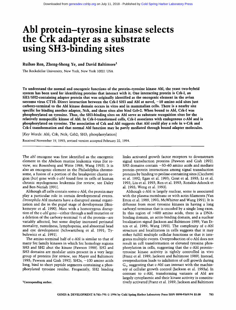
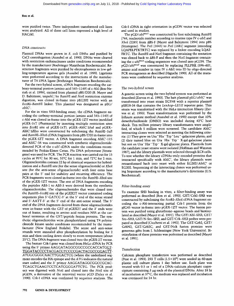
DNA sequence analysis revealed that the nine c-Abl- interacting clones identified by the two-hybrid screen belonged to three genes. The DNA sequences were com- pared with sequences from GenBank. Two of them (clone A, representing six independent clones, and clone C) were found to be novel genes. The DNA sequence of clone B, representing 2 of 10 primary clones, was iden- tical to part of the human proto-oncogene Crk-I cDNA {Fig. 1).
The Crk oncogene was originally isolated as the onco- genic element of avian retrovirus CT10 [Mayer et al. 1988] and ASV-1 (Tsuchie et al. 1989). The human c-Crk proto-oncogene encodes two proteins, Crk-I and Crk-II, through altemative splicing pathways (Matsuda et al. 1992). Crk-I and Crk-II proteins are adapter molecules containing primarily SH2 and SH3 domains: Crk-II has one SH2 and two SH3 domains; Crk-I lacks the carboxy-
T a b l e 1. Clones interacting with carboxy-terminal portion of Abl in yeast two-hybrid system
~-Gal activity in y e a s t a
Frequency with with {in 5 x l0 s p E G 2 0 2 / pEG202/ with
Clone transformantsl AblC' Abl pEG202
A 6 + + -
B 2 + + -
C 1 + + - D 1 + + +
a~-Gal activity in yeast was detected in X-gal plates. Entries indicate the presence or absence of blue color with transfor- mants.
784 GENES & D E V E L O P M E N T
Cold Spring Harbor Laboratory Press on July 11, 2018 - Published by genesdev.cshlp.orgDownloaded from

Crk binds to Abl
CRK-II
SH2 N-SH3 C-SH3 1 ~ 04
CRK-I 1 ~ 204
88 C l o n e - B ~ ~ ~ V ~ 1 2 0 4
Figure 1. Schematic representation of the c-Abl-interacting Crk protein identified by genetic screening. The domain struc- tures of human Crk-I and Crk-II (Matsuda et al. 1992) are com- pared with the primary structure of clone B found to interact in the two-hybrid screen with Abl.
terminal SH3 domain. The Abl-interacting clone B cDNA lack the sequence encoding the amino-terminal 87 amino acids of Crk-I cDNA, thus producing a partial Crk-I protein from which the SH2 domain was mostly deleted (Fig. 1). The Crk/Abl interaction therefore ap- peared to be mediated by the Crk-I SH3 domain (CrkI- SH3).
CrkI-SH3 binds c-AbI in vitro
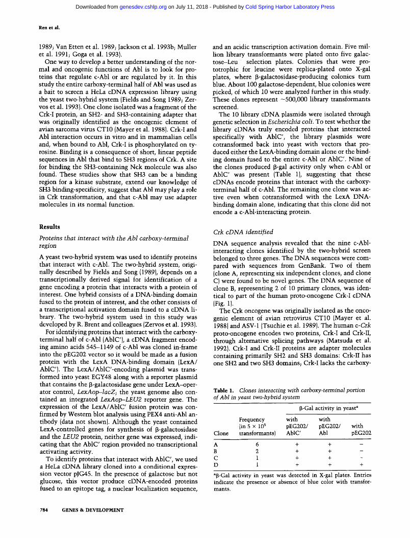
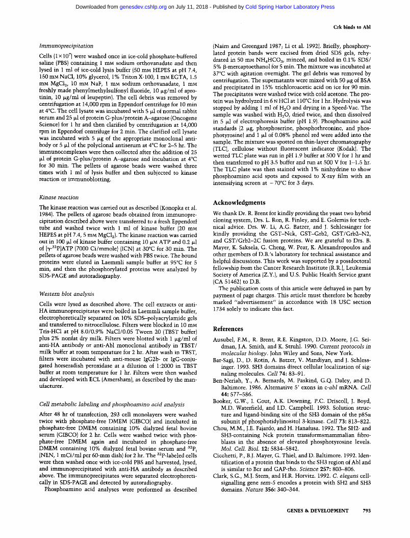
To examine whether CrkI-SH3 bound Abl directly, an in vitro filter assay was employed. For this assay, the cloned partial Crk-I protein was expressed as a glutathi- one S-transferase fusion protein in E. coli (referred as GST/CrkI-SH3). GST/CrkI-SH3 was purified using glu- tathione-agarose beads, biotinylated as described (Mayer et al. 19911, and used as a probe on filters containing Abl protein.
The cDNA encoding AblC' was expressed in E. coli by cloning it in-frame carboxy-terminal to GST. Crk bind- ing to the GST/AblC' fusion protein was examined fol- lowing electrophoretic fractionation of crude E. coli ex- tracts, transfer to a filter, and probing with biotinylated GST/CrkI-SH3. As revealed by anti-GST antibody (Fig. 2) and PEX4 anti-Abl antibody (data not shown), the GST/AblC' fusion protein expressed in bacteria had an apparent molecular mass of 110 kD. However, the ma- jority of the GST/AblC' fusion protein was degraded into polypeptides with various sizes. Because AblC' was tagged with GST, this natural degradation of GST/AblC' facilitated mapping of the binding sites for Crk and other SH3-containing proteins.
The GST/CrkI-SH3 probe bound to the full-length GST/AblC' fusion protein and to a number of its degra- dation products (Fig. 2). Binding of the GST/CrkI-SH3 probe to GST and the GST probe to the GST/AblC' fu- sion protein was not detected, indicating that the inter- action of GST/CrkI-SH3 and GST/AblC' was through the binding of CrkI-SH3 to AblC'. The GST/AblC' deg- radation products that bound to CrkI-SH3 were similar to those bound to anti-GST antibody, suggesting that the CrkI-SH3-binding site is located in the amino-terminal portion of AblC'.
216 ---
1 0 5 ~
7 0 ~
4 3 ~
28--"
CRK1 Grb2 Nck Abi Sr¢ N-Src GST Anti- SH3 SH3 SH3 SH3 GST A S A ~ ' A " ~ " A B A B A B A S A S
Figure 2. In vitro binding of the c-Abl carboxy-terminal half to various SH3 domains and SH3-containing proteins. Proteins from induced lysates of bacteria that expressed the GST-AblC' fusion protein (A) or GST (B) were probed with anti-GST anti- body or with biotinylated GST or GST fusion proteins as indi- cated above each pair of lanes. Numbers (left) represent the molecular size in kilodaltons.
The CrkI-SH3 probe used to examine the Crk-Abl in- teraction in vitro contained flanking sequences of the SH3 domain. To confirm that the Crk-Abl interaction was through the Crk-I SH3 domain alone, the first SH3 domain of murine c-Crk (Cicchetti et al. 1992) was tested for binding to AblC'. The murine Crk SH3 bound to AblC' as effectively as did CrkI-SH3 (data not shown). Both the murine Crk SH3 domain and cloned human CrkI-SH3 could also bind overexpressed full-length c-Abl from NIH-3T3 cells (data not shown).
CrkI-SH3-binding sites are in a region between the Abl kinase domain and its nuclear localization signal
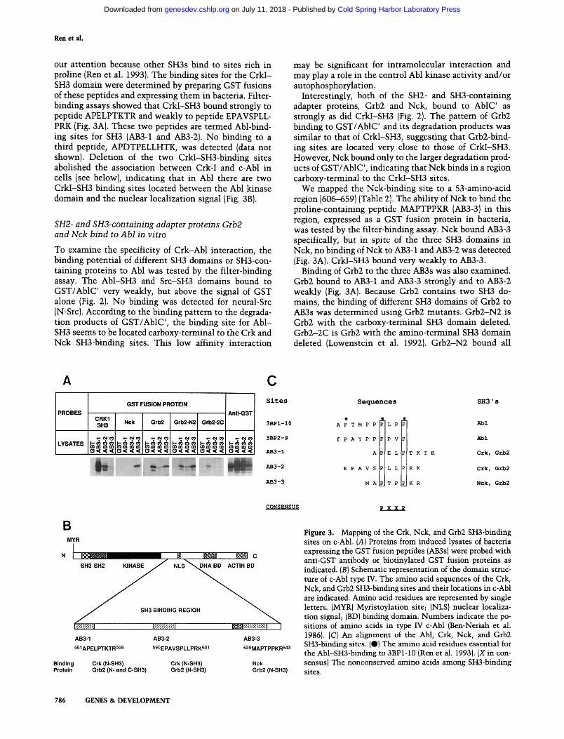
The pattern of CrkI-SH3 binding to the degradation products of the GST/AblC' fusion protein suggested that the SH3-binding site was located in the amino-terminal region of AblC'. Sequential deletion analysis of AblC' from its carboxyl terminus mapped the CrkI-SH3-bind- ing site to a 56-amino-acid region (Table 2). In this region there are three proline-containing peptides (APELPT- KTR, EPAVSPLLPRK, and APDTPELLHTK) that drew
Table 2. Mapping of the Crk, Nck, and Grb2 SH3-binding sites
SH3 binding a
GST fusion pept ides CrkI-SH3 Nck Grb2
AblC' (545-1149} b + + + AblC'•Sal (545-987) + + + AblC'aXho (545-763) + + + AblC'115 (545-659) + + + AblC'56 (551-606) + - +
aDetermined by filter-binding assay. bNumbers indicate the amino acid sequences in type IV c-Abl.
GENES & DEVELOPMENT 785
Cold Spring Harbor Laboratory Press on July 11, 2018 - Published by genesdev.cshlp.orgDownloaded from
Ren et al.
our attention because other SH3s bind to sites rich in proline (Ren et al. 1993). The binding sites for the CrkI- SH3 domain were determined by preparing GST fusions of these peptides and expressing them in bacteria. Filter- binding assays showed that CrkI-SH3 bound strongly to peptide APELPTKTR and weakly to peptide EPAVSPLL- PRK (Fig. 3A). These two peptides are termed Abl-bind- ing sites for SH3 (AB3-1 and AB3-2). No binding to a third peptide, APDTPELLHTK, was detected (data not shown). Deletion of the two CrkI-SH3-binding sites abolished the association between Crk-I and c-Abl in cells {see below}, indicating that in Abl there are two CrkI-SH3 binding sites located between the Abl kinase domain and the nuclear localization signal (Fig. 3B).
SH2- and SH3-containing adapter proteins Grb2 and Nck bind to Abl in vitro
To examine the specificity of Crk-Abl interaction, the binding potential of different SH3 domains or SH3-con- taining proteins to Abl was tested by the filter-binding assay. The Abl-SH3 and Src-SH3 domains bound to GST/AblC' very weakly, but above the signal of GST alone (Fig. 2). No binding was detected for neural-Src (N-Src). According to the binding pattern to the degrada- tion products of GST/AblC', the binding site for Abl- SH3 seems to be located carboxy-terminal to the Crk and Nck SH3-binding sites. This low affinity interaction
may be significant for intramolecular interaction and may play a role in the control Abl kinase activity and/or autophosphorylation.
Interestingly, both of the SH2- and SH3-containing adapter proteins, Grb2 and Nck, bound to AblC' as strongly as did CrkI-SH3 (Fig. 2). The pattem of Grb2 binding to GST/AblC' and its degradation products was similar to that of CrkI-SH3, suggesting that Grb2-bind- ing sites are located very close to those of CrkI-SH3. However, Nck bound only to the larger degradation prod- ucts of GST/AblC', indicating that Nck binds in a region carboxy-terminal to the CrkI-SH3 sites.
We mapped the Nck-binding site to a 53-amino-acid region (606-659) (Table 2). The ability of Nck to bind the proline-containing peptide MAPTPPKR (AB3-3) in this region, expressed as a GST fusion protein in bacteria, was tested by the filter-binding assay. Nck bound AB3-3 specifically, but in spite of the three SH3 domains in Nck, no binding of Nck to AB3-1 and AB3-2 was detected (Fig. 3A). CrkI-SH3 bound very weakly to AB3-3.
Binding of Grb2 to the three AB3s was also examined. Grb2 bound to AB3-1 and AB3-3 strongly and to AB3-2 weakly (Fig. 3A). Because Grb2 contains two SH3 do- mains, the binding of different SH3 domains of Grb2 to AB3s was determined using Grb2 mutants. Grb2-N2 is Grb2 with the carboxy-terminal SH3 domain deleted. Grb2-2C is Grb2 with the amino-terminal SH3 domain deleted (Lowenstein et al. 1992}. Grb2-N2 bound all
A C
GST FUSION PROTEIN
PROBES AntI-GST CRK1 SH3 Nck Grb2 Grb2-N2 Grb2-2C
( 3 < < < t 3 < < < [ ( 3 < < < ( 3 < [ < < ( 3 < < < ( 3 < < <
Sites Sequences SH3 ' s
3BP1-10 A P T H P P L P Abl
3BP2-9 f P A Y P P P V Abl
AB3-1 A E L T K T R Crk, Grb2
AB3-2 E P A V S L L R K Crk, Grb2
AB3-3 M A T P K R Nck, Grb2
B MYR
N C
~ ~ G~ ~" ~- LL~ t I~t-~,~'~'~,~-~-~-~-~,~'~t I
AB3-1 AB3-2 AB3-3
551APELPTKTR559 5 S 0 E P A V S P L L P R K 6 0 1 636MAPTPPKR643
Binding Crk (N-SH3) Crk (N-SH3) Nck Protein Grb2 (N- and CoSH3) Grb2 (N-SH3) Grb2 (N-SH3)
p X X P
Figure 3. Mapping of the Crk, Nck, and Grb2 SH3-binding sites on c-Abl. (A) Proteins from induced lysates of bacteria expressing the GST fusion peptides (AB3s) were probed with anti-GST antibody or biotinylated GST fusion proteins as indicated. (B) Schematic representation of the domain struc- ture of c-Abl type W. The amino acid sequences of the Crk, Nck, and Grb2 SH3-binding sites and their locations in c-Abl are indicated. Amino acid residues are represented by single letters. {MYR) Myristoylation site; (NLS) nuclear localiza- tion signal; (BD) binding domain. Numbers indicate the po- sitions of amino acids in type IV c-Abl (Ben-Neriah et al. 1986). [C} An alignment of the Abl, Crk, Nck, and Grb2 SH3-binding sites. (e} The amino acid residues essential for the Abl-SH3-binding to 3BP1-10 (Ren et al. 1993). (X in con- sensus) The nonconserved amino acids among SH3-binding sites.
786 G E N E S & D E V E L O P M E N T
Cold Spring Harbor Laboratory Press on July 11, 2018 - Published by genesdev.cshlp.orgDownloaded from
Crk binds to Abl
three AB3s: AB3-3 was strongest and AB3-2 weakest (Fig. 3A). The binding of Grb2-N2 to AB3-1 was weaker than that of wild-type Grb2. Grb2-2C bound weakly and only to AB3-1. The different specificities of the two SH3 do- mains of Grb2 suggested that they may bind Abl coop- eratively in a specific orientation.
The locations of the CrkI-SH3, Grb2, and Nck-binding sites in c-Abl are shown in Figure 3B.
Two proteins that bind specifically to the Abl-SH3 domain were isolated previously by screening a hgt l l cDNA expression library using the GST/Abl-SH3 fusion protein (Cicchetti et al. 1992). The SH3-binding sites of the two SH3-binding proteins were localized to a 10- amino-acid stretch very rich in proline residues (Ren et al. 1993). In the 3BP1 site, proline residues at positions 2, 7, and 10, were crucial to the binding. A number of SH3- binding proteins have been identified since then (Egan et al. 1993; Gout et al. 1993; Li et al. 1993; Liu et al. 1993; Rozakis-Adcock et al. 1993; G. Cheng, S.-Z. Ye, and D. Baltimore, in prep.; K. Alexandropoulos and D. Balti- more, unpubl.). All of the SH3-binding sites have been characterized by multiple proline residues. Although the Abl-SH3- and CrkI-SH3-binding sites are very different in amino acid sequence, they can be aligned with two conserved proline residues spaced by two nonconserved amino acids (Fig. 3C). The Crk, Nck, and Grb2 SH3- binding sites share basic amino acids at their carboxyl terminus not found in the Abl sites.
Crk-I associates with both c-Abl and transforming Abl in mammaBan cells
The studies described above demonstrated that trun- cated Crk, consisting of the SH3 domain and its flanking sequences, interacts with c-Abl both in yeast and in vitro. To determine whether Crk associates with Abl in vivo, full-length human Crk-I cDNA was cloned by re- verse transcriptase (RT)-PCR. For identification pur- poses, the Crk-I protein was tagged with an influenza virus hemagglutinin (HA) epitope at its amino terminus,
producing the HACrkI chimera. The gene for this chi- meric protein was subcloned into mammalian expres- sion vector pGD, in which the Moloney leukemia viral long terminal repeat (LTR) directs RNA synthesis.
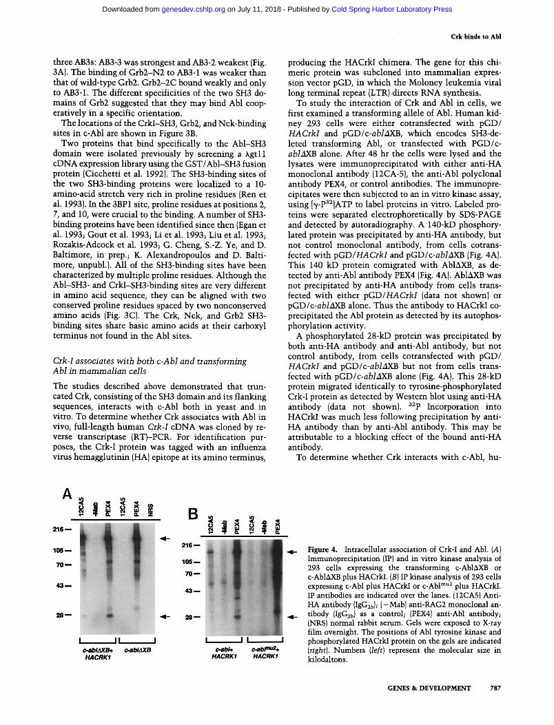
To study the interaction of Crk and Abl in cells, we first examined a transforming allele of Abl. Human kid- ney 293 cells were either cotransfected with pGD/ HACrkI and pGD/c-abMXB, which encodes SH3-de- leted transforming Abl, or transfected with PGD/c- abMXB alone. Mter 48 hr the cells were lysed and the lysates were immunoprecipitated with either anti-HA monoclonal antibody (12CA-5), the anti-AN polyclonal antibody PEX4, or control antibodies. The immunopre- cipitates were then subjected to an in vitro kinase assay, using [~/-Pa2]ATP to label proteins in vitro. Labeled pro- teins were separated electrophoretically by SDS-PAGE and detected by autoradiography. A 140-kD phosphory- lated protein was precipitated by anti-HA antibody, but not control monoclonal antibody, from cells cotrans- fected with pGD/HACrkI and pGD/c-ablAXB (Fig. 4A). This 140 kD protein comigrated with AblAXB, as de- tected by anti-Abl antibody PEX4 (Fig. 4A). AblAXB was not precipitated by anti-HA antibody from cells trans- fected with either pGD/HACrkI {data not shown) or pGD/c-ablAXB alone. Thus the antibody to HACrkI co- precipitated the Abl protein as detected by its autophos- phorylation activity.
A phosphorylated 28-kD protein was precipitated by both anti-HA antibody and anti-Abl antibody, but not control antibody, from cells cotransfected with pGD/ HACrkI and pGD/c-ablAXB but not from cells trans- letted with pGD/c-abMXB alone (Fig. 4A). This 28-kD protein migrated identically to tyrosine-phosphorylated Crk-I protein as detected by Western blot using anti-HA antibody (data not shown), a~p Incorporation into HACrkI was much less following precipitation by anti- HA antibody than by anti-Abl antibody. This may be attributable to a blocking effect of the bound anti-HA antibody.
To determine whether Crk interacts with c-Abl, hu-
A
2 1 6 - -
1 0 5 - -
7 0 - -
4 3 , -
2 8 - -
~- O. 0. Z B
I
c.ablAXB+ HACRK1
216 - -
105--.
7 0 - -
43, , . -
28.--
I I i I I I c-abhU(B c.ab~
HACRK1
Q. D.
I
c.abPm2+ HACRK1
Figure 4. Intracellular association of Crk-I and Abl. (A) Immunoprecipitation (IP} and in vitro kinase analysis of 293 cells expressing the transforming c-Abl~kXB or c-Abl~XB plus HACrkI. (B) IP kinase analysis of 293 cells expressing c-Abl plus HACrkI or c-Abl m"2 plus HACrkI. IP antibodies are indicated over the lanes. {12CA5) Anti- HA antibody (IgG2b); ( -Mab) anti-RAG2 monoclonal an- tibody (IgG2b) as a control; (PEX4) anti-Abl antibody; (NRS) normal rabbit serum. Gels were exposed to X-ray film overnight. The positions of Abl tyrosine kinase and phosphorylated HACrkI protein on the gels are indicated (right). Numbers (left) represent the molecular size in kilodaltons.
GENES & DEVELOPMENT 787
Cold Spring Harbor Laboratory Press on July 11, 2018 - Published by genesdev.cshlp.orgDownloaded from
Ren et al.
man kidney 293 cells were cotransfected with pGD/ HACrkI and pGD/c-abI, and 32P-labeled, immunopre- cipitated proteins were detected after phosphorylation in vitro. As with transforming Abl, labeled Crk-I and c-Abl were specifically precipitated by both anti-HA and anti- Abl antibody [Fig. 4B), indicating that Crk-I associates with c-Abl in cells.
The SH3-binding sites on Abl are needed for Crk interaction in cells
The in vitro studies mapped the Crk SH3-binding site on Abl to a region between the Abl kinase domain and its nuclear localization signal. To determine whether the association of Crk and Abl in vivo was directly mediated by this interaction, the CrkI-SH3-binding sites were mu- tated in c-Abl by deletion. Mutation of the higher affin- ity CrkI-SH3 binding site, AB3-1, gave the c-Abl mul mu- tant Abl protein. Mutation of both CrkI-SH3-binding sites, AB3-1 and AB3-2, gave c-Abl mu2 mutant Abl pro- tein. The expression and in vitro kinase activity of the mutant Abl proteins were similar to that of c-Abl (Fig. 4B). To examine whether HACrkI interacts with the mu- tant Abl proteins, they were coexpressed in 293 cells. Immunoprecipitation and in vitro kinase assay showed that HACrkI and c-Abl r"ul still form a complex in cells (data not shown). Association of HACrkI and c-Abl mu2 however, was not detected by immunoprecipitation and kinase assay (Fig. 4B), indicating that Crk-I interacts with Abl through the CrkI-SH3 domain's binding to the defined sites in Abl.
Crk is phosphorylated on tyrosine upon binding to Abl
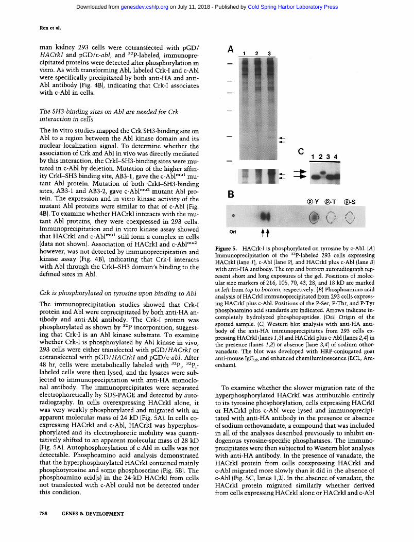
The immunoprecipitation studies showed that Crk-I protein and Abl were coprecipitated by both anti-HA an- tibody and anti-Abl antibody. The Crk-I protein was phosphorylated as shown by 32p incorporation, suggest- ing that Crk-I is an Abl kinase substrate. To examine whether Crk-I is phosphorylated by Abl kinase in vivo, 293 cells were either transfected with pGD/HACrkI or cotransfected with pGD/HACrkl and pGD/c-abl. After 48 hr, cells were metabolically labeled with 32P i. 32P i- labeled ceils were then lysed, and the lysates were sub- jected to immunoprecipitation with anti-HA monoclo- hal antibody. The immunoprecipitates were separated electrophoretically by SDS-PAGE and detected by auto- radiography. In cells overexpressing HACrkI alone, it was very weakly phosphorylated and migrated with an apparent molecular mass of 24 kD [Fig. 5A). In cells co- expressing HACrkI and c-Abl, HACrkI was hyperphos- phorylated and its electrophoretic mobility was quanti- tatively shifted to an apparent molecular mass of 28 kD [Fig. 5A). Autophosphorylation of c-Abl in cells was not detectable. Phosphoamino acid analysis demonstrated that the hyperphosphorylated HACrkI contained mainly phosphotyrosine and some phosphoserine (Fig. 5B). The phosphoamino acidIs) in the 24-kD HACrkI from ceils not transfected with c-Abl could not be detected under this condition.
788 GENES & DEVELOPMENT
A 1 2 3
B
C 1 2 3 4
@-Y (~T (~-S
Ori ~
Figure 5. HACrk-I is phosphorylated on tyrosine by c-Abl. (A) Immunoprecipitation of the 32p-labeled 293 cells expressing HACrkI (lane 1), c-Abl {lane 2), and HACrkI plus c-Abl {lane 3) with anti-HA antibody. The top and bottom autoradiograph rep- resent short and long exposures of the gel. Positions of molec- ular size markers of 216, 105, 70, 43, 28, and 18 kD are marked at left from top to bottom, respectively. (B I Phosphoamino acid analysis of HACrkI immunoprecipitated from 293 cells express- ing HACrkI plus c-Abl. Positions of the P-Ser, P-Thr, and P-Tyr phosphoamino acid standards are indicated. Arrows indicate in- completely hydrolyzed phosphopeptides. (Off) Origin of the spotted sample. (C) Western blot analysis with anti-HA anti- body of the anti-HA immunoprecipitates from 293 cells ex- pressing HACrkI (lanes 1,3) and HACrkI plus c-Abl (lanes 2,4) in the presence (lanes 1,2) or absence (lane 3,4) of sodium othor- vanadate. The blot was developed with HRP-conjugated goat anti-mouse IgG2b and enhanced chemiluminescence (ECL, Am- ersham I.
To examine whether the slower migration rate of the hyperphosphorylated HACrkI was attributable entirely to its tyrosine phosphorylation, cells expressing HACrkI or HACrkI plus c-Abl were lysed and immunoprecipi- tated with anti-HA antibody in the presence or absence of sodium orthovanadate, a compound that was included in all of the analyses described previously to inhibit en- dogenous tyrosine-specific phosphatases. The immuno- precipitates were then subjected to Western blot analysis with anti-HA antibody. In the presence of vanadate, the HACrkI protein from cells coexpressing HACrkI and c-Abl migrated more slowly than it did in the absence of c-Abl {Fig. 5C, lanes 1,2}. In the absence of vanadate, the HACrkI protein migrated similarly whether derived from cells expressing HACrkI alone or HACrkI and c-Abl
Cold Spring Harbor Laboratory Press on July 11, 2018 - Published by genesdev.cshlp.orgDownloaded from
Crk binds to Abl
(lanes 3,4). Only the slower migrating HACrkI contained phosphotyrosine, as detected by antiphosphotyrosine an- tibody {data not shown). These data indicate that the Abl-generated electrophoretic mobility shift of HACrkI in SDS-PAGE is attributable to tyrosine phosphoryla- tion.
Using the mutants of c-Abl described above, we could show that the phosphorylation of Crk depended on its ability to interact with Abl. Compared with wild type {Fig. 6, lane 2), less HACrkI protein was tyrosine phos- phorylated in cells coexpressing HACrkI and c-Abl r"ul (lane 3) and very little phosphorylated form was evident when both CrkI-SH3-binding sites were deleted in c-Abl mu2 {lane 4). The expression of c-Abl and mutant c-Abl proteins in these cells was about equal (data not shown), directly demonstrating that the HACrk/Abl SH3-mediated interaction makes HACrk a substrate for the Abl kinase domain.
Crk-I associates with c-Abl and is phosphorylated in Crk-I-transformed fibroblast cells
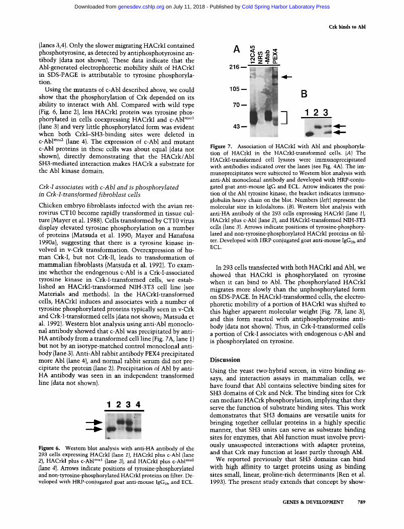
Chicken embryo fibroblasts infected with the avian ret- rovirus CT10 become rapidly transformed in tissue cul- ture (Mayer et al. 1988). Cells transformed by CT10 virus display elevated tyrosine phosphorylation on a number of proteins (Matsuda et al. 1990; Mayer and Hanafusa 1990a), suggesting that there is a tyrosine kinase in- volved in v-Crk transformation. Overexpression of hu- man Crk-I, but not Crk-II, leads to transformation of mammalian fibroblasts (Matsuda et al. 1992). To exam- ine whether the endogenous c-Abl is a Crk-I-associated tyrosine kinase in Crk-I-transformed cells, we estab- lished an HACrkI-transformed NIH-3T3 cell line (see Materials and methods). In the HACrkI-transformed cells, HACrkI induces and associates with a number of tyrosine phosphorylated proteins typically seen in v-Crk and Crk-I-transformed cells {data not shown; Matsuda et al. 1992). Western blot analysis using anti-Abl monoclo- nal antibody showed that c-Abl was precipitated by anti- HA antibody from a transformed cell line (Fig. 7A, lane 1) but not by an isotype-matched control monoclonal anti- body {lane 3). Anti-Abl rabbit antibody PEX4 precipitated more Abl (lane 4), and normal rabbit serum did not pre- cipitate the protein {lane 2). Precipitation of Abl by anti- HA antibody was seen in an independent transformed line (data not shown).
1 2 3 4
Figure 6. Western blot analysis with anti-HA antibody of the 293 cells expressing HACrkI (lane 1), HACrkI plus c-Abl (lane 2), HACrkI plus c-Abl mul (lane 3), and HACrkI plus c-Abl mu2 (lane 4). Arrows indicate positions of tyrosine-phosphorylated and non-tyrosine-phosphorylated HACrkI proteins on filter. De- veloped with HRP-conjugated goat anti-mouse IgG2b and ECL.
A
2 1 6 - - -
1 0 5 - -
7 0 - - .
4 3 - - -1
B
1 2 3
Figure 7. Association of HACrkI with Abl and phosphoryla- tion of HACrkI in the HACrkI-transformed cells. (A) The HACrkI-transformed cell lysates were immunoprecipitated with antibodies indicated over the lanes (see Fig. 4A). The im- munoprecipitates were subjected to Western blot analysis with anti-Abl monoclonal antibody and developed with HRP-conju- gated goat anti-mouse IgG and ECL. Arrow indicates the posi- tion of the Abl tyrosine kinase; the bracket indicates immuno- globulin heavy chain on the blot. Numbers (left} represent the molecular size in kilodaltons. (B). Western blot analysis with anti-HA antibody of the 293 cells expressing HACrkI {lane I), HACrkI plus c-Abl (lane 2), and HACrkI-transformed NIH-3T3 ceils (lane 3). Arrows indicate positions of tyrosine-phosphory- lated and non-tyrosine-phosphorylated HACrkI proteins on fil- ter. Developed with HRP-conjugated goat anti-mouse IgG2b and ECL.
In 293 cells transfected with both HACrkI and Abl, we showed that HACrkI is phosphorylated on tyrosine when it can bind to Abl. The phosphorylated HACrkI migrates more slowly than the unphosphorylated form on SDS-PAGE. In HACrkI-transformed cells, the electro- phoretic mobility of a portion of HACrkI was shifted to this higher apparent molecular weight (Fig. 7B, lane 3), and this form reacted with antiphosphotyrosine anti- body (data not shown). Thus, in Crk-I-transformed cells a portion of Crk-I associates with endogenous c-Abl and is phosphorylated on tyrosine.
Discussion
Using the yeast two-hybrid screen, in vitro binding as- says, and interaction assays in mammalian cells, we have found that Abl contains selective binding sites for SH3 domains of Crk and Nck. The binding sites for Crk can mediate HACrk phosphorylation, implying that they serve the function of substrate binding sites. This work demonstrates that SH3 domains are versatile units for bringing together cellular proteins in a highly specific manner, that SH3 units can serve as substrate binding sites for enzymes, that Abl [unction must involve previ- ously unsuspected interactions with adapter proteins, and that Crk may function at least partly through Abl.
We reported previously that SH3 domains can bind with high affinity to target proteins using as binding sites small, linear, proline-rich determinants (Ren et al. 1993). The present study extends that concept by show-
GENES & DEVELOPMENT 789
Cold Spring Harbor Laboratory Press on July 11, 2018 - Published by genesdev.cshlp.orgDownloaded from
Ren et al.
ing that many SH3s have binding sites, each with a dif- ferent specificity not necessarily involving more than two prolines. Thus, SH3 domains can provide the spec- ificity to mediate aggregation of many highly specific protein complexes. Studies of the interaction of SH3 do- mains on Grb2 with proline-rich sites on Sos protein have also extended the range of action of SH3 regions (ref. in McCormick 1993). There, it appears that the function of SH3 is to bring the Sos protein to the cell membrane where it can find its substrate Ras. Similarly, SH3 domains were implicated in directing cellular local- ization of PLC7 and Grb2 (Bar-Sagi et al. 1993). The GT- Pase dynamin binds to and is activated by SH3 domains on other proteins {Gout et aI. 1993). Furthermore, we have found recently that SH3 domains on Fyn, Lyn, and Hck can bring these protein-tyrosine kinases to the Btk kinase (G. Cheng, Z.-S. Ye, and D. Baltimore, in prep.). Thus, SH3 binding can apparently serve to bring sub- strates to enzymes, to organize protein complexes at the cell membrane and probably elsewhere in the cell, and to regulate enzymatic activities. Given the nature of mol- ecules that contain SH3s, the purpose of these interac- tions must be to facilitate the transmission of signals through the cytoplasm and nucleus of the cell.
The function of the Crk-SH3 interaction with Abl ap- pears to be, at least in part, to bring one or more tyrosines of Crk into the catalytic site of the Abl kinase domain. In vitro studies with the Abl kinase (Foulkes et al. 1985; Frackelton 1985), as well as its extensive phosphoryla- tion of bacterial proteins (Wang et al. 1982), led us to conclude previously that it was a kinase with very little intrinsic substrate specificity. We can now suggest that this relative nonspecificity is a characteristic of the ac- tive site but that specificity can be mediated by interac- tions elsewhere in the molecule. Abl not only has its own SH2 and SH3 regions amino-terminal to the ki- nase--the SH2 having been implicated previously in bringing substrates to the kinase (Mayer and Baltimore, 1994)--but it has the SH3-binding sites carboxy-terminal to the kinase, which capture substrates. In both cases, the substrates would be expected to dissociate from the enzyme relatively slowly compared with dissociation from enzymes that have substrate binding sites as inte- gral parts of their catalytic machinery. This has three consequences: The substrate will stay near the enzyme so that phosphorylation can predominate over dephos- phorylation by phosphatases; the enzyme will have a very low turnover number because the substrate will not dissociate easily; and other substrates can also be brought to the enzyme through secondary domain asso- ciations. Crk in particular has an SH2 domain that can interact with other proteins, potentially mediating for- mation of complex, multiprotein complexes. It has been shown previously that the SH2 domains can mediate complex formation, particularly on receptor protein-ty- rosine kinases, and we now must see SH3 as equally important in such interactions, especially because the Abl example shows us that one protein can have both an SH3 domain and an SH3-binding site.
The limitation of turnover number caused by separat-
ing the catalytic site from the substrate site has an im- portant implication for signal transduction pathways: A signal will only be passed in a limited fashion, with rel- atively little amplification. By independent evolution of the substrate binding sites in transduction pathways, the extent of signal amplification can be controlled. A sim- ilar example is the Raf-Mek interaction, where Ra/ki- nase stably binds its substrate Mek kinase through its carboxyl catalytic domain (Crews and Erikson 1993; Van Aelst et al. 1993).
In cells coexpressing HACrkI and c-Abl, HACrkI was phosphorylated by c-Abl as described; however, auto- phosphorylation of c-Abl in cells was not detected. This observation suggest that c-Abl may be activated without autophosphorylation, and the autophosphorylation, of- ten seen in transforming variants of Abl, may provide other signals, for example, recruiting SH2-containing signaling molecules.
Crk structure and function
The human crk proto-oncogene encodes two proteins through alternative splicing (Matsuda et al. 1992). In this study the association of Crk-I and Abl through the SH3 interaction was demonstrated. Crk-II contains the Crk-I SH3 domain, making it likely that Crk-II will also asso- ciate with Abl in cells. Consistent with this possibility, the endogenous Crk-II proteins become phosphorylated on tyrosine in cells overexpressing transforming Abl lB. Mayer, unpubl.). Crk-II is the predominant form of Crk in most cells and differs in having a second SH3 domain of unknown specificity [Matsuda et al. 1992; Tanaka et al. 1993). Overexpression of human Crk-I leads to trans- formation of mammalian fibroblasts, whereas Crk-II is not transforming and therefore the second SH3 could serve to modulate the function of the first [Matsuda et al. 19921. Microinjection of Crk-I into PC-12 cells induces neuronal differentiation, whereas Crk-II has a much weaker effect in PC-12 cells (Tanaka et al. 1993), again suggesting that the second SH3 is inhibitory to Crk func- tion. Thus, if Crk-I is a mediator of growth and differen- tiation events, Crk-II may have a more temperate activ- ity.
Role of the Crk/Abi association in v-Crk transformation
Avian v-Crk is composed of viral Gag protein fused to a Crk-I-like domain containing one SH2 domain and one SH3 domain (Mayer et al. 19881. Chicken embryo fibro- blasts infected with the avian retrovirus CT10 become rapidly transformed in tissue culture and induce tumors when injected into chickens (Mayer et al. 1988). Al- though v-Crk protein does not have a tyrosine kinase domain, cells transformed by CT10 virus display ele- vated tyrosine phosphorylation on a number of proteins {Matsuda et al. 1990; Mayer and Hanafusa 1990a), sug- gesting that there is a tyrosine kinase involved in v-Crk transformation. Tyrosine kinase activity was found to associate with Crk {Mayer and Hanafusa 1990a), and
790 GENES & DEVELOPMENT
Cold Spring Harbor Laboratory Press on July 11, 2018 - Published by genesdev.cshlp.orgDownloaded from
Crk binds to Abl
both Crk SH2 and SH3 domains were found to be re- quired for transformation (Mayer and Hanafusa 1990b). The demonstration of Crk and Abl association in this study indicates that the c-Abl tyrosine kinase is a Crk- associated tyrosine kinase and may play a critical role in Crk transformation.
Binding of other adapter proteins to Abl
Nck and Grb2 both interact with Abl in vitro. Nck was originally isolated from a melanoma expression eDNA library (Lehmann et al. 1990). It contains primarily three SH3 and one SH2 domains and is oncogenic. Overexpres- sion of Nck leads to transformation of mammalian fibro- blasts (Chou et al. 1992; Li et al. 1992). Nck is phospho- rylated on both tyrosine and serine and threonine in re- sponse to epidermal growth factor (EGF), platelet-derived growth factor {PDGF), and nerve growth factor (NGF) stimulation, in response to activation of T-cell receptor and membrane IgM receptor and in v-Src-transformed cells {Chou et al. 1992; Li et al. 1992; Meisenhelder and Hunter 1992; Park and Rhee 1992). It is a common target for many receptor and nonreceptor tyrosine kinases. Nck binds to tyrosine-phosphorylated EGF receptor or PDGF receptor through its SH2 domain (Li et al. 1992). Studies here suggest that Nck associates with c-Abl through one or more of its SH3 domains. This association may also lead to tyrosine phosphorylation of Nck and could be important for signal transduction as well as Nck trans- formation.
Grb2 (Lowenstein et al. 1992), also known as ASH, Sere5, or Drk {Clark et al. 1992; Matuoka et al. 1992; Simon et al. 1993) is an SH2- and SH3-containing adapter that links activated protein-tyrosine kinase receptors to the Ras activator protein Sos. Consistent with our in vitro data, Pendergast et al. recently showed that both Grb2 SH3 domains can bind to c-Abl in vitro (Pendergast et al. 1993). The functional significance of this binding is uncertain because Grb2 is involved in other interactions with its SH3 domains, particularly with the Ras activa- tor protein Sos. The Grb2 SH2, however, binds to a ty- rosine-phosphorylated site in Bcr-Abl that is critical for transformation (Pendergast et al. 1993), providing the op- portunity for a multipoint interaction among these pro- teins.
The Crk, Nck, and Grb2 SH3-binding sites were mapped close to the nuclear localization signal. The as- sociation of these adapter molecules with Abl may block the translocation of Abl into the nucleus and, therefore, retain Abl in cytoplasm. Alternatively, the association of Crk with c-Abl may bring Crk into nucleus. Transloca- tion of proteins in cells is an important mechanism for transducing signals.
c-Abl has one close relative, Arg, that might also be a target for binding by the adapters that bind to Abl. Arg is also a protein-tyrosine kinase with SH2, SH3, and a long carboxy-terminal extension {Kruh et al. 1986). The car- boxy-terminal portion of Arg is less homologous to c-Abl than the amino terminus, but the Crk, Nck, and Grb2 SH3-binding sites identified in c-Abl are conserved, sug-
gesting that these adapter molecules should also bind Arg.
Characteristics of SH3-binding sites
The solution and crystal structure of the SH3 domains of spectrin, Src, Fyn, p85et, and PLC~ have been solved (Musacchio et al. 1992; Yu et al. 1992; Booker et al. 1993; Kohda et al. 1993; Koyama et al. 1993~ Noble et al. 1993). All of these SH3 domains are formed from five antipar- allel f~-strands. The perturbation of resonances by bound proline-rich peptides indicates that the binding site is a hydrophobic surface region of the SH3 domain that con- sists of aromatic residues flanked by two charged loops {Booker et al. 1993; Yu et al. 1992).
Like the Abl-SH3-binding sites, the Crk, Nck, and Grb2 SH3-binding sites in c-Abl were mapped to proline- containing short stretches of amino acids. The CrkI- SH3-binding sites were proved to mediate the associa- tion of Crk-I and Abl in cells. The specificity of the SH3 interactions is evident. However, is there any common structural motif in the SH3-binding sites that might fit into a conserved element of SH3 structure.~ An align- ment of the Abl, Crk, Nck, and Grb2 SH3-binding sites shows that two proline residues spaced by two noncon- served amino acids are conserved. The consensus PXXP was also observed among some other SH3-binding sites {Egan et al. 1993; Gout et al. 1993; Li et al. 1993; Liu et al. 1993; Rozakis-Adcock et al. 1993; G. Cheng, Z.-S. Ye, and D. Baltimore, in prep.; K. Alexandropoulos, unpubl.), suggesting that PXXP may serve as a scaffold for the SH3 interaction. The amino acids around these two highly conserved proline residues might then determine SH3- binding specificity. For example, the nonconserved se- quences amino-terminal to the PXXP consensus, partic- ularly the critical proline at position 2 in the 3BP1 site, may play an important role for Abl-SH3-binding speci- ficity. The basic amino acid{s) carboxy-terminal to PXXP in the CrkI, Grb2, and Nck SH3-binding sites could play a similar role.
Mater ia l s and m e t h o d s
Cells and antibodies
293T ceils {Pear et al. 1993) were grown in Dulbecco's modified Eagle medium {DMEM) containing 10% fetal bovine serum, 100 U/ml of penicillin, and 100 mg/ml of streptomycin. Mouse monoclonal antibody 12CA-5 (Berkeley Antibody Company), anti-Abl monoclonal antibody 24-21 (Oncogene Science), and goat anti-mouse IgG-HRPO and IgG2b-HRPO (Southern Bio- technology Associates, Inc} were used in this study. Abl-specific PEX4 antiserum was described previously {Konopka et al. 1984). The control monoclonal antibody used was an anti-RAG2 monoclonal antibody (Spanopoulou et al. unpubl.).
HACrkI-transformed cell lines were established by infecting NIH-3T3 cells with an HACrkI retrovirus. The helper-free HACrkI retrovirus was generated by transiently transfecting BOSC-23 cells with pGD/HACrkI as described {Pear et al. 1993). NIH-3T3 cells [2x l0 s) infected with 5x 10 s HACrkI ret- rovirus were transfered into 0.3% soft agar 2 days after infec- tion. Colonies were picked after 3 weeks. The transformed cells
GENES & DEVELOPMENT 791
Cold Spring Harbor Laboratory Press on July 11, 2018 - Published by genesdev.cshlp.orgDownloaded from
Ren et al.
were purified twice. Three independent transformed cell lines were analyzed. All of these cell lines expressed a high level of HACrkI.
DNA constructs
Plasmid DNAs were grown in E. coli DHSc~ and purified by CsC1 centrifugation (Ausubel et al. 1990). DNAs were cleaved with restriction endonucleases under conditions recommended by the manufacturer (Boehringer Mannheim Biochemicals). Re- striction fragments were purified by electrophoresis in low-gel- ling-temperature agarose gels (Ausubel et al. 1990). Ligations were performed according to the instructions of the manufac- turer of T4 DNA ligase (Boehringer Mannheim Biochemicals).
For the two-hybrid screen, eDNA fragment encoding the car- boxy-terminal portion (amino acid 545-1149) of c-Abl (Ben-Ne- riah et al. 1986), excised from plasmid pBS/D20 {B. Mayer and D. Baltimore, unpubl.I by BamHI and NotI restriction enzyme digestion, was cloned in-frame into pEG202 vector with an EcoRI-BamHI linker. This plasmid was designated as pEG/ ablC'.
For the in vitro SH3-binding assay, the eDNA fragment en- coding the carboxy-terminal portion (amino acid 545-1149) of oAbl was cloned in-frame into the pGEX-1ZT vector (modified pGEX-IKT IPharmacia) by inserting multiple restriction sites into EcoRI site of pGEX-IKT), pGEX-AblC'ASal and pGEX- AblC'AXho were constructed by subcloning the BamHI-SalI and BamHI-XhoI cDNA fragments from pBS/D20 in-frame into the pGEX-1ZT vector. A DNA fragment encoding AblC'- l l5 and AblC'-56 was constructed with synthetic oligonucleotide- directed PCR of the c-abl cDNA under the conditions recom- mended by Perkin-Elmer Cetus. Pfu DNA polymerase (Strata- gene) was used for PCR. The PCR reaction was carried out for 15 cycles at 95°C for 30 sec, 55°C for 1 min, and 72°C for 2 min. Oligonucleotides contain 22 bp of identical sequence for hybrid- ization and a BarnHI site (for the sense oligonucleotide) or XhoI site (for the anti-sense oligonucleotide) with four extra base pairs at the 5' end for stability and recutting efficiency. The PCR fragments were cloned in-frame into the BamHI-XhoI site of the pGEX-1ZT vector. The rest of DNA fragments encoding the peptides AB3-1 to AB3-3 were derived from the synthetic oligonucleotides. The oligonucleotides that were cloned into the BamHI-EcoRI site of the pGEX2T vector contained coding sequences plus 5'-GATCC-3' at the 5' end of the sense strand and 5'-AATT-3' at the 5' end of the anti-sense strand. The 5' end of the DNA fragments derived from these oligonucleotides were in-frame with the GST of pGEX2T and the 3' ends were out of frame, resulting in amino acid residues NSS at the car- boxyl terminus of the GST/peptide fusion proteins. The syn- thetic oligonucleotides were phosphorylated using T4 polynu- cleotide kinase under conditions recommended by the manu- facturer (New England Biolabs). The sense and anti-sense strands were annealed after phosphorylation by boiling for 2 rain and then cooling down slowly to room temperature (about 25°C). The DNA fragment was cloned into the pGEX-2T vector.
The human Crk-I gene was cloned from HeLa cDNA by PCR using the 5' primer AAGGATAGGCGGCCGCCACCATGGT- TGGATACCCCTACGACGTCCCCGACTACGCCGGAGTT- ATGGCGGGCAACTTGGACTCG (where the underlined seg- ment encodes the HA epitope and the A TG indicates the natural start codon) and the 3' primer AAGGATAGGCGGCCGCAC- TCAGCTGAAGTCCTCATCGGGATTCTG. The PCR prod- uct was digested with NotI and cloned into the NotI site of pGDN, a derivative of the retroviral vector pGD (Daley et al. 1990). Crk-I cDNA was confirmed by sequence analysis. The
Crk-I cDNA in right orientation in pGDN vector was selected and used in studies.
The pGD/abl mu~ was constructed by first subcloning BamHI (744, nucleotide number according to murine type IV c-abl) and XhoI (2285) from ABS-F (Mayer and Baltimore 1994) into pBS (Strategene). The PstI (1645) to PstI (1681) segment (encoding LQAPELPTKTRTC) was replaced by a linker encoding LQAE- FRTC. The BamHI and NarI fragment containing the mutation was cloned back to ABS-F and then the NotI fragment contain- ing the c-ablmu~-coding sequences was cloned into pGDN. The pGD/abl mu2 was constructed by replacing PLLPRK (596-601, amino acid number in type IV c-Abll into ID by oligo-directed PCR mutagenesis as described (Higuchi 1990). All of the muta- tions were confirmed by sequence analysis.
The two-hybrid screen
A genetic screen using the two-hybrid system was performed as described (Zervos et al. 1993). The bait plasmid pEG/ablC' was transformed into yeast strain EGY48 with a reporter plasmid pSH18-34 that contains the LexAop-LEU2 reporter gene. This strain was transformed with the HeLa interaction library (Zer- vos et al. 1993). Yeast transformation was performed by the lithium acetate method (Ausubel et al. 1990) except that 10% dimethylsulfoxide (DMSO) was included during 42°C heat shock. Ten million primary library transformants were ampli- fied, of which 5 million were screened. The candidate AblC' interacting clones were selected as meeting the following crite- ria: ( 1 ) They grew on Ura- His - Trp - Leu- -galactose plates; and (21 they turned blue on U r a - H i s - T r p - X-gal-galactose plates but not on U r a - H i s - T r p - X-gal-glucose plates. Plasmids from the candidate yeast strains were isolated (Hoffman and Winston 1987), and the library plasmids were selected through KC8 cells. To test whether the library cDNAs truly encoded proteins that interacted specifically with AblC', the library plasmids were retransformed back into yeast with either EG202/AblC' or EG202. Sequencing of the interacting clones was performed us- ing Sequenase according to the manufacturer's directions (U.S. Biochemicall.
Filter-binding assay
To examine SH3 binding in vitro, a filter-binding assay was performed as described (Ren et al. 1993). GST/CrkI-SH3 was constructed by subcloning the EcoRI-XhoI eDNA fragment en- coding the c-Abl-interacting partial Crk-I protein from the pJG45 vector in-frame into pGEX-1ZT vector. The fusion pro- tein was purified using glutathione-agarose beads and biotiny- lated as described (Mayer et al. 1991). The GST/Abl-SH3, GST/ Src-SH3, GST/N-Src-SH3, and GST/Crk-SH3 probes were pre- pared as described (Cicchetti et al. 1992). The GST/Grb2, GST/ GrbN2, GST/Grb2C, and GST/Nck fusion proteins were generous gifts from J. Schlessinger (New York University). Bi- otinylation of these proteins was performed as described (Mayer et al. 1991).
Transfection
Calcium phosphate transfection was performed as described (Pear et al. 1993). 293 T cells (1.5x 10⁶) were seeded in 60-mm plastic cell culture plates 1 day before use. Each plate was treated with 0.5 or 1 ml of a DNA-calcium phosphate copre- cipitate containing 3 }ag each of the plasmid DNAs. After 18 hr of incubation at 37°C, the medium was replaced and incubation was continued for 24 hr.
792 GENES & DEVELOPMENT
Cold Spring Harbor Laboratory Press on July 11, 2018 - Published by genesdev.cshlp.orgDownloaded from
Crk binds to Abl
Immunoprecipitation
Cells (1 x l0 z) were washed once in ice-cold phosphate-buffered saline (PBS) containing 1 mM sodium orthovanadate and then lysed in 1 ml of ice-cold lysis buffer {50 mM HEPES at pH 7.4, 150 mM NaC1, 10% glycerol, 1% Triton X-100, 1 mM EGTA, 1.5 mM MgC12, 10 mM NaF, 1 mM sodium orthovanadate, 1 mM fleshly made phenylmethylsulfonyl fluoride, 10 ~g/ml of apro- tinin, 10 ~g/ml of leupeptin). The cell debris was removed by centrifugation at 14,000 rpm in Eppendorf centrifuge for 10 min at 4°C. The cell lysate was incubated with 5 lal of normal rabbit serum and 25 Ixl of protein G-plus/protein A-agarose (Oncogene Science) for 1 hr and then clarified by centrifugation at 14,000 rpm in Eppendorf centrifuge for 2 rain. The clarified cell lysate was incubated with 5 ~g of the appropriate monoclonal anti- body or 5 ixl of the polyclonal antiserum at 4°C for 2-5 hr. The immunocomplexes were then collected after the addition of 25 ~1 of protein G-plus/protein A-agarose and incubation at 4°C for 30 rain. The pellets of agarose beads were washed three times with 1 ml of lysis buffer and then subjected to kinase reaction or immunoblotting.
Kinase reaction
The kinase reaction was carried out as described {Konopka et al. 1984). The pellets of agarose beads obtained from immunopre- cipitation described above were transferred to a fresh Eppendorf tube and washed twice with 1 ml of kinase buffer (20 mM HEPES at pH 7.4, 5 mM MgC12). The kinase reaction was carried out in 100 ~1 of kinase buffer containing 10 ~M ATP and 0.2 ~1 of [~/-32P]ATP {7000 Ci/mmole) (ICN) at 30°C for 30 min. The pellets of agarose beads were washed with PBS twice. The bound proteins were eluted in Laemmli sample buffer at 95°C for 5 rain, and then the phosphorylated proteins were analyzed by SDS-PAGE and autoradiography.
Western blot analysis
Cells were lysed as described above. The cell extracts or anti- HA immunoprecipitates were boiled in Laemmli sample buffer, electrophoretically separated on 10% SDS--polyacrylamide gels and transferred to nitrocellulose. Filters were blocked in 10 mM Tris-HC1 at pH 8.0/0.9% NaC1/0.05 Tween 20 (TBST buffer) plus 2% nonfat dry milk. Filters were blotted with 1 ~g/ml of anti-HA antibody or anti-Abl monoclonal antibody in TBST/ milk buffer at room temperature for 2 hr. After wash in TBST, filters were incubated with anti-mouse IgG2b- or IgG-conju- gated horseradish peroxidase at a dilution of 1:2000 in TBST buffer at room temperature for 1 hr. Filters were then washed and developed with ECL (Amersham), as described by the man- ufacturer.
Cell metabolic labeling and phosphoamino acid analysis
After 48 hr of transfection, 293 cell monolayers were washed twice with phosphate-flee DMEM (GIBCO) and incubated in phosphate-free DMEM containing 10% dialyzed fetal bovine serum (GIBCO] for 2 hr. Cells were washed twice with phos- phate-flee DMEM again and incubated in phosphate-flee DMEM containing 10% dialyzed fetal bovine serum and 32P i INEN, 1 mCi /ml per 60-ram dish) for 2 hr. The 32p-labeled cells were then washed once with ice-cold PBS and harvested, lysed, and immunoprecipitated with anti-HA antibody as described above. The immunoprecipitates were separated electrophoreti- caUy in SDS-PAGE and detected by autoradiography.
Phosphoamino acid analyses were performed as described
(Naim and Greengard 1987; Li et al. 1992). Briefly, phosphory- lated protein bands were excised from dried SDS gels, rehy- drated in 50 mM NH4HCO3, minced, and boiled in 0.1% SDS/ 5% [3-mercaptoethanol for 5 rain. The mixture was incubated at 37°C with agitation overnight. The gel debris was removed by centrifugation. The supematants were mixed with 50 lag of BSA and precipitated in 15% trichloroacetic acid on ice for 90 rain. The precipitates were washed twice with cold acetone. The pro- tein was hydrolyzed in 6 N HC1 at 110°C for 1 hr. Hydrolysis was stopped by adding 1 ml of H20 and drying in a Speed-Vac. The sample was washed with H20, dried twice, and then dissolved in 5 ~xl of electrophoresis buffer {pH 1.9). Phosphoamino acid standards (2 ~g, phosphoserine, phosphothreonine, and phos- photyrosine) and 1 ~xl of 0.08% phenol red were added into the sample. The mixture was spotted on thin-layer chromatography {TLC), cellulose without fluorescent indicator (Kodak). The wetted TLC plate was run in pH 1.9 buffer at 500 V for 1 hr and then transferred to pH 3.5 buffer and run at 500 V for 1-1.5 hr. The TLC plate was then stained with 1% ninhydrine to show phosphoamino acid spots and exposed to X-ray film with an intensifying screen at - 70°C for 3 days.
A c k n o w l e d g m e n t s
We thank Dr. R. Brent for kindly providing the yeast two hybrid cloning system, Drs. L. Ron, R. Finley, and E. Golemis for tech- nical advice. Drs. W. Li, A.G. Batzer, and J. Schlessinger for kindly providing the GST-Nck, GST-Grb2, GST/Grb2-N2, and GST/Grb2-2C fusion proteins. We are grateful to Drs. B. Mayer, K. Saksela, G. Cheng, W. Pear, K. Alexandropoulos and other members of D.B.'s laboratory for technical assistance and helpful discussions. This work was supported by a postdoctoral fellowship from the Cancer Research Institute (R.R.), Leukemia Society of America IZ.Y.), and U.S. Public Health Service grant (CA 51462) to D.B.
The publication costs of this article were defrayed in part by payment of page charges. This article must therefore be hereby marked "advertisement" in accordance with 18 USC section 1734 solely to indicate this fact.
R e f e r e n c e s
Ausubel, F.M., R. Brent, R.E. Kingston, D.D. Moore, J.G. Sei- dman, J.A. Smith, and K. Struhl. 1990. Current protocols in molecular biology. John Wiley and Sons, New York.
Bar-Sagi, D., D. Rotin, A. Batzer, V. Mandiyan, and J. Schless- inger. 1993. SH3 domains direct cellular localization of sig- naling molecules. Cell 74: 83-91.
Ben-Neriah, Y., A. Bernards, M. Paskind, G.Q. Daley, and D. Baltimore. 1986. Alternative 5' exons in c-abl mRNA. Cell 44: 577-586.
Booker, G.W., I. Gout, A.K. Downing, P.C. Driscoll, J. Boyd, M.D. Waterfield, and I.D. Campbell. 1993. Solution struc- ture and ligand-binding site of the SH3 domain of the p85a subunit pf phosphotidylinositol 3-kinase. Cell 73: 813-822.
Chou, M.M., J.E. Fajardo, and H. Hanafusa. 1992. The SH2- and SH3-containing Nck protein transformsmammalian fibro- blasts in the absence of elevated phosphotyrosine levels. Mol. Cell. Biol. 12: 5834-5842.
Cicchetti, P., B.J. Mayer, G. Thiel, and D. Baltimore. 1992. Iden- tification of a protein that binds to the SH3 region of Abl and is similar to Bcr and GAP-rho. Science 257: 803--806.
Clark, S.G., M.J. Stem, and H.R. Horvitz. 1992. C. elegans cell- signalling gene sere-5 encodes a protein with SH2 and SH3 domains. Nature 356: 340--344.
GENES & DEVELOPMENT 793
Cold Spring Harbor Laboratory Press on July 11, 2018 - Published by genesdev.cshlp.orgDownloaded from

Ren et al.
Crews, C.M. and R.L. Erikson. 1993. Extracellular signals and reversible protein phosphorylation: What to Mek of it all. Cell 74: 215-217.
Daley, G.Q. and Y. Ben-Neriah. 1991. Implicating the Bcr-Abl gene on the pathogenesis of philadelphia chromosome posi- tive human leukemia. Adv. Cancer Res. 57: 151-184.
Daley, G.Q., R.A. Van Etten, and D. Baltimore. 1990. Induction of chronic myelogenous leukemia in mice by the P210 bcr/ abl gene of the philadelphia chromosome. Science 247: 824- 830.
Egan, S.E., B.W. Giddings, M.W. Brooks, L. Buday, A.M. Size- land, and R.A. Weinberg. 1993. Association of Sos Ras ex- chane protein with Grb2 is implicated in tyrosine kinase signal transduction and transformation. Nature 363: 45-51.
Fields, S. and O. Song. 1989. A novel genetic system to detect protein-protein interaction. Nature 340: 245-246.
Foulkes, J.G., B. Mathey-Prevot, B.C. Guild, R. Prywes, and D. Baltimore. 1985. A comparison of the protein-tyrosine ki- nases encoded by Abelson murine leukemia virus and Rous sarcoma virus. Cancer Cells 3: 329-337.
Frackelton, A.R., Jr. 1985. Characterization of phosphotyrosyl proteins in cells transformed by abelson murine leukemia virus: Use of a monoclonal antibody to phosphotyrosine. Cancer Cells 3: 339-345.
Franz, W.M., P. Berger, and J.Y.J. Wang. 1989. Deletion of an N-terminal regulatory domain of the c-abl tyrosine kinase activates its oncogenic potential. EMBO J. 8: 137-147.
Goga, A., I. Mclaughlin, A. Pendergast, K. Parmar, A. Muller, N. Rosenberg, and O. Witte. 1993. Oncogenic activation of c-Abl by mutation within its last exon. Mol. Cell. Biol. 13: 4967-4975.
Gout, I., R. Dhand, I.D. Hiles, M.J. Fry, G. Panayotou, P. Das, O. Truong, N.F. Totty, J. Hsuan, G.W. Booker, I.D. Campbell, and M.D. Waterfield. 1993. The GTPase dynamin binds to and is activated by a subset of SH3 domains. Cell 75: 25-36.
Henkemeyer, M., S.R. West, F.B. Gertier, and F.M. Hoffman. 1990. A novel tyrosine kinase-independant function of Drosophila Abl correlates with proper subcellular localiza- tion. Cell 65: 451-464.
Higuchi, R. 1990. Recombinant PCR. In PCR protocols: A guide to methods and applications. {ed. M.A. Inns, D.H. Gelfand, J.J. Sninsky, and T.J. White), pp. 177-183. Academic Press, San Diego CA.
Hoffman, C.S. and F. Winston. 1987. A ten minute DNA prep- aration from yeast efficiently releases autonomus plasmids for transformation of E. coll. Gene 57: 267-272.
Jackson, P. and D. Baltimore. 1989. N-terminal mutations acti- vate the leukemogenic potential of the myristoylated form of c-abl. EMBO ]. 8: 449-456.
Jackson, P., D. Baltimore, and D. Picard. 1993a. Hormone-con- ditional transformation by fusion proteins of c-Abl and its transforming variants. EMBO ]. 12:2809-2819.
Jackson, P.K., M. Paskind, and D. Baltimore. 1993b. Mutation of a phenylalanine conserved in SH3-containing tyrosine ki- nases activates the transforming ability of c-Abl. Oncogene 8: 1943-1956.
Kohda, D., H. Hatanaka, M. Odaka, V. Mandiyan, A. Ullrich, J. Schlessinger, and F. Inagaki. 1993. Solution structure of the SH3 domain of phospholipase C-~. Cell 72: 953-960.
Konopka, J.B., R.L. Davis, S.M. Watanabe, A.S. Ponticelli, L. Schiff-Maker, N. Rosenberg, and O.N. Witte. 1984. Only site-directed antibodies reactive with the highly conserved src-homologous region of the v-abl protein neutralize kinase activity. ]. Virol. 51: 223-232.
Koyama, S., H. Yu, D.C. Dalgamo, T.B. Shin, L.D. Zydowsky, and S.L. Schreiber. 1993. Structure of the PI3K SH3 domain
and analysis of the SH3 family. Cell 72: 945-952. Kruh, G.D., C.R. King, M.H. Kraus, N.C. Popescu, S.C. Am-
sbaugh, W.O. McBride, and S.A. Aaronson. 1986. A novel human gene closely related to the abl proto-oncogene. Sci- ence 234: 1545-1548.
Lehmann, J.M., G. Riethmuller, and I. Johnson. 1990. Nck, a melanoma cDNA encoding a cytoplasmic protein consisting of the src homology units SH2 and SH3. Nucleic Acids Res. 18: 1048.
Li, N., A. Batzer, R. Daly, V. Yajnik, E. Skolnik, P. Chardin, D. Bar-Sagi, B. Margolis, and J. Schlessinger. 1993. Guanine- nucleotide-releasing factor hSosl binds to Grb2 and links receptor tyrosine kinases to Ras signalling. Nature 363: 85- 88.
Li, W., P. Hu, E.Y. Skolnik, A. Ullrich, and J. Schlessinger. 1992. The SH2 and SH3 domain-containing Nck protein is onco- genic and a common target for phosphorylation by different surface receptors. Mol. Cell. Biol. 12: 5824-5833.
Liu, X., L.E.M. Marengere, C.A. Koch, and T. Pawson. 1993, The v-Src SH3 domain binds phosphatidyinositol 3'-kinase. Mol. Cell. Biol. 13: 5225-5232.
Lowenstein, E.J., R.J. Daly, A. Batzer, D. Bar-Sagi, and J. Schless- inger. 1992. The SH2 and SH3 domains containing protein GRB2 links receptor tyrosine kinases to ras signaling. Cell 70: 431-442.
Matsuda, M., B.J. Mayer, Y. Fukui, and H. Hanafusa. 1990. Bind- ing of transforming protein, P47 gag-crk, to a broad range of phosphotyrosine-containing proteins. Science 248: 1537- 1539.
Matsuda, M., S. Tanaka, S. Nagata, A. Kojima, T. Kurata, and M. Shibuya. 1992. Two species of human CRK cDNA encode proteins with distinct biological activities. Mol. Cell. Biol. 12: 3482-3489.
Matuoka, K., M. Shibata, A. Yamakawa, and T. Takenawa. 1992. Cloning of ASH, aubiquitous protein composed of one Src homology region {SH) 2 and two SH3 domains, from human and rat cDNA libraries. Proc. Natl. Acad. Sci. 89: 9015-9019.
Mayer, B. and D. Baltimore. 1993. Signalling through SH2 and SH3 domains. Trends Cell Biol. 3: 8-13.
1994. Mutagenic analysis of the roles of SH2 and SH3 domains in regulation of the Abl tyrosine kinase. Mol. Cell. Biol. (in press).
Mayer, B.J. and H. Hanafusa. 1990a. Association of the v-crk oncogene product with phosphotyrosine-containing proteins and protein kinase activity. Proc. Natl. Acad. Sci. 87: 2638- 2642.
1990b. Mutagenic analysis of the v-crk oncogene: re- quirement for SH2 and SH3 domains and correlation be- tween increased cellular phosphotyrosine and transforma- tion. 1. Virol. 64: 3581-3589.
Mayer, B.J., M. Hamaguchi, and H. Hanafusa. 1988. A novel viral oncogene with structural similarity to phospholipase C. Nature 332: 272-275.
Mayer, B.I., P.K. Jackson, and D. Baltimore. 1991. The noncat- alytic src homology region 2 segment of abl tyrosine kinase binds to tyrosine-phosphorylated cellular proteins with high affinity. Proc. Natl. Acad. Sci. 88: 627-631.
McCormick, F. 1993. How receptors turn Ras on. Nature 363: 15-16.
McWhirter, J.R. and J.Y.J. Wang. 1991. Activation of tyrosine kinase and microfilament binding functions of c-Abl by Bcr sequences in Bcr/Abl fusion proteins. Mol. Cell. Biol. 11: 1553-1565.
Meisenhelder, J. and T. Hunter. 1992. The SH2/SH3 domain- containing protein Nck is recognized by certain anti phos-
794 GENES & DEVELOPMENT
Cold Spring Harbor Laboratory Press on July 11, 2018 - Published by genesdev.cshlp.orgDownloaded from
Crk binds to Abl
pholipase C~/1 monoclonal antibodies, and its phosphoryla- tion on tyrosine is stimulated by pletelet-derived growth fac- tor or epidermal growth factor treatment. Mol. Cell. Biol. 12: 5843--5856.
Muller, A.J., J.C. Young, A. Pendergast, M. Pondel, N.R. Landau, D.R. Littman, and O.N. Witte. 1991. BCR first exon se- quences specifically activate the bcr/abl tyrosine kinase on- cogene of Philadelphia chromosome-positive human leuke- rajas. Mol. Cell. Biol. 11: 1785-1792.
Musacchio, A., M. Noble, R. Pouptit, R. Wierenga, and M. Sa- raste. 1992. Crystal structure of a Src-homology 3 {SH3) do- main. Nature 359: 851-855.
Nairn, A.C. and P. Greengard. 1987. Purification and character- ization of Ca 2 ÷/calmodulin-dependent protein kinase I from bovine brain. 1. Biol. Chem. 262: 7273-7281.
Noble, M.E.M., A. Musacchio, M. Saraste, S.A. Courtneidge, and R.K. Wierenga. 1993. Crystal structure of the SH3 do- main in human Fyn; comparison of the three-dimensional structures of SH3 domains in tyrosine kinases and spectrin. EMBO ]. 12: 2617-2624.
Park, D.J. and S.G. Rhee. 1992. Phosphorylation of Nck in re- sponse to a variety of receptors, phorbol myristate acetate, and cyclic AMP. Mol. Cell. Biol. 12: 5816-5823.
Pawson, T. 1988. Non-catalytic domains of cytoplasmic pro- tein-tyrosine kinases: regulatory elements in signal trans- duction. Oncogene 3: 491-495.
Pawson, T. and G. Gish. 1992. SH2 and SH3 domains: From structure to function. Cell 71: 359-362.
Pear, W.S., G.P. Nolan, M.L. Scott, and D. Baltimore. 1993. Production of high-titer helper-flee retroviruses by transient transfection. Proc. Natl. Acad. Sci. 90: 8392-8396.
Pendergast, A.M., L.A. Quilliam, L.D. Cripe, C.H. Bassing, Z. Dai, N. Li, A. Batzer, K.M. Rabun, C.J. Der, J. Schlessinger, and M. Gishizky. 1993. BCR-ABL-induced oncogenesis is mediated by direct interaction with the SH2 domain of the Grb-2 adaptor protein. Cell 75: 175-185.
Ren, R., B.J. Mayer, P. Cicchetti, and D. Baltimore. 1993. Iden- tification of a 10-amino acid proline-rich SH3 binding site. Science 259:1157-1161.
Rosenberg, N. and O.N. Witte. 1988. The viral and cellular forms of the Abelson (abl) oncogene. In Advances in virus research, pp.39-81. Academic Press, New York.
Rozakis-Adcock, M., R. Fernley, 1. Wade, T. Pawson, and D. Bowtell. 1993. The SH2 and SH3 domains of mammalian Grb2 couple the EGF receptor to the Ras activator mSosl. Nature 363: 83--85.
Schwartzberg, P.L., A.M. Stall, J.D. Hardin, K.S. Bowdish, T. Humaran, S. Boast, M.L. Harbison, E.J. Robertson, and S.P. Golf. 1991. Mice homozygous for the ablml mutation show poor viability and depletion of selected B and T cell popula- tions. Cell 65: 1165-1175.
Simon, M.A., G.S. Dodson, and G.M. Rubin. 1993. An SH3-SH3- SH3 protein is required for p21 ~S activation and bind to sevenless and Sos proteins in vitro. Cell 73: 169-177.
Tanaka, S., S. Hattori, T. Kurata, K. Nagashima, Y. Fukui, S. Nakamura, and M. Matsuda. 1993. Both the SH2 and SH3 domain of human CRK protein are required for neuronal differentiation of PC 12 cells. Mol. Cell. Biol. 13: 4409-4415.
Tsuchie, H., C.H.W. Chang, M. Yoshida, and P. Vogt. 1989. A newly isolated avian sarcoma virus, ASV-1, carries the crk oncogene. Oncogene 4: 1281-1284.
Tybulewicz, V.L.J., C.E. Crawford, P.K. Jackson, R.T. Bronson, and R.C. Mulligan. 1991. Neonatal lethality and lymphope- nia in mice with a homozygous disruption of the c-abl proto- oncogene. Cell 65: 1153-1163.
Van Aelst, L., M. Barr, S. Marcus, A. Polverino, and M. Wigler.
1993. Complex formation between RAS and RAF and other protein kinases. Proc. Natl. Acad. Sci. 90: 6213-6217.
Van Etten, R.A., P. Jackson, and D. Baltimore. 1989. The mouse type IV c-abl gene product is a nuclear protein, and activa- tion of transforming ability is associated with cytoplasmic localization. Cell 58: 669-678.
Van Etten, R.A., P.K. Jackson, D. Baltimore, M. Sanders, P.T. Matsudaira, and P.A. Janmey. 1993. The C-terminus of the c-Abl tyrosine kinase contains distinct F- and G-actin bind- ing domains with bundling activity. [. Cell. Biol. 124: 325- 340.
Wang, J.Y.J. 1993. Abl tyrosine kinase in signal transduction and cell-cycle regulation. Curr. Opin. Genet. Dev. 3: 35--43.
Wang, J.Y.J., C. Queen, and D. Baltimore. 1982. Expression of an Abelson murine leukemia virus-encoded protein in Esche- richia coli causes extensive phosphorylation of tyrosine res- idues. ]. Biol. Chem. 257: 13181-13184.
Weng, Z., J.A. Taylor, C.E. Turner, J.S. Brugge, and C. Seidel- Dugan. 1993. detection of Src homology 3-binding proteins, including paxillin, in normal and v-Src-transformed Balb/c 3T3 cells. ]. Biol. Chem. 268: 14956-14963.
Yu, H., M.K. Rosen, T.B. Sin, C. Seidel-Dugan, J.S. Brugge, and S. Schreiber. L. 1992. Solution structure of the SH3 domain of src and identification of its ligand binding site. Science 258: 1665-1668.
Zervos, A.S., J. Gyuris, and R. Brent. 1993. Mxil, a protein that specifically interacts with Max to bind Myc-Max recogni- tion sites. Cell 72: 223--232.
GENES & DEVELOPMENT 795
Cold Spring Harbor Laboratory Press on July 11, 2018 - Published by genesdev.cshlp.orgDownloaded from
10.1101/gad.8.7.783Access the most recent version at doi: 8:1994, Genes Dev.
R Ren, Z S Ye and D Baltimore using SH3-binding sites.Abl protein-tyrosine kinase selects the Crk adapter as a substrate
References
http://genesdev.cshlp.org/content/8/7/783.full.html#ref-list-1
This article cites 64 articles, 25 of which can be accessed free at:
License
ServiceEmail Alerting
click here.right corner of the article or
Receive free email alerts when new articles cite this article - sign up in the box at the top
Copyright © Cold Spring Harbor Laboratory Press
Cold Spring Harbor Laboratory Press on July 11, 2018 - Published by genesdev.cshlp.orgDownloaded from


![Immersible Pumps 60 Hz - cff3.com · CRK 4 30 CRK 8 32 CRK 16 34 Additional data Loss curves for CRK 8 and CRK 16 36 ... 23 45 67 8 10 12[in] Number of chambers x 10 20 z 6 3/4 30](https://img.pdfslide.net/doc/110x75/5c8df90409d3f216698b5b8e/immersible-pumps-60-hz-cff3-crk-4-30-crk-8-32-crk-16-34-additional-data-loss.jpg)