Embed Size (px)
Citation preview

Page 1 of 19
Abnormal calcifications of the urinary tract
Poster No.: C-0833
Congress: ECR 2014
Type: Educational Exhibit
Authors: R. A. Costa1, C. M. Oliveira2, M. A. C. D. Abreu2, F. Caseiro
Alves2; 1PT, 2Coimbra/PT
Keywords: Urinary Tract / Bladder, Kidney, Conventional radiography, Plainradiographic studies, Diagnostic procedure, Calcifications / Calculi
DOI: 10.1594/ecr2014/C-0833
Any information contained in this pdf file is automatically generated from digital materialsubmitted to EPOS by third parties in the form of scientific presentations. Referencesto any names, marks, products, or services of third parties or hypertext links to third-party sites or information are provided solely as a convenience to you and do not inany way constitute or imply ECR's endorsement, sponsorship or recommendation of thethird party, information, product or service. ECR is not responsible for the content ofthese pages and does not make any representations regarding the content or accuracyof material in this file.As per copyright regulations, any unauthorised use of the material or parts thereof aswell as commercial reproduction or multiple distribution by any traditional or electronicallybased reproduction/publication method ist strictly prohibited.You agree to defend, indemnify, and hold ECR harmless from and against any and allclaims, damages, costs, and expenses, including attorneys' fees, arising from or relatedto your use of these pages.Please note: Links to movies, ppt slideshows and any other multimedia files are notavailable in the pdf version of presentations.www.myESR.org
Page 2 of 19
Learning objectives
1. Review and illustrate a variety of urinary tract calcifications and theirdifferential diagnoses.
2. Demonstrate the important role of conventional radiology in their detection.
Background
Urinary tract calcifications are very common and can be due to a large spectrum ofcauses. They can be characterized according to their location (Kidney, Ureteral orBladder), appearance and relation to various pathologic conditions.
Calculi, the most common cause of urinary tract calcification, are usually radiopaque dueto their calcium content. Other common causes include Nephrocalcinosis, cystic or solidmasses, and, less frequently, Xanthogranulomatous pyelonephritis, Schistosomiasis andTuberculosis.
Findings and procedure details
Renal Calcification
Nephrolithiasis
Renal calculi are the most common cause of calcification in the kidney. It has beenestimated that 12% of the population will have a urinary tract stone at some time in theirlives. There are four major types of stones differentiated by their major components.Those different components induce different radiopacity (Table 1).
Composition Frequency (%)
Calcium
Calcium phosphate (pure)
Calcium oxalate/phosphate
Calcium oxalate (pure)
70-80
5-10
30-45
20-30

Page 3 of 19
Struvite
Cystine
Uric acid
15-20
1-3
5-10
Note - Stones are listed in order of decreasing radiopacity.Table 1 - Frequency of Major Stone Types (Source - reference 4)
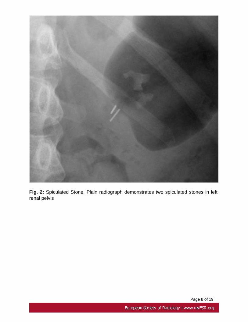
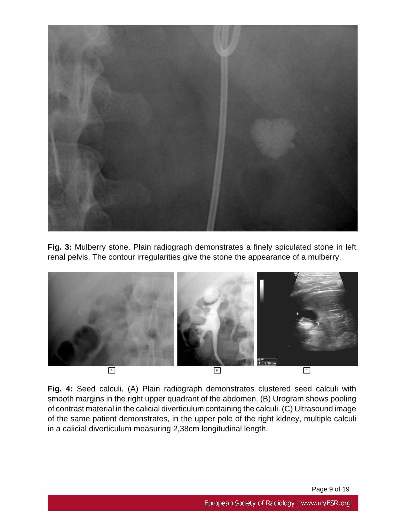
Renal calculi are radiopaque due to their calcium content. Calcium oxalate stones mayoccur in a pure monohydrate or dihydrate form. Pure calcium oxalate monohydratestones, like pure calcium phosphate, are usually small and highly radiopaque for theirsize (Fig. 1 on page 6). Calcium oxalate dihydrate stones may appear spiculated(Fig. 2 on page 7) (similar to a child's toy jack) or mamillated (Fig. 3 on page 8)("mulberry" stones).
Stones formed into small cavities such as caliceal diverticula are all of similar size andare known as "seed calculi" (Fig. 4 on page 9).
The "milk of calcium" pattern of stone (Fig. 5 on page 9) results from a suspensionof calcium carbonate in a renal cyst or caliceal diverticulum. The suspension is heavierthan urine and therefore layers in the most dependent portion of the cyst or diverticula.
A staghorn stone (Fig. 6 on page 10) is a branched renal calculus that resembles theantlers of a stag. They are usually composed of struvite and, less commonly, cystineor uric acid. Staghorn calculi are associated with recurrent urinary tract infections frombacterial pathogens that produce alkaline urine. Therefore it's the only type of calculusthat is more common in women. These stones are less opaque than calcium ones.
Nephrocalcinosis
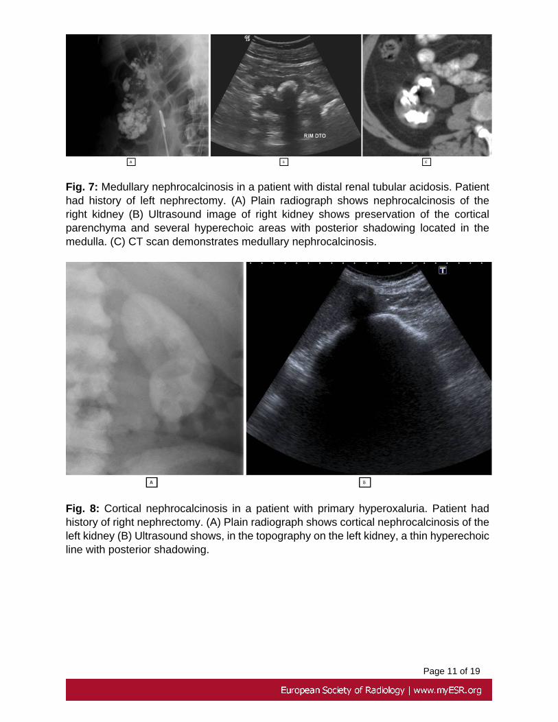
Deposition of calcium in the renal parenchyma is a common pathologic finding known asnephrocalcinosis. It is characterized according to the anatomic area involved, medullaor cortex.
The most common form is medullary nephrocalcinosis, and it has a wide variety of causes(table 2). Uniform and bilateral deposition of calcium in the medullary portion of thekidney is most common seen secondary to hyperparathyroidism or distal renal tubularacidosis (Fig. 7 on page 10). On the other hand, an asymmetric form of medullarynephrocalcinosis is commonly seen in medullar sponge kidney.

Page 4 of 19
Cortical nephrocalcinosis (Fig. 8 on page 11) may also be attributable to a variety ofcauses (table 2) but, it is usually secondary to chronic glomerulonephritis or acute corticalnecrosis.
Medullary Nephrocalcinosis Cortical Nephrocalcinosis
Bartter syndrome
Bone metastases
Chronic pyelonephritis
Cushing syndrome (endogenous,exogenous)
Hyperparathyroidism
Hyperthyroidism
Idiopatic hypercalcemia
Malignancy
Medullar sponge kidney
Nephrotoxic drugs
Primary hyperoxaluria
Renal papillary necrosis
Renal tuberculosis
Renal tubular acidosis
Sarcoidosis
Sickle cell disease
Vitamin D excess
Acute cortical necrosis
Alport syndrome
Chronic glomerulonephritis
Chronic hypercalcemic states
Ethylene glycol poisoning
Methoxyflurane toxicity
Oxalosis
Rejected renal transplant
Sickle cell disease
Table 2 - Common causes of Medullary and Cortical Nephrocalcinosis (source -reference 1)
Cyst or Solid Mass

Page 5 of 19
Simple renal cysts can show intracystic or wall calcifications (Fig. 9 on page 11).Intracystic calcifications are usually thin and peripheral and are often described as havingan "eggshell" appearance.
The most common primary solid neoplasm of the kidney is renal cell carcinoma (Fig. 10on page 12) and it calcifies in about 20% of the cases. Calcifications may also be seenin other primary renal neoplasms (e.g. renal osteosarcoma) or in metastatic lesions ofthe kidney. Renal masses with calcification should always lead to further investigation inorder to exclude malignancy.
Miscellaneous Renal Calcifications
Several pathologic conditions such as diabetes mellitus and severe atheroscleroticdisease may cause calcification of the renal vasculature (Fig. 11 on page 12).
Renal tuberculosis is a result of secondary hematogenous infection from the lungs.The genitourinary system is often affected, being the second most common site oftuberculosis infection. In the end stage of renal tuberculosis, we can see a small,totally calcified and nonfunctional kidney resulting in autonephrectomy. The radiologicappearance of this condition has been described as putty kidney (Fig. 12 on page 13).
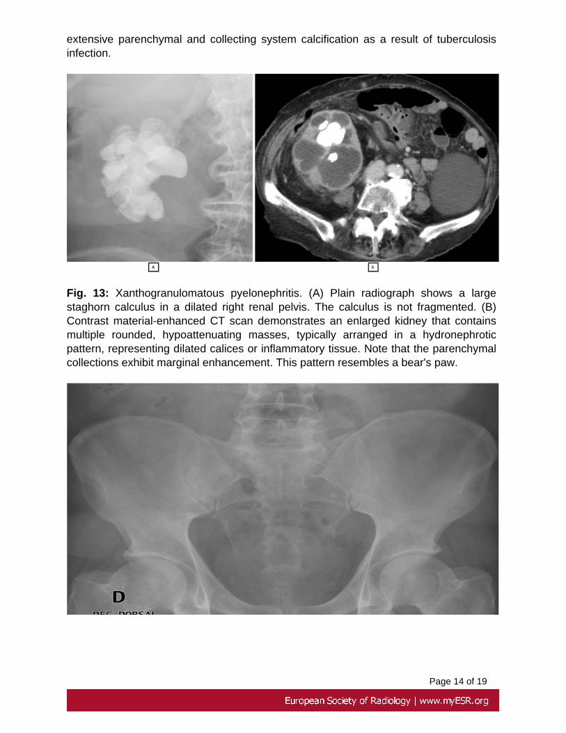
Xanthogranulomatous pyelonephritis (Fig. 13 on page 14) is an uncommoncomplication of long term urinary tract obstruction with a superimposed chronic infection.On classic excretory urography it is characterized by an obstructing staghorn calculusthat may be fragmented, renal enlargement and nonexcretion of contrast material fromthe involved kidney. At CT, there is replacement of the renal parenchyma by an infectiousprocess that produces hypoattenuating masses arranged in a hydronephrotic pattern.There may be enhancement in the margins of these masses after contrast materialadministration. This appearance on CT scans has been described as the bear paw sign.
Ureteral Calcification
Stones that migrate from kidney are the most common cause of ureteral calcification.Therefore ureteral stones show the same composition as the ones from the kidneys.Stones usually become impacted at points of anatomic narrowing such as theureteropelvic junction, the ureteral crossing of iliac vessels and the ureterovesicaljunction.

Page 6 of 19
In the pelvis, phleboliths may be mistaken with ureteral stones but, while stones (Fig. 14on page 14, Fig. 15 on page 15, Fig. 16 on page 15) usually have a uniformradiopacity and are often angulated along the course of the ureter, phleboliths (Fig. 17on page 16) tend to be multiple and centrally radiolucent.
Although uncommon, inflammatory conditions like schistosomiasis and tuberculosis maycause calcification of the ureteral wall.
Bladder Calcification
Bladder calculi (Fig. 18 on page 16) are the most common cause of bladdercalcification. Nowadays, in developed countries, they are not as common as they usedto be, and are usually associated with bladder outlet obstruction. Like renal calculi, theyusually consist of a mixture of calcium oxalate and calcium phosphate. Bladder calculiradiopacity reflects their composition.
Calcifications within the bladder wall can be seen in patients with primary neoplasms ofthe bladder.
Other rare entities that can cause bladder calcification are schistosomiasis (Fig. 19 onpage 16), tuberculosis, alkaline incrusted cystitis and radiation therapy.
In the pelvic region we can also see calcifications of the prostate gland (Fig. 20 on page17) and seminal vesicles (Fig. 21 on page 17).
Images for this section:

Page 7 of 19
Fig. 1: Highly opaque calculus. (A) Plain radiograph shows a small and highly opaquecalculus in right renal pelvis. (B) Plain radiograph demonstrates a highly opaque calculusin right renal pelvis
Page 8 of 19
Fig. 2: Spiculated Stone. Plain radiograph demonstrates two spiculated stones in leftrenal pelvis
Page 9 of 19
Fig. 3: Mulberry stone. Plain radiograph demonstrates a finely spiculated stone in leftrenal pelvis. The contour irregularities give the stone the appearance of a mulberry.
Fig. 4: Seed calculi. (A) Plain radiograph demonstrates clustered seed calculi withsmooth margins in the right upper quadrant of the abdomen. (B) Urogram shows poolingof contrast material in the calicial diverticulum containing the calculi. (C) Ultrasound imageof the same patient demonstrates, in the upper pole of the right kidney, multiple calculiin a calicial diverticulum measuring 2,38cm longitudinal length.

Page 10 of 19
Fig. 5: Milk of calcium stones in a renal cyst. (A) Plain radiograph shows a calcifydensity in the right upper quadrant. (B) Ultrasound and (C) CT scan of the same patientdemonstrates, in the right kidney, a cyst with a calcified image inside that makes a liquid-calcium level.
Fig. 6: Staghorn calculus. (A) Plain radiograph shows a staghorn calculus in left renalpelvis. (B) Plain radiograph of a different patient also shows a staghorn calculus in leftrenal pelvis and collecting system.
Page 11 of 19
Fig. 7: Medullary nephrocalcinosis in a patient with distal renal tubular acidosis. Patienthad history of left nephrectomy. (A) Plain radiograph shows nephrocalcinosis of theright kidney (B) Ultrasound image of right kidney shows preservation of the corticalparenchyma and several hyperechoic areas with posterior shadowing located in themedulla. (C) CT scan demonstrates medullary nephrocalcinosis.
Fig. 8: Cortical nephrocalcinosis in a patient with primary hyperoxaluria. Patient hadhistory of right nephrectomy. (A) Plain radiograph shows cortical nephrocalcinosis of theleft kidney (B) Ultrasound shows, in the topography on the left kidney, a thin hyperechoicline with posterior shadowing.

Page 12 of 19
Fig. 9: Cyst Calcifications (A) Plain radiograph shows multiple rim calcifications in theleft kidney. (B) (C) CT scan shows multiple cysts with rim calcification in the left kidney.
Fig. 10: Renal cell carcinoma (A) Plain radiograph shows a spotty calcification in theleft upper quadrant of the abdomen. (B) Ultrasound shows in the left upper pole of theright kidney a nodular formation with multiple and millimetric hyperechoic areas. (C)Contrast material-enhanced CT scan demonstrates a mass containing multiple punctuatecalcifications in the upper pole of the let kidney.

Page 13 of 19
Fig. 11: Renal vascular calcification in diabetes mellitus. Plain radiograph showsextensive calcification of both main renal arteries and their peripheral branches.
Fig. 12: Putty Kidney in a patient with renal tuberculosis. (A) Plain radiograph of theabdomen shows extensive calcification in the left kidney, which was nonfunctional,consistent with autonephrectomy from tuberculosis. (B) CT scan demonstrates the
Page 14 of 19
extensive parenchymal and collecting system calcification as a result of tuberculosisinfection.
Fig. 13: Xanthogranulomatous pyelonephritis. (A) Plain radiograph shows a largestaghorn calculus in a dilated right renal pelvis. The calculus is not fragmented. (B)Contrast material-enhanced CT scan demonstrates an enlarged kidney that containsmultiple rounded, hypoattenuating masses, typically arranged in a hydronephroticpattern, representing dilated calices or inflammatory tissue. Note that the parenchymalcollections exhibit marginal enhancement. This pattern resembles a bear's paw.

Page 15 of 19
Fig. 14: Ureteral Stone. Plain radiograph shows a ureteral stone on the medial portionof the left ureter. Sometimes these calculi may be difficult to detect when overlying bonystructures, such as the sacrum, as shown in this radiograph.
Fig. 15: Ureteral Stones. (A) Plain radiograph and (B) CT scan apparently show a giantureteral stone in left distal ureter. Later, after surgical approach, it was verified that thepatient had multiple small calculi instead.

Page 16 of 19
Fig. 16: Ureteral Stone. (A) Plain radiograph shows a round opacity (ureteral calculus)in the right side of the pelvis that could easily be mistaken for a phlebolith. (B) Urogramdemonstrates that it is localized inside the ureter confirming the diagnosis.
Fig. 17: Phleboliths. (A) (B) Plain radiographs show, in the pelvis, several areas of highopacity with a central radiolucency suggestive of phleboliths. This diagnosis was laterconfirmed by CT scan.
Fig. 18: Large bladder calculus. Plain radiograph (A) and Ultrasound (B) show a largecalculus in the bladder.

Page 17 of 19
Fig. 19: Schistosomiasis. Plain radiograph (A) shows a heterogeneous calcification inthe pelvic region. CT scans (B) (C) show a rim of calcification within the bladder wall asa sequel of a previous infection.
Fig. 20: Plain radiograph shows calcification of the prostate gland.

Page 18 of 19
Fig. 21: Plain radiograph shows calcification of the seminal vesicles.

Page 19 of 19
Conclusion
Urinary tract calcifications are very common. Usually they present with specific imagingcharacteristics on conventional radiology that suggest certain diagnoses. Nevertheless,correct interpretation of these findings is not an easy task and the use of other imagingmodalities should be considered whenever necessary.
It is our role, as radiologists, to be aware of these findings in order to allow an adequatemanagement of the patient.
Personal information
References
1. Dyer R.B. et all; Abnormal Calcifications in the Urinary Tract; Radiographics,1998.
2. Dyer R.B. et all; Classical Signs in Uroradiology; Radiographics, October2004.
3. Kawashima A. et all; CT of Renal Inflammatory Disease; Radiographics1997.
4. Van Arsdalen, K.N.; Pathogenesis of Renal Calculi; Urologic Radiologoly,1984.