Embed Size (px)
Citation preview
Abnormalities Of The Testis And Scrotum
Ahmed Al-Sayyad
Embryology
Testicular differentiation is initiated in the 7th week of gestation by the SRY gene
At 4 to 6 weeks’ gestation, the genital ridges organize. This is followed by migration of primordial germ cells
At 7 to 8 weeks’ both sertoli and leydig cells have developed
Embryology
During the 8th week, the fetal testis begins to secrete testosterone and MIS independent of pituitary hormonal regulation
MIS is secreted by the Sertoli cells and causes degeneration of the müllerian structures after the 8th week of gestation
The gubernaculum appears at the 7th week of embryologic development where its cranial aspect envelops the cauda epididymis and lower pole of the testis and extends caudally into the inguinal canal, where it maintains a firm attachment
Cryptorchidism
3% of full-term male newborns and 30.3% incidence in premature infants
More prevalent among preterm, small-for-gestational-age, low-birth-weight, and twin neonates
Approximately 70% to 77% of cryptorchid testes will spontaneously descend by 3 months of age
By 1 year of age, the incidence of cryptorchidism declines to about 1% and remains constant throughout adulthood
Descent Factors
Hormonal: androgens,MIS,estrogen,descendin Gubernaculum GFN and CGRP Epididymis Intra-abdominal pressure
Terminology
Undescended Ascended Gliding Retractile Ectopic
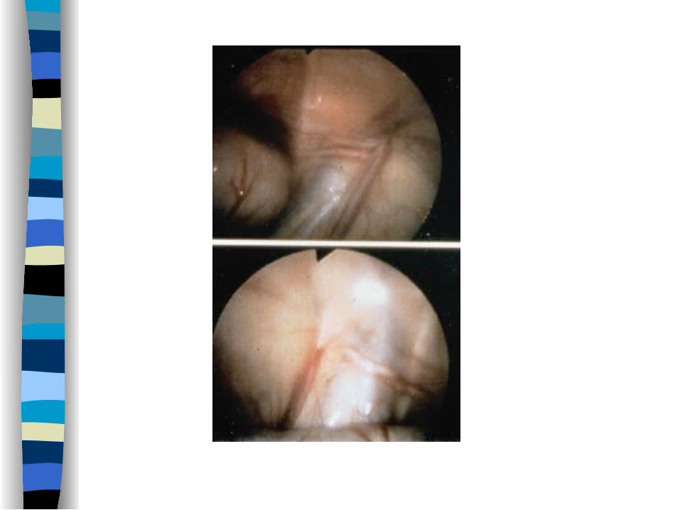
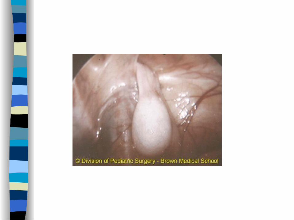
Nonpalpable testis
Intra-abdominal Vanishing Atrophic Missed on examination Bilateral nonpalpable work-up
Consequences of Cryptorchidism
Infertility Neoplasia Hernia Torsion Trauma Cosmetic
Work-UP
Maternal history including the use of gestational steroids, Perinatal history, including documentation of a scrotal examination at birth,PMH,PSH,FH
Examine in a warm room,supine,squatting etc Look for genital abnormalities,scrotal
size,contralateral hypertrophy

Investigations
Hormones US CT MRI Laparoscopy
Hormonal Therapy
HCG or GnRH can be used The lower the pretreatment position the better the
results Self limiting side effects Overall success rate < 20% Limited indications if any
Surgical Intervention
When Inguinal orchiopexy Laparoscopic orchiopexy Fowler-Stephens orchiopexy Staged orchiopexy Microvascular autotransplantation
Hydrocele
Normally, the processus vaginalis is obliterated from the internal inguinal ring to the upper scrotum, leaving a small potential space in the scrotum that partially surrounds the testis
Embryologic misadventures may occur and results in (hydrocele, hydrocele of the cord, and communicating hydrocele).
Simple Hydrocele
Simple (scrotal) hydrocele is an accumulation of fluid within the tunica vaginalis
Results from persistence of or delayed closure of the processus vaginalis
Commonly seen at birth, frequently bilateral, may be quite large. They transilluminate and may seem quite tense but not painful
Most resolve during the first 2 years of life If surgical repair is elected, an inguinal approach should be
used
Communicating Hydrocele
Persistence of the processus vaginalis which allows peritoneal fluid to communicate with the scrotum
The classic description is that of a hydrocele that changes in size
It can be compressible during examination All should be fixed using an inguinal approach Do it bilateral if patient got VP shunt or on
peritoneal dialysis
Hydrocele of the cord
Segmental closure of the processus, which leaves a loculated hydrocele of the cord
Presents as a painless groin mass which is mobile and transilluminates
Inguinal exploration and high ligation is curative


Acute Scrotum
Differential Diagnosis
Torsion testis Torsion appendix testis Torsion appendix epididymis Epididymo-orchitis Hernia Trauma Vasculitis Dermatological
Testicular Torsion
True surgical emergency of the highest order Irreversible ischemic injury may begin as soon as
4 hours after occlusion of the cord Intravaginal torsion, result from lack of normal
fixation of the testis and epididymis to the fascial and muscular coverings that surround the cord
This creates an abnormally mobile testis that hangs freely within the tunical space (a "bell-clapper deformity")
Testicular Torsion
Happens in any age but most commonly in prepubertal males
Presentation: Pain,N\V,Poor appetite,previous episodes
Examination:Swelling,Tenderness,High riding,transverse orientation,Loss of cremasteric reflex
Testicular Torsion
Doppler US may help in the diagnosis Manual detorsion may be attempted in ER Scrotal exploration is mandatory Detorte the affected testis and pex the other side
while waiting for the testis to pink up If the testis is still alive pex it , if not do an
orchiectomy

Intermittent Torsion
Recurrent episodes of acute, self-limited scrotal pain
Normal physical examination will be found in-between
If the suspicion is strong , elective scrotal exploration and bilateral orchiopexy should be performed
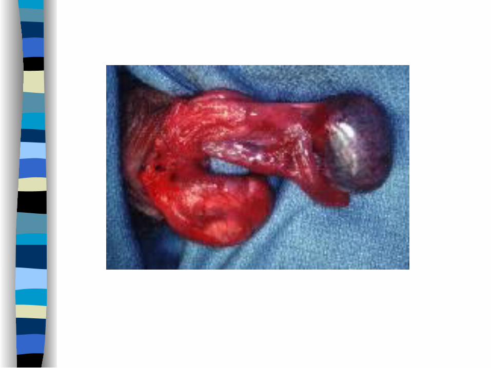
Prenatal testicular torsion
Extravaginal torsion Presents at birth as a hard,nontender testis fixed to
the scrotal skin which is usually discolored Doppler US may help in the diagnosis Management is controversial: observation Vs
exploration
Torsion Appendix Testis
presentation is extremely variable, from an insidious onset of scrotal discomfort to an acute presentation identical to torsion testis
Exam:Tenderness or mass in the upper pole,Blue dot sign,cremasteric reflex usually present
Doppler US may help in diagnosis Management:conservative,pain meds,limit activity
Epididymitis
Rare in pediatrics Presentation:pain,swelling,erethyma,LUTS,fever,
urethral discharge,STDs Investigations:pyuria, bacteriuria, positive urine
culture, increased flow on doppler IV Abx given if systematically ill then oral for
total of 10-14 days Screening US usually indicated ? VCUG

Varicocele
Dilated and tortuous veins of the pampiniform plexus
Found in approximately 15% of male adolescents, with a marked left-sided predominance
Etiology:increased venous pressure in the left renal vein, incompetent valves of the internal spermatic vein
varicocele
Unilateral varicocele may affect testicular function bilaterally
Toxic effect of varicocele may manifest as testicular growth failure, semen abnormalities, Leydig cell dysfunction, and histologic changes
Possible mechanisms:reflux of adrenal metabolites, hyperthermia, hypoxia, local testicular hormonal imbalance, and intratesticular hyperperfusion injury
varicocele
Presentation:asymptomatic,pain,scrotal mass,infertility,atrophy
Grading on physical examination Obtain scrotal US Treat if there is loss of volume (> 2 mls or > 20%)
Treatment Alternatives
Inguinal Ligation and Subinguinal Ligation Retroperitoneal and Laparoscopic Ligation Transvenous Occlusion Complications:hydrocele,recurrence,testicular
atrophy


![[PPT]SISTEM REPRODUKSI PRIA - From Heart to Heart ... · Web viewAlatkelaminlaki-lakiterbagiatas 3 bagian: Organ reproduksiluar Penis Scrotum Organ reproduksidalam Testis Epididimis](https://img.pdfslide.net/doc/110x75/5af335947f8b9ac24691aac4/pptsistem-reproduksi-pria-from-heart-to-heart-viewalatkelaminlaki-lakiterbagiatas.jpg)