Embed Size (px)
Citation preview
P a g e | 1
2015 ARDMSPediatric Echocardiography
Job Task Analysis Summary Report
© American Registry for Diagnostic Medical Sonography (ARDMS)

P a g e | 2
Table of ContentsABOUT THE REPORT.....................................................................................................................3METHODOLOGY............................................................................................................................ 3
Job Task Analysis (JTA) Working Group......................................................................................3Survey Questionnaire Development............................................................................................3Survey Administration.................................................................................................................3Data Analysis............................................................................................................................... 3
SURVEY RESULTS......................................................................................................................... 5Demographics and Backgrounds of Participants........................................................................5
Country of Practice.................................................................................................................. 5Educational Background..........................................................................................................5Work Experience...................................................................................................................... 5Work Environment................................................................................................................... 6
Task Descriptions........................................................................................................................ 7
P a g e | 3
ABOUT THE REPORTThe American Registry for Diagnostic Medical Sonography (ARDMS) is the globally recognized standard of excellence in sonography. It is responsible for the preparation of valid and reliable certification examinations in sonography. The performance of job task analysis (JTA) at the national level assists ARDMS in evaluating the current practice expectations and performance requirements of the specialty. The 2015 Pediatric Echocardiography (PE) JTA was designed to collect information on the sonography-related work activities sonography registrants actually perform in practice. The results were used in the development of the test content outline that guides content distribution of the PE Examination. This report details the methodology, data collection & analysis and survey results. It also includes the test content outline that resulted from the JTA.
METHODOLOGYJob Task Analysis (JTA) Working GroupA JTA Working Group consisting of four subject matter experts (SMEs) led this project. All fifteen JTA Working Group members were Exam Development Task Force (EDTF) members and volunteers. Survey Questionnaire Development
ARDMS facilitated a process whereby the JTA Working Group developed the task list and demographic items for the survey. Tasks and demographic items from previous job task surveys were used as a starting point in this development. The JTA Working Group reached a consensus on a list of 105 tasks to be used in the survey. These tasks were divided into six domains: Anatomy and Physiology; Abnormal Pathology and
Pathophysiology; Congenital Anomalies; Postoperative (surgically corrected/palliated) Anatomy; Clinical Standards and Guidelines; and Measurement Techniques and Quantification. All task statements and response options were relevant to RDCS credentialed sonographers.
The survey questionnaire was pilot-tested with a group of fifteen individuals from the PE EDTF and volunteers.
Survey AdministrationThe survey was made available to participants as a web-based survey through the survey platform Qualtrics®. An invitation to participate in the study was sent via email to the members.
ARDMS sent the job task analysis survey to 2,538 registrants credentialed since 2010. These registrants were selected randomly using a stratified sampling method so that the sample is representative of all ARDMS RDCS registrants in terms of specialty, gender, and geographic region. The survey was made available to the participants for four weeks between June 29th and July 17th, 2015. The participants responded anonymously and all responses were kept confidential.
A total of 1381 (54.4% of those sampled) sonographers responded to the survey. Of these, 1261 (49.68%) reported that they are currently performing Pediatric Echocardiography. The data analysis were based on the responses from the 1261 sonographers.
P a g e | 4
Data AnalysisRespondents were asked the following questions for each task: How frequently do you perform the task in your practice, and how important is the task in affecting clinical decisions and patient outcomes? The frequency and importance rating scales were scored 1-5. The response options for the frequency scale were Never, Rarely, Occasionally, Often, and Frequently. The response options for the importance scale were Not Important, Somewhat Important, Moderately Important, Very Important, and Critically Important.
P a g e | 5
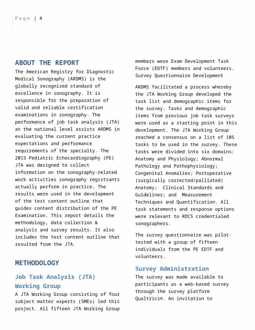
SURVEY RESULTSDemographics and Backgrounds of ParticipantsGenderApproximately 75% of the respondents were female and 25% were male (Figure 1).
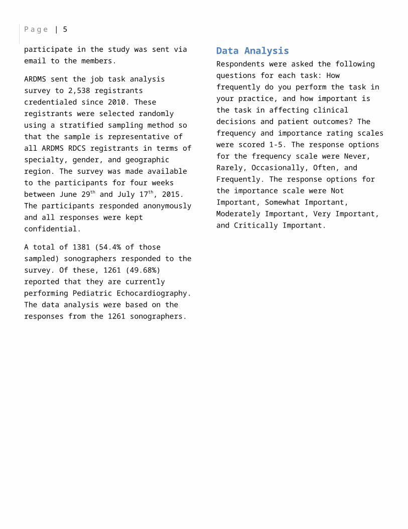
Location of PracticeMost of the respondents reside in the United States. About a third of the respondents practice in the southern region of the United States.(Figure 2).
PrerequisiteApproximately one-half of the candidates met eligibility requirements under Prerequisite 1 or Prerequisite 2. The other one-half was spread among eight other prerequisites (Figure 3).
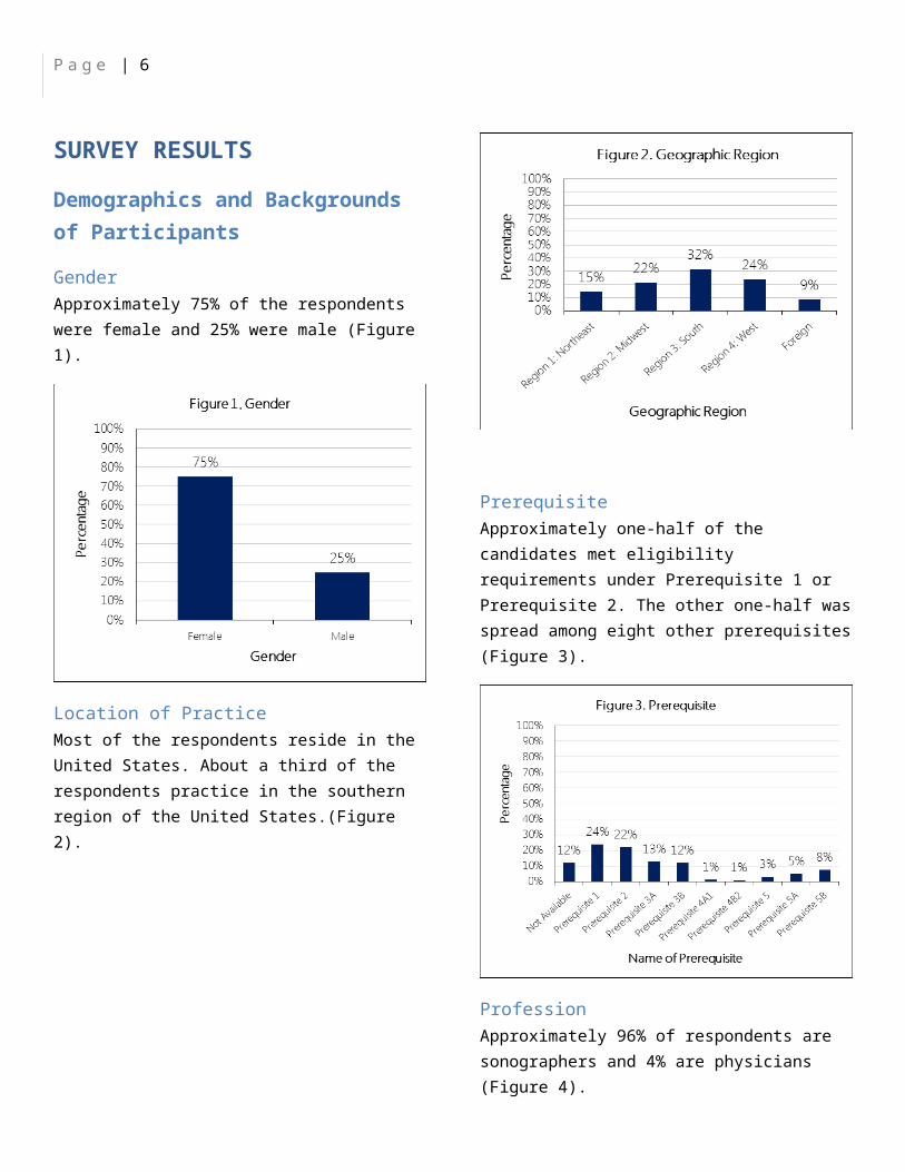
ProfessionApproximately 96% of respondents are sonographers and 4% are physicians (Figure 4).
Work SettingThe majority (88%) of respondents are currently practicing Pediatric Echocardiography (Figure 5). Approximately
P a g e | 6
74% of respondents are currently a sonography educator (Figure 6).
Approximately 43% of the respondents perform 0-50 PE exams per month and about 56% perform more than 50 exams per month (Figure 7).
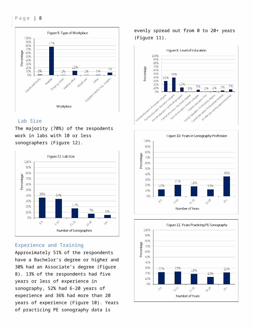
The respondents were asked to indicate the type of environment they perform most of their PE sonographic examiniations. The highest frequencides were seen in hospitals (Figure 9).
Lab SizeThe majority (70%) of the respodents work in labs with 10 or less sonographers (Figure 12).
P a g e | 7
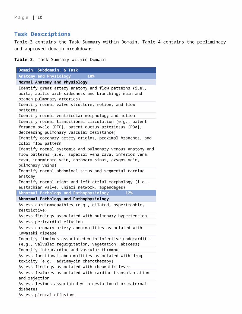
Experience and TrainingApproximately 51% of the respondents have a Bachelor’s degree or higher and 30% had an Associate’s degree (Figure 8). 13% of the respondents had five years or less of experience in sonography, 52% had 6-20 years of experience and 36% had more than 20 years of experience (Figure 10). Years of practicing PE sonography data is evenly spread out from 0 to 20+ years (Figure 11).
P a g e | 8
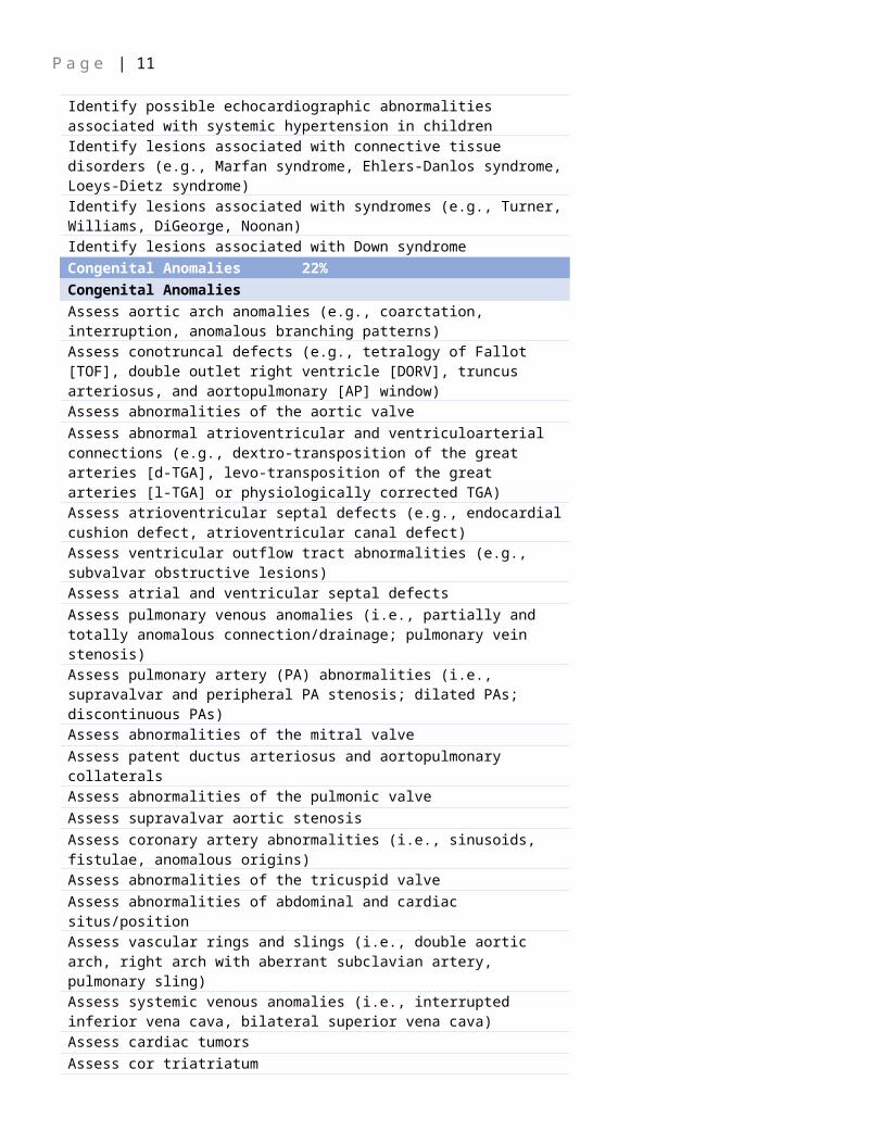
Task Descriptions Table 3 contains the Task Summary within Domain. Table 4 contains the preliminary and approved domain breakdowns.
Table 3. Task Summary within Domain
Domain, Subdomain, & TaskAnatomy and Physiology 10%Normal Anatomy and PhysiologyIdentify great artery anatomy and flow patterns (i.e., aorta; aortic arch sidedness and branching; main and branch pulmonary arteries)Identify normal valve structure, motion, and flow patternsIdentify normal ventricular morphology and motionIdentify normal transitional circulation (e.g., patent foramen ovale [PFO], patent ductus arteriosus [PDA], decreasing pulmonary vascular resistance)Identify coronary artery origins, proximal branches, and color flow patternIdentify normal systemic and pulmonary venous anatomy and flow patterns (i.e., superior vena cava, inferior vena cava, innominate vein, coronary sinus, azygos vein, pulmonary veins)Identify normal abdominal situs and segmental cardiac anatomyIdentify normal right and left atrial morphology (i.e., eustachian valve, Chiari network, appendages)Abnormal Pathology and Pathophysiology 12%Abnormal Pathology and PathophysiologyAssess cardiomyopathies (e.g., dilated, hypertrophic, restrictive)Assess findings associated with pulmonary hypertensionAssess pericardial effusionAssess coronary artery abnormalities associated with Kawasaki diseaseIdentify findings associated with infective endocarditis (e.g., valvular regurgitation, vegetation, abscess)Identify intracardiac and vascular thrombusAssess functional abnormalities associated with drug toxicity (e.g., adriamycin chemotherapy)Assess findings associated with rheumatic feverAssess features associated with cardiac transplantation and rejectionAssess lesions associated with gestational or maternal diabetesAssess pleural effusionsIdentify possible echocardiographic abnormalities associated with systemic hypertension in childrenIdentify lesions associated with connective tissue disorders (e.g., Marfan syndrome, Ehlers-Danlos syndrome, Loeys-Dietz syndrome)Identify lesions associated with syndromes (e.g., Turner, Williams, DiGeorge, Noonan)Identify lesions associated with Down syndromeCongenital Anomalies 22%

P a g e | 9
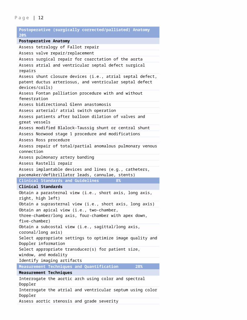
Congenital AnomaliesAssess aortic arch anomalies (e.g., coarctation, interruption, anomalous branching patterns)Assess conotruncal defects (e.g., tetralogy of Fallot [TOF], double outlet right ventricle [DORV], truncus arteriosus, and aortopulmonary [AP] window)Assess abnormalities of the aortic valveAssess abnormal atrioventricular and ventriculoarterial connections (e.g., dextro-transposition of the great arteries [d-TGA], levo-transposition of the great arteries [l-TGA] or physiologically corrected TGA)Assess atrioventricular septal defects (e.g., endocardial cushion defect, atrioventricular canal defect)Assess ventricular outflow tract abnormalities (e.g., subvalvar obstructive lesions)Assess atrial and ventricular septal defectsAssess pulmonary venous anomalies (i.e., partially and totally anomalous connection/drainage; pulmonary vein stenosis)Assess pulmonary artery (PA) abnormalities (i.e., supravalvar and peripheral PA stenosis; dilated PAs; discontinuous PAs)Assess abnormalities of the mitral valveAssess patent ductus arteriosus and aortopulmonary collateralsAssess abnormalities of the pulmonic valveAssess supravalvar aortic stenosisAssess coronary artery abnormalities (i.e., sinusoids, fistulae, anomalous origins)Assess abnormalities of the tricuspid valveAssess abnormalities of abdominal and cardiac situs/positionAssess vascular rings and slings (i.e., double aortic arch, right arch with aberrant subclavian artery, pulmonary sling)Assess systemic venous anomalies (i.e., interrupted inferior vena cava, bilateral superior vena cava)Assess cardiac tumorsAssess cor triatriatumPostoperative (surgically corrected/palliated) Anatomy 20%Postoperative AnatomyAssess tetralogy of Fallot repairAssess valve repair/replacementAssess surgical repair for coarctation of the aortaAssess atrial and ventricular septal defect surgical repairsAssess shunt closure devices (i.e., atrial septal defect, patent ductus arteriosus, and ventricular septal defect devices/coils)Assess Fontan palliation procedure with and without fenestrationAssess bidirectional Glenn anastomosisAssess arterial/ atrial switch operationAssess patients after balloon dilation of valves and great vesselsAssess modified Blalock-Taussig shunt or central shuntAssess Norwood stage 1 procedure and modificationsAssess Ross procedureAssess repair of total/partial anomalous pulmonary venous
P a g e | 10
connection Assess pulmonary artery bandingAssess Rastelli repairAssess implantable devices and lines (e.g., catheters, pacemaker/defibrillator leads, cannulae, stents)Clinical Standards and Guidelines 8%Clinical Standards Obtain a parasternal view (i.e., short axis, long axis, right, high left)Obtain a suprasternal view (i.e., short axis, long axis)Obtain an apical view (i.e., two-chamber, three-chamber/long axis, four-chamber with apex down, five-chamber)Obtain a subcostal view (i.e., sagittal/long axis, coronal/long axis)Select appropriate settings to optimize image quality and Doppler informationSelect appropriate transducer(s) for patient size, window, and modalityIdentify imaging artifactsMeasurement Techniques and Quantification 28%Measurement Techniques Interrogate the aortic arch using color and spectral DopplerInterrogate the atrial and ventricular septum using color DopplerAssess aortic stenosis and grade severityInterrogate the pulmonary venous return using color and spectral DopplerInterrogate the pulmonary artery and branches using color and spectral DopplerAssess right ventricular pressure using tricuspid and pulmonary regurgitant jet velocitiesAssess pulmonary stenosis and grade severityInterrogate systemic venous return using color and spectral DopplerAssess tricuspid regurgitation and grade severityAssess aortic regurgitation and grade severityAnalyze ventricular regional wall motion qualitatively using two-dimensional imaging and/or M-modeAssess mitral regurgitation and grade severityAssess mitral stenosis and grade severityAssess ventricular septal defect gradientsCalculate maximal pressure gradients using the modified Bernoulli equationAssess pulmonary regurgitation and grade severityDemonstrate echocardiographic findings at specific times during the electrocardiogram (cardiac) cycleAssess atrial septal shunting gradientsAssess tricuspid stenosis and grade severityMeasure chamber sizes and wall thickness using M-modeCalculate fractional shortening using M-modePerform linear measurements using two-dimensional imaging methodsCalculate ejection fraction using two-dimensional imaging
P a g e | 11
methodsCalculate indices of diastolic function (e.g., E/A ratio, E/E' ratio, mitral valve inflow pattern, pulmonary venous flow pattern)Measure chamber sizes and wall thickness using two-dimensional imaging methods
P a g e | 12
Table 4. Content Outline Breakdown by Domain
Domain Percentage of Examination
Anatomy and Physiology 10%Abnormal Pathology and Pathophysiology 12%Congenital Anomalies 22%Postoperative (surgically corrected/palliated) Anatomy 20%
Clinical Standards and Guidelines 8%Measurement Techniques and Quantification 28%
Total 100%
Note. Forms built to this outline may not match approved percentages exactly.


![Jta User Guide[1]](https://img.pdfslide.net/doc/110x75/577d248a1a28ab4e1e9cb1ab/jta-user-guide1.jpg)