Embed Size (px)
Citation preview
Hindawi Publishing CorporationInternational Journal of DentistryVolume 2010, Article ID 326124, 4 pagesdoi:10.1155/2010/326124
Research Article
Absorption of Nickel, Chromium, and Iron by the Root Surface ofPrimary Molars Covered with Stainless Steel Crowns
David Keinan,1 Eliyahu Mass,2 and Uri Zilberman1, 2
1 Laboratory of Bioanthropology and Ancient DNA, School of Dental Medicine, The Hebrew University of Jerusalem,P.O. Box 12272, Jerusalem 91120, Israel
2 Clinic of Pediatric Dentistry, Barzilai Medical Center, 2nd Hahistadrut Street, Ashkelon 78278, Israel
Correspondence should be addressed to Uri Zilberman, [email protected]
Received 29 September 2010; Revised 27 November 2010; Accepted 22 December 2010
Academic Editor: Toru Nikaido
Copyright © 2010 David Keinan et al. This is an open access article distributed under the Creative Commons Attribution License,which permits unrestricted use, distribution, and reproduction in any medium, provided the original work is properly cited.
Objective. The purpose of this study was to analyze the absorption of metal ions released from stainless steel crowns by root surfaceof primary molars. Study Design. Laboratory research: The study included 34 primary molars, exfoliated or extracted duringroutine dental treatment. 17 molars were covered with stainless-steel crowns for more than two years and compared to 17 intactprimary molars. Chemical content of the mesial or distal root surface, 1 mm apically to the crown or the cemento-enamel junction(CEJ), was analyzed. An energy dispersive X-ray spectrometer (EDS) was used for chemical analysis. Results. Higher amounts ofnickel, chromium, and iron (5-6 times) were found in the cementum of molars covered with stainless-steel crowns comparedto intact molars. The differences between groups were highly significant (P < .001). Significance. Stainless-steel crowns releasenickel, chromium, and iron in oral environment, and the ions are absorbed by the primary molars roots. The additional burdenof allergenic metals should be reduced if possible.
1. Introduction
Nickel sensitivity is common and increasing in prevalence.Nickel was named the “contact allergen of the year” in2008 by the American Contact Dermatitis Society (ACDS)because of its significant public health importance [1].Nickel has been the most frequently detected allergen inpatch-test populations worldwide, and in North America theprevalence of nickel sensitivity has been increasing steadilysince the mid-1980s [2]. Contact dermatitis to nickel cansignificantly limit an individual’s lifestyle, and allergy tonickel has health implications because of the use of nickel inimplanted medical devices and in dentistry.
Nickel, an abundant natural element, is a hard, silvery-white material in its pure state, which can be combined withother metals, for example, iron, copper, chromium, and zinc,to form alloys and stainless steel. All soil contains nickel andit is accumulated in plants. It is found in meteorites andon the ocean floor and is emitted from volcanoes. Smallamounts of nickel are naturally found in drinking water andfood. Smokers have a high nickel uptake through their lungs.
In humans, most ingested and unabsorbed nickel is excretedin the feces [3]. Nickel absorbed from the gastrointestinaltract is excreted in the urine. Elimination half time averages28 ± 9 hours [4]. The US Department of Health andHuman Services has determined that metallic nickel maybe a carcinogen. The International Agency for Research onCancer has concluded that some nickel compounds andmetallic nickel are carcinogenic to humans.
In Europe and North America, nickel sulphate is themost common sensitizer [5–8]. Although prevalence variesfrom one country to another, sensitization rates range from9.2% in Germany to 14.3% in the United States, 15% inUAE, up to 19.1% in Turkey, and 19.9% in Singapore [9].Overall, nickel sensitivity is 4 to 10 times more commonin women than in men, which can be explained by thecustom of girls piercing their ears at a very young age.Sensitivity begins between the ages of 2 and 5 years andincreases to a peak at 10–15 years [10]. In North America,the frequency of positive patch-test reaction in children (age0–18 years) was higher (28.3%) than that of adults (17.2%)[11].
2 International Journal of Dentistry
Nickel-containing alloys are used in dental care, eitherin construction of restorations or as endodontic instrumentsand orthodontic appliances. Dental restorations leak nickeland chromium, another well-known allergenic compound.The oral manifestations of the contact allergy to nickel usedin dentistry included lichen planus or stomatitis [12]. Theblood concentration of nickel and chromium in patients withremovable partial dentures made of nickel and chromiumcontaining stainless steel showed a significant increased level[13, 14].
Stainless steel orthodontic materials and stainless steelcrowns (SSC) are the two major devices in pediatric dentistrythat contain nickel. Fixed orthodontic appliances releasemeasurable amounts of nickel and chromium in the salivaand serum, without reaching toxic levels [14]. Nickel releasefrom orthodontic appliances made from nickel-titaniumand stainless steel increased over the first week after place-ment and then decreased over time [15]. The influenceof chromium and nickel concentrations in saliva and theireffects on gingival tissues during orthodontic treatment hasbeen reported [16]. After 3 months, 20% of the females and10% of the males showed an allergic reaction in a formof gingivitis, which disappeared a month after applianceremoval. There is some evidence that oral contact with nickeland chromium may induce a state of partial tolerance tothese materials in nonsensitized individuals, but the evidenceis not significant statistically [17].
Prefabricated stainless steel crowns (SSCs) cemented toprimary molars are the second most common dental devicecontaining stainless steel used in children. Nickel sensitivityhas been reported in children treated with old generationSSCs with high (up to 72%) nickel content [18]. Based onthese findings, the new generation of SSC contains only 9%–12% nickel [19]. The aim of this study was to investigate thehypothesis that the new SSCs also release nickel, chromium,and iron that are absorbed by the roots of the primary molarson which the SSCs are cemented.
2. Material and Methods
The study group consisted of 17 primary molars coveredwith stainless steel crowns due to extensive loss of toothmaterial (SSC-ION 3M/ESPE co. St. Paul MN, USA) for atleast 24 months and had 2 mm or more of mesial or distalroot below the crown margins. The crowns were cementedwith carboxylate cement (Durelon, 3M/ESPE AG, Seefeld,Germany). The teeth were collected after normal exfoliation(12 molars) or extraction performed due to orthodonticrequests (5 molars). The control group consisted of 17 intactprimary molars with at least 2 mm of mesial or distal rootbelow the CEJ, normally exfoliated (14 molars) or extractedfor orthodontic reasons (3 molars).
The molars were kept dry in a plastic tube till thechemical examination was performed. An energy dispersiveX-ray spectrometer (EDS) was used for chemical analysis. Inorder to enable a more accurate analysis of trace elements wehad to work in a high-vacuum mode and high pressure. Theteeth were coated with pure gold in order to keep the surfaceof the teeth intact in these conditions. The coating was of an
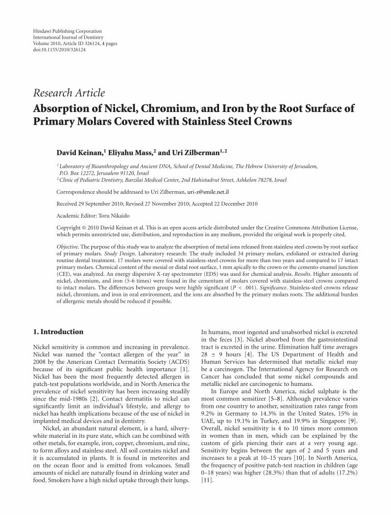
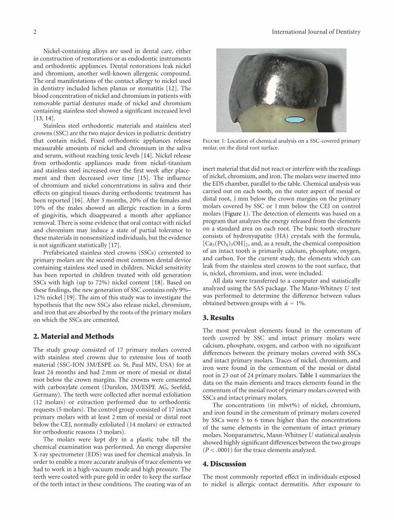
Figure 1: Location of chemical analysis on a SSC-covered primarymolar, on the distal root surface.
inert material that did not react or interfere with the readingsof nickel, chromium, and iron. The molars were inserted intothe EDS chamber, parallel to the table. Chemical analysis wascarried out on each tooth, on the outer aspect of mesial ordistal root, 1 mm below the crown margins on the primarymolars covered by SSC or 1 mm below the CEJ on controlmolars (Figure 1). The detection of elements was based on aprogram that analyzes the energy released from the elementson a standard area on each root. The basic tooth structureconsists of hydroxyapatite (HA) crystals with the formula,[Ca5(PO4)3OH]2, and, as a result, the chemical compositionof an intact tooth is primarily calcium, phosphate, oxygen,and carbon. For the current study, the elements which canleak from the stainless steel crowns to the root surface, thatis, nickel, chromium, and iron, were included.
All data were transferred to a computer and statisticallyanalyzed using the SAS package. The Mann-Whitney U testwas performed to determine the difference between valuesobtained between groups with α = 1%.
3. Results
The most prevalent elements found in the cementum ofteeth covered by SSC and intact primary molars werecalcium, phosphate, oxygen, and carbon with no significantdifferences between the primary molars covered with SSCsand intact primary molars. Traces of nickel, chromium, andiron were found in the cementum of the mesial or distalroot in 23 out of 24 primary molars. Table 1 summarizes thedata on the main elements and traces elements found in thecementum of the mesial root of primary molars covered withSSCs and intact primary molars.
The concentrations (in mlwt%) of nickel, chromium,and iron found in the cementum of primary molars coveredby SSCs were 5 to 6 times higher than the concentrationsof the same elements in the cementum of intact primarymolars. Nonparametric, Mann-Whitney U statistical analysisshowed highly significant differences between the two groups(P < .0001) for the trace elements analyzed.
4. Discussion
The most commonly reported effect in individuals exposedto nickel is allergic contact dermatitis. After exposure to
International Journal of Dentistry 3
Table 1: The quantity (in mlwt%) of Ni, Cr, and Fe in thecementum of the mesial root of primary molars.
Ni Cr Fe
SSC Intact P SSC Intact P SSC Intact P
Mean 0.86 0.16 <.0001 0.59 0.11 <.0001 1.13 0.20 <.0001
N 17 17 17 17 17 17
SD 0.42 0.10 0.37 0.07 0.77 0.13
Min 0.40 0.00 0.20 0.00 0.40 0.00
Max 1.50 0.30 1.70 0.20 2.70 0.40
Note. SSC: molars covered with stainless steel crowns, Intact: intact molars,P: P value, N: number of molars examined, and SD: standard deviation.
nickel, the endothelial cells that line the blood vessels pro-duced immune-response-mediating molecules (cytokines)within 24 hours, suggesting that nickel itself was acting asthe signal for T cells recruitment [20]. Nickel triggers aninflammatory response by directly activating human Toll-like receptor 4 (TLR4), and this activation was speciesspecific [21]. The risk of developing an allergic reaction tonickel from stainless steel devices used as dental orthodonticappliances is well documented [22–26]. The reports aregenerally of patients with presensitization to nickel, thatis, from earrings containing nickel. Both new and recycledbrackets will corrode in the oral environment [27], andtherefore, metal brackets should be made more resistantto corrosion and recycled brackets should not be used toavoid clinical side effects. In one study, [22] it was foundthat from 40 students without skin piercing, 4 out of 11with a history of permanent braces had developed nickelallergy compared with none out of 22 without orthodontictreatment, suggesting the possibility of sensitization throughdental devices. Oral exposure to nickel-containing metallicorthodontic appliances before sensitization to nickel (earpiercing) may have reduced the frequency of nickel hyper-sensitivity [28].
Toxicity and carcinogenicity of certain nickel compoundsmay be related to their uptake, transport, distribution, andretention at a cellular level [29].
The present study measured the absorption of metal ionsreleased from SSC by the cementum of primary molars.Nickel is found normally in the saliva [30] and can beabsorbed by intact teeth, but this level is significantly smallerwhen compared to the level detected after placement of fixeddental appliances containing nickel. A substantial amount ofnickel, chromium, and iron was released from the stainlesssteel crowns and absorbed by the cementum of the root ofprimary molars below the crowns. The amount observedin molars covered with SSC was 5-6 times greater than theconcentration in the cementum of intact primary molars andthe differences were significant statistically.
In vivo aged SSC surfaces showed significant morpho-logic alterations with wear and occlusal perforations as themost common findings. When the metal concentrations ofnew and in vivo aged SSC were compared, no statisticallysignificant differences were found [31]. The quantitativeanalysis of the SSC components was analyzed as percentage
of the component and not as the absolute amount. Theauthors stated that “The results of this study should not beviewed as a conclusive evidence of no compositional alter-ation of prefabricated pediatric metal crowns under clinicalconditions” (p. 219). The release of metal components fromthe SSCs in the oral cavity will not affect the concentration ofthe elements in the SSCs.
The present study showed that the amount of metallicions absorbed by cementum was 0.4–1.5 mlwt% (Ni), 0.2–1.7 mlwt% (Cr), and 0.4–2.7 mlwt% (Fe). If this amountis reduced from the original content of the SSC (the lownickel crowns), Fe—72%, Cr—18%, and Ni—10%, it can beunderstood why the differences between new and in vivo agedSSC showed no significance statistically [31].
5. Conclusion
The chemical analysis showed that nickel, chromium, andiron were released from SSC and absorbed into the cemen-tum of teeth covered with SSC. Since SSCs are also used tocover permanent molars, the influence of the release of theseelements on the systemic health of children should be furtherinvestigated.
References
[1] R. Kornik and K. A. Zug, “Nickel,” Dermatitis, vol. 19, no. 1,pp. 3–8, 2008.
[2] J. G. Marks Jr., D. V. Belsito, V. A. DeLeo et al., “NorthAmerican Contact Dermatitis Group patch-test results, 1998to 2000,” American Journal of Contact Dermatitis, vol. 14, no.2, pp. 59–62, 2003.
[3] M. Patriarca, T. D. B. Lyon, and G. S. Fell, “Nickel metabolismin humans investigated with an oral stable isotope,” AmericanJournal of Clinical Nutrition, vol. 66, no. 3, pp. 616–621, 1997.
[4] F. W. Sunderman Jr., S. M. Hopfer, K. R. Sweeney, A. H.Marcus, B. M. Most, and J. Creason, “Nickel absorption andkinetics in human volunteers,” Proceedings of the Society forExperimental Biology and Medicine, vol. 191, no. 1, pp. 5–11,1989.
[5] O. B. Christensen, “Nickel dermatitis. An update,” Dermato-logic Clinics, vol. 8, no. 1, pp. 37–40, 1990.
[6] A. Sertoli, S. Francalanci, M. C. Acciai et al., “Epidemiologicalsurvey of contact dermatitis in Italy (1984–1993) by GIRDCA(Gruppo Italiano Ricerca Dermatiti da Contatto e Ambien-tali),” American Journal of Contact Dermatitis, vol. 10, no. 1,pp. 18–30, 1999.
[7] J. E. R. Britton, S. M. Wilkinson, J. S. C. English et al., “TheBritish standard series of contact dermatitis allergens: vali-dation in clinical practice and value for clinical governance,”British Journal of Dermatology, vol. 148, no. 2, pp. 259–264,2003.
[8] L. A. Garner, “Contact dermatitis to metals,” DermatologicTherapy, vol. 17, no. 4, pp. 321–327, 2004.
[9] A. T. J. Jin and C. L. Goh, “Metal allergy in Singapore,” ContactDermatitis, vol. 52, no. 3, pp. 130–132, 2005.
[10] T. H. Clayton, S. M. Wilkinson, C. Rawcliffe, B. Pollock, andS. M. Clark, “Allergic contact dermatitis in children: shouldpattern of dermatitis determine referral? A retrospective studyof 500 children tested between 1995 and 2004 in one U.K.
4 International Journal of Dentistry
centre,” British Journal of Dermatology, vol. 154, no. 1, pp. 114–117, 2006.
[11] K. A. Zug, D. McGinley-Smith, E. M. Warshaw et al.,“Contact allergy in children referred for patch testing: NorthAmerican contact dermatitis group data, 2001–2004,” Archivesof Dermatology, vol. 144, no. 10, pp. 1329–1336, 2008.
[12] U. Raap, M. Stiesch, H. Reh, A. Kapp, and T. Werfel,“Investigation of contact allergy to dental metals in 206patients,” Contact Dermatitis, vol. 60, no. 6, pp. 339–343, 2009.
[13] S. Denizoglu and Z. Y. Duymus, “Evaluation of cobalt,chromium, and nickel concentrations in plasma and bloodof patients with removable partial dentures,” Dental MaterialsJournal, vol. 25, no. 2, pp. 365–370, 2006.
[14] G. Agaoglu, T. Arun, B. Izgu, and A. Yarat, “Nickel andchromium levels in the saliva and serum of patients with fixedorthodontic appliances,” Angle Orthodontist, vol. 71, no. 5, pp.375–379, 2001.
[15] R. D. Barrett, S. E. Bishara, and J. K. Quinn, “Biodegradationof orthodontic appliances. Part I. Biodegradation of nickeland chromium in vitro,” American Journal of Orthodontics andDentofacial Orthopedics, vol. 103, no. 1, pp. 8–14, 1993.
[16] A. A. Ramadan, “Effect of nickel and chromium on gingivaltissues during orthodontic treatment: a longitudinal study,”World Journal of Orthodontics, vol. 5, no. 3, pp. 230–235, 2004.
[17] C. S. Jensen, S. Lisby, O. Baadsgaard, A. Vølund, and T. Menne,“Decrease in nickel sensitization in a Danish schoolgirlpopulation with ears pierced after implementation of a nickel-exposure regulation,” British Journal of Dermatology, vol. 146,no. 4, pp. 636–642, 2002.
[18] W. H. Feasby, E. R. Ecclestone, and R. M. Grainger, “Nickelsensitivity in pediatric dental patients,” Pediatric dentistry, vol.10, no. 2, pp. 127–129, 1988.
[19] R. C. Randall, “Preformed metal crowns for primary andpermanent molar teeth: review of the literature,” PediatricDentistry, vol. 24, no. 5, pp. 489–500, 2002.
[20] M. Goebeler, A. Trautmann, A. Voss, E. B. Brocker, A. Toksoy,and R. Gillitzer, “Differential and sequential expression ofmultiple chemokines during elicitation of allergic contacthypersensitivity,” American Journal of Pathology, vol. 158, no.2, pp. 431–440, 2001.
[21] M. Schmidt, B. Raghavan, V. Muller et al., “Crucial role forhuman Toll-like receptor 4 in the development of contactallergy to nickel,” Nature Immunology, vol. 11, no. 9, pp. 814–819, 2010.
[22] K. Kalimo, L. Mattila, and H. Kautiainen, “Nickel allergy andorthodontic treatment,” Journal of the European Academy ofDermatology and Venereology, vol. 18, no. 5, pp. 543–545,2004.
[23] A. L. Counts, M. A. Miller, M. L. Khakhria, and S. Strange,“Nickel allergy associated with a transpalatal arch appliance,”Journal of Orofacial Orthopedics, vol. 63, no. 6, pp. 509–515,2003.
[24] P. D. Pigatto and G. Guzzi, “Systemic contact dermatitisfrom nickel associated with orthodontic appliances,” ContactDermatitis, vol. 50, no. 2, pp. 100–101, 2004.
[25] J. C. Schultz, E. Connelly, L. Glesne, and E. M. Warshaw,“Cutaneous and oral eruption from oral exposure to nickel indental braces,” Dermatitis, vol. 15, no. 3, pp. 154–157, 2004.
[26] G. Schuster, R. Reichle, R. R. Bauer, and P. M. Schopf,“Allergies induced by orthodontic alloys: incidence and impacton treatment. Results of a survey in private orthodontic officesin the federal state of Hesse, Germany,” Journal of OrofacialOrthopedics, vol. 65, no. 1, pp. 48–59, 2004.
[27] T. H. Huang, S. J. Ding, Y. Min, and C. T. Kao, “Metalion release from new and recycled stainless steel brackets,”European Journal of Orthodontics, vol. 26, no. 2, pp. 171–177,2004.
[28] H. Kerosuo, A. Kullaa, E. Kerosuo, L. Kanerva, and A.Hensten-Pettersen, “Nickel allergy in adolescents in relation toorthodontic treatment and piercing of ears,” American Journalof Orthodontics and Dentofacial Orthopedics, vol. 109, no. 2,pp. 148–154, 1996.
[29] K. S. Kasprzak, F. W. Sunderman Jr., and K. Salnikow, “Nickelcarcinogenesis,” Mutation Research, vol. 533, no. 1-2, pp. 67–97, 2003.
[30] R. M. de Souza and L. M. de Menezes, “Nickel, chromiumand iron levels in the saliva of patients with simulated fixedorthodontic appliances,” Angle Orthodontist, vol. 78, no. 2, pp.345–350, 2008.
[31] S. Zinelis, T. Lambrinaki, K. Kavvadia, and L. Papagiannoulis,“Morphological and compositional alterations of in vivoaged prefabricated pediatric metal crowns (PMCs),” DentalMaterials, vol. 24, no. 2, pp. 216–220, 2008.

Submit your manuscripts athttp://www.hindawi.com
Hindawi Publishing Corporationhttp://www.hindawi.com Volume 2014
Oral OncologyJournal of
DentistryInternational Journal of
Hindawi Publishing Corporationhttp://www.hindawi.com Volume 2014
Hindawi Publishing Corporationhttp://www.hindawi.com Volume 2014
International Journal of
Biomaterials
Hindawi Publishing Corporationhttp://www.hindawi.com Volume 2014
BioMed Research International
Hindawi Publishing Corporationhttp://www.hindawi.com Volume 2014
Case Reports in Dentistry
Hindawi Publishing Corporationhttp://www.hindawi.com Volume 2014
Oral ImplantsJournal of
Hindawi Publishing Corporationhttp://www.hindawi.com Volume 2014
Anesthesiology Research and Practice
Hindawi Publishing Corporationhttp://www.hindawi.com Volume 2014
Radiology Research and Practice
Environmental and Public Health
Journal of
Hindawi Publishing Corporationhttp://www.hindawi.com Volume 2014
The Scientific World JournalHindawi Publishing Corporation http://www.hindawi.com Volume 2014
Hindawi Publishing Corporationhttp://www.hindawi.com Volume 2014
Dental SurgeryJournal of
Drug DeliveryJournal of
Hindawi Publishing Corporationhttp://www.hindawi.com Volume 2014
Hindawi Publishing Corporationhttp://www.hindawi.com Volume 2014
Oral DiseasesJournal of
Hindawi Publishing Corporationhttp://www.hindawi.com Volume 2014
Computational and Mathematical Methods in Medicine
ScientificaHindawi Publishing Corporationhttp://www.hindawi.com Volume 2014
PainResearch and TreatmentHindawi Publishing Corporationhttp://www.hindawi.com Volume 2014
Preventive MedicineAdvances in
Hindawi Publishing Corporationhttp://www.hindawi.com Volume 2014
EndocrinologyInternational Journal of
Hindawi Publishing Corporationhttp://www.hindawi.com Volume 2014
Hindawi Publishing Corporationhttp://www.hindawi.com Volume 2014
OrthopedicsAdvances in