Embed Size (px)
Citation preview
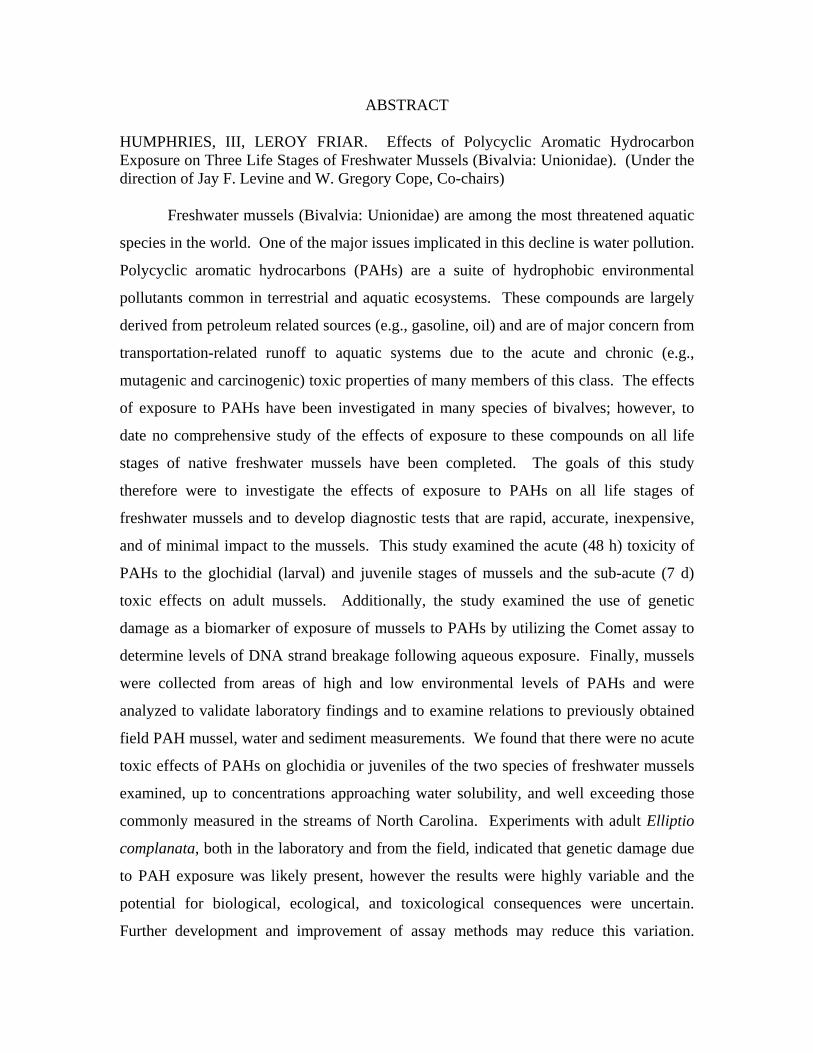
ABSTRACT HUMPHRIES, III, LEROY FRIAR. Effects of Polycyclic Aromatic Hydrocarbon Exposure on Three Life Stages of Freshwater Mussels (Bivalvia: Unionidae). (Under the direction of Jay F. Levine and W. Gregory Cope, Co-chairs)
Freshwater mussels (Bivalvia: Unionidae) are among the most threatened aquatic
species in the world. One of the major issues implicated in this decline is water pollution.
Polycyclic aromatic hydrocarbons (PAHs) are a suite of hydrophobic environmental
pollutants common in terrestrial and aquatic ecosystems. These compounds are largely
derived from petroleum related sources (e.g., gasoline, oil) and are of major concern from
transportation-related runoff to aquatic systems due to the acute and chronic (e.g.,
mutagenic and carcinogenic) toxic properties of many members of this class. The effects
of exposure to PAHs have been investigated in many species of bivalves; however, to
date no comprehensive study of the effects of exposure to these compounds on all life
stages of native freshwater mussels have been completed. The goals of this study
therefore were to investigate the effects of exposure to PAHs on all life stages of
freshwater mussels and to develop diagnostic tests that are rapid, accurate, inexpensive,
and of minimal impact to the mussels. This study examined the acute (48 h) toxicity of
PAHs to the glochidial (larval) and juvenile stages of mussels and the sub-acute (7 d)
toxic effects on adult mussels. Additionally, the study examined the use of genetic
damage as a biomarker of exposure of mussels to PAHs by utilizing the Comet assay to
determine levels of DNA strand breakage following aqueous exposure. Finally, mussels
were collected from areas of high and low environmental levels of PAHs and were
analyzed to validate laboratory findings and to examine relations to previously obtained
field PAH mussel, water and sediment measurements. We found that there were no acute
toxic effects of PAHs on glochidia or juveniles of the two species of freshwater mussels
examined, up to concentrations approaching water solubility, and well exceeding those
commonly measured in the streams of North Carolina. Experiments with adult Elliptio
complanata, both in the laboratory and from the field, indicated that genetic damage due
to PAH exposure was likely present, however the results were highly variable and the
potential for biological, ecological, and toxicological consequences were uncertain.
Further development and improvement of assay methods may reduce this variation.

Generally, mussels from streams with higher average daily traffic counts (ADTC)
exhibited greater levels of genetic damage compared to mussels from streams with lower
ADTC values. Data obtained from the laboratory study generally showed increasing
DNA damage relative to increasing PAH concentration. Based on the data generated,
however, PAHs are not likely contributing to acute toxicity of mussels in North Carolina
streams, but the chronic, long-term pervasive effect of PAHs on native freshwater
mussels remains uncertain.

EFFECTS OF POLYCYCLIC AROMATIC HYDROCARBON EXPOSURE ON THREE LIFE STAGES OF FRESHWATER MUSSELS (BIVALVIA:
UNIONIDAE)
by LEROY F. HUMPHRIES, III
A thesis submitted to the Graduate Faculty of North Carolina State University
in partial fulfillment of the requirements for the Degree of
Master of Science
COMPARATIVE BIOMEDICAL SCIENCE
Raleigh, NC
2006
APPROVED BY:
________________________________ _________________________________
Jay F. Levine, DVM, MPH W. Gregory Cope, Ph.D.
Co-Chair of Advisory Committee Co-Chair of Advisory Committee
________________________________
Arthur E. Bogan, Ph.D.

Dedication
This work is dedicated to my wife Diane, and my children Leah and Tyler,
without whose support and love I would be lost, and to my grandmother, the late Mary
Winifred K. Solomons, who always believed in me and encouraged me to do my best and
to reach my goals. I also wish to dedicate this work to the memories of Mrs. Faye Battle
and Mr. Barry Butts, both of whom inspired me as a student and a teacher.
ii
Biography
I am a 1987 graduate of Mullins High School in Mullins, SC. Following
graduation, I enlisted in the United States Navy and served aboard USS Richmond K.
Turner (CG-20) for 4 years. During my tour of duty I participated in “Operation Desert
Shield/Desert Storm” and “Operation Provide Comfort” in the Middle East. Upon
returning to the States after the war, I received an Honorable Discharge from active duty
and entered active reserve service, during which I participated in numerous drug
interdiction exercises in the Caribbean Sea. I attended Horry-Georgetown Technical
College where I earned an Associates degree in Science in 1995. After graduation from
HGTC I enrolled at Coastal Carolina University where I earned a Bachelors of Science
degree in Marine Science with minors in Biology and Environmental Science in 1998.
Following graduation from CCU I moved to North Carolina to work as a Laboratory
Technician for the NC Division of Marine Fisheries in Morehead City, NC where I
conducted testing to determine the prevalence of the oyster parasite Perkinsus marinus in
NC waters. In 2001 I came to NCSU and enrolled in Dr. Levine’s Comparative
Biomedical Science graduate program. Currently I teach Biology at Overhills High
School in Springlake, NC. My wife and I reside in Angier, NC with our two children.
iii

Acknowledgements
I wish to thank my co-authors and mentors, Drs. Jay Levine, Greg Cope, and
Arthur Bogan for all their advice, assistance, and, above all, their patience and
understanding. I also wish to thank Chris Eads, Dr. Robert Bringolf, Peter Lazaro, and
the rest of the “Mussel Crew” for all their help in sample collection and processing.
Furthermore, I wish to acknowledge Dr. Ray Tice from Integrated Laboratory Systems in
Research Triangle Park, NC for technical advice on the use of the Comet Assay. The
NC Department of Transportation funded this work.
iv
Table of Contents
List of Tables .................................................................................................vi List of Figures ................................................................................................vii 1. Introduction and Literature Review.........................................................1 2. Materials and Methods ............................................................................10
2.1. Study organisms ...............................................................................10 2.2. Collection of study organisms .........................................................11 2.3. Field Study Site selection ................................................................12 2.4. Test solutions and supplies ..............................................................13 2.5. Test protocols ...................................................................................14
2.5.1. Glochidial tests.......................................................................14 2.5.2. Juvenile tests ........................................................................15 2.5.3. Adult tests .............................................................................15
2.5.3.1.Positive Control Experiment............................................15 2.5.3.2.Laboratory Exposure Study .............................................16 2.5.3.3.Field Study.......................................................................17
2.6. Test procedures.................................................................................18 2.6.1. Comet Assay .........................................................................19 2.6.2. Contaminant Analysis ...........................................................20
3. Statistical Analyses ..................................................................................21 3.1. Acute Toxicity Tests on Glochidia ...................................................21 3.2. Juvenile Tests ...................................................................................22 3.3. Comet Assay ....................................................................................22
4. Results .....................................................................................................22
4.1. Glochidial Tests ...............................................................................22 4.2. Juvenile Tests ...................................................................................23 4.3. Adult tests ......................................................................................23
4.3.1. Positive Control Experiment .................................................23 4.3.2. Adult Mussel PAH Experiment .............................................23 4.3.3. Field Study ............................................................................24
5. Discussion................................................................................................26
6. Conclusions..............................................................................................31 7. References................................................................................................32 8. Appendix 1: Tables ..................................................................................39 9. Appendix 2: Figures.................................................................................41
v
List of Tables Page Table 1 Sites selected for use in this study .................................................39 Table 2 Measured levels of PAHs in glochidia and juvenile test solutions in µg/L. .....................................................................39 Table 3 Tissue levels of PAHs in mussels exposed in the laboratory and water concentrations (day 14). ...............................40 Table 4 Average sum of PAH contamination measured from PSDs .........40
vi
List of Figures Page Figure 1 Tail and Olive Moment values (µm) for E. complanata hemocytes exposed for 4 and 24 hours to 4-NQO compared to unexposed cells ..........................................................................41 Figure 2 Tail (2a) and Olive Moment (2b) values for laboratory study .......41 Figure 3 Percent DNA in comet tails per treatment......................................42 Figure 4 Regression of mussel tissue PAH concentration versus Log Tail Moment (um) from the Laboratory study ...............................42 Figure 5 Regression of Average Daily Traffic Count versus estimated water PAH contamination (µg/L) from PSD data ..........................43 Figure 6 Sum of 48 PAH in 20 streams estimated from PSD residue ..........43 Figure 7 Mean Petrogenic/pyrogenic PAH ratio in the 20 streams used in this study ....................................................................................44 Figure 8 Tail and Olive Moment (µm) values for the streams sampled for the field portion of the study.....................................................44 Figure 9 Predicted concentrations (ng/g lipid) of total PAHs in mussel tissue estimated from passive sampling devices (PSDs) placed at the study sites .............................................................................45
vii
1. Introduction
Polycyclic aromatic hydrocarbon (PAH) compounds are a class of hydrophobic
environmental pollutants widespread in terrestrial and aquatic environments. Many of
the compounds in this group are of major concern to environmental agencies and
researchers worldwide due to their mutagenic and carcinogenic properties (Baumard et
al., 1999). Polycyclic aromatic hydrocarbons enter the environment via natural
(biogenic) processes (e.g., forest or grass fires, natural petroleum seeps, etc.), and
anthropogenic processes, including accidental spills or releases of petroleum compounds
into the environment (e.g., tanker spills, oil platform releases) and high temperature
combustion (petrogenic and pyrolitic) processes (e.g., the burning of fossil fuels,
industrial activities) (Eisler, 1987; Fernandes et al., 1997; Baumard et al., 1998; Piccardo
et al., 2001). Higher molecular weight PAH compounds are mainly generated by high
temperature combustion of organic matter, therefore anthropogenic activities are
generally considered to be the major source of higher molecular weight PAH
environmental contamination (Piccardo et al., 2001). Low molecular weight PAH
compounds may be produced by fossil fuel combustion, but are also major components of
petroleum products (Fernandes et al., 1997), and natural processes (Eisler, 1987).
Polycyclic aromatic hydrocarbon compounds are considered to be highly
hazardous to the environment and to human health. Higher molecular weight four- to
seven-ring PAHs are highly mutagenic and carcinogenic, and lower molecular weight
two- to three-ring PAHs, although less mutagenic, can be highly toxic (Eisler, 1987,
Fernandes et al., 1997). In many cases the parent compounds are relatively inert, but the
metabolites exert toxicity. Low molecular weight PAHs, dominant in fossil fuel
1

assemblages, are more labile and readily volatilize into the atmosphere from the air/water
interface. With increasing molecular weight comes decreasing water solubility, with the
result that low molecular weight PAHs are preferentially adsorbed to particles in the
water column (Baumard et al., 1999). Low molecular weight PAH compounds in the
water column may therefore be more bioavailable to organisms. Polycyclic aromatic
hydrocarbons can interact with cells to produce toxic responses by binding to lipophilic
sites in cells and interfering with cellular processes (Neff, 1979). In light of the toxicity
and carcinogenicity issues to terrestrial organisms, aquatic organisms, and to humans, the
monitoring of PAH contamination in the environment is critical.
Polycyclic aromatic hydrocarbons enter aquatic environments by many routes,
including domestic and industrial effluents, surface runoff from land, atmospheric
deposition, and spillage from petroleum operations (Eisler, 1987; Piccardo et al., 2001).
Runoff from impervious surface can be one of the main carriers of these pollutants to
surface waters, and a wide range of organic and heavy metal contamination has been
detected in waters adjacent to paved roads (Federal Highway Administration, 1981;
Hoffman et al., 1985). Maltby and co-workers (1995) demonstrated that stormwater
runoff from a motorway in the United Kingdom was toxic to the benthic amphipod
Gammarus pulex. Heavy metals and PAHs at levels that could significantly impact
aquatic biota were detected in runoff from a bridge in Canada (Marsalek et al., 1997).
Beasely and Kneale (2002) noted that there is considerable evidence that heavily
trafficked roads are an important source of toxicants to streams. Hallhagen (1973) and
Wakeham (1977) indicated that urban storm water runoff was responsible for a
significant level of hydrocarbon contamination to aquatic systems. Data from a previous
2

study of roadway crossing structures in North Carolina found that there were elevated
levels of PAH compounds downstream of 18 bridges and 2 culverts, and that this increase
in contaminant levels correlated to decreased freshwater mussel abundance in stream
reaches directly downstream of the crossing structure (Shea et al. 2004).
Biomonitoring of environmental conditions, especially in relation to
measurements of hydrophobic contaminants, is more cost effective and accurate
compared to direct environmental sampling. For instance, direct analyses of water
samples for PAH contaminations are time-consuming, require large sample sizes (and are
therefore innately more expensive), and do not necessarily represent the bioavailable
fraction present in the water column (Gewurtz et al., 2002). Beasely and Kneale (2002)
state that “snapshot” monitoring represents conditions only at a single point in time and
may imply a greater grade of water quality than actually present. Additionally, these
types of samples may miss periods of high contamination due to pulsed events (i.e., storm
runoff). Sentinel organisms, however, concentrate contaminants within their tissues
making trace levels of contaminants easier to monitor (Baumard et al., 1998). According
to Pereira and co-workers (1996), bed sediments and lipid tissues of aquatic organisms
integrate hydrophobic contaminants over seasonal or yearly timeframes, indicating that
the biota of a stream may be more effective as monitors of water quality than the water in
which they reside. However, this accumulation of organic contaminants is a complex
function of the physiochemical properties of the contaminant, its distribution within the
system, and the feeding behavior and metabolism of the aquatic organism used as
biomonitors.
3

Due to their primarily sessile lifestyles and filter feeding activities, bivalve
mollusks are among the most sensitive aquatic species to environmental contamination
(Dame, 1996; Suavé et al., 2002). In fact, some mollusks may be more susceptible to the
effects of PAHs compared to vertebrate species due to the lack of efficient enzymes for
metabolizing and detoxifying PAH compounds and metabolites (Eisler, 1987). The
Cytochrome P-450 (CYP450) mono-oxygenase system is an apparently universally
distributed system involved in the metabolism of xenobiotics, including PAH
compounds. Increases in the activity of the CYP450 system are routinely used as a
means of detecting exposure to PAHs and other pollutants in fish, although the response
in bivalves is less obvious. Porte and co-workers (2001) attempted to develop an
integrated monitoring strategy using CYP450 activity, benzo(a)pyrene hydroxylase
(BPH) activity, and stress-70 proteins. They found that exposure to PAH-contaminated
environments did not elicit a CYP450 response, but there was a clear induction of stress
proteins (stress-70). It is important to note, however, that stress proteins are induced by a
great number of environmental factors (e.g., UV light, salinity, temperature, oxidizing
agents, etc.) in addition to contaminants (Sanders, 1993). In one study, no clear evidence
of changes in activity of respiratory enzymes due to exposure to petroleum hydrocarbons
were observed in the blue mussel (Mytilus edulis planatus) (Long et al., 2003).
The use of bivalves as sentinel organisms in aquatic environments has proved to
be an effective method of monitoring chemical contaminant levels. Oysters (Crassostrea
sp.) and mussels (Mytilus sp.) are commonly used sentinel organisms in marine
ecosystems (Baumard et al., 1999; Piccardo et al., 2001; Geffard et al., 2002). For many
years freshwater bivalves have been used as biomonitors of pollutant contamination in
4
waterways (Renaud et al., 1995; Gagné et al., 2002; Gewurtz et al., 2002). Cataldo and
co-workers (2001) used juvenile Corbicula fluminea survival to monitor sediment
pollutant levels in Argentina. They determined that C. fluminea mortality rates
corresponded well with sediment pollutant levels. Gewurtz and co-workers (2002) used
the mussel Elliptio complanata to perform quantitative biomonitoring of PAHs. In
another study, E. complanata and Dreissena polymorpha were used to study the effects
of exposure to pollutants dispersed in a municipal effluent plume (Gagné et al., 2002).
Assays of tissue body burden conducted on Mytilus sp., Anodonta anatina, Unio tumidus,
and E. complanata have demonstrated that mussels bioaccumulate PAHs and are reliable
sentinel organisms (Cossu et al., 1997; Anderson et al., 1999; Gewurtz et al., 2002;
Hyötyläinen et al., 2002; Thorsen et al., 2004).
A major objective of toxicology-related epidemiological testing is to provide
reliable and specific information concerning the effects of exposure to a particular agent.
The possibility of using biomarkers of exposure to substitute for classical endpoints (e.g.,
disease incidence or mortality) in molecular epidemiological studies is promising
(Bonassi and Au, 2002). However, documentation of exposure may be non-existent or
difficult to obtain in many cases. Bonassi and Au (2002) suggested that the use of
biomarkers of exposure may provide a more precise method of obtaining that
information, and that the data, if correctly collected, may be utilized to calculate the
internal exposure doses and to determine the dose-response relationship. According to
Porte et al. (2001), mussels exhibit a series of sub-lethal biochemical responses to
pollutants, making them excellent choices for pollution monitoring studies of chemical
5
analysis of tissue burden and biomarkers of exposure. In instances of non-lethal
exposures, biomarkers may be used to determine exposure level of sentinel organisms.
The primary routes of exposure for bivalves are across the gill and digestive gland
membranes. Biomarkers of PAH exposure for bivalves have included growth and
development (Geffard et al., 2002; Widdows et al., 2002), CYP450 induction (Anderson
et al., 1999; Porte et al., 2001; Gagné et al., 2002), respiratory enzymes (Long et al.,
2003), embryogenisis and larval development (Beiras et al., 1998; Geffard et al., 2002),
hemocyte phagocytosis (Fournier et al., 2000; Blaise et al., 2002), antioxidant enzymes,
glutathione and lipid peroxidation (Cossu et al., 1997; Doyette et al., 1997), digestive
acini (Le Pennec and Le Pennec, 2001), and DNA damage in hemocytes, digestive tissues
and somatic cells (Sasaki et al., 1997; Wilson et al., 1998; Pavlica et al., 2001; Coughlan
et al., 2002; Hamoutene et al., 2002; Large et al., 2002; Rank and Jensen, 2003; Klobučar
et al., 2003; Siu et al., 2004). In one study, RNA arbitrarily primed PCR was used to
look for genomic aberrations in digestive tissues of Unio tumidus exposed to effluent
from a cokery plant on the Fensch River (France) known to be responsible for PAH
contamination (Rodius et al., 2002).
Previous studies of biomarkers of contaminant exposure have utilized gill and
digestive gland dissected from mussels (Cossu et al., 1997; Doyotte et al., 1997; Long et
al., 2003) or whole body analyses (Anderson et al., 1999; Porte et al., 2001) for CYP450
and stress-70 protein induction (Porte et al., 2001), respiratory enzyme activity (Long et
al., 2003), and hemocyte phagocytosis (Fournier et al., 2000; Blaise et al., 2002) in
mussels exposed to contaminants. However, the results of some of these experiments
have been inconclusive. Cytochrome P450 activity in bivalves is believed ineffective in
6

relation to the metabolism of PAH compounds, hence the propensity of these chemicals
to bioaccumulate in mollusks (Eisler, 1987). Stress-70 proteins are non-specific (i.e.,
elicited by a variety of stressors) and are thus offer little predictive value in determining
exposure to specific contaminants (Porte et al., 2001). Additionally, the use of
respiratory enzymes has proved to be an unreliable measure of contaminant exposure in
bivalve mollusks (Long et al., 2003). A comparison of lethal and non-lethal biomarker
techniques is needed. One such non-lethal technique, the sampling of bivalve hemocytes
may yield reliable results with minimal impact on the animals (Gustafson, et al., 2005).
Hemocytes may be sampled from the hemolymph extracted from the adductor muscle of
bivalves with minimal effort and adverse impact to the animal.
Bivalve mollusks are vital members of aquatic ecosystems. They function as
living filters, trapping food and particles in the water column as they filter-feed (Dame,
1996). Particles not ingested are excluded in pseudo-feces and effectively removed from
the water column, enhancing the removal of particle-associated contaminants from the
system. Native freshwater mussels (Bivalvia: Unionidae) filter large volumes of water on
a daily basis, removing suspended particles and pollutants at a rate faster than accounted
for through normal settling. Furthermore, freshwater mussels are important components
of aquatic food webs, forming a major portion of the diet of muskrats, otters, raccoons,
and other carnivorous animals that use rivers and streams as feeding areas.
Biomonitoring of freshwater mussels may provide an early detection of potential
problems arising from exposure to environmental contaminants. This would allow a
potential pollution problem to be addressed prior to reaching levels within the system that
would pose a threat to humans, agricultural animals, and other wildlife.
7

As a group, native freshwater mussels are among the most threatened aquatic
animal species in North America (Williams et al., 1993; Lydeard et al., 2004). The
National Native Mussel Conservation Committee (1998) estimated that 67% of the nearly
300 species of native North American mussels are either vulnerable to extinction or are
already extinct, and recognized water pollution as a major factor in unionid decline.
Despite this speculation, little documentation of the effects of major aquatic pollutants on
these animals exists (Moulton et al., 1996). Major sources of water pollution in streams
and rivers home to freshwater mussels include agricultural runoff containing various
pesticides and chemicals, roadway runoff, municipal wastewater treatment plants
(Goudreau et al., 1993), and industrial effluent. In particular, roadway runoff and
municipal wastewater discharges can carry heavy loads of PAHs into an aquatic system.
This may be particularly hazardous to mussels during their reproductive period.
Freshwater mussels have a stage of development during which they are obligate
parasites on fish (Huebner and Pynnönen, 1992; Pynnönen, 1995; McMahon and Bogan,
2001). The female mussel broods her larvae, called glochidia, inside specially adapted
chambers within her gills known as marsupia. When mature, the female mussel will
either release the glochidia into the water in a mucosal conglutinate packet or attempt to
attract an appropriate host organism using a section of her mantle as a lure designed to
mimic a prey item (Jacobson et al., 1997; McMahon and Bogan, 2001) depending on the
species of mussel. When the glochidia come into contact with a potential host organism
they rapidly snap their valves together and attach themselves to either a fin or to the gills
of the host. Once attached, the host rapidly forms a cyst around the glochidia. The
period of encystment on the host varies between species and many species of mussel have
8
a specific suite of fish hosts. Upon encystment on the host fish, the mussel glochidia are
assumed to be well protected from stressful environmental conditions. However, in the
period of development prior to or just after release into the environment, glochidia may
be at risk of exposure to toxic compounds in the water column.
Experiments utilizing the glochidia of freshwater mussels have demonstrated
sensitivity of glochidia to many toxic compounds, including PAHs. Huebner and
Pynnönen (1992), Pynnönen (1995), and Hanstén et al. (1996) demonstrated that
glochidia of Anodonta sp. were sensitive to sub-lethal exposure concentrations of heavy
metals and low pH, and that these exposures could significantly impact viability and
survival. Keller et al. (1998) tested the toxicity of diesel fuel contaminated sediments on
the glochidia of Lampsilis siliquoidea and Lasmigona costata and juvenile Villosa
villosa, with ambiguous results. It should be noted, however, that the contaminant levels
in these experiments were below the documented ‘lowest effects level’ from the
literature. Weinstein (2000) tested the glochidia of Utterbackia imbecillis to characterize
the acute toxicity of photo-activated fluoranthene. He found that the glochidia rapidly
accumulated the contaminant within their tissues and the presence of low UV intensities
made the glochidia >45 times more sensitive to fluoranthene. In 2001, Weinstein and
Polk repeated this experiment with anthracene and pyrene on the same species of mussel
with similar results. Tests on juveniles of many bivalve species have produced
comparable results (McKinney and Wade, 1996; Ahrens et al., 2002). However, little is
known about the toxicity of PAHs found at relatively low levels in streams with little
urbanization in the watersheds.
9

This study was conducted as part of a larger study funded by the NC Department
of Transportation examining the impact of crossing structures on freshwater mussels and
their habitat. The primary goal of this effort was to examine the effects of exposure to
PAHs on various life stages (glochidia, juvenile, and adult) of freshwater bivalves.
Mussels at each of the three different life stages were analyzed to assess toxicity and to
evaluate biomarkers of exposure and genotoxic effects resulting from exposure to PAH
compounds. The secondary goal of this study was to explore and develop non-lethal
sampling regimes and test procedures for working with this rapidly declining group of
aquatic macro-invertebrates. The specific objectives of this study were:
1) To quantify in the laboratory and the field the effects of exposure to PAH
compounds on all life stages of freshwater mussels;
2) To explore the use of non-lethal techniques that are accurate, rapid,
inexpensive, and have minimal adverse impact on the animals being sampled
in determining exposure history of freshwater bivalves to PAH compounds.
2. Materials and Methods
2.1 Study organisms
Three species of unionid mussels (Elliptio complanata, Lampsilis fasciola, and
Lampsilis siliquoidea) were used in glochidia, juvenile, and adult tests. Lampsilis
fasciola and Lampsilis siliquoidea were used in glochidia and juvenile tests, and Elliptio
complanata was used in adult tests. Lampsilis fasciola glochidia were obtained from
gravid females collected from the Little Tennessee River near Franklin, NC. Juvenile L.
fasciola were obtained from individuals transformed in the Freshwater Mussel Rearing
10
Facility at the NCSU College of Veterinary Medicine. Lampsilis siliquoidea, an interior
drainage mussel found in the Midwestern United States, glochidia and juveniles were
obtained from Dr. Chris Barnhart at Missouri State University in Springfield, MO.
Elliptio complanata is a common mussel found in many Atlantic slope drainage streams
in North Carolina, and is a tachytictic brooder (Bogan, 2002). Lampsilis siliquoidea and
L. fasciola are sexually dimorphic and are bradytictic brooders. Elliptio complanata and
L. fasciola represent different reproductive strategies and mussel habitats found in North
Carolina. Voucher specimens of all species used were obtained from the North Carolina
Museum of Natural History archive collection to define the standards and verify species
identification.
2.2 Collection of study organisms
For glochidial tests, gravid mussels were collected by hand and kept damp, cool,
and dark for transport to the laboratory where they were placed in an indoor closed,
recirculating holding facility at an ambient air temperature of 21ºC and a 12:12 light:dark
cycle. Gravid mussels brought into the laboratory were placed into a tank equipped with
a chiller system to maintain a water temperature of 12ºC to reduce the possibility of
premature release of glochidia prior to use. Collection time of gravid mussels varied
between species, based on the time of year for the maturation of glochidia within the
marsupia.
All adult mussels brought into the lab were measured (total length to the nearest
mm), weighed (to the nearest g), and marked with an identifying number with a rotary
grinding tool. Mussels were held in the laboratory prior to testing in closed, re-
circulating tanks with aerated tap water from the City of Raleigh conditioned with
11
sodium thiosulfate to remove chloramine ions. Mussels in the laboratory were fed a diet
of Chlorella sp. cultured at our facility in 150L batches. Glochidia used for testing were
flushed directly from the marsupia of gravid females collected from the field after the
depuration period. Female mussels were returned to their native stream following
extraction of glochidia.
Adult Elliptio complanata collected for use in the adult PAH toxicity test were
obtained from a relatively uncontaminated stream in Central North Carolina (based on
data obtained in a previous NCDOT funded study) and transported to the laboratory in a
45.5L cooler filled approximately half full with ambient water from their native stream.
The mussels were acclimated by replacing roughly half the volume of ambient water with
ASTM moderately hard re-constituted water (ASTM, 1993) every hour until the entire
volume had been replaced. Mussels were weighed and measured as previously described,
and their shells were scrubbed to remove attached debris. The mussels were randomly
assigned a number (I or II) and following acclimation overnight, one mussel from each
number group was randomly distributed to an experimental unit (test chamber).
2.3 Field Study Site selection
Twenty streams in North Carolina were randomly selected from the 50 streams
utilized in the NCDOT funded study for use in the Toxicology portion of the study. The
sites for the intensive field study were a subset of 6 randomly selected streams out of the
20 used in the Toxicology portion of the study (Table 1). At all 20 sites, passive
sampling devices (PSDs) were deployed upstream and downstream of the crossing
structure to determine baseline levels of stream contamination with PAH compounds.
Passive sampling devices have been shown to be a good surrogate for mussel tissues in
12
determining PAH contaminant levels within a stream (Shea et al., 2004). Toxicity data
was compared to that obtained from 18 bridges and 2 culverts in a previous study.
2.4 Test solutions and supplies
Baseline data from a previous study funded through NCDOT was used to
determine polycyclic aromatic hydrocarbon (PAH) levels in test solutions (1, 10, 50, 100,
and 200 µg/L). The test concentrations were based on the mean PAH levels measured at
relatively uncontaminated sites (agricultural/rural/forested) and highly contaminated sites
(urban) in a previous study, and designed to cover a range of potential contaminant levels
(Shea, et al., 2004), up to solubility of most of the higher molecular weight PAH
compounds in water. The stock PAH test solutions were prepared using a mixture of
Alaskan North Slope crude oil and creosote dissolved in dichloromethane (DCM). Test
concentrations consisted of stock solutions diluted with ASTM moderately hard re-
constituted water (ASTM, 2002). Controls consisted of ASTM water and 200µl DCM +
ASTM water. Positive control treatments consisted of 4-Nitroquinoline-N-oxide +
ASTM water. All test treatments were conducted in triplicate.
Test containers were borosilicate glass dishes washed with HPLC grade reverse-
osmosis water, acetone-rinsed, and oven-baked between trials to remove organic residues
and other contaminants. Glass containers were used to minimize loss of PAH compounds
due to adsorption onto the surface of containers. Test containers consisted of 120 x
90mm dishes for glochidia and juvenile tests, and 3L glass jars for adult trials.
Glochidia were gently flushed from one marsupia of each female mussel using a
50cc hypodermic syringe with a 10-gauge needle and ASTM water. Glochidia and
juvenile experiments were conducted at 21oC with a 12:12 light:dark cycle. Adult
13
experiments were conducted at 20oC ambient air temperature and aerated gently with a
16:8 light:dark cycle. Water quality variables (temperature, dissolved oxygen, pH, and
conductivity) were measured daily in each test chamber. Mussels were not fed during
any of the experiments.
2.5 Test protocols
2.5.1 Glochidial tests
Acute (48h) toxicity tests were conducted on glochidia during summer 2004,
depending on the mussel species and availability of glochidia. Each brood was tested for
viability with the addition of 2-3 drops of saturated NaCl solution to a sub-sample of the
brood. When exposed to NaCl solution glochidia snap closed, viability is determined
based on the percent of glochidia within the sub-sample that close following NaCl
exposure. Broods with less than 90% viability were not used in experiments. Once
viability was determined the broods were pooled to minimize any between animal
associated bias and about 150 glochidia were added to each test chamber. Glochidia
were added to the test containers by gently swirling the holding container and
withdrawing ~0.5cc into a borosilicate glass pipette to obtain a random sample. At 24
and 48h of exposure to PAHs, a sub-sample of ~50 glochidia was tested for viability
using the NaCl method, and the test solutions were renewed (2/3 volume) with new stock
solution in ASTM water. Lampsilis fasciola glochidia not used in the acute toxicity tests
were used to infest fish hosts (largemouth bass, Micropterus salmoides) to obtain
laboratory-reared juveniles. Concentrations of PAHs in test solutions were measured as
described below to ensure that the target contaminant level was reached.
14
Largemouth bass were infested with glochidia by either pipetting glochidia
directly onto the gills or by placing the fish with glochidia in a 10-gallon aquarium
rapidly aerated to mix the water well. The period of encystment of the glochidia on the
host fish varies per species of mussel, but lasts only a few weeks
(http://news.fws.gov/mussels.html). During the encystment period the fish hosts were
maintained in recirculating 10-gallon aquaria in the Mussel Barn at the NC State
University College of Veterinary Medicine. Aquaria were siphoned daily beginning one
week post-infestation to collect transformed juvenile mussels.
2.5.2 Juvenile tests
Acute toxicity testing was performed on recently (<30 day old) transformed
mussels of both species, depending on transformation success from fish hosts and
availability of juveniles from the supplier, during summer 2004, and on >60d old L.
siliquiodea. Test duration was 96h and viability assessment was conducted at 48 and 96h
of exposure. Viability was determined during a 5-minute observation period and based
on foot movement inside or outside of the shell. Seven juvenile mussels were used per
replicate, and all PAH treatments were conducted in triplicate. PAH test solutions were
renewed daily (2/3 volume) with new stock solution in ASTM water.
2.5.3 Adult tests
2.5.3.1 Positive Control Experiment
Adult E. complanata (N=4) were sampled from a relatively uncontaminated
reference site (Richland Creek, Wake County, NC) on 16 Feb 2005. Approximately
0.5ml of hemolymph was drawn from the anterior adductor muscle of each mussel
(Gustafson et al., 2005). Hemolymph samples were pooled to account for between
15
animal variation, and allocated in 100µl aliquots to 4 tubes for a positive control
experiment using 4-nitroquinoline-N-oxide, a known genotoxic compound (Le Pennec
and Le Pennec, 2001; Conners and Black, 2004). Treatments consisted of 2 control
(100µl untreated hemolymph) tubes and 2 treatment tubes (100µl hemolymph + 10µl
0.25mg/ml 4-nitroquilonline-N-oxide) and placed in a refrigerator. One tube from each
treatment was sampled after 4h exposure and the second tube was sampled at 24h. Two
samples were taken from each tube for comet assay analysis.
2.5.3.2 Laboratory Exposure Study
Adult E. complanata (N=62, 6 per treatment, 8 treatments, plus an additional 13
to obtain baseline data) collected on 03 March 2005 from a relatively uncontaminated
reference site on the Eno River were exposed in the laboratory to the PAH test
concentrations for 14d following a 24h depuration and acclimation period in the
laboratory. Pre-exposure hemolymph samples were taken to determine baseline levels of
genetic damage in the population. Three mussels were sampled in the field to determine
pre-acclimation levels of genetic damage. Hemolymph was removed from the anterior
adductor muscle and placed in 1ml plastic tubes and stored dark and cold for transport to
the laboratory. Once in the lab, these samples were processed immediately for the Comet
assay to minimize loss due to cellular degradation. The remaining mussels were
acclimated to laboratory conditions for 24h prior to use. Mussels were scrubbed with a
soft bristled brush to remove particulate matter attached to the shells to prevent particle
adsorption of test solutions and randomly labeled with either an “I” or “II” for allocation
to treatments. Following the 24h acclimation period, one mussel from each group was
randomly allocated to each of the treatments (control, positive control, solvent control,
16

PAH 1, 10, 50, 100, and 200µg/L). Hemolymph (0.25ml) was drawn from 10 mussels
post-acclimation and processed immediately for Comet assay. These same 10 mussels
were removed from their shells, and the tissues frozen at –80oC for later tissue PAH
analysis. Hemolymph from each of the experimental mussels was repeatedly sampled on
days 3, 7, and 14. On d14 all experimental mussels were removed from their shells, and
frozen at –80oC for PAH tissue analysis. Test solutions of PAH were renewed daily (2/3
volume) with the exception of the positive control which was only renewed on d7. Water
quality measurements (temperature, dissolved oxygen, and pH) were taken daily in each
test chamber and water in the test containers was completely changed on d7. Composite
waters samples (100ml per treatment) were taken for PAH analysis on d0, d7, and d14.
2.5.3.3 Field Study
Adult E. complanata (N=6 per stream, 36 total) were collected from 6 streams
(Table 1) out of the 20 chosen for study in a NC Department of Transportation (NCDOT)
funded study examining the effects of culvert style crossing structures on freshwater
mussels. Mussels were collected between 25 – 50m upstream and downstream of each of
the road crossing structures from 15–17 December 2004. Two streams were considered
reference sites and corresponded to a low average daily traffic count (e.g., <500
vehicles). Two streams were from suburban areas and corresponded to moderate average
daily traffic volume (e.g., 500-1000 vehicles). The remaining 2 streams were from high
traffic areas (>10,000 ADTC): one stream passed beneath Interstate 40 between Raleigh
and Research Triangle Park, the other passed beneath Interstate 40 at Raleigh Durham
International Airport and is directly beneath the runway flight path of the airport.
Streams chosen for this portion of the study were matched as closely as possible
17

regarding geomorphological structure (e.g., drainage area, flow, size, substrate
composition) to minimize potential variation due to non-contaminant related variables.
Mussels collected from these streams were processed immediately for testing. Mussels
were weighed and measured as previously described, and ~1.0ml of hemolymph was
drawn to obtain hemocytes for use in the Comet assay.
Passive sampling devices (PSDs) were deployed at these study sites upstream and
downstream of the crossing structure following the methods of Shea et al. (2004).
Briefly, PSDs were constructed using approximately 12.7µm thick low-density
polyethylene (PE) tubing, containing no plasticizers or additives. The PE tubing (5cm x
30cm, surface area of 300cm2) was pre-extracted with hexane for 48h prior to use and
fixed inside a protective polyethylene cage. Two PSDs were placed in each cage and
deployed within a 50m zone upstream and downstream from the crossing structure and
retrieved approximately 30d later. Previous work has demonstrated that a 30d
deployment time allows the 12.7µm PE to reach equilibrium with water. Following
deployment, one of the PSDs was archived at -20ºC and the second was cleaned with de-
ionized water and a soft brush, followed by a rinse in acetone to rigorously solvent
remove material from the surface of the LDPE prior to extraction. Data collected from
the PSDs, directly related to PAH contaminant levels found within the streams, was used
for comparison with DNA damage levels in adult mussels sampled from the same stream.
2.6 Test procedures
This study utilized acute toxicity and DNA strand breakage to explore the effects
of PAH exposure on the glochidial, juvenile, and adult life stages of freshwater mussels
and to explore the use of non-traditional tissue types (i.e., hemocytes) for use in the
18
Comet assay for determination of levels of genetic damage in relation to exposure level.
The goal was to develop accurate, rapid, and cost effective non-lethal sampling
procedures to determine effects of exposure of mussels to PAHs.
Hemolymph was drawn from the anterior adductor muscle of adult mussels.
Following hemolymph extraction, mussels were dissected and the tissue frozen at -80oC
for tissue body burden analysis in the Environmental Toxicology Laboratory at NCSU.
2.6.1 Comet Assay
The single-cell gel electrophoresis assay (Comet assay) was performed to
determine the extent of genetic damage due to exposure to PAHs. This assay measures
the level of DNA damage in single cells and has been reliably used on a variety of
organisms (Cotelle and Férard, 1999). Slides were prepared using an adaptation of the
methods outlined by Woods et al. (1999) and Coughlan et al. (2002). Microscope slides
were prepared by dipping each slide in 1.5% normal-melting agarose in phosphate
buffered saline followed by air-drying and storage in a desicator until use. All of the
following steps were conducted under low light conditions to prevent confounding DNA
damage due to ultra-violet radiation exposure. To prepare the sample, 100µl of mussel
hemolymph was mixed with 100µl of 1.3% low melting point agarose (LMPA). The
tubes were vortexed gently to mix the sample then 100µl were drawn off and placed on
the slide, a 40 x 60 mm coverslip added, and the gels allowed to set on ice. Once the cell
layer had set, the coverslips were removed, a third layer of 1.5% NMA was added and
allowed to set as before.
Once the gels were set, the cover slips were removed and the cells lysed in a high
salt buffer (2.5 M NaCl, 10 mM Tris, 100 mM EDTA, 1% (v/v) Triton X-100, and 10%
19
(v/v) DMSO, pH 10.0) for at least 90 min to 8h in coplin jars at 4ºC in the dark.
Following the lysis period, the slides were rinsed 3 times with DI water for 5 minutes and
gently placed in a horizontal electrophoresis tank and covered with an alkaline solution
(0.3 M NaOH, 1 mM EDTA; pH >12) for 15 min at 4ºC in the dark to allow for
unwinding of the DNA. Without changing the electrolysis solution, a 25 V, 300 mA
current was applied for 15 min, followed by neutralization three times with Tris buffer
(0.4 M Tris-HCl; pH 7.4) at 5-minute intervals followed by rinsing with cold EtOH.
Slides were then stored in a desicator until visual microscopic analysis. When ready to
be read, slides were stained with 2–3 drops of ethidium bromide for 5 min, the coverslips
were replaced and randomly selected nucleoids were photographed at 100x magnification
using an Olympus BH-2 epifluorescence microscope fitted with a Fuji Finepix S5100
digital camera. DNA damage was expressed in terms of tail moment (TM, determined as
the product of the tail length and the fraction of DNA in the tail), olive moment (OM, the
summation of Tail Intensity profile values multiplied by their relative distances to the
Head Center, divided by Total Comet Intensity), and percent DNA in tail .
2.6.2 Contaminant Analysis
Mussel tissue and PSD samples were extracted for PAH analysis as described by
Thorsen et al. (2004) and Luellen and Shea (2003). Samples were shaker-extracted (200
rpm) for 24-h using dichloromethane (DCM) for mussels and PSDs. Concentrated
extracts were fractionated using high performance gel permeation chromatography to
remove high molecular weight matrix components (e.g., lipids, polyethylene waxes). The
extracts were solvent exchanged into hexane and then further purified on a 3-g silica
column. Mussel lipid content was determined by passing extracts through a gel
20
permeation chromatography (GPC) column, collecting the lipid fraction, evaporating and
weighing. Samples were analyzed for 48 PAH analytes including the 16 USEPA priority
PAHs.
Instrumental analysis on water samples and tissue and PSD extracts was
conducted following the methods outlined in Shea et al. (2004). Briefly, the purified
extracts were analyzed for total PAHs using an Agilent 6890 gas chromatograph (GC)
connected to an Agilent 5973N MSD utilizing a Restek 30m x 0.25mm Rtx-5 (film 5
thickness 0.25 µm) MS w/Integra-Guard column. The pressure was ramped to 40 psi
before injection with a 1-min hold time. The flow was then dropped to give a constant
flow of 1mL/min for the duration of the run. The temperature program for PAH analysis
was as follows: initial temperature 40 °C for 1 min with a ramp of 6 °C /min to 290 °C
and a final hold time of 30 min; injector temperature 300 °C, detector temperature 280
°C. Selected ion monitoring (SIM) was used for analysis.
3. Statistical Analyses 3.1 Acute Toxicity Tests on Glochidia
Data from the 48h acute toxicity tests on glochidia were used to determine “No
Observed Effects Concentration” (NOEC) and “Lowest Observed Effects Concentration”
(LOEC) curves using PROC PROBIT in SAS based on 48h survival (Weinstein, 2000;
Weinstein and Polk, 2001). Additionally, ToxStat software (Gulley and WEST, Inc.,
1994) was used to determine LC50 values using the Spearman-Karber method and 95%
confidence intervals. Tests were considered valid if mortality was <20% in the controls
during the duration of the test. In the event that no total kills were observed, EC50 values
were calculated instead of LC50.
21
3.2 Juvenile Tests
Data from the 96h acute toxicity tests on juvenile mussels were used to determine
NOEC and LOEC curves using PROC PROBIT in SAS (Weinstein, 2000; Weinstein and
Polk, 2001) and the Spearman-Karber method in ToxStat (Gulley and WEST, Inc., 1994)
based on 96h survival, as stated previously. Again, if no total kills were observed EC50
values were used instead of LC50 values.
3.3 Comet Assay
Comet images were analyzed using CometScore™ software (TriTek Corporation,
http://tritekcorp.com). Data were exported from CometScore into Microsoft Excel and
then to JMP (SAS Corporation, Cary, NC) for statistical analysis. The average distance
of strand migration of ~50 nuclei per slide were used in data analysis using a one-way
analysis of variance (ANOVA). When conducting the comet assay, the slides, not the
individual cells on the slide, were considered the smallest unit of measure (i.e. the means
of all of the cells measured on a given slide are used for analysis, not the individual
cells). The Tukey-Kramer HSD method for pairwise comparisons of means between
treatments was used for statistical analysis. This procedure requires a single value for
judging the significance of differences between measured parameters. Statistical
significance was considered at p < 0.05. Data were normalized by logarithmic
transformation, where necessary.
4. Results
4.1 Glochidial Tests
In repeated tests, no significant mortality was observed in glochidia exposed to
any of the experimental concentrations of PAHs, therefore no LOEC curves could be
22

calculated. Experimental exposures with L. fasciola and L. siliquoidea indicated EC50
values greater than solubility of most PAH compounds in water (e.g., EC50 > 200µg/L for
both species). Measured concentrations of contaminants were on target at time 0, but
declined some over time (Table 2). It is important to note however that samples for
contaminant analysis were taken just prior to replenishment of the solutions in the test
chambers.
4.2 Juvenile Tests
Studies with L. fasciola and L. siliquoidea juveniles (n = 14) indicated no acute
toxicity to any PAH treatment after 96h of exposure. Acute toxicity tests indicated LC50
values greater than solubility of most PAH compounds in water (EC50 > 200µg/L for both
species). Although not quantified as a test endpoint, some lethargy was observed in
mussels exposed to the greatest PAH concentration (200µg/L).
4.3 Adult tests
4.3.1 Positive Control Experiment
Hemocytes exposed to 4-NQO for only 4 hours exhibited significantly greater
levels of genetic damage compared to controls (Fig. 1) expressed in terms of tail moment
and olive moment (as defined previously). This trend continued at 24h, although levels
of genetic damage in both the control and treatment samples were reduced.
4.3.2 Adult Mussel PAH Experiment
Samples of hemolymph (1.0ml) were taken from test mussels on days 3, 7, and
14. However, the first set of slides made from the d14 samples was compromised when
nearly all of the gels slipped off of the slides during the 24h lysis period, possibly due to
an error in slide preparation. The slides were immediately remade utilizing new
23
ingredients, however the cells appeared to have degraded and therefore, the data obtained
from the d14 samples has not been reported. Most comet parameters at d7 showed
distinct trends towards increasing levels of DNA damage with increasing PAH exposure
levels. Trends in tail moment and olive moment increased with increasing PAH
concentration over time, compared to control values (Fig. 2). The data were highly
variable resulting in low levels of statistical significance in both comet parameters.
Solvent control treatments did not exhibit any significant difference from control
treatments. Levels of DNA damage in the positive control treatments did vary
significantly from control treatments, particularly in samples from d3.
Other comet parameters demonstrated similar increasing trends with exposure
level and time. Most notably, the percent DNA in the comet tails (%DNA in Tail)
increased over time compared to controls (Fig. 3).
Tissue analysis on exposed mussels demonstrated that the mussels were taking up
and bioaccumulating PAHs in the test chambers. The tissue concentrations exhibited a
clear concentration dependent relationship, increasing in the mussels with respect to
exposure level (Table 3). Water samples from day 14 indicated that the desired levels of
PAH concentration in the water in the treatment chambers were slightly off target,
however the samples were taken 24 hr after replenishment (Table 3). Levels of genetic
damage, in terms of Tail Moment, expressed a positive correlation to mussel tissue PAH
levels (Fig. 4)
4.3.3 Field Study
Data obtained from PSDs deployed upstream and downstream of the 20 crossing
structures indicated a general positive relationship between contamination level and
24

average daily traffic count (Fig. 5). Stream G29 was omitted from the analysis of the
PSD data because sewer line construction and paving in the vicinity lead to
concentrations of PAHs that were unusually high compared to other streams with similar
traffic loads. When site G29 was included in the analysis the regression equation was:
y = 0.0027x + 112.11 (R2 = 0.1244).
Estimated water concentrations for 48 PAHs are shown in Fig. 6. There was no
significant difference between petrogenic and pyrogenic PAHs between low, medium, or
high ADTC groups of streams, although there were differences between individual
streams, even within ADTC groups (Fig. 7).
Levels of genetic damage in mussel hemocytes from field-collected mussels
generally increased with average daily traffic count (ADTC) (Fig. 8), measured as vehicle
crossings per day. As in the laboratory adult mussel PAH exposure study, the data were
highly variable, but the trend towards increasing genetic damage in relation to water
column PAH concentration was distinct. The lone exception to this trend was stream
A338. This stream represented the lowest average daily traffic count of any site in the
field study (Table 1), but the PSD data indicated an extremely high level of PAH
contamination relative to streams of comparable ADTC (Table 4). Despite the high PAH
contamination at this site, mussels sampled from A338 exhibited the lowest levels of
DNA damage measured in the field study. Stream O263 had the second highest ADTC
of the streams in this study (Table 1), yet the PSD data indicated that the PAH levels
were slightly less than streams with significantly lower ADTC values (Table 4). Levels
of DNA damage in mussels sampled at this location, however, reflected the trend of
25
increasing levels of genetic damage with increasing ADTC. The predicted levels of
mussel tissue PAH concentration based on PSD data are shown in Fig. 9.
5. Discussion
Freshwater mussel populations are in decline throughout their range within North
America (Bogan 1993; Williams et al., 1993; Lydeard et al., 2004). Although the decline
is well documented and extensive, the factors associated with these changes in population
abundance and density are poorly understood. In some locations, urban and suburban
development has been associated with declining populations, and road and bridge
construction are often components of the related land-use changes. In many of these
instances, polycyclic aromatic hydrocarbons are generally present in alarmingly high
concentrations due to paving activities, fuel spills and leaks, or burning of debris.
Polycyclic aromatic hydrocarbons have previously been associated with decreases in
embryo growth, development, and survival, and adult mortality in a variety of aquatic
species. Bivalve mollusks have been utilized for many studies of the effects of PAH
compounds in aquatic ecosystems due to their primarily sessile lifestyles and lack of
efficient metabolizing enzymes. Freshwater mussels are of particular concern as many of
their habitats are in high traffic or developing areas.
Data from the glochidial and juvenile tests appeared to contradict previous
published information, however these studies were conducted with other freshwater
mussel species. Weinstein (2000) and Weinstein and Polk (2001) reported high
sensitivity and mortality of U. imbecillis glochidia to relatively high levels of several
different PAH compounds (fluoranthene, pyrene, and anthracene) following
photoactivation with ultraviolet light. This study utilized total PAHs and used a 16:8
26
light/dark cycle with no UV photoactivation of the PAHs. The measured levels of
fluoranthene, pyrene, and anthracene from rivers used in this study were 0.01073,
0.01022, and 0.00027 µg/L respectively. Weinstein’s studies indicated sensitivity of U.
imbecillis glochidia to levels of these same compounds that were significantly higher
(lowest concentrations 1.2, 0.9, and 0.7 µg/L respectively). Based on the field data, the
levels of the individual PAHs used in this study were considerably lower than the
concentrations reported by the previous works. It is possible, therefore, that this study
presents a more natural scenario (i.e., more like the naturally occurring conditions) than
the Weinstein studies, as levels of individual PAHs measured in streams in NC are lower
than experimental values.
The experiments with glochidia did not yield any evidence of acute toxicity to
PAHs and suggested that LC50 levels for total PAHs may be above solubility of the
compounds in water. The measured endpoint, however, was simply survival of glochidia
during a 48h exposure. It is possible that sub-lethal effects, or the EC50 value, (e.g.,
delayed response to stimuli, lethargy, genetic damage, or delayed development) occurred
due to exposure, although no quantification attempts were made. No attempt at
measuring single strand DNA breaks using the Comet assay with glochidia or juveniles
was successful. In methods development trials with U. imbecillis, attempts were made to
duplicate the methods utilized by Conners and Black (2004) to test for genetic damage
with limited success. Further work in refining methods of removing tissue from the
minute shell fragments of the glochidia and juveniles will present greater opportunities
for determining genotoxic effects on these life stages of mussels. More complete
separation of tissue from shell fragments could be accomplished using chemical or
27
mechanical means (i.e., use of chemicals to disassociate tissue from shell fragments, or
more complete pulverization of the minute shells).
Experiments with juvenile mussels did not yield any evidence of acute toxicity of
PAHs; therefore no LC50 value could be calculated. Mortality in PAH treatments was not
significantly different from that of controls. Although some lethargy was observed, no
quantification of this or other sub-lethal effects (e.g., response to stimuli, time to
movement, growth and development) were made in the studies. A direct method of
quantification of sub-lethal effects due to exposure would be to measure time to first
movement. Lethargy could thereby be quantified and related to exposure level. In the
wild, lethargic responses due to exposure to contaminants could directly impact the
survival of juvenile mussels by delaying closing response initiated by the proximity of a
potential predator.
The data obtained from the positive control experiment indicate that mussel
hemocytes may be affected by exposure to environmental genotoxic contaminants and
therefore may be a viable alternative to traditionally sampled tissue types such as gill or
digestive gland tissues from mussels. The decrease in levels of genetic damage over the
24h period of the positive control experiment may be due to a reduction in cell viability
over time. The data concur with the findings of Siu and co-workers (2004) and Klobučar
and co-workers (2003). Both of these studies found that hemocytes were sensitive to
genotoxins and that the use of hemocytes was a sensitive and valuable tool in monitoring
of these compounds in the environment. Additionally, hemocytes are rapidly and easily
sampled with minimal impact on the organism (Gustafon et al., 2005). During the
laboratory portion of this study, 0.25ml of hemolymph was sampled from mussels 3
28
times during a 2-week period. No mussels died during the experiment, suggesting that
repeated sampling of small amounts of hemolymph is not detrimental to short-term
survival of the mussel.
The PAH exposure study with adult eastern elliptio demonstrated clear time and
concentration dependant effects on levels of genetic damage in mussel hemocytes,
although the results exhibited a high degree of variation. Previous in vivo studies (Siu et
al., 2004; Rank and Jensen, 2003; Klobučar et al., 2003) have found that mussel
hemocytes withdrawn from exposed mussels are as sensitive as tissues (gill, digestive
gland, etc.) in detecting DNA damage in the mussels. However, based on observations
from this study, hemocytes drawn from mussels should be utilized soon after collection,
as viability tends to decrease over time. Still, the results indicate that rapid, cost
effective, and non-lethal hemolymph sampling (Gustafson et al., 2005) may be a viable
alternative to whole mussel or tissue sampling methods for assessing the effects of
genotoxic compounds. Levels of DNA damage, expressed in terms of Tail Moment
corresponded well to levels of PAH tissue contamination, which were high due to
bioaccumulation of the PAHs over the course of the experiment. This indicates that
chronic exposure to PAHs is detrimental to the long-term health of the animals and may
relate to survival of animals in contaminated streams.
Data obtained from the field portion of the study demonstrated a distinct trend in
increasing levels of genetic damage in relation to average daily traffic load, and thus
presumably PAH exposure. This concurs with evidence presented by Maltby and co-
workers that motorway runoff was toxic to the benthic amphipod G. pulex. Other
researchers have presented evidence that PAH levels in streams crossed by heavily
29

trafficked roads were at levels that could significantly impact biota within the waterway
(Marsalek et al., 1997; Beasely and Kneals, 2002; Hallhagen, 1973, Wakeham, 1977) The
results of this study appear to reinforce this evidence. Generally, ADTC on a roadway
corresponded well to PAH concentrations within the stream. The exceptions to this
relationship were likely due to other factors such as land use patterns in the watershed,
atmospheric deposition influenced by regional weather patterns, or other anthropogenic
activities upstream of the crossing structure. Therefore, based on the data, sampling and
analysis of mussel hemocytes for genotoxic compounds may yield important information
about contaminant loading in a stream and its effects on the biota within the stream.
The data obtained from the laboratory and field portions of this study indicate that
there is a good correlation between traffic load on a roadway, water contaminant load,
and tissue body burden of mussels in the stream. The Comet assay data, although
variable, indicates a positive correlation between tissue burden of contaminants and DNA
damage in the mussels (Fig. 4). Shea and coworkers (2004) found that there was a
positive correlation between contaminant loading in streams in North Carolina and a
decrease in mussel biomass downstream of the roadway crossing structure. The data
from this study indicate that this decrease is not due to acute effects of exposure, but due
to chronic effects (e.g., reduction in DNA viability over time due to exposure).
Although much of the data obtained from the Comet assay in this study were
highly variable, the positive control exposure experiment indicates that mussel hemocytes
present a potential alternative to lethal methods of testing. The data obtained from the
laboratory and field portions of this study indicate that mussel hemocytes are sensitive to
PAH exposure in the environment. However, methods need to be refined and attempts
30

made to reduce variability. Although additional testing is required to refine assay
methods, this study indicates that the methods are robust and that PAH contamination in
streams may be negatively affecting freshwater mussels.
6. Conclusions
Overall, we found that there were no acute toxic effects of PAHs on glochidia or
juveniles of the two species of freshwater mussels examined, up to concentrations
approaching water solubility, and well exceeding those commonly measured in the
streams of North Carolina. Experiments with adult Elliptio complanata, both in the
laboratory and from the field, indicated that genetic damage due to PAH exposure was
likely present, however the results were highly variable and the potential for biological,
ecological, and toxicological consequences were uncertain. Further development and
improvement of assay methods may reduce this variation. Generally, mussels from
streams with higher average daily traffic counts (ADTC) exhibited greater levels of
genetic damage compared to mussels from streams with lower ADTC values. Data
obtained from the laboratory study generally showed increasing DNA damage relative to
increasing PAH concentration. Based on the data generated, however, PAHs are not
likely contributing to acute toxicity of mussels in North Carolina streams, but the chronic,
long-term pervasive effect of PAHs on native freshwater mussels remain uncertain.
31

References Ahrens MJ, Nieuwenhuis R, Hickey CW (2002) Sensitivity of Juvenile Macomona liliana
(Bivalvia) to UV-Photoactivated Fluoranthene Toxicity. Enviro Tox, 17: 567 – 577. American Society for Testing and Materials (ASTM) (2002) Standard guide for conducting acute
toxicity tests on test materials with fishes, macroinvertebrates, and amphibians (ASTM E729-96). ASTM annual book of standards volume 11.05, ASTM, West Conshohocken, PA.
Anderson JW, Jones JM, Steinert S, Sanders B, Means J, McMillin D, Vu T, Tukey R (1999)
Correlation of CYP1A1 induction, as measured by the P450 RGS biomarker assay, with high molecular weight PAHs in mussels deployed at various sites in San Diego Bay in 1993 and 1995. Mar Enviro Res, 48: 389 – 405.
Baumard P, Budzinski H, Garrigues P (1998) PAHs in Arcachon Bay, France: Origin and
Biomonitoring with Caged Organisms. Mar Poll Bull, 36 (8): 577 – 586. Baumard P, Budzinski H, Garrigues P, Narbonne JF, Burgeot T, Michel X, Bellocq J (1999)
Polycyclic aromatic hydrocarbon (PAH) burden of mussels (Mytilus sp.) in different marine environments in relation with sediment PAH contamination, and bioavailability. Mar Enviro Res, 47: 415 – 439.
Beasley G, Kneale P (2002) Reviewing the impact of metals and PAHs on macroinvertebrates in
urban watercourses. Prog Phys Geo, 26(2): 236 – 270. Beiras, R, His E, Seaman MNL (1998) Effects of storage temperature and duration on
toxicity of sediments assessed by Crassostrea gigas oyster embryo bioassay. Enviro Tox & Chem, 17(10): 2100 – 2105.
Blaise C, Trottier S, Gagné F, Lallement C, Hansen P-D (2002) Immunocompetence of Bivalve
Hemocytes as evaluated by a Miniaturized Phagocytosis Assay. Enviro Tox, 17: 160 – 169.
Bogan, AE (1993) Freshwater bivalve extinctions (Mollusca:Unionoida): a search for causes.
American Zoologist, 33: 599-609. Bogan AE (2002) Workbook and key to the freshwater bivalves of North Carolina. North
Carolina Museum of Natural Sciences, Raleigh, NC, 101 pp, 10 color plates. Bonassi S, Au WW (2002) Biomarkers in molecular epidemiology studies for health risk
prediction. Mut Res, 511: 73 – 86. Cataldo D, Columbo JC, Boltovskoy D, Bilos C, Landoni P (2001) Environmental toxicity
assessment in the Parana River delta (Argentina): simultaneous evaluation of selected pollutants and mortality rates of Corbicula fluminea (Bivalvia) early juveniles. Enviro Poll, 112: 379 – 389.
32
Conners DE, Black MC (2004) Evaluation of lethality and genotoxicity in the freshwater mussel Utterbackia imbecillis (Bivalvia: Unionidae) exposed singly and in combination to chemicals used in lawn care. Arch Enviro Cont Tox, 46(3): 362-371.
Cossu C, Doyette A, Jacquin MC, Babut M, Exinger A., Vasseur P (1997) Glutathione
Reductase, Selenium-Dependent Glutathione Peroxidase, Glutathione Levels, and Lipid Peroxidation in Freshwater Bivalves, Unio tumidus, as Biomarkers of Aquatic Contamination in Field Studies. Ecotox Enviro Safety, 38: 122-131.
Cotelle S, Férard JF (1999) Comet Assay in Genetic Ecotoxicology: A Review. Enviro Mol Mut,
34: 246 – 255. Coughlan BM, Hartl MGJ, O’Reilly SJ, Sheehan D, Morthersill C, van Pelt FNAM, O’Halloran
J, O’Brien NM (2002) Detecting genotoxicity using the Comet assay following chronic exposure of Manila clam Tapes semidecussatus to polluted estuarine sediments Mar Pol Bul, 44: 1359 – 1365.
Dame RF (1996) Ecology of Marine Bivalves: an Ecosystems Approach. CRC Press, Boca
Raton, FL. Doyette A, Cossu C, Jacquin M-C, Babut M, Vasseur P (1997) Antioxidant enzymes, glutathione,
and lipid peroxidation as relevant biomarkers of experimental of field exposure in the gills and the digestive gland of the freshwater bivalve Unio tumidus. Aqua Tox, 39: 93 – 110.
Eisler R (1987) Polycyclic aromatic hydrocarbon hazards to fish, wildlife, and invertebrates: a
synoptic review. U.S. Fish Wildlife Service Biological Report 85(1.11). 81 pp. Federal Highway Administration (1981) Constituents of highway runoff. Vol. VI. Executive
Summary Final Report. Report No. FHWA/RD-G1/047. Environmental Division, Federal Highway Administration, Washington, DC.
Fernandes MB, Sicre M-A, Boireau A, Tronczynski J (1997) Polyaromatic Hydrocarbon (PAH)
Distributions in the Seine River and its Estuary. Mar Pol Bul, 34(11); 857 – 867. Fournier M, Cyr D, Blakely B, Boermans H, Brousseau P (2000) Phagocytosis as a Biomarker of
Immunotoxicity in Wildlife Species Exposed to Environmental Xenobiotics. Amer Zoo, 40: 412 – 420.
Gagné F, Blaise C, Aoyama I, Luo R, Gagnon C, Couillard Y, Cambell P, Salazar M (2002)
Biomarker Study of a Municipal Effluent Dispersion Plume in Two Species of Freshwater Mussels. Enviro Tox, 17: 149 – 159.
Geffard O, Budzinski H, His E (2002) The Effects of Elutriates from PAH and Heavy Metal
Polluted Sediments on Crassostrea gigas (Thunberg) Embryogenesis, Larval Growth, and Bioaccumulation by the Larvae of Pollutants from Sedimentary Origin. Ecotox, 11: 403 – 416.
Gewurtz SB, Drouillard KG, Lazar R, Haffner GD (2002) Quantitative Biomonitoring of PAHs
Using the Barnes Mussel (Elliptio complanata). Arch Enviro Cont Tox, 43: 497 – 504.
33

Goudreau SE, Neves RJ, Sheehan RJ (1993) Effects of wastewater treatment plant effluents on freshwater bivalves in the upper Clinch River, Virginia, USA. Hydrobiologia, 252(3): 211-230.
Gulley DD, WEST, Inc. (1994) TOXSTAT 3.4. WEST, Inc., Cheyenne, WY. Gustafson LL, Stoskopf MK, Bogan AE, Showers W, Kwak TJ, Hanlon S, Levine JF (2005)
Evaluation of a nonlethal technique for hemolymph collection in Elliptio complanata, a freshwater bivalve (Mollusca: Unionidae). Dis Aqua Org, 65: 159-165.
Hallhagan A (1973) Survey of present knowledge and discussion of input of petroleum to the
marine environment in Sweden. Paper presented at workshop on Inputs, Fates, and Effects of Petroleum in the Marine Environment, National Academy of Sciences.
Hamoutene D, Payne JF, Rahimtula A, Lee K (2002) Use of the Comet assay to assess DNA
damage in hemocytes and digestive gland cells of mussels and clams exposed to water contaminated with petroleum hydrocarbons. Mar Enviro Res, 54: 471 – 474.
Hanstén C, Heino M, and Pynnönen K (1996) Viability of glochidia of Anodonta anatina
(Unionidae) exposed to selected metals and chelating agents. Aqua Tox, 34: 1 – 12. Hoffman EJ, Latimer JS, Hunt CD, Mills GL, Quinn JG (1985) Stormwater runoff from
highways. Water, Air, and Soil Pol, 25: 349 - 364. Huebner JD, Pynnönen KS (1992) Viability of glochidia of two species of Anodonta exposed to
low pH and selected metals. Can J Zoo, 70: 2348 – 2355. Hyötyläinen T, Karels A, Oikari A (2002) Assessment of bioavailability and effects of chemicals
due to remediation actions with caging mussels (Anodonta anatina) at a creosote-contaminated lake sediment site. Water Res, 36: 4497 – 4504.
Jacobson PJ, Neves RJ, Cherry DS, Farris JL (1997) Sensitivity of glochidial stages of freshwater
mussels (Bivalvia: Unionidae) to copper. Enviro Tox Chem, 16(11): 2384 – 2392. Keller AE, Ruessler DS, Chaffee CM (1998) Testing the toxicity of sediments contaminated with
diesel fuel using glochidia and juvenile mussels (Bivalvia: Unionidae). Aqua Eco Health Manage, 1: 37 – 47.
Klobučar GIV, Pavlica M, Erben R, Papeš D (2003) Application of the micronucleus and comet
assays to mussel Dreissena polymorpha haemocytes for genotoxicity monitoring of freshwater environments. Aqua Tox, 64: 15 – 23.
Large AT, Shaw JP, Peters LD, McIntosh AD, Webster L, Mally A, Chipman JK (2002)
Different levels of mussel (Mytilus edulis) DNA strand breaks following chronic field and acute laboratory exposure to Polycyclic aromatic hydrocarbons. Mar Enviro Res, 54: 493 – 497.
Le Pennec G, Le Pennec M (2001) Evaluation of the toxicity of chemical compounds using the
digestive acini of the bivalve mollusk Pecten maximus L. maintained alive in vitro. Aqua Tox, 53: 1 – 7.
34

Long SM, Ryder KJ, Holdway DA (2003) The use of respiratory enzymes as biomarkers of petroleum hydrocarbon exposure in Mytilus edulis planatus. Ecotox Enviro Saf, 53: 261 – 270.
Luellen DR, Shea D (2003) Semipermeable membrane devices accumulate conserved ratios of
sterane and hopane petroleum biomarkers. Chemo, 53:705-713. Lydeard C, Cowie RH, Ponder WF, Bogan AE, Bouchet B, Clark SA, Cummings KS, Frest TJ,
Gargominy O, Herbert DG, Hershler R, Perez KE, Roth B, Seddon M, Strong EE, Thompson FG (2004) The global decline of nonmarine mollusks. Bioscience, 54(4): 321 – 330.
Maltby L, Alistair ABA, Boxall BA, Forrow DM, Calow P, Betton CI (1995) The effects of
motorway runoff on freshwater ecosystems: 2. Identifying major toxicants. Enviro Tox Chem, 14(6): 1093 – 1101.
Marsalek J, Brownlee B, Mayer T, Lawal S, Larkin GA (1997) Heavy Metals and PAHs in
Stormwater Runoff from the Skyway Bridge, Burlington, Ontario. Water Qual Res J Canada, 32(4): 815-827.
McKinney AD, Wade DC (1996) Comparative response of Ceriodaphnia dubia and juvenile
Anodonta imbecillis to pulp and paper mill effluents discharged to the Tennessee River and its tributaries. Enviro Tox, 15(4): 514 – 517.
McMahon RF, Bogan AE (2001) Mollusca: Bivalvia. In Ecology and Classification of North
American Freshwater Invertebrates, 2nd Edition. Academic Press, pp. 331 - 430. Moulton CA, Fleming WJ, Purnell CE (1996) Effects of two cholinesterase-inhibiting pesticides
on freshwater mussels. Enviro Tox Chem, 15(2): 131-137. National Native Mussel Conservation Committee (1998) National Strategy for the Conservation
of Native Freshwater Mussels. J Shell Res, 17(5): 1419-1428. Neff JM (1979) Polycyclic Aromatic Hydrocarbons in the Aquatic Environment: Sources, Fates,
and Biological Effects. Applied Science, Barking, Essex. Pavlica M, Klobučar GIV, Mojaš N, Erben R, Papeš D (2001) Detection of DNA damage in
haemocytes of zebra mussel using comet assay. Mut Res, 490: 209 – 214. Pereira WE, Domagalski JL, Hostettler FD, Brown LR, Rapp JB (1996) Occurrence and
accumulation of pesticides and organic contaminants in river sediment, water, and clam tissues from the San Joaquin River and tributaries, California. Enviro Tox Chem, 15(2): 172 – 180.
Piccardo MT, Coradeghini R, Valerio F (2001) Polycyclic Aromatic Hydrocarbon Pollution in
Native and Caged Mussels. Mar Pol Bul, 42(10): 951 – 956. Porte C, Biosca X, Solé M, Albaigés J (2001) The integrated use of chemical analysis,
cytochrome P450 and stress proteins in mussels to assess pollution along the Galician coast (NW Spain). Enviro Pol, 112: 261 – 268.
35
Pynnönen K (1995) Effect of pH, hardness and maternal pre-exposure on the toxicity of Cd, Cu, and Zn to the glochidial larvae of a freshwater clam Anodonta cygnea. Water Res, 29(1): 247 – 254.
Rank J, Jensen K (2003) Comet assay on gill cells and hemocytes from the blue mussel Mytilus
edulis. Ecotox Enviro Saf, 54: 323-329. Renaud CB, Kaiser KLE, Comba ME, Metcalfe-Smith JL (1995) Comparison between lamprey
ammocoetes and bivalve molluscs as biomonitors of organochlorine contaminants. Ca. J Fish Aquat Sc., 52: 276-282.
Roduis F, Hammer C, Vasseur V (2002) Use of RNA Arbitrarily Primed PCR to Identify
Genomic Alterations in the Digestive Gland of the Freshwater Bivalve Unio tumidus at a contaminated site. Enviro Tox, 17: 538 – 546.
Sanders BM (1993) Stress proteins in aquatic organisms: an environmental perspective. Crit
Revs Tox, 23: 49 – 75. Sasaki YF, Izumiyama F, Nishidate E, Ishibashi S, Tsuda S, Matsusaka N, Asano N, Saotome K,
Sofuni T, Hayashi M (1997) Detection of genotoxicity of polluted seawater using shellfish and the alkaline single-cell gel electrophoresis (SCE) assay: a preliminary study. Mut Res, 393: 133 - 139.
Sauve S, Brousseau P, Pellerin J, Morin Y, Senecal L, Goudreau P, Fournier M (2002)
Phagocytic activity of marine and freshwater bivalves: in vitro exposure of hemocytes to metals (Ag, Cd, Hg and Zn) Aqua Tox, 58(3-4): 189 – 200.
Shea D, Cope WG, Lazaro P, Thorsen W, Forestier D (2004) Highway Runoff as a Source of
Contaminants to Freshwater Mussels. Final report to NC Dept. of Transportation. NCSU Dept. of Environmental and Molecular Toxicology, 59 pp.
Siu WHL, Cao J, Jack RW, Wu RSS, Richardson BJ, Xu L, Lam PKS (2004) Application of the
comet and micronucleus assays to the detection of B[a]P genotoxicity in haemocytes of the green-lipped mussel (Perna viridis). Aqua Tox, 66: 381- 392.
Thorsen WA, Cope WG, Shea D (2004) Bioavailability of PAHs: Effects of Soot Carbon and
PAH Source. Environ Sci Technol. 38: 2029-2037. Wakeham SG (1977) A characterization of the sources of petroleum hydrocarbons in Lake
Washington. J Water Pol Control Fed, 49: 1680 – 1689. Weinstein JE (2000) Characterization of the acute toxicity of photoactivated fluoranthene to
glochidia of the freshwater mussel, Utterbackia imbecillis. Environ Tox Chem, 20(2): 412 – 419.
Weinstein JE, Polk KD (2001) Phototoxicity of anthracene and pyrene to glochidia of the
freshwater mussel Utterbackia imbecillis. Environ Tox Chem, 20(9): 2021 – 2028. Widdows J, Donkin P, Staff FJ, Matthiessen P, Law RJ, Allen YT, Thain JE, Allchin CR, Jones
BR (2002) Measurement of stress effects (scope for growth) and contaminant levels in mussels (Mytilus edulis) collected from the Irish Sea. Mar Environ Res, 53: 327-356.
36
Williams JD, Warren ML, Cummings KS, Harris JL, Neves RJ (1993) Conservation status of
freshwater mussels of the United States and Canada. Fisheries, 18: 6 – 22. Wilson JT, Pascoe PL, Parry JM, Dixon DR (1998) Evaluation of the comet assay as a method for
the detection of DNA damage in the cells of a marine invertebrate, Mytilus edulis L. (Mollusca: Pelecypoda). Mut Res, 399: 87 – 95.
Woods JA, O’Leary KA, McCarthy RP, O’Brien NM (1999) Preservation of comet assay slides:
comparison with fresh slides. Mut Res, 429: 181 – 187.
37

Appendixes
38
Appendix 1: Tables
Table 1: Sites selected for use in this study. Sites where mussels were sampled for Comet assay are highlighted in gray.
River Basin County Bridge
Number Creek Road Average Daily Traffic Volume (vehicles/day
Cape Fear Alamance 74 UT to Back Creek Jimmie Kerr Road 55 Cape Fear Randolph 339 Reedy Creek Jugtown Rd 90 Dan Person 211 Mayo Creek Mayo Lake Rd. 90 Cape Fear Alamance 338 Poppaw Creek Foster's Store Rd 100 Neuse Person 38 Lick Creek Willie Gray Road 130 Tar Granville 177 Shelton Cr Sunset Rd < 500 Tar Granville 9 Coon Creek Mountain Road (Horner Siding Rd) 440 Cape Fear Randolph 459 Reed Creek Low Bridge Road 1100 Cape Fear Alamance 204 Rock Creek Friendship Patterson Rd (Walt Shoe Rd) 1500 Cape Fear Chatham 12 Terrell's Creek NC 87 2300 Cape Fear Chatham 18 Dry Creek NC 87 2550 Cape Fear Alamance 20 Mary's Creek NC 87 3500 Tar Granville 28 North Fork US 158 3500 Neuse Orange 30 North Fork Little River NC 57 3600 Tar Franklin 62 Fox Creek NC 56 10000 Neuse Johnston 2052 Buffalo Creek NC 42 13000 Tar Granville 29 Coon Creek Business 158 (in Oxford, NC) 13000 Neuse Wake 561 Terrible Creek US 401 24000 Cape Fear Orange 263 New Hope Creek I-40 56000 Neuse Wake 49 Brier Creek I-40 126000
Table 2: Measured levels of PAHs in glochidia and juvenile test solutions in µg/L. Concentrations in test solutions at t0 were on target. Note that the t48 and t96 water
samples were taken post-incubation just prior to replenishment.
Treatment and Time
Date Type PAH50 t0 PAH100 t0 PAH200 t0 PAH50 t48 PAH100 t48 PAH200 t48 PAH 100 t96 PAH 200 t96
19-Jul-04 Glochidia 105.07 206.81 44.40 70.54
11-Aug-04 Glochidia 91.58 219.40 22.83 54.52
5-Mar-04 Glochidia 46.73 90.34 164.32 12.82 18.52 34.38
2-Aug-04 Juvenile 96.14 201.71 20.25 42.68 19.69 50.16
39
Table 3: Tissue levels of PAHs in mussels exposed in the laboratory and water concentrations (day 14).
Mussel Water Target Conc. Tissue Conc.
(ug/L) Conc. (ug/g) (ug/L)CTRL 0.203 Not SampledPAH 1 3.977 Not Sampled
PAH 10 25.812 1.326 PAH 50 101.464 Not SampledPAH 100 223.637 14.515 PAH 200 289.309 69.002
Table 4: Average sum of PAH contamination measured from PSDs. Sampling devices were deployed above and below the crossing structure for streams where mussels were
sampled for Comet assay testing. Streams are listed in order of ADTC.
StreamAve. Sum PAH
(ng/L)A338 300.37 G177 25.11 C12 63.11 O30 58.80
O263 48.81 W49 486.38
40

Appendix 2: Figures
0
10
20
30
40
50
60
T4C T4+C T24C T24+C
Treatment
Tail
and
Oliv
e M
omen
ts (u
m) Tail Moment
Olive Moment
Figure 1. Tail and Olive Moment values (µm) for E. complanata hemocytes exposed for 4 and 24 hours to 4-NQO compared to unexposed cells, with 95% confidence intervals. (C = Control sample, +C = Positive Control)
2a
-0.2
0.0
0.2
0.4
0.6
0.8
1.0
1.2
1.4
Control PosC SolvC 1ug/L 10ug/L 50ug/L 100ug/L 200ug/L
Treatment
Log
Tail
Mom
ent (
um)
PreAccD0D3D7
2b
-0.2
0.0
0.2
0.4
0.6
0.8
1.0
1.2
1.4
Cont rol PosC SolvC 1ug/ L 10ug/ L 50ug/ L 100ug/ L 200ug/ L
Treatment
Log
Oliv
e M
omen
t (um
)
Figure 2. Tail (2a) and Olive Moment (2b) values for laboratory study, logarithmically transformed with 95% confidence intervals.
41

-5
0
5
10
15
20
25
Control PosC SolvC 1ug/L 10ug/L 50ug/L 100ug/L 200ug/L
Treatment
%D
NA
in T
ail
PreAccD0D3D7
Figure 3. Percent DNA in comet tails per treatment, with 95% confidence intervals from the laboratory study.
y = 1E-06x + 0.567R2 = 0.5201
0.0
0.1
0.2
0.3
0.4
0.5
0.6
0.7
0.8
0.9
1.0
0.00E+00 5.00E+04 1.00E+05 1.50E+05 2.00E+05 2.50E+05 3.00E+05 3.50E+05
Tissue PAH Concentration (ng/g dry weight)
Log
Tail
Mom
ent (
um)
Figure 4. Regression of mussel tissue PAH concentration versus Log Tail Moment (um) from the Laboratory study.
42
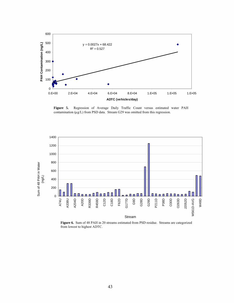
y = 0.0027x + 68.422R2 = 0.527
0
100
200
300
400
500
600
0.E+00 2.E+04 4.E+04 6.E+04 8.E+04 1.E+05 1.E+05 1.E+05
ADTC (vehicles/day)
PAH
Con
tam
inat
ion
(ng/
L)
Figure 5. Regression of Average Daily Traffic Count versus estimated water PAH contamination (µg/L) from PSD data. Stream G29 was omitted from this regression.
0
200
400
600
800
1000
1200
1400
A74U
A338
U
A204
D
A20D
R33
9D
R45
9D
C12
D
C18
D
F62D
G17
7D
G9D
G28
D
G29
D
P211
D
P38D
O30
D
O26
3D
J205
2D
W56
1D-A
VG
W49
D
Stream
Sum
of 4
8 P
AH
in W
ater
(ng/
L)
Figure 6. Sum of 48 PAH in 20 streams estimated from PSD residue. Streams are categorized from lowest to highest ADTC.
43

0.00
1.00
2.00
3.00
4.00
5.00
6.00
7.00
8.00
9.00
A74
R33
9
P211
A338 P3
8
G17
7
G9
R45
9
A20
C12
C18 A2
0
G28
O30 F62
J205
2
G29
W56
1
O26
3
W49
l----- Low ADTC -----l l----- Medium ADTC -----l l----- High ADTC -----l
Stream
Petr
ogen
ic/P
yrog
enic
Rat
io
Figure 7. Mean Petrogenic/pyrogenic PAH ratio in the 20 streams used in this study. Streams are categorized from lowest ADTC to highest. There is no significant difference between ADTC groups.
-5
0
5
10
15
20
25
A338 C12 G177 O30 O263 W49
Stream
Tail MomentOlive Moment
Figure 8. Tail and Olive Moment (µm) values for the streams sampled for the field portion of the study, with 95% confidence intervals.
44
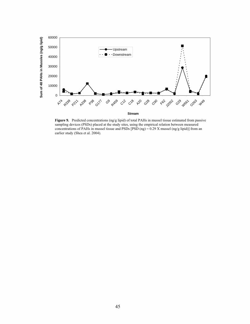
0
10000
20000
30000
40000
50000
60000
A74R33
9P21
1A33
8P38
G177 G9
R459
C12 C18 A20 G28 O30 F62J2
052
G29W
561
O263
W49
Stream
Sum
of 4
8 PA
Hs
in M
ussl
es (n
g/g
lipid
)
UpstreamDownstream
Figure 9. Predicted concentrations (ng/g lipid) of total PAHs in mussel tissue estimated from passive sampling devices (PSDs) placed at the study sites, using the empirical relation between measured concentrations of PAHs in mussel tissue and PSDs [PSD (ng) = 0.29 X mussel (ng/g lipid)] from an earlier study (Shea et al. 2004).
45