Embed Size (px)
Citation preview

29
ABSTRACTSOF THE 78th SCIENTIFIC CONFERENCE OF THE UNIVERSITY OF LATVIA
January – February 2020
Druvietis I., Hartmane K., Kokorīte I., Dobkeviča L.Succession of phytoplankton communities in small urban lake situated in Riga city, Latvia 31
Gailīte A., Ruņģis D.E.Study of genetic diversity of species from the genus Vaccinium 33
Štrausa D., Poppels A.Survival of benthic communities of inland standing waters in summer low water period of 2019 in Zemgale region, Latvia 35
Nikolajeva V., Seņkovs M., Volperts A., Dobele G., Kleperis J.Nitrogen-doped carbon electrodes for microbial fuel cells 36
Krivmane B., Girgžde E., Samsone I., Ruņģis D.Development of molecular markers for assessment of juvenility during micropropagation of silver birch (Betula pendula) 37
Aleksejevs Ē., Bērziņš E.Changes in the structure of ichthyocenosis of the Lake Rāznas 39
Andersone-Ozola U., Ineta Samsone I., Karlsons A., Osvalde A., Ievinsh G.Heavy metal tolerance and accumulation potential of Armeria maritima plants from a dry coastal meadow 41
Ievinsh G., Landorfa-Svalbe Z., Andersone-Ozola U., Bule A.Wild Rumex species as models in ecophysiological studies: effect of Na/K salts and nitrogen compounds on growth and electrolyte accumulation 43
Andersone-Ozola U., Ievinsh G., Landorfa-Svalbe Z., Karlsons A., Osvalde A.Wetland species Ranunculus sceleratus from a sea coast: heavy metal tolerance and accumulation potential 45
Ievinsh G., Andersone-Ozola U., Landorfa-Svalbe Z., Karlsons A., Osvalde A.New model species in studies with coastal plants: Hypochaeris maculata, Mentha aquatica, Veronica beccabunga, Tripleurospermum maritimum 47
Ievinsh G., Andersone-Ozola U.Does sand burial promote growth of dune-forming grass species Leymus arenarius and Ammophila arenaria? 49
Bajinskis J., Aleksejevs Ē., Ozoliņa Z., Začs D.The composition and quality of European eel Anguilla anguilla stock in Lake Rāznas 51
Seņkovs M., Bērziņa Z., Nikolajeva V., Nakurte I.Analysis of lipopeptides produced by Bacillus subtilis 53
Rudzīte M., Rudzītis M.Problems of protection of the freshwater mussels in Latvia and the world 54
Mališevs A., Makarova S., Konvisers G., Pūle D., Valciņa O., Grantiņa-Ieviņa L.Free-living protozoa and Legionella spp. coexistence and identification in drinking water systems in Riga apartment houses 55
Kassaliete J., Zīle A., Ozoliņa Z., Kokina A., Liepiņš J.Use of baker’s yeast purine auxotrophs for adenine quantification 57
Boroduske A.Callus culture of black elder (Sambucus nigra): initiation and basic chemical characterization 59
Environmental and Experimental Biology ISSN 2255-9582
https://doi.org/10.22364/eeb.18.05

30

31
Succession of phytoplankton communities in small urban lake situated in Riga city, Latvia
Ivars Druvietis1,3*, Kitija Hartmane2, Ilga Kokorīte1,3, Linda Dobkeviča1,3
1University of Latvia, Faculty of Biology, Jelgavas 1, Riga LV–1004, Latvia2University of Latvia, Faculty of Geography and Earth Sciences, Jelgavas 1, Riga LV-1004, Latvia3Institute of Biology, Miera 3, Salaspils LV–2169, Latvia*Corresponding author, E-mail: [email protected]
Key words: phytoplankton, seasonal succession, urban lake.
Environmental and Experimental Biology (2020) 18: 31–32 Abstract of the 78th Scientific Conference of the University of Latvia
The aim of our study was to detect development of algae groups and taxa forming phytoplankton community in small lake Linezers surrounded by urban territory of Riga city. Previous studies of phytoplankton succession were performed in Riga reservoir (Druvietis 2018). Together with phytoplankton sampling, some analyses of physical and chemical data characterising water quality in the lake were obtained. It is poossible to characterise the lake as very shallow, soft water, polyhumic (Table 1).
Eight algae divisions represented by 52 algae taxa were observed in the Lake Linezers.From begining (May 31, 2018) till end of this study (October 30, 2018) dominating algae group forming phytoplankton biomass was cyanobacteria (Cyanophyta; Fig. 1).
Development of phytoplankton communities in Lake Linezers in vegetation period of 2018 began in May, when lake waters were in early summer clear water stage with low development of Chrysophytes Dinobryon stipitatum and Dinobryon sertularia, green algae Scenedesmus spp. and Ankistrodesmus spp., which was supplemented by development of diatoms Nitzchia acicularis. In eaerly summer period Cyanobacteria in low amount were represented by Aphanocapsa sp., Gomphosphaeria sp. and Planktothrix sp., which in midsummer period was replaced by cyanobacteria Aphanizomenon flos-aquae and Microcystis aeruginosa. Some dinophytes such as Ceratium hirundinella were detected. Midsummer and late summer
phytoplankton was characterised by high development of cyanobacteria: Anabaena flos-aquae, Anabaena spiroides, Aphanizomenon flos-aquae, Gomphosphaeria sp., Planktothrix sp., Microcystis aeruginosa, Microcystis incerta, Microcystis viridis, Microcystis wessenbergii. These species formed high biomass (5.84 mg L–1) while total nitrogen level was low (1.18 mg L–1; Table 1).
In July and August due to cyanobacteria blooms phytoplankton index EQR showed low ecological quality (EQR = 0.33). However, in the begining of summer and during autumn period phytoplankton index EQR showed
Table 1. Physical and chemical characteristics of water of the Lake Linezers
Sampling date Electrical conductivity
(µS cm–1)
Color (PtCo) Temperature (°C)
Oxygen (mg L–1) Total P (mg L–1) Total N (mg L–1)
May 31, 2018 103.6 133 21.8 6.25 0.214 5.02July 18, 2018 93 108 25.2 6.15 – 2.44August 3, 2018 96.7 121 26.2 6.72 0.299 1.97August 29, 2018 114 99 18.3 7.21 0.164 1.18October 2, 2018 121 91 11.3 8.71 0.255 1.72October 30, 2018 115.7 85 4.9 9.62 0.231 1.77
Fig. 1. Percentage (%) of phytoplankton biomass-forming dominant algal groups.

32
good ecological quality (EQR = 0.8). Seasonal phytoplankton succession of Lake Linezers depended on low water level in summer and changes in water temperature in August. At the end of August, Raphidophyte algae Gonyostomum semen characteristic for humic waters was detected in small amounts. Autumn phytoplankton showed development of Chrysophyta Dinobryon divergens; Dinophyta Peridinium spp. and Gymnodinium sp.; Euglenophyta Euglena sp.; Cyanophyta Planktothrix sp., Microcystis spp. and Gomphosphaeria aponina; Bacillariophyta Aulacoseira italica and Nitzschia acicularis; Chlorophyta Pediastrum spp., Scenedesmus spp., Ankistrodesmus spp., Botryococcus braunii and Koliella sp. in very small amounts.
ReferencesDruvietis I. 2018. Phytoplankton of Riga Reservoir (1978 – 2017).
Abstract of the 75th Scientific Conference of the University of Latvia. Enviro. Exp. Biol. 16: 83.
Fig. 2. Biomass (mg L–1) of phytoplankton-forming algae groups.

33
Study of genetic diversity of species from the genus Vaccinium
Agnese Gailīte*, Dainis Edgars Ruņģis
Genetic Resource Centre, Latvian State Forest Research Institute “Silava”, Rigas 111, Salaspils LV–2169, Latvia*Corresponding author, E-mail: [email protected]
Key words: Vaccinium, genetic diversity.
Environmental and Experimental Biology (2020) 18: 33–34 Abstract of the 78th Scientific Conference of the University of Latvia
Population and genetic diversity studies are important sources of information for the development of conservation strategies for crop wild relatives (CWR) and wild harvested plants (WHP). The main CWR and WHP plant groups in Latvia are forage grasses, aromatic and medicinal plants, as well as forest fruits and berries.
In this study two species from the genus Vaccinium were analysed: Vaccinium myrtillus (bilberries) and Vaccinium vitis-idaea (lingonberries). Both are dwarf shrubs typical in the northern hemisphere (Nestby et al. 2010) and can propagate vegetatively and generatively.
An investigation of the population structure and genetic diversity of plants from the genus Vaccinium has not been previously undertaken in Latvia. The majority of molecular studies, including the use of EST-SSR markers, have been done on species of the section Cyanococcus (Rowland et al. 2003; Boches et al. 2005). The species endemic to Latvia belong to other sections (Nestby et al. 2010). RAPD (Bjedov et al. 2015), ISSR (Debnath 2007; Zoratti et al. 2015) and AFLP (Albert et al. 2003) markers have been used in studies on these species. In this study EST SSR markers (Boches et al. 2005) were used, and analyses were performed with eight markers on bilberry samples and 10 markers on lingonberry collected within Latvia (21 and 20 locations respectively), Estonia (seven locations) and Lithuania (nine locations). In addition, accessions from V. myrtillus var. leucocarpum (white fruited bilberries) collected in three sites in Latvia were analysed. Genotyping was done with
Applied Biosystems ABI Prism 3100xl Genetic Analyzer. SSR genotype data were analysed with GenAlEx 6.501 (Peakall, Smouse 2012) and alleles frequency, heterozygosity, analysis of molecular variance (AMOVA), PCoA, genetic and geographic distance correlation were estimated.
Most of the genetic diversity was found within individuals, differences among regions – Latvia, Lithuania and Estonia – were not found (Fig. 1, 2.). The number of alleles unique to one population was low, heterozygosity (Ho) varied from 0.318 to 0.526 in bilberries and 0.4 to 0.638 in lingonberries. The correlation between genetic and geographic distances between populations is positive, indicating genetic differentiation of Latvian bilberry and lingonberry populations due to isolation by distance.
Only one genet of V. myrtillus var. leucocarpum with many ramets was found in each of three analysed sites. It seems that this variety originated spontaneously within each population.
AcknowledgementsThis work was supported by the European Regional Development Fund Postdoctoral research aid Nr.1.1.1.2/VIAA/1/16/123 “Investigation of Vaccinium genetic resources in Latvia”. We thank Lelde Stirna and Lāsma Lasmane for help with information about V. myrtillus var. leucocarpum sites. We thank Külli Annamaa from Estonian Crop Research Institute Plant Gene Bank and Bronislovas Gelvonauskis, Raimondas Baltrenas, Laima Šveistyté from Plant Gene Bank of Lithuania for assistance in collecting of leaf material in Estonia and Lithuania. We thank Anita Gaile
Fig. 1. AMOVA of bilberries. Fig. 2. AMOVA of lingonberries.

34
for her help to prepare a map of collecting sites. We also thank our colleagues Anna Korica, Krišs Bitenieks, Viktorija Beļēviča un Baiba Krivmane for technical assistance with DNA extraction.
ReferencesAlbert T., Raspe O., Jacquemart A.L. 2003. Clonal structure in
Vaccinium myrtillus L. revealed by RAPD and AFLP markers. Int. J. Plant Sci. 164: 649–655.
Bjedov I., Obratov-Petković D., Mišić D., Šiler B., Aleksić J.M. 2015. Genetic patterns in rangeedge populations of Vaccinium species from the central Balkans: implications on conservation prospects and sustainable usage. Silva Fenn. 49: 1283.
Boches P. S., Bassil N.V., Rowland L.J. 2005. Microsatellite markers for Vaccinium from EST and genomic libraries. Mol. Ecol.
Notes 5: 657–660. Debnath S.C. 2007. Inter simple sequence repeat (ISSR) to assess
genetic diversity within a collection of wild lingonberry (Vaccinium vitis-idaea L) clones. Can. J. Plant Sci. 87: 337–344.
Nestby R., Percival D., Martinussen I., Opstad N., Rohloff J. 2011. The European blueberry (Vaccinium myrtillus L.) and the potential for cultivation. A review. Eur. J. Plant Sci. Biotechnol. 5: 5–16.
Peakall R., Smouse P.E. 2012. GenAlEx 6.5: genetic analysis in Excel. Population genetic software for teaching and research – an update. Bioinformatics 28: 2537–2539.
Zoratti L., Palmieri L., Jaakola L., Häggman H. 2015. Genetic diversity and population structure of an important wild berry crop. AoB Plants 7: plv117.

35
Survival of benthic communities of inland standing waters in summer low water period of 2019 in Zemgale region, Latvia
Diāna Štrausa1*, Arkādijs Poppels2
1University of Latvia, Faculty of Biology, Jelgavas 1, Riga LV–1004, Latvia2Riga Zoo, Meža Prospekts 1, Riga LV–1014, Latvia*Corresponding author, E-mail: [email protected]
Key words: low water level, macrozoobenthos, small shallow lakes.
Environmental and Experimental Biology (2020) 18: 35 Abstract of the 78th Scientific Conference of the University of Latvia
The aim of this study was to detect impact of summer low water level period on survival of macrozoobenthos communities in littoral zone. It would be possible that climate change and global qarming would cause changes in biodiversity (Klavins et al. 2008; Jeppesen et al. 2014). Macrozoobenthoss communities were observed in littoral zones of four lakes in low water level period in the second part of July 2019 and August 2019 in Zemgale region, Latvia. Biological diversity of macrophytes and macrozoobenthos animals were investigated. Summer of 2019 was characterised as arid. It caused decline of water level in water objects. As a result water level sharply decreased and former littoral zone in three lakes become dry (Fig. 1).
Due to high water temperature (more than 26 C°) littoral zone shifted to all aquatoria, where macrophytes formed compact stands, which gave suitable conditions for zoobenthos animals to survive.
Dominated macrozoobenthos groups were Mollusca, Chironomidae, Malacostraca, Odonata and Varia. Mollusca were represented by Bihynia tentaculata, Anisus spp. and Pisidium amnicum. Shallow and partly dry former littoral zone in places were covered by immmobile dead big mussels Anodonta spp. Younger stages of Chironomidae
were represented in high amounts. Division Malacostraca were formed by Asellus aquaticus, which is relatively resistant against low water period. Division Odonata was represented by species characteristic for all the lakes aquatoria. Group Varia was dominated by water blazers, water ticks and common water striders. According former observations sustained low water level periods implement corrections in development of macrophyte and zoobenthoss communities. Species which fail to adapt under pressure of climate changes are partly subjected to perish.
ReferencesJeppesen E., Meerhof M., Davidson T., Trolle D, Sondergaard M,
Lauridsent T., Beklioģlu M., Brucets S.,Volta P., Gonzalez-Bergonzoni I, Nielsen A. 2014. Climate change impacts on lakes: an integrated ecological perspective based on a multi-faceted approach, with special focus on shallow lakes. J. Limnol. 73: 84–107.
Kļaviņš M., Blumberga D., Bruņiniece I., Briede A., Grišule G., Andrušaitis A., Āboliņa K. 2008. Climate Change and Global Warming. LU Akadēmiskais apgāds, Rīga, 174 p. /in Latvian/
Fig. 1. Small lake in Zemgale region in late summer of 2019. Photo: D. Štrausa.
Fig. 2. Impact of water level lowering on survival of mollusca (%) in observed lakes.

36
Nitrogen-doped carbon electrodes for microbial fuel cells
Vizma Nikolajeva1*, Māris Seņkovs1, Aleksandrs Volperts2, Gaļina Dobele2, Jānis Kleperis3
1Faculty of Biology, University of Latvia, Jelgavas 1, Riga LV–1004, Latvia2Latvian State Institute of Wood Chemistry, Dzērbenes 27, Riga LV–1006, Latvia3Institute of Solid State Physics, University of Latvia, Ķengaraga 8, Riga LV–1063, Latvia*Corresponding author, E-mail: [email protected]
Key words: electricity generation, electrode materials, microbial fuel cells.
Environmental and Experimental Biology (2020) 18: 36 Abstract of the 78th Scientific Conference of the University of Latvia
Microbial fuel cells (MFC) are electrochemical devices that use bacteria as the catalysts to oxidize organic and inorganic matter to generate current (reviewed in Logan et al. 2006). Traditionally, different carbon materials such as carbon paper, carbon felt and cheaper graphite electrodes are used in MFC (reviewed in Wei et al. 2011).
The aim of the study was to test the efficiency of newly synthesized nitrogen-doped carbon material in MFC with ceramic separator. Earthen pot as a low-cost separator (instead of cation exchange membrane such as Nafion) was adapted in the experiments. The wall of pot itself act as medium for transfer of protons from anode to cathode (Behera et al. 2010). Synthetic wastewater containing acetate and yeast extract was chosen as a substrate and sapropel as an inoculum.
Electrode materials were tested in a single chamber mediator-less air cathode MFC using earthen pot with wall thickness of 5 mm and fed-batch mode of operation at temperature of 20 to 24 °C. The working volume of the anode chamber was 150 mL. The anode was made up of AvCarb P50 carbon fiber paper with a surface area of 16 cm2. Air cathodes were prepared by covering the outer ceramic surfaces with synthesized carbon materials without added
catalyst. The cathode surface area was 181 cm2. Electrodes were connected to the data acquisition system (Velleman PCRU01), which was connected to a personal computer, and voltage measurements were recorded. Relatively stable power output was recorded after seven days of operation. The highest power densities observed were 35.3 W m–3 for nitrogen-doped carbon cathode (Table 1).
Part of the experiments was carried out in single chamber air cathode MFC using earthen pot with wall thickness of 7 mm and outer anode chamber. Detected open circuit voltage in case of wood-based alkali-activated nanoporous carbon electrode was 0.462 ± 0.077 V; the same but nitrogen-doped carbon gave 0.432 ± 0.053 V, and non-activated small porous carbon gave 0.168 ± 0.069 V.
The results showed that nanostructured, nitrogen doped carbon materials are suitable for MFC cathode formation. Membrane porosity played an important role because excessive porosity promoted growth of facultative anaerobic bacteria instead of strictly anaerobic bacteria including electro-active bacteria such as Geobacter due to more oxygen diffusion to the anode chamber. In future experiments, great attention should be paid to the quality of attachment of cathodes to the ceramic separator to decrease the internal resistance of MFC.
AcknowledgementsThis research is funded by the Latvian Council of Science, project NN-CARMA, project No. lzp-2018/1-0194.
ReferencesBehera M., Jana P.S., Ghangrekar M.M. 2010. Performance
evaluation of low cost microbial fuel cell fabricated using earthen pot with biotic and abiotic cathode. Bioresour. Technol. 101: 1183–1189.
Logan B.E., Hamelers B., Rozendal R., Schröder U., Keller J., Freguia S., Aelterman P. 2006. Microbial fuel cells: methodology and technology. Environ. Sci. Technol. 40: 5181–5192.
Wei J., Liang P., Huang X. 2011. Recent progress in electrodes for microbial fuel cells. Bioresour. Technol. 102: 9335–9344.
Table 1. Performance of the ceramic MFC with two cathode materials
Parameter Carbon cathode Nitrogen-doped carbon cathode
U (mV) 20 230I (μA) 20 23P (mW) 0.4 5.3P (mW m–2) 250 3306W m–3 2.7 35.3mA m–2 13.0 14.4mA m–3 133 153
37
Development of molecular markers for assessment of juvenility during micropropagation of silver birch (Betula pendula)
Baiba Krivmane, Elva Girgžde, Ineta Samsone, Dainis Ruņģis*
Latvian State Forest Research Institute “Silava”, Rigas 111, Salaspils LV–2169, Latvia*Corresponding author, E-mail: [email protected]
Key words: Betula pendula, gene expression, microclonal propagation, microRNA, rejuvenation.
Environmental and Experimental Biology (2020) 18: 37–38 Abstract of the 78th Scientific Conference of the University of Latvia
Silver birch (Betula pendula Roth) is one of the most economically important tree species in Latvia. For establishing highly productive and qualitative birch stands, tree breeding is carried out, which increases the added value of timber and contributes to ensuring the competitiveness and sustainable development of the forest industry.
Vegetative propagation (including micropropagation) of mature birch trees enables capture of genetic gain more rapidly than by sexual reproduction, due to selection and maintenance of both additive and non-additive gene effects (George et al. 2008). Vegetatively propagated material can then be used for further breeding to vegetatively propagate selected genotypes, which are then used for controlled crosses, thereby shortening the breeding cycle by 10 to 15 years. Evaluation of birch according to phenotypic parameters is carried out when trees have reached their mature phase. In vitro culture initiation and shoot rejuvenation from birche trees in their reproductive phase are difficult and often unsuccessful. There is a lack of understanding of the mechanisms controlling rejuvenation and the factors affecting it.
Recent studies with annual model species indicate that the main endogenous signals that regulate juvenility are microRNAs (miRNAs), miR156/157, miR172 and their target genes (Wu et al. 2009; Wang et al. 2011).
The aims of this study were to develop a technology (molecular markers) for determining juvenility during the micropropagation process of silver birch based on miRNAs and their target gene expression changes, and to investigate factors affecting juvenility of birch genotypes with different in vitro morphogenic ability.
Plant material for miRNA and target gene expression analysis has been collected for total RNA isolation and real time PCR analysis. Total RNA was extracted from leaves from 25-year-old mature silver birch (mature control; sample 11C), from leaves of rejuvenated in vitro shoots (1B), from seedlings (juvenile control; 13C) and two types of mature in vitro shoots. One exhibited signs of a mature in vitro culture (thick stem, large and thick leaves, inability to
grow and other signs; 8E). The other, which was significantly different (8E2), initially exhibited signs of a mature in vitro culture, but subsequently started growing in length and developing axillary or adventitious shoots, which showed morphological signs of juvenility (thin and long stems, thin leaves, ability to grow, high reproductive capacity). However, eventually, this in vitro culture again exhibited morphological signs of maturity. RNA was extracted using a standard phenol/chloroform/isoamyl alcohol protocol (Rubio-Piña, Zapata-Pérez 2011).
Expression levels of miRNAs and target genes were determined using real-time PCR analysis of 10 miRNA and four target gene primers. Initial results indicate that two miR156 primers and two miR172 and three target gene primers (SPL1, SPL9 and AP2) can be used for detection of juvenility state. miR156 primers showed up-regulated expression in sample 13C (juvenile control) and 1B, and down-regulated in 11C (mature control), but expression in sample 8E was lower than expression in sample 8E2, reflecting the morphological juvenility signs in these in vitro cultures. The expression of miR156 target genes SPL1 and SPL9 was down-regulated in sample 13C and 8E2, but up-regulated in sample 11C. In the case of miR172 and target gene AP2, the opposite expression levels were observed. These results indicate that the developed miRNA and target gene primers can be used to assess juvenility state in silver birch. Further investigations will enable a further understanding of these process in silver birch and the applicability of the developed methods to other forest tree species.
AcknowledgementsThis research is funded by the Latvian Council of Science, project “Development of molecular markers for assessment of juvenility during micropropagation of silver birch (Betula pendula Roth.)”, project No. lzp-2019/1-0387.
ReferencesGeorge E.F., Hall M.A., De Klerk G.J. 2008. Plant Propagation by
Tissue Culture 3rd Ed. Vol. 1, The Background. Dordrecht, 501

38
pp.Wang J.W., Park M.Y., Wang L.J., Koo Y., Chen X.Y., Weigel D.,
Poethig R.S. 2011. MiRNA control of vegetative phase change in trees. PLoS Genet. 7: e1002012.
Wu G., Park M.Y., Conway S.R., Wang J., Weigel D., Poethig R.S. 2009. The sequential action of miR156 and miR172 regulates
developmental timing in Arabidopsis. Cell 138: 750–759.Rubio-Piña J.A., Zapata-Pérez O. 2011. Isolation of total RNA
from tissues rich in polyphenols and polysaccharides of mangrove plants. Electron. J. Biotechnol. 14.

39
Changes in the structure of ichthyocenosis of the Lake Rāznas
Ēriks Aleksejevs*, Edmunds Bērziņš
Scientific Institute of Food Safety, Animal Health and Environment “BIOR”, Lejupes 3, Rīga LV–1076, Latvia*Corresponding author, E-mail: [email protected]
Key words: ichthyocenosis, Lake Rāznas.
Environmental and Experimental Biology (2020) 18: 39–40 Abstract of the 78th Scientific Conference of the University of Latvia
Lake Rāznas is the largest natural lake in Latvia with a water surface of 5756.4 ha, maximum depth of 17 m and an average depth of 7 m. It is located in the Latgale highlands south-eastern region of the country (56° 19’ 37’’ N, 27° 26’ 45’’ E). Several small watercourses flow into Lake Rāznas, but the Rēzekne River flows out.
Information about the ichthyocenosis obtained from various sources of literature from 1925, “BIOR” databases on fishery statistics from 1950, as well as field research performed from 1989 to 2019. In the field research fishing nets with varying mesh sizes (8 to 70 mm), a beach seine (mesh size in the codend 5 mm) and electro-fishing equipment have been used.
The relatively large surface, depth and connection with the rivers of Lake Rāznas determine its relatively high diversity of ichthyofauna. From 1947 to 2019, a total of 25 fish species have been identified in fisheries research at Lake Rāznas: bleak Alburnus alburnus, bream Abramis brama, bullhead Cottus gobio, burbot Lota lota, carp Cyprinus carpio, crucian carp Carassius carassius, eel Anguilla anguilla, gudgeon Gobio gobio, ide Leuciscus idus, perch Perca fluviatilis, pike Esox lucius, pike-perch Sander lucioperca, Prussian carp Carassius gibelio, roach Rutilus rutilus, rudd Scardinius erythrophthalmus, ruffe Gymnocephalus cernua, silver bream Blicca bjoerkna, smelt Osmerus eperlanus, spined loach Cobitis taenia, stone loach Barbatula barbatula, sunbleak Leucaspius delineatus, tench Tinca tinca, vendace Coregonus albula, weather loach Misgurnus fossilis, whitefish Coregonus sp.
Literature in the twenties and thirties of the last century reports that vendace, smelt and bleak live on Lake Rāznas (Zandbergs 1925; Gaņģis 1939).
The first known fish survey of the lake in 1947 found 14 fish species: bleak, bream, burbot, crucian carp, eel, ide, perch, pike, roach, rudd, ruffe, smelt, vendace and white bream (Savina 1948).
The formation and existence of the eel population to nowadays is apparently determined by the release of glass eels from 1925 to 2005. As the eel does not reproduce in the lake, its population will gradually decline and disappear
as a result of natural and fishing mortality. The continued existence of the eel population depends on their possible releases in the future.
The establishing of the pike-perch population is determined by its release from 1956 to 1990. The lake has a self-sustaining pike-perch population, which is not too large. Lakes with low water transparency are usually better suited for pike-perch. As a result of further eutrophication of Lake Rāznas, the population of pike-perch is expected to increase.
The introduction of Lake Rāznas from 1925 to 1971 has resulted in the creation of a small population of naturally reproducing whitefish. Further eutrophication and climate warming of the lake is expected to result in further decline and eventual extinction of its population.
Climate change and anthropogenic eutrophication are also adversely affecting other populations of cold-loving smelt and vendace, which have declined significantly since the middle of the last century.
Carp and Prussian carp have appeared in the lake as a result of the releases from 1955 to nowadays. These two species are thought to have no or only very low reproductive populations.
Lake Rāznas is experiencing a gradual increase in the size and overall proportion of the tench population in ichthyocenosis, caused by climate warming and eutrophication of the lake.
Changes in the size and proportion of populations of other indigenous fish species compared to the middle of the last century are not significant.
Given the fact that Lake Rāznas is predominantly fisheries researched, information on non-commercial species of fish (such as bullhead, gudgeon, spined loach, stone loach, sunbleak, and weather loach) is of limited magnitude and does not allow for estimation of possible changes in the size of their populations. It can only be assumed that the populations of bullhead and weather loach are very small, as in the 1950-ies, because nowadays have not been found in fishery surveys in Lake Rāznas anymore.

40
AcknowledgementsWe would like to thank our recent and past colleagues Jānis
Aizups, Toms Zalāns, Amanda Tropa and Raimonds Reščenko for help in the field works.
ReferencesGaņģis U. 1939. Fish diversity in Latgale. Fisheries Monthly
Magazine 12: 521–522. /in Latvian/
Savina N. 1948. Survey of some lakes of the Republic of Latvia. Fisheries XXIV/6: 29–31. /in Russian/
Sloka J. 1959. Fish biology of Lake Rāznas. Proceedings of the Latvian SSR Academy of Sciences 10/147: 139–146. /in Latvian/
Zandbergs A. 1925. Breeding of Lake Peipus whitefish in our lakes. Earth Power. 1925.01.07. Supplement to the newspaper “Free Land” 7/8. /in Latvian/

41
Heavy metal tolerance and accumulation potential of Armeria maritima plants from a dry coastal meadow
Una Andersone-Ozola1, Ineta Samsone1, Andis Karlsons2, Anita Osvalde2, Gederts Ievinsh1*
1Department of Plant Physiology, Faculty of Biology, University of Latvia, Jelgavas 1, Rīga LV–1004, Latvia2Institute of Biology, University of Latvia, Miera 3, Salaspils LV-2169, Latvia
*Corresponding author, E-mail: [email protected]
Key words: accumulation potential, Armeria maritima, heavy metals, tolerance.
Environmental and Experimental Biology (2020) 18: 41–42 Abstract of the 78th Scientific Conference of the University of Latvia
Armeria maritima is a perennial rosette-forming species with a complicated taxonomy. According to Lefèbvre (1974), there are four subspecies of A. martima: A. maritima subsp. alpina, growing exclusively in mountain regions 1600 to 3000 m above sea level; A. maritima subsp. elongata, characteristic on acidic sandy soils both in coastal and inland areas; A. maritima subsp. halleri, growing as metallophyte on heavy metal contaminated soils; A. maritima subsp. maritima, characteristic species of coastal salt marshes. However, it is still under scientific debate whether high metal toelrance and metal accumulation capacity is an exclusive fetaure of A. maritima subsp. halleri only or, as an alternative, species-wide tolerance are present. So far, only limited number of studies have tried to experimentally assess this problem on a comparative basis. When Zn tolerance and accumulation capacity of three ecotypes of A. maritima (acidic sand, salt marsh and heavy metal) were compared in artificial soil system during long-term experiment, all ecotypes tolerated 182 mg kg–1 Zn (Köhl 1997). Plants of all ecotypes accumulated comparable concentrations of Zn, with maximum level reaching 11 427 mg kg–1 in roots and 1 697 mg kg–1 in leaves. Also in other studies no tendency for increased metal accumulation in aboveground parts of A. maritima has been shown. The aim of the present study was to analyze heavy metal tolerance of A. maritima subsp. elongata accession from a dry coastal
meadow of the Baltic Sea in the Southern Sweden near Nybrostrand.
Plants were brought in culture by seeds collected in natural population. Seeds were germinated in closed containers on sterile garden soil at 10/20 °C thermoperiod. Seedlings were gradually transplanted to larger containers filled with a mix of commercial garden soil (Biolan, Finland) and quartz sand (1:1, vol/vol). Final cultivation was performed in a 400 mL plastic containers in an automated greenhouse with 16 h photoperiod (additional photosynthetically active radiation of 380 µmol m–2 s–1) and night/day temperature 15/20 °C. Plant germination and initial growth was extremely slow, taking three months from the start of germination untill the start of treatments. After treatment with gradually increasing concentrations of heavy metals within the next month, plants were cultivated for another two months before termination of the experiment. Plants were treated with Mn, Zn and Cd in a form of respective sulphate salts and Pb in a form of nitrate at final concentration of the metals reaching 0.2, 0.5 and 1.0 g L–1 for Mn; 0.2, 0.5 and 1.0 g L–1 for Zn; 0.005, 0.02, 0.1 g L–1 for Cd; 0.1, 0.2, 0.5 g L–1 for Pb. Low level of substrate moisture (30 to 40 %) was provided with deionized water on individual basis. Plants were fertilized with Kristalon Green soluble mineral fertilizer once a month. During termination of the experiment, individual plants were
Fig. 1. Morphology of Armeria maritima plants cultivated for 2 months in the presence of different heavy metal concentration in substrate. From left to right: control, 0.2, 0.5, 1.0 g L–1 Mn; 0.2, 0.5, 1.0 g L–1 Zn; 0.005, 0.02, 0.1 g L–1 Cd; 0.1, 0.2, 0.5 g L–1 Pb.

42
Fig. 2. Effect of increasing metal concentration in substrate on accumulation of Mn (A), Zn (B), Cd (C), and Pb (D) in different parts of Armeria maritima plants. Dotted line indicate concentration of hyperaccumulation threshold for the respective metal.
extracted from substrate and separated in roots, leaves, flower stalks and inflorescences (flowers). All parts were carefully washed and blotted dry. Both fresh and dry mass was measured. Analysis of metals was performed in dry-ashed samples using atomic absorption spectrophotometry.
Plant growth and morphology was not affected by increasing doses of heavy metals (Fig. 1). There was no significant effect of any of the treatments on both fresh and dry mass of different plant parts (roots, leaves, flower stalks, flowers). Total number of leaves also was not affected. Also, number of senescent leaves was extremely low and it was not affected by the treatments.
There was a clear substrate concentration-dependence for accumulation of metals in plants, with metal-specific significant differences between different plant parts (Fig. 2). Mn was predominantly accumulated in leaves of A. maritima, with concentration exceeding 10 g kg–1, which is a hyperaccumulation threshold for this metal (Fig. 2A). Mn concentration in roots was only about 20% from that in leaves, with significanly lower levels in flower stalks and flowers. At lower concentration treatments, plants accumulated similar concentration of Zn in roots and leaves, but at 1.0 g L–1, accumulation potential was significanly higher in leaves, reaching 15 g kg–1 (Fig. 2B). Hyperaccumulation concentration threshold for Zn was reached at 0.5 g L–1 substrate concentration. Level of Zn in generative parts was significantly lower. A. maritima plants had extremely high potential for Cd accumulation, which was at identical levels both in roots and leaves, but Cd was efficiently excluded from generative parts (Fig. 2C). Similarly, concentration of Pb was low in flower stalks and flowers, with the highest level in leaves and roots (Fig. 2D). Hyperaccumulation concentration threshold of Pb in plant leaves (1.0 g kg–1) was reached at 0.2 g L–1 Pb in substrate.
It is evident that A. maritima subsp. elongata plants growing naturally on dry coastal soils with no increased level of heavy metals had extremely high tolerance against both biogenous (Mn and Zn) and nonbiogenous (Cd and Pb) heavy metals in substrate during relatively long-term experiments in controlled conditions. Cd, Zn and Pb was accumulated in high concentration in both plant roots and leaves, but Mn was predominantly accumulated in leaves. All metals were efficiently excluded from generative structures, showing high level of physiological adaptation. Most importantly, respective hyperaccumulation thresholds for all heavy metals used in the study were exceeded,
emphasizing extreme potential of the particular accession of A. maritima for phytoextraction. It seems that heavy metal tolerance and accumulation ability is a characteristic feature of A. maritima plants irrespective of their origin, but further studies using different accessions need to be performed to fully confirm species-wide nature of the phenomenon.
AcknowledgementsThe study was supported by the University of Latvia project “Functional diversity of ecosystems and their contribution to ecosystem services II”.
ReferencesKöhl K.L. 1997. Do Armeria maritima (Mill.) Willd. ecotypes
from metalliferous soils and non-metalliferous soils differ in growth response under Zn stress? A comparison by a new artificial soil method. J. Exp. Bot. 48: 1959–1967.
Lefèbvre C. 1974. Population variation and taxonomy in Armeria maritima with special reference to heavy-metal-tolerant populations. New Phytol. 73: 209–219.

43
Wild Rumex species as models in ecophysiological studies: effect of Na/K salts and nitrogen compounds on growth and electrolyte accumulation
Gederts Ievinsh*, Zaiga Landorfa-Svalbe, Una Andersone-Ozola, Annija Bule
Department of Plant Physiology, Faculty of Biology, University of Latvia, Jelgavas 1, Rīga LV–1004, Latvia*Corresponding author, E-mail: [email protected]
Key words: coastal accessions, developmental strategy, electrolyte accumulation, nitrogen, Rumex, salinity tolerance
Environmental and Experimental Biology (2020) 18: 43–44 Abstract of the 78th Scientific Conference of the University of Latvia
Wild plants species from natural salt-affected habitats, as these on a sea coast, represent a useful resource in salinity tolerance studies. Relationship between Na+ and K+ in salt tolerance physiology of plants seems to be an important aspect, but it is not throughly explored in species native to salt-affected habitats (Percey et al. 2016). Besides osmotic effects, electrolytic activity is a main characteristic of both Na and K ions, with K+ being the main contributor to ionic strength, necessary for maintanence of cellular activities. Therefore, it is interesting to understand how cellular electrolyte balance is maintained for plants growing on soils with significantly fluctuating Na and K concentration. An ability of Na+ to substitute K+ has been suggested as an important feature of salinity tolerance of halophytic species (Belkheiri, Mulas 2013). It can be proposed that mineral nutrient availability in general as well as level of nitrogen in particular, can affect plant responses to salinity. The aim of the present study was to establish model system for studying salinity tolerance and electrolyte accumulation using Rumex plant species from salt-affected habitats.
Three Rumex species with accessions from coastal habitats of the Baltic Sea with relatively well water supply (Rumex hydrolapathum, Rumex longifolius and Rumex maritimus) were chosen as models for ecophysiological experiments in controlled conditions. For comparison, Rumex confertus, a cosmopolytic species, not occurring in saline soils was used. Seeds of R. hydrolapathum were collected in sea-affected coastal wetland in Mērsrags, Latvia. Seeds of R. longifolius and R. maritimus were collected on shingle beach at island of Saaremaa, Estonia. Seeds of R. confertus were collected in moderately wet meadow near pond in Salaspils, Latvia. Three consecutive experiments in controlled conditions were performed: (i) analysis of developmental and physiologcial differences between the four species in control conditions; (ii) exploring of salt tolerance and electrolyte accumulation potential of the four species under the effect of Na and K in a form of chloride, nitrate and nitrite; (ii) assessment of possible nitrophilic character of R. hydrolapathum using different levels of
mineral nutrient availability and additional treatment with nitrate or ammonia.
When development and physiological status of the four Rumex species was compared in control conditions (without elevated soil salinity), significant differences were observed between the species. All species at initial stages of seedling development had very strong vertical dominance, with leaf appearance order-related differences in leaf morphology and distribution (Fig. 1). Further, vertical dominance decreased with time, fastest for R. maritimus, followed by R. longifolius. Plants of R. hydrolapathum showed the strongest vertical dominance and physiological gradient according to leaf age, followed by R. confertus. Chlorophyll fluorescence parameter Performance Index Total was the only fluorescence indicator showing significant leaf age-dependent differences for all species, with the highest level in youngest fully developed leaf. There were significant differences in Na+ and K+ concentration and electrolyte accumulation between leaves of different age when estimated on dry mass basis, but due to significant changes in leaf water content in leaves of different age, the differences in electrolyte concentration were smoothed out
Fig. 1. Morphological differences of 3-week-old plants of different Rumex species. From left to right, Rumex hydrolapathum, Rumex maritimus, Rumex confertus, Rumex longifolius.

44
when the concentrations were expressed on tissue water basis.
In general, plant leaf number and leaf dry mass was relatively little affected by 2 g Na+ and 3.4 g K+ treatment in a form of chloride salts (Fig. 2). Both parameters were negatively affected by both teratments for R. confertus, but dry mass accumulation was stimulated by NaCl for R. hydrolapathum and KCl for R. maritimus. High growth increase by respective nitrate salts was observed for all Rumex species, but nitrite salts were inhibitory only for R. confertus and R. hydrolapathum in the case of Na. K nitrite significantly stimulated leaf dry mass accumulation only in R. longifolius and R. maritimus. Root growth was negatively affected by both nitrite salts for all species.
Analysis of Na+ and K+ concentration and electrolytic activity in plant tissues showed that all Rumex species can accumulate high level of electrolytes, consisting of different proportions of Na+ and K+ as affected by substrate concentration of the respective ion and other electrolytically active ions for equilibration, and the particular summary level of the activity was adjusted by changes in water content (Fig. 3). All Rumex species were able to grow and develop over a wide range of internal K+/Na+ concentration ratio. The most extreme cases were R. maritimus plants with the higher biomass accumulation potential, treated with Na nitrate (K+/Na+ ratio 0.05) and K nitrate (K+/Na+ ratio 17.00).
Increase in general mineral nutrient availability significantly stimuated growth of leaves and roots R. hydrolapathum plants, but both additional nitrogen in a form of nitrate or ammonia resulted in further growth increase at both mineral nutrient levels, suggesting the nitrophilous nature of the species.
It is usually thought that Na+ as a metal has special characteristics, leading to its significant toxicity, in comparison to K+. The results of the present study clearly indicated that while the effect of the two ions differed in some cases, as, for example, number of leaves was more severely reduced in R. confertus plants by KCl in comparison to NaCl, and leaf dry mass was lower for R. maritimus plants treated with NaNO2 in comparison to KNO2, plant growth was most drastically affected by a nature of anionic component of the salt. In general, both Na and K nitrites were toxic, chlorides were relatively inactive, and nitrates were highly stimulative. Most importantly, there were no relationship between accumulation capacity of the ion in plant tissues and its growth-related effect.
AcknowledgementsThe study was supported by the University of Latvia project
“Functional diversity of ecosystems and their contribution to ecosystem services II”.
ReferencesBelkheiri O., Mulas M. 2013. The effects of salt stress on growth,
water relations and ion accumulation in two halophyte Atriplex species. Environ. Exp. Bot. 86: 17–28.
Percey W.J., Shabala L., Wu Q., Su N., Breadmore M.C., Guijt R.M., Bose J., Shabala S. 2016. Potassium retention in leaf mesophyll as an element of salinity tissue tolerance in halophytes. Plant Physiol. Biochem. 109: 346–354.
Fig. 2. Relative leaf dry mass of Rumex species as affected by 2.0 g L–1 Na and 3.4 g L–1 K in a form of respective chloride, nitrate and nitrite salts.
Fig. 3. Electrolytical activity (measured as electrical conductivity of water extracts and expressed on tissue water content basis) in largest leaf group of Rumex species as affected by 2.0 g L–1 Na and 3.4 g L–1 K in a form of respective chloride, nitrate and nitrite salts.

45
Wetland species Ranunculus sceleratus from a sea coast: heavy metal tolerance and accumulation potential
Una Andersone-Ozola1, Gederts Ievinsh1*, Zaiga Landorfa-Svalbe1, Andis Karlsons2, Anita Osvalde2
1Department of Plant Physiology, Faculty of Biology, University of Latvia, Jelgavas 1, Rīga LV–1004, Latvia2Institute of Biology, University of Latvia, Miera 3, Salaspils LV-2169, Latvia*Corresponding author, E-mail: [email protected]
Key words: accumulation potential, heavy metals, Ranunculus sceleratus, tolerance.
Environmental and Experimental Biology (2020) 18: 45–46 Abstract of the 78th Scientific Conference of the University of Latvia
Ranunculus sceleratus is a semi-aquatic species of northern hemisphere with circumpolar distribution often found in wetland habitats. The species is extremely resistant to soil flooding, with a survival strategy based on leaf petiole elongation to promote leaf blade contact with aerial environment to sustain photosynthesis (Smulders, Horton 1991) and constitutive presence of aerenchyma in roots (He et al. 1999). R. sceleratus has been used in artificail wetland systems because of ability to remove dissolved nitrogen and phosphorus. However, no studies have explored heavy metal accumulation potential of R. sceleratus in controlled conditions. Previously we have shown that coastal accession of R. sceleratus has high salinity tolerance and good Na accumulation capacity in aerial parts (Landorfa-Svalbe et al. 2019). Therefore, the same accession was used in the present study to explore tolerance against biogenous (Mn, Zn) and nonbiogenous (Cd, Pb) heavy metals and their accumulation potential.
R. sceleratus plants were grown from seeds collected in controlled conditions and cultivated in a 400 mL plastic containers in an automated greenhouse with 16 h photoperiod (additional photosynthetically active radiation of 380 µmol m–2 s–1) and night/day temperature 15/20 °C. Plants were treated with gradually increasing concentration of Cd, Mn and Zn in a form of sulphate and Pb in a form of nitrate. Final concentration of the metals reached 0.2, 0.5 and 1.0 g L–1 for Mn; 0.2, 0.5 and 1.0 g L–1 for Zn; 0.005, 0.02, 0.1 g L–1 for Cd; 0.1, 0.2, 0.5 g L–1 for Pb.
Plant growth was not significantly affected by Mn, Zn and Cd in a form of sulphate salts, but it was significantly
stimulated by increasing concentration of Pb in a form of nitrate (Fig. 1). Mn preferentially accumulated in rosette leaves of R. sceleratus plants, with significantly lower concentration in stems, followed by generative structures, and with the lowest concentration in roots (Fig. 2A). Maximum concentration of Mn in leaves (7 g kg–1) was reached already at 0.5 g L–1. In contrast, accumulation potential of Zn was relatively similar in roots and leaves, with somehow higher levels in roots (Fig. 2B). Hyperaccumulation threshold for Zn accumulation was exceeded at 0.2 g L–1 for roots and 0.5 g L–1 for leaves. Significantly lower level of Zn was accumulated in stems, and this metal was almost completely excluded from generative parts. Cd was accumulated preferentially in plant roots reaching extremely high concentration (0.7 g kg–1), but hyperaccumulation threshold concentration was reached also for leaves at the highest substrate Cd level (Fig. 2C). The highest concentration of Pb also was observed in plant roots, with the level in leaves being about half of that (Fig. 2D). Concentration of Pb in generative parts was only negligible.
When the effect of Pb in a form of nitrate or acetate on growth of R. sceleratus plants was compared, it was evident that nitrate-treated plants showed more vigorous growth and higher leaf chlorophyll concentration in comparison to that in acetate-treated plants (Fig. 3). Total dry mass of shoots under Pb nitrate treatment, but not in Pb acetate treatment, increased significantly in a concentration-dependent manner, confirming the nitrophilous nature of the species. Small rosette leaves in Pb acetate-treated
Fig. 1. Morphology of Ranunculus sceleratus plants cultivated for 4 weeks in presence of different heavy metal concentration in substrate. From left to right: control, 0.2, 0.5, 1.0 g L–1 Mn; 0.2, 0.5, 1.0 g L–1 Zn; 0.005, 0.02, 0.1 g L–1 Cd; 0.1, 0.2, 0.5 g L–1 Pb.

46
Fig. 3. Morphology of Ranunculus sceleratus plants cultivated for 7 weeks in the presence of different Pb concentration in substrate. From left to right: control, 0.2 g L–1 Pb as acetate, 0.2 g L–1 Pb as nitrate, 0.5 g L–1 Pb as acetate, 0.5 g L–1 Pb as nitrate, 1.0 g L–1 Pb as acetate, 1.0 g L–1 Pb as nitrate.
Fig. 2. Effect of increasing metal concentration in substrate on accumulation of Mn (A), Zn (B), Cd (C), and Pb (D) in different parts of Ranunculus sceleratus plants. Dotted line indicate hyperaccumulation threshold concentration for the metal.
R. sceleratus plants accumulated as much as 3 g kg–1 Pb, with the respective level in large rosette leaves reaching hyperaccumulation threshold of 1 g kg–1 for plants growing at the highest level of Pb in substrate.
It seems that accession of R. sceleratus from coastal habitats represent either ecotype or physiological type of individuals well-adapted to chemical soil heterogeneity (both Na+ and heavy metals) with extremely high accumulation potential in both roots and aboveground parts. Consequently, in contrast to other studies, showing preferential accumulation of different heavy metals in roots, with shoot concentration of Mn, Pb, Cu and Zn being only 6, 10, 12 and 31% from that in roots, respectively (Farahat, Galal 2018), R. sceleratus plants from a salt-adapted coastal accession showed relatively high degree of shoot translocation for Mn, Zn and Pb, making them good candidates for phytoextraction, phytodesalination and phytodeeutrophication systems in wet or flooded soil conditions.
AcknowledgementsThe study was supported by the University of Latvia project “Functional diversity of ecosystems and their contribution to ecosystem services II”.
ReferencesFarahat E.A., Galal T.M. 2018. Trace metal accumulation by
Ranunculus sceleratus: implications for phytostabilization. Environ. Sci. Pollut. Res. 25: 4214–4222.
Landorfa-Svalbe Z., Andersone-Ozola U., Miesniece E., Ievinsh G. 2019. Does Ranunculus sceleratus from coastal wetlands is potential electrolyte-accumulating species? Environ. Exp. Biol. 17: 65–66.

47
New model species in studies with coastal plants: Hypochaeris maculata, Mentha aquatica, Veronica beccabunga, Tripleurospermum maritimum
Gederts Ievinsh1*, Una Andersone-Ozola1, Zaiga Landorfa-Svalbe1, Andis Karlsons2, Anita Osvalde2
1Department of Plant Physiology, Faculty of Biology, University of Latvia, Jelgavas 1, Rīga LV–1004, Latvia2Institute of Biology, University of Latvia, Miera 3, Salaspils LV-2169, Latvia*Corresponding author, E-mail: [email protected]
Key words: coastal species, heavy metal tolerance, metal accumulation, salinity tolerance.
Environmental and Experimental Biology (2020) 18: 47–48 Abstract of the 78th Scientific Conference of the University of Latvia
Due to need for recultivation of degraded or contaminated lands, selection of appropriate plant species that can tolerate different soil-related problems is of great practical interest. It has been suggested that plant species with a large ecological amplitude have a significant potential to evolve such characteristic as a metal resistance (Ernst et al. 2004). Coastal habitats are charaterized by extreme spatial and temporal variability in environmental conditions (Ievinsh 2006), with soil chemical heterogeneity being one of the most important manifestation of this variability (Karlsons et al. 2011). In this respect, widespread plant species that can occur also in coastal habitats could have significant potential, as being adapted to soil heterogeneity, including fluctuations in salinity. Therefore, the aim of the present study was to find new wild plant species from coastal habitats of the Baltic Sea useful as a models for studies concerned with chemical tolerance, including that to heavy metal and nitrogen contamination.
Hypochaeris maculata (Asteraceae, syn. Trommsdorfia maculata) is a species characetrizing such habitats as EU 2180 Wooded coastal dunes and EU 9060 Coniferous forests on, or connected to, glaciofluvial eskers. However, it has been frequently found also in dry semi-natural
grasslands (Eriksson 1997). In Britain, the species occur also on calcareous grasslands and maritime cliffs. H. maculata individuals with ripe seeds were found on the coastal side of dune pine forest near Vaide, Latvia and were estabilshed for cultivation in controlled conditions. Plants were cultivated in an automated greenhouse at two levels of mineral nutrient availability and in presence of several chemical substances in substrate: NaCl (0.378 g L–1 Na), NaNO3 (0.378 g L–1 Na, 0.6 g L–1 NO3), KNO3 (0.6 g L–1 NO3), Pb(NO3)2 (1.0 g L–1 Pb, 0.6 g L–1 NO3), Pb(CH3COO)2 (1.0 g L–1 Pb). At the lower mineral nutrient availability, plant growth was significantly stimulated by NaCl (by 25%), NaNO3 (by 88%), KNO3 (by 87%), Pb(NO3)2 (by 30%), but no effect on growth was evident for Pb(CH3COO)2 (Fig. 1). Doubled mineral nutrient dose resulted in plant growth stimulation by 40%. At the higher mineral nutrient availability plant growth was largely unaffected by treatments except significant stimulation by KNO3 (by 36%) and significant inhibition by Pb(CH3COO)2 (by 22%, down to the control level of plants at the lower mineral availability). Plants accumulated maximum 1470 mg kg–1
Pb in roots, but accumulation potential in leaves was low (maximum 25.3 mg kg–1).
Fig. 1. Morphology of Hypochaeris maculata plants cultivated for 2 months at different mineral nutrient availability and in the presence of different chemical substances in substrate. Above line, 50% mineral nutrition; below line, 100% mineral nutrition. From left to right: control, NaCl, NaNO3, KNO3, Pb(NO3)2, Pb(CH3COO)2.
48
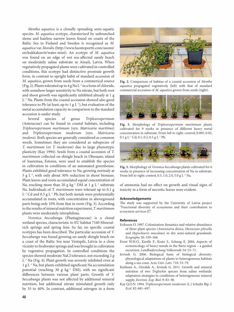
Mentha aquatica is a clonally spreading semi-aquatic species. M. aquatica ecotype, charaterized by unbranched stems and hairless narrow leaves found on coasts of the Baltic Sea in Finland and Sweden is recognized as M. aquatica var. litoralis (http://www.luontoportti.com/suomi/en/kukkakasvit/water-mint). An ecotype of M. aquatica was found on an edge of wet sea-affected sandy beach on moderately saline substrate in Ainaži, Latvia. When vegetatively propagated plants were cultivated in controlled conditions, this ecotype had distinctive prostrate growth form, in contrast to upright habit of standard accession of M. aquatica, grown from seeds from a commerical source (Fig. 2). Plants tolerated up to 4 g Na L–1 in a form of chloride, with somehow larger sensitivity to Na nitrate, but both root and shoot growth was significantly inhibited already at 1 g L–1 Na. Plants from the coastal accession showed also good tolerance to Pb (at least, up to 1 g L–1), but evaluation of the metal accumulation capacity in comparison to the standard accession is under study.
Several species of genus Tripleurospermum (Asteraceae) can be found in coastal habitats, including Tripleurospermum martimum (syn. Matricaria maritima) and Tripleurospermum inodorum (syn. Matricaria inodora). Both species are generally considered as common weeds. Sometimes they are considered as subspecies of T. maritimum (or T. inodorum) due to large phenotypic plasticity (Kay 1994). Seeds from a coastal accession of T. maritimum collected on shingle beach in Ohesaare, island of Saaremaa, Estonia, were used to establish the species in cultivation in conditions of an automated greenhouse. Plants exhibited good tolerance to Na, growing normaly at 5 g L–1, with only about 30% reduction in shoot biomass. Plant leaves and roots accumulated equial concentration of Na, reaching more than 20 g kg–1 DM at 1 g L–1 substrate Na. Individuals of T. maritimum were tolerant up to 0.1 g L–1 Cd and 0.5 g L–1 Pb, but both metals were preferentially accumulated in roots, with concentration in aboveground parts being only 10% from that in roots (Fig. 3). According to the results of mineral nutrition experiment, T. maritimum plants were moderately nitrophilous.
Veronica beccabunga (Plantaginaceae) is a clonal wetland species, characteristic to EU habitat 7160 Mineral-rich springs and spring fens. So far, no specific coastal ecotypes has been described. The particular accession of V. beccabunga was found growing on sandy shingle beach on a coast of the Baltic Sea near Ventspils, Latvia in a close vicinity to freshwater springs and was brought in cultivation by vegetative propagation. In controlled conditions the species showed moderate NaCl tolerance, not exceeding 2 g L–1 Na (Fig. 4). Plant growth was severely inhibited even at 1 g L–1 Na, but plants exhibited significant Na accumulation potential (reaching 30 g kg–1 DM), with no significant differences between various plant parts. Growth of V. beccabunga plants was not affected by additional mineral nutrition, but additional nitrate stimulated growth only by 35 to 40%. In contrast, additional nitrogen in a form
Fig. 2. Comparison of habitus of a coastal accession of Mentha aquatica propagated vegetatively (left) with that of standard commercial accession of M. aquatica grown from seeds (right).
Fig. 3. Morphology of Tripleurospermum maritimum plants cultivated for 9 weeks in presence of different heavy metal concentration in substrate. From left to right: control; 0.005, 0.02, 0.1 g L–1 Cd; 0.1, 0.2, 0.5 g L–1 Pb.
Fig. 3. Morphology of Veronica beccabunga plants cultivated for 6 weeks in presence of increasing concentration of Na in substrate. From left to right: control, 0.5, 1.0, 2.0, 5.0 g L–1 Na.
of ammonia had no effect on growth and visual signs of toxicity in a form of necrotic leaves were evident.
AcknowledgementsThe study was supported by the University of Latvia project “Functional diversity of ecosystems and their contribution to ecosystem services II”.
ReferencesEriksson O. 1997. Colonization dynamics and relative abundance
of three plant species (Antennaria dioica, Hieracium pilosella and Hypochoeris maculata) in dry semi-natural grasslands. Ecography 20: 559–568.
Ernst W.H.O., Knolle F., Kratz S., Schung E. 2004. Aspects of ecotoxicology of heavy metals in the Bartz region – a guided excursion. Landbaiforschung Volkenrode 54: 53–71.
Ievinsh G. 2006. Biological basis of biological diversity: physiological adaptations of plants to heterogeneous habitats along a sea coast. Acta Univ. Latv. 710: 53–79.
Karlsons A., Osvalde A., Ievinsh G. 2011. Growth and mineral nutrition of two Triglochin species from saline wetlands: adaptation strategies to conditions of heterogeneous mineral supply. Environ. Exp. Biol. 9: 83–90.
Kay Q.O.N. 1994. Tripleurospermum inodorum (L.) Schultz Bip. J. Ecol. 82: 681–697.

49
Does sand burial promote growth of dune-forming grass species Leymus arenarius and Ammophila arenaria?
Gederts Ievinsh*, Una Andersone-Ozola
Department of Plant Physiology, Faculty of Biology, University of Latvia, Jelgavas 1, Rīga LV–1004, Latvia*Corresponding author, E-mail: [email protected]
Key words: dune-forming species, growth stimulation, sand burial.
Environmental and Experimental Biology (2020) 18: 49–50 Abstract of the 78th Scientific Conference of the University of Latvia
Sand burial is one of the environmental factors on coastal sand dune habitats with significant effect both at the level of distribution of plant species as well as plant physiological status (Ievinsh 2006). Many sand dune species show extreme tolerance to burial by sand, including Alyssum montanum subsp. gmelinii, Honckenya peploides, Lathyrus japonicus subsp. maritimus, Linaria loeselii, being able to flower and bear viable seed even in conditions of almost complete burial. Two distinctive growth habits of plants with particular efectiveness in dune formation process have been described, e.g. true clonal species with potentially unlimited growth in a form of horizontal rhizomes, and species exhibiting induced clonality in a form of vertical rhizome or stem growth. Several grass species are so called dune-building species, showing not only high tolerance to sand accretion but also significant growth stimulation in conditions of sand burial. The aim of the present study was to compare growth responses to sand burial of two coast-specific dune-building grass species from the dunes of the Baltic sea, Leymus arenarius and Ammophila arenaria.
Plants were propagated by seeds collected in natural coastal habitats and further cultivated in a mixture of garden soil and quartz sand for two weeks in an automated greenhouse. For L. arenarius, four burial depths were used, at 7, 13, 21 and 31% intensity relative to plant height (Fig. 1). For A. arenaria, five burial depths were used, at 13, 23,
37, 46 and 60% intensity (Fig. 3). Changes in plant height were compared relative to unburied control (0%), but for A. arenaria, two additional (more shaded) controls were used, with extended containers at 0% and 23%. Burial was performed by dry quartz sand as a single treatment. After that, L. arenarius plants were cultivated for 9 weeks and A. arenaria plants for 11 weeks.
Shoot elongation of L. arenarius plants following burial was rapid, but timing of growth stimulation depended on burial depth (Fig. 2). The fastest growth response was evident for 7 and 13% treatments, followed by 21% treatment. In the case of 21% burial, first stimulation phase was short and was followed by a second phase with maximum growth at 4th week. At the same time there was a peak of growth stimulation of most intensively buried plants (31%), which extended for the next two weeks. Plant height at the end of the experiment was significanly larger in 13 to 31% burial treatments, but there was no dependence of the height on
Fig. 1. Morphology of Leymus arenarius plants buried by sand to different depths expressed as % from seedling height at the time of burial. From left to right: control (0%), 7%, 13%, 21%, 31%.
Fig. 2. Changes in relative growth increase of Leymus arenarius plants buried by sand to different depths during sultivation period. Data are means from 10 replications.

50
burial depth. In contrast, root mass linearly decreased with increase in burial depth, but total dry matter accumulation in shoots did not increase. However, there was a relative mass reallocation to leaf sheaths (significant at 13 to 31% burial intensity) and terminal leaf (significant at 21 and 31% burial intensity). In addition, number of leaves decreased at 21 to 31% burial intensity.
There were no coordinated burial-depth dependent growth stimulation for leaves of A. arenaria after sand burial, as there were extremely large differences of growth responses at the level of individual plants. Final shoot height increased linearly with increasing burial depth up to 46% intensity. Number of individual tillers significantly
decreased linearly with increasing burial depth only at 46 and 60% burial. Dry mass of shoots was significanly higher at 13% intensity, but significant decrease was evident at 60% intensity (Fig. 4). In contrast, root mass increased at 13% burial intensity and from 37 to 60% intensity. Buried plants allocated relatively larger part of resources in buried roots and burried shoots, but relative mass of roots in initial substrate gradually decreased.
Two different developmental strategies of the two functionally related dune-building grass species following sand burial are evident. While individuals of L. arenarius translocate resources from roots to leaf sheaths and terminal leaves, individuals of A. arenaria accumulate resources in roots and stems of the buried zone. These differences could be related to different clonal propagation strategies of the two species. A. arenaria plants are able to develop vertically expanding rhizomes, resulting in dense clonal expnasion, while L. arenarius plants rely on horizontal spread of rhizomes (Pavlik 1983; Reijers et al. 2020).
AcknowledgementsThe study was supported by the University of Latvia project “Functional diversity of ecosystems and their contribution to ecosystem services II”.
ReferencesIevinsh G. 2006. Biological basis of biological diversity:
physiological adaptations of plants to heterogeneous habitats along a sea coast. Acta Univ. Latv. 710: 53–79.
Pavlik B.M. 1983. Nutrient and productivity relations of the dune grasses Ammophila arenaria and Elymus mollis. III. Spatial aspects of clonal expansion with reference to rhizome growth and the dispersal of buds. Bull. Torrey Bot. Club 110: 271–279.
Reijers V.C., Lammers C., de Rond A.J.A., Hoetjes S.C.S., Lamers L.P., van der Heide T. 2020. Resilience of beach grasses along a biogeomorphic successive gradient: resource availability vs. clonal integration. Oecologia 192: 201–212.
Fig. 3. Morphology of Ammophila arenaria plants buried by sand to different depths expressed as % from seedling height at the time of burial. From left to right: control (0%), shaded control (0%), 13%, 23%, shaded control 25%, 37%, 46%, 60%.
Fig. 4. Effect of burial depth on dry mass of shoots and roots of Ammophila arenaria plants. Data are means from 10 replications.

51
The composition and quality of European eel Anguilla anguilla stock in Lake Rāznas
Jānis Bajinskis*, Ēriks Aleksejevs, Zanda Ozoliņa, Dzintars Začs
Institute of Food Safety, Animal Health and Environment “BIOR”, Lejupes 3, Riga LV–1076, Latvia*Corresponding author, E-mail: [email protected]
Key words: eel quality, European eel, Lake Rāznas, stock composition.
Environmental and Experimental Biology (2020) 18: 51–52 Abstract of the 78th Scientific Conference of the University of Latvia
Lake Razna ranks second in Latvia in terms of average eel landings in the last decade, but the commertial productivity of the eel in this lake is lower than that of Lake Usmas, Lake Cirmas and Lake Sīvers. Eel commertial productivity in this lake in time period from 1950 to 2018 ranged from 0.01 to 4.3 kg ha–1. Particularly intense eel fishing took place in the 1990-ies. Changes in eel landing volume in Lake Rāznas are mainly related to changes in fishing effort and the efficiency of the restocking of eel.
Nowadays, Lake Rāzna is not accessible to eel natural migration due to multiple hydroelectric power stadions and other migration obstacles. The eels were periodically released in Lake Rāznas from 1925 to 2005 (Fig. 1).
In Lake Rāznas in the Institute BIOR studies eels have been found from 2006 to 2019. Eels sampled with electrofishing, gill nets and fyke nets. According to the results of control fishing conducted in Lake Rāznas and Rēzekne River in 2019, the eel stock at present is mainly comprised of 14- to 17-year-old eels, which corresponds to the restocking in 2002 and 2005. The dominant age group was 14-year-old eels (age determined using etched and colored otolith thin sections), which accounted for 80% of the total control landings.
As in 2019, prior control fishing landings in 2010 were dominated by eel restocked in 2002 and 2005, but also some eel released in 1999 and 1995 were found, whereas in control fishing in 2018, only eels from 2005 release were recorded.
The average eel growth rate observed in this study (Fig.
2) is not significantly different from the results obtained at Lake Rāznas in 1971 by Volkova and Tarkač (1971).
In the control fishing carried out in 2019, the length of eel caught ranged from 42.5 to 45.5 cm for males, but from 45.5 to 100 cm for females, while body weights ranged from 91 to 116 g for males and 109 to 1997 g for females. As evidenced by the results of this study, in Lake Rāznas eel reach minimum commercial length in 5 to 7 years. The average growth rate of female eels has been higher than that of males. Ninety-six of the eel caught were females at different stages of development. Only two of the eels caught in 2019 corresponded to the silver eel stage and were ready for spawning migration, the rest of the eels were resident or pre-migratory.
Eel is a long-lived benthic carnivorous fish with a wide diet range that carries a high risk of bioaccumulation of environmental pollution. The aims of this study were to evaluate quality of eel in Lake Rāznas and to evaluate the feasibility and effectiveness of transporting eel from Lake Rāznas to the waters accessible for migration, the so-called “trap and transport”, because several studies indicate that in many parts of Europe the quality of eel is low and this is an important factor influencing migration and spawning success.
Chemical analyses of 30 eels migrating downstream from Lake Rāznas in 2019 were made. Inductively coupled plasma mass spectrometry was used for elemental concentration
Fig. 1. Eel landings in fisheries and restocked eel amounts in Lake Rāznas 1950 – 2018.
Fig.2. Eel growth progression in Lake Rāznas based on control fishing data for 2010, 2018 and 2019 (n = 162).
52
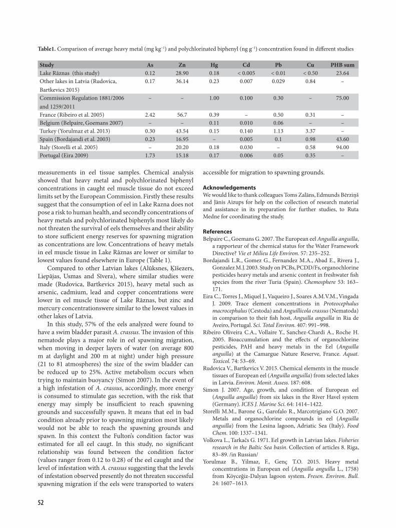
measurements in eel tissue samples. Chemical analysis showed that heavy metal and polychlorinated biphenyl concentrations in caught eel muscle tissue do not exceed limits set by the European Commission. Firstly these results suggest that the consumption of eel in Lake Razna does not pose a risk to human health, and secondly concentrations of heavy metals and polychlorinated biphenyls most likely do not threaten the survival of eels themselves and their ability to store sufficient energy reserves for spawning migration as concentrations are low. Concentrations of heavy metals in eel muscle tissue in Lake Rāznas are lower or similar to lowest values found elsewhere in Europe (Table 1).
Compared to other Latvian lakes (Alūksnes, Ķīšezers, Liepājas, Usmas and Sīvera), where similar studies were made (Rudovica, Bartkevics 2015), heavy metal such as arsenic, cadmium, lead and copper concentrations were lower in eel muscle tissue of Lake Rāznas, but zinc and mercury concentrationswere similar to the lowest values in other lakes of Latvia.
In this study, 57% of the eels analyzed were found to have a swim bladder parasit A. crassus. The invasion of this nematode plays a major role in eel spawning migration, when moving in deeper layers of water (on average 800 m at daylight and 200 m at night) under high pressure (21 to 81 atmospheres) the size of the swim bladder can be reduced up to 25%. Active metabolism occurs when trying to maintain buoyancy (Simon 2007). In the event of a high infestation of A. crassus, accordingly, more energy is consumed to stimulate gas secretion, with the risk that energy may simply be insufficient to reach spawning grounds and successfully spawn. It means that eel in bad condition already prior to spawning migration most likely would not be able to reach the spawning grounds and spawn. In this context the Fulton’s condition factor was estimated for all eel caugt. In this study, no significant relationship was found between the condition factor (values ranger from 0.12 to 0.28) of the eel caught and the level of infestation with A. crassus suggesting that the levels of infestation observed presently do not threaten successful spawning migration if the eels were transported to waters
accessible for migration to spawning grounds.
AcknowledgementsWe would like to thank colleagues Toms Zalāns, Edmunds Bērziņš and Jānis Aizups for help on the collection of research material and assistance in its preparation for further studies, to Ruta Medne for coordinating the study.
ReferencesBelpaire C., Goemans G. 2007. The European eel Anguilla anguilla,
a rapporteur of the chemical status for the Water Framework Directive? Vie et Milieu Life Environ. 57: 235–252.
Bordajandi L.R., Gomez G., Fernandez M.A., Abad E., Rivera J., Gonzalez M.J. 2003. Study on PCBs, PCDD/Fs, organochlorine pesticides heavy metals and arsenic content in freshwater fish species from the river Turia (Spain). Chemosphere 53: 163–171.
Eira C., Torres J., Miquel J., Vaqueiro J., Soares A.M.V.M., Vingada J. 2009. Trace element concentrations in Proteocephalus macrocephalus (Cestoda) and Anguillicola crassus (Nematoda) in comparison to their fish host, Anguilla anguilla in Ria de Aveiro, Portugal. Sci. Total Environ. 407: 991–998.
Ribeiro Oliveira C.A., Vollaire Y., Sanchez-Chardi A., Roche H. 2005. Bioaccumulation and the effects of organochlorine pesticides, PAH and heavy metals in the Eel (Anguilla anguilla) at the Camargue Nature Reserve, France. Aquat. Toxicol. 74: 53–69.
Rudovica V., Bartkevics V. 2015. Chemical elements in the muscle tissues of European eel (Anguilla anguilla) from selected lakes in Latvia. Environ. Monit. Assess. 187: 608.
Simon J. 2007. Age, growth, and condition of European eel (Anguilla anguilla) from six lakes in the River Havel system (Germany). ICES J. Marine Sci. 64: 1414–1422.
Storelli M.M., Barone G., Garofalo R., Marcotrigiano G.O. 2007. Metals and organochlorine compounds in eel (Anguilla anguilla) from the Lesina lagoon, Adriatic Sea (Italy). Food Chem. 100: 1337–1341.
Volkova L., Tarkačs G. 1971. Eel growth in Latvian lakes. Fisheries research in the Baltic Sea basin. Collection of articles 8. Riga, 83–89. /in Russian/
Yorulmaz B., Yilmaz, F., Genç T.O. 2015. Heavy metal concentrations in European eel (Anguilla anguilla L., 1758) from Köyceğiz-Dalyan lagoon system. Fresen. Environ. Bull. 24: 1607–1613.
Table1. Comparison of average heavy metal (mg kg–1) and polychlorinated biphenyl (ng g–1) concentration found in different studies
Study As Zn Hg Cd Pb Cu PHB sumLake Rāznas (this study) 0.12 28.90 0.18 < 0.005 < 0.01 < 0.50 23.64Other lakes in Latvia (Rudovica, Bartkevics 2015)
0.17 36.14 0.23 0.007 0.029 0.84 –
Commission Regulation 1881/2006 and 1259/2011
– – 1.00 0.100 0.30 – 75.00
France (Ribeiro et al. 2005) 2.42 56.7 0.39 – 0.50 0.31 –Beļgium (Belpaire, Goemans 2007) – – 0.11 0.010 0.06 – –Turkey (Yorulmaz et al. 2013) 0.30 43.54 0.15 0.140 1.13 3.37 –Spain (Bordajandi et al. 2003) 0.23 16.95 – 0.005 0.1 0.98 43.60Italy (Storelli et al. 2005) – 20.20 0.18 0.030 – 0.58 94.00Portugal (Eira 2009) 1.73 15.18 0.17 0.006 0.05 0.35 –

53
Analysis of lipopeptides produced by Bacillus subtilis
Māris Seņkovs1*, Zane Bērziņa2, Vizma Nikolajeva1, Ilva Nakurte2
1Faculty of Biology, University of Latvia, Jelgavas 1, Riga LV–1004, Latvia2Faculty of Chemistry, University of Latvia, Jelgavas 1, Riga LV–1004, Latvia*Corresponding author, E-mail: [email protected]
Key words: Bacillus subtilis, chromatography-mass spectrophotometry, lipopeptides.
Environmental and Experimental Biology (2020) 18: 53 Abstract of the 78th Scientific Conference of the University of Latvia
The lipopeptides are compounds formed from lipid and peptide residues that are most often derived from bacterial cultures. They may be isolated as metabolites by microorganisms such as Pseudomonas, Bacillus, Streptomyces (Waalia et al. 2015). Several types of lipopeptide groups are distinguished: surfactins, iturines, fengicins, etc., which differ in their chemical and physical properties (Meena et al. 2015). Such compounds have intrinsic properties that can be widely used in biotechnological and pharmaceutical applications. The lipopeptides are also widely used in agriculture as plant protection products and have antifungal and antibacterial activity against undesirable soil microorganisms (Alajlani et al. 2016).
The aim of this work was to perform the identification of surfactin and inturin lipopeptides produced by two strains of Bacillus subtilis by chromatography-mass spectrophotometry method.
Two strains of B. subtilis (MSCL 897 and MSCL 1441) were cultivated for the study, in two different growth media (nutrient broth medium and Miller-Hinton medium). Cultivation was carried out in the Department of Microbiology and Biotechnology, Faculty of Biology, University of Latvia. Two different sample
preparation methods were used for initial lipopeptide extraction from bacterial cultivation liquid (truncated extraction – acid precipitation of peptides for 30 min at 4 °C; prolonged extraction for 12 h at 4 °C). Agilent1290 Infinity series UHPLC liquid chromatography equipment, which was connected to an Agilent 6230 TOF LC / MS mass spectrometer, was used for chromatography-mass spectrophotometry at the Department of Physical Chemistry, University of Latvia. Chromatographic analysis was performed under two gradient conditions (Gradient 1 for surfactin lipopeptide analysis; Gradient 2 for iturine and fengycin lipopeptide analysis) using an Extend column. The column temperature was maintained at 35 °C. The mobile phase consisted of formic acid in deionized water and formic acid in acetonitrile at a flow rate of 0.3 mL min–1.
By extraction involving acid precipitation of peptides and chromatography-mass spectrophotometry, it is possible to isolate and identify various lipopeptides produced by B. subtilis. The B. subtilis products studied were found to contain surfactin (Fig. 1) and fengicin lipopeptides, but no iturine lipopeptides were detected. B. subtilis strains and culture medium do not significantly affect the chemical profile of the lipopeptides.
By taking mass spectra and analyzing individual molecules with certain values of their m/z (scanning range), it is possible to determine the structure of an unknown lipopeptide based on the basic principles of fragmentation and information available in the literature. Prolonged extraction provided 7 to 82% increase in the quantitative result of the signal, resulting in an extended 39% more efficient extraction than the truncated one.
ReferencesAlajlani M., Shiekh A., Hasnain S., Brantner A. 2016. Purification
of bioactive lipopeptides produced by Bacillus subtilis strain BIA. Chromatographia 79: 1527–1532.
Meena K.R., Kanwar S.S. Lipopeptide as the antifungal and antibacterial agents: applications in food safety and therapeutics. BioMed Res. Int. 2015: 473050.
Waalia N.K., Cameotra S.S. 2015. Lipopeptides: biosynthesis and applications. J. Microb. Biochem. Technol. 7: 103–107.
Fig. 1. Chemical profile of lipopeptides of surfactin group in Bacillus subtilis LMKK 897 sample cultured on Miller-Hinton medium.

54
Problems of protection of the freshwater mussels in Latvia and the world
Mudīte Rudzīte*, Māris Rudzītis
Museum of the University of Latvia, Raiņa bulvāris 19, Rīga LV–1586, Latvia*Corresponding author, E-mail: [email protected]
Key words: assessment, freshwater mussels, indicators; Latvia, management, protection statuss, threats, Unionida.
Environmental and Experimental Biology (2020) 18: 54 Abstract of the 78th Scientific Conference of the University of Latvia
A new list of protected molluscs in Latvia – the New Red List – has been prepared (Rudzīte et al. 2018). It includes three species of Unionida: Margaritifera margaritifera in the first category of the IUCN Red List of Threatened Animals (IUCN 2017), Unio crassus in the second and Pseudanodonta complanata in the third. Another four Unionida species occurring in Latvia (Anodonta anatina, Anodonta cygnaea, Unio pictorum, Unio tumidus) are included in the LC category (least concern, there is no need for special protection measures for these species; however, the dynamics of their populations should be monitored).
The main factors affecting the status of these species in Latvia are fragmentation of natural habitats that restrict populations and does not allow the species to spread further; decrease and degradation of natural habitat areas as a result of the human activity or ecological succession; decrease and degradation of natural habitats due to spreading of an expansive species; water pollution due to human activities, intentional use in the interests of humans (Rudzīte et al. 2018)
Freshwater mussels are experiencing a global decline, and establishing efficient conservation strategies is critical. Here, we provide a global assessment of freshwater mussel status, develop a portfolio of research priorities, and suggest a framework to prioritize conservation actions. Conservation priorities can vary widely, ranging from avoiding imminent extinction to maintaining long-term monitoring efforts. To address this, we developed a research priorities portfolio using a hierarchical trait framework, using both internal traits as indicators of organism or population status, and external traits encompassing environmental conditions and threats such as water quality and habitat modification. Internal traits are useful in monitoring, but external traits are necessary when internal traits suggest that conservation status is inadequate, such as when a population is declining. This approach can serve as a guide for conservation status assessments prior to the establishment of priority species and conservation actions (Ferreira-Rodríguez et al. 2019).
Latvia has a problem that is not typical elsewhere in the world. The excessive proliferation of beaver Castor fiber also poses a threat to habitat of mussels. Beavers destroy habitats of mussels in small rivers, reducing the survival possibilities of populations (Rudzite 2005; Rudzite et al. 2017). In some places in Eastern Europe, beavers have been reintroduced into areas where they have disappeared.
In the framework of the former USSR forest management, beaver-friendly living conditions were promoted. But now they numbers have grown significantly due to the lack of natural predators (Balodis 1990).
ReferencesBalodis M. 1990. The beaver. Biology and the Importance in the
Economy. Rīga. 271 p. /in Russian/Ferreira-Rodríguez N., Akiyama Y.B., Aksenova O.V., Araujo
R., Barnhart M.C., Bespalaya Y.V., Bogan A.E., Bolotov I.N., Budha P.B., Clavijo C., Clearwater S.J., Darrigran G., Do V.T., Douda K., Froufe E., Gumpinger C., Henrikson L. , Humphrey C.L., Johnson N.A., Klishko O., Klunzinger M.W., Kovitvadhi S., Kovitvadhi U., Lajtner J., Lopes-Lima M., Moorkens E.A., Nagayama S., Nagel K.-O., Nakano M., Negishi J.N., Ondina P., Oulasvirta P., Prié V., Riccardi N., Rudzīte M., Sheldon F., Sousa R., Strayer D.L., Takeuchi M., Taskinen J., Teixeira A., Tiemann J.S., Urbańska M., Varandas S., Vinarski V.M., Wicklow B.J., Zając T., Vaughn C.C. 2019. Research priorities for freshwater mussel conservation assessment. Biol. Conserv. 231: 77–87.
IUCN. 2017. The IUCN Red List of Threatened Species. Version 2017-3. Downloaded on 16 March 2018. http://www. iucnredlist.org
Rudzīte M. 2005. Assessment of the condition of freshwater pearl mussel Margaritifera margaritifera (Linnaeus 1758) populations in Latvia. Acta Univ. Latv. 691: 121–128.
Rudzīte M., Boikova E., Dreijers E., Jakubāne I., Parele E., Pilāte D., Rudzītis M. 2018. The New Red List of the molluscs of Latvia. Environ. Exp. Biol. 16: 55–59.
Rudzīte M., Rudzītis M., Birzaks J. 2017. The Populations of the Freshwater Pearl Mussel, Margaritifera margaritifera (Linnaeus, 1758), and the Thick Shelled River Mussel, Unio crassus Philipsson, 1788, in Latvia. Biol. Bull. 44: 99–107.

55
Free-living protozoa and Legionella spp. coexistence and identification in drinking water systems in Riga apartment houses
Artjoms Mališevs*, Svetlana Makarova, Genadijs Konvisers, Daina Pūle, Olga Valciņa, Lelde Grantiņa-Ieviņa
Institute of Food Safety, Animal Health and Environment “BIOR”, Lejupes 3, Riga LV–1076, Latvia*Corresponding author, E-mail: [email protected]
Key words: apartment houses, drinking water, Legionella, protozoa, water system.
Environmental and Experimental Biology (2020) 18: 55–56 Abstract of the 78th Scientific Conference of the University of Latvia
Legionella pneumophila is a causative agent of Legionnaires’ disease, while free-living protozoa (FLP) can provide shelter for Legionella spp. which may become intracellular pathogens. FLP is able to create biofilms in water pipes and can protect pathogenic bacteria from high temperatures, disinfectants, can serve as a reservoir for bacterial populations. The interaction of bacteria and protozoa can increase the resistance of microorganisms to antibiotics and their virulence (Muchesa et al. 2018).
The aim of this study was to investigate the coexistence of FLP and Legionella spp. in the drinking water supply systems of Riga apartment houses, identifying the main FLP genera and to detect FLP and Legionella spp. occurrence depending on various factors: water temperature, floor of the building, type of hot water supply method (individual water heaters or central supply system) address location (right or left bank of the river Daugava). Last point is important because that part of Riga which is at right bank have water supply from underground sources, but at left bank is using purified water from Daugava river.
Hot and cold drinking water samples (n = 59) were
tested for the presence of L. pneumophila according to the standard (ISO 11731: 2017) method and real time PCR, and the presence of free-living protozoa by isolating and cultivating on Page’s Amoeba saline with peptone yeast extract glucose (Vaerewijck et al. 2010). FLP genus were identified by microscopy and molecular biology methods with Acanthamoeba (Schroeder et al. 2001), Vahlkampfidae, Amoebaidae (Calvez et al. 2012) and Hartmanella (Solgi et al. 2012) specific primers. The average cold water temperature of analyzed samples was 16.61 °C, hot water 48.39 °C.
FLP were detected in 31 of 59 (52.5%) water samples, while L. pneumophila or Legionella rubrilucens were detected in 30 (50.8%) water samples.
Of the Legionella spp. positive cold water samples, 30% were also positive for FLP (9 samples) and 20% (6 samples) of the hot water samples. Using Pearson correlation test it was found that correlations between legionella quantity (CFU L–1) and cold or hot water temperature are weak, but there was moderately strong correlation with the floor: the higher was the floor of building, the larger was the
Fig. 1. Correlation between floor of building and Legionella spp. quantity (CFU L–1) (A) and correlation between floor of building and Ct at real time PCR reaction (B)
56
legionella quantity (Fig. 1A) and lower Ct of the real time PCR reaction (Fig. 1B; r = 0.42 un r = –0.53). This can be connected with water circulation and lower water pressure in pipes at higher building floors where biofilms can be formed more easily.
From eight locations with individual water heaters 50% were positive for legionella (three samples from hot water), but five samples were FLP positive.
In total, 80% of water samples from left bank of river Daugava and 52% from right bank were Legionella-positive. FLP positive were 70 and 63% of samples, respectively. From these results we can see that at the left bank, where water supply is proceeded from Daugava river, there are more positive samples.
From the kitchen, 47.06% of the samples were Legionella spp. positive and 58.82% FLP positive, from the shower 52.38 and 50%, respectively. Moreover, 43.33 and 56.67% of cold, 58.62 and 48.28% of hot water were Legionella spp. and FLP positive, respectively. In cold water samples are more FLP and Legionella spp. coexistence occurences than in hot water samples (30 vs 20%). The free-living protozoa genera Acanthamoeba, Vahlkampfia and Hartmanella (Vermamoeba) were identified.
The implementation of water treatment and disinfection strategies for the inactivation of protozoa should also improve the control of pathogenic microorganisms (Lasjerdi et al. 2011). From previous studies in BIOR it is known that mostly galvanized steel pipes are used in Riga. From study of Jing et al. 2018 it can be seen that biofilm
formation is the slowest in pipes from stainless steel. This is not a cheap material, so polyethylene and polyvinyl chloride with quite similar effect can be used as well.
ReferencesMuchesa P., Leifels M., Jurzik L., Barnard T.G., Bartie C. 2018.
Detection of amoeba-associated Legionella pneumophila in hospital water networks of Johannesburg. South African J. Infect. Dis. 33: 72–75.
Schroeder J.M, Booton G.C, Hay J. 2001. Use of subgenic 18S ribosomal DNA PCR and sequencing for genus and genotype identification af Acanthamoebae from humans with keratitis and from sewage sludge. J. Clin. Microbiol. 39: 1903–1911.
Calvez T., Trouilhe M.C, Humeau O. 2012. Detection of free-living amoebae by using multiplex quantitative PCR. Mol. Cell. Probes 26: 116–120.
Solgi R., Niyyati M., Haghihgi A., Mojarad N.E. 2012 Occurence of thermotolerant Hartmanella vermiformis and Naegleria spp in hot springs of Ardebil Province, northwest Iran. Iranian J. Parasitol. 7: 47–52.
Vaerewijck M.J.M., Sabbe K., Hende J., Bare J., Houf K. 2010. Sampling strategy, occurence and diversity of free-living protozoa in domestic refrigerators. J. Appl. Microbiol. 109: 1556–1578.
Jing H., Sahle-Demessie E., Sorial A.G. 2018. Inhibition of biofilm growth on polymer -MWCNTs composites and metal surfaces. Sci. Total Environ. 633: 167–178.
Lasjerdi Z., Niyyati M., Haghighi A., Shahabi S., Biderouni F.T., Taghipour N., Eftekhar M., Mojarad E.N. 2011. Potentially pathogenic free-living amoebae isolated from hospital wards with immunodeficient patients in Tehran, Iran. Parasitol. Res. 109: 575–580.

57
Use of baker’s yeast purine auxotrophs for adenine quantification
Jana Kassaliete*, Anitra Zīle, Zane Ozoliņa, Agnese Kokina, Jānis Liepiņš
Institute of Microbiology and Biotechnology, University of Latvia, Jelgavas 1, Riga LV–1004, Latvia*Corresponding author, E-mail: [email protected]
Key words: adenine, auxotrophy, optical density, Saccharomyces cerevisiae.
Environmental and Experimental Biology (2020) 18: 57–58 Abstract of the 78th Scientific Conference of the University of Latvia
Purines and pyrimidines are nitrogen bases found in DNA and RNA nucleotides and in energy carriers – ATP and GTP. They are also part of NAD, coenzyme-A and other cofactors. Cells can obtain nucleotides in two ways: salvage and de novo biosynthesis pathway. Purine synthesis is strictly regulated and highly conserved in all organisms (Agmon et al. 2020). However, some parasites and cancer cells have lost the ability to synthesise their own adenine (Kao, Puck 1975; Frame et al. 2015). Any gene (ADE1 to ADE8) knockout in the adenine synthesis pathway leads to adenine auxotrophy (Armitt, Woods 1970). Auxotrophs are organisms that require specific compounds in order to ensure their uninterrupted growth (Russel 1994). Auxotrophic organisms can be used to quantify specific compounds, for example, quantification of B9 and B12 vitamins are done by specific Lactobacillus bacteria strains auxotrophic for intermediates of folate or cobalamin synthesis (reviewed by Eitenmiller et al. 2008).
In our study a microbiological assay for adenine quantification was developed in two budding yeast
Saccharomyces cerevisiae strain backgrounds – W303 2832-1B MatA and CEN.PK 113-7D MatA using five purine synthesis pathway gene knockouts: ade4, ade8, ade6, ade2 and ade1. Knockouts were generated by using linear KanMX cassettes with flanking homologous regions, as described (Janke et al. 2004). Cell cultures were grown in synthetic dextrose (SD) media (Miller et al. 2017), with an additional 100 mg L–1 of adenine. Cultures were grown to an exponential growth phase (2 × 10⁷ cells mL–1). Afterwards cells were washed with sterile water to remove media and excess adenine. Cells were reinoculated in 96-well microplates in SD media with adenine in different concentrations: 36.6, 18.3, 9.15, 4.6, 2.3, 1.15, 0.6 mg L–1 for W303 strain and 20, 5, 2.5, 1.25, 0.6, 0.3 mg L–1 for CEN.PK strain.
Culture growth was measured as optical density (OD) at 590 nm wavelength in microplate reader Tecan Infinite M200 PRO (Austria). Growth curves obtained from the microplate reader were transformed into biomass growth speed (Fig. 1A). Thus the maximum speed corresponds
Fig. 1. W303 ade6 rate of increase of biomass in adenine concentrations in the media (A). Adenine quantification standard lines (B).

58
to the end of exponential growth phase and is a hallmark of purine depletion, as all other media compounds were added in surplus. Growth curves in cultures with adenine concentration of 10 mg L–1 or more overlapped. It could not be determined if growth in these cases stopped due to depletion of adenine, glucose or due to saturation of OD signal. If less than 1 mg L–1 adenine was added, noise of signal was too large to accurately determine the end of the exponential phase. From this data adenine quantification standard lines were created (Fig. 1B). Adenine quantification standard lines show a good fit of the regression model (R2 > 0.95) in auxotrophs ade4, ade1 and ade6 in W303 strain; ade2 and ade1 in CEN.PK strain.
We conclude that yeast adenine auxotrophs can be used for adenine quantification, based on their growth suspension in adenine limitation conditions if external adenine concentration is between 1 and 10 mg L–1. Budding yeast ade1 knockouts are best-suited for microbiological assays as biomass yield is not affected by different adenine concentrations and adenine quantification standard lines of these knockouts in two strain backgrounds have almost equal fit to regression model. Therefore, budding yeast ade1 knockout could be used to quantify extracellular purines.
AcknowledgementsThis study was supported by Latvian Council of Science, Project LZP-2018/2-0213.
ReferencesAgmon N., Temple J., Tang Z., Schraink T., Baron M., Chen J.,
Paolo Mita P., Martin J.A., Tu B.P., Yanai I., Fenyö D., Boeke J.D. 2020. Phylogenetic debugging of a complete human biosynthetic pathway transplanted into yeast. Nucl. Acids Res. 48: 486–499.
Armitt S., Woods R.A. 1970. Purine-excreting mutants of Saccharomyces cerevisiae: I. Isolation and genetic analysis. Gen. Res. 15: 7–17.
Eitenmiller R., Ye L., Landen W.O. 2008. Vitamin Analysis for the Health and Food Sciences. CRC Press, Taylor & Francis Group, New York, 637 p.
Frame I.J., Deniskin R., Arora A., Akabas M.H. 2015. Purine import into malaria parasites as a target for antimalarial drug development. Ann. N Y Acad. Sci. 1342: 19–28.
Janke C., Magiera M.M., Rathfelder N., Taxis C., Reber S., Maekawa H., Moreno-Borchart A., Doenges G., Schwob E., Schiebel E., Knop M. 2004. A versatile toolbox for PCR-based tagging of yeast genes: new fluorescent proteins, more markers and promoter substitution cassettes. Yeast 21: 947–962.
Kao F.T., Puck T. 1975. Mutagenesis and genetic analysis with Chinese hamster auxotrophic cell markers. Genetics 79: 343–352.
Miller A.W., Mitchell E., Befort C., Dunham M.J. 2017. Dunham Lab Ministat Manual. http://dunham.gs.washington.edu/2017MinistatManual.pdf
Russel P.J. 1994. Fundamentals of Genetics. New York, HarperCollins College Publishers, 527 p.

59
Callus culture of black elder (Sambucus nigra): initiation and basic chemical characterization
Anete Boroduske*
Department of Microbiology and Biotechnology, Faculty of Biology, University of Latvia, Jelgavas 1, Riga LV–1004, Latvia*Corresponding author, E-mail: [email protected]
Key words: anti-radical activity, black elder, callus culture, Sambucus nigra, total phenolic content..
Environmental and Experimental Biology (2020) 18: 59–60 Abstract of the 78th Scientific Conference of the University of Latvia
Limited success of chemically produced pharmaceuticals and consumer rising demand in environmentally friendly alternatives has renewed interest in plant-based substances as ingredients for pharmaceutical and cosmetic industries. Plant tissue culture-based products are viewed as an effective and interesting source of plant-derived substances with a potential for high added value product development (Barbulova et al. 2014). Recently a concept of plant cell culture-based food products also has been established (Nordlund et al. 2018). Black elderberry Sambucus nigra L. is a medicinal plant, traditionally well known for its antioxidative, antifever and antiviral properties (Ulbricht et al. 2014). More recent scientific studies have also proved skin antiageing (Lin et al. 2019), antiinflammatory and antidiabetic (Ho et al. 2017) activity of S. nigra extracts. Taking into account beneficial properties of S. nigra and technical advantages offered by plant tissue culture approach, S. nigra callus culture could become an alternative source of S. nigra-derived substances for production of cosmetic and pharmaceutical products. Previously in vitro cultures of S. nigra have been studied mainly with regard to propagation of horticultural material, therefore scientific literature mainly contains references to establishment of in vitro shoot cultures (Charlebois, Brassard 2013), leaving S. nigra callus culture establishment neglected. Here we report results of study testing different explant surface sterilization agents and plant growth regulator combinations for initiation and growth of S. nigra callus culture. In addition, preliminary basic characterization of chemical properties of S. nigra callus culture is reported.
In our study we tested six different explant surface sterilization treatments. For all treatments, after brief immersion in 70% ethanol, one of the following methods were used: (1) 15 min in 3% (w/v) calcium hypochlorite (CaOCl), (2) 30 min in 10% (v/v) domestic bleach containing <5% sodium hypochlorite, (3) 20 min in 1% (w/v) silver nitrate (AgNO3). For all treatments, explants were thoroughly rinsed in sterile distilled water for three times. Half of the explants from each sterilization treatment were directly plated on growth media but half were subjected to 4 h treatment with 5% (v/v) plant preservative mixture
(PPM). Shoot fragments with one to two buds were used as explants. Developing young leaves of established shoot in vitro cultures were used for callus initiation experiments. For initiation of S. nigra callusogenesis in vitro McCown woody plant solid medium containing 30 g L–1 sucrose, 0.2% (v/v) PPM and one of four different combinations of plant growth regulators were tested: thidiazuron (TDZ) 2 mg L–1 plus 1-naphtaleneacetic acid (NAA) 2 mg L–1; NAA 1 mg L–1 plus kinetin (KIN) 0.1 mg L-1, NAA 0.2 mg L-1 plus KIN 1 mg L–1, 2,4-dichlorophenoxyacetic acid (2,4-D) 0.2 mg L–1 plus KIN 1 mg L–1.
For basic chemical characterization of S. nigra callus cultures, total phenolic content and antiradical activity were assessed using Folin-Ciocalteu reagent-based assay (Zhang et al. 2006) and DPPH-based assay adjusted for multi-well test plates (Herald et al. 2012), respectively. Total phenolic content was expressed as mg gallic acid equivalents (GAE) per g dry mass (DM) of calli. Antiradical activity was quantified based on ascorbic acid calibration curve and expressed as ascorbic acid equivalents (AAE). Callus extracts for chemical characterization were derived by shaking freeze-dried callus material with 50% (v/v) ethanol for 24 h at 180 rpm at room temperature.
The lowest infection rate was achieved applying AgNO3 for surface sterilization of explants (Table 1). However, this treatment cannot be viewed as optimal for S. nigra in vitro culture establishment since the majority of explants were strongly oxidized and did not show any further development. Highest rate of explant survival was achieved using CaOCl.
Out of four tested growth regulator combinations the highest rate of S. nigra callus initiation (89% of plated explants showed at least some degree of callus formation) was achieved using 2,4-D 0.2 mg L–1 in combination with KIN 1 mg L–1. However, calli initiated by this combination of growth regulators were not vigorous, cells proliferated slowly and gradually after two months of growth started to suffer from browning despite of frequent transfer to fresh medium. Combination of TDZ 2 mg L–1 and NAA 2 mg L–1 yielded rapidly proliferating, soft, light green calli that continued to proliferate after four months in in vitro.

60
Morphological traits of established callus cultures suggest that calli initiated by combination of TDZ 2 mg L–1 and NAA 2 mg L–1 might be suitable for cell suspension culture establishment.
Total phenolic content of established callus cultures varied from 2.68 ± 0.07 mg GAE g–1 DM to 1.49 ± 0.14 mg GAE g–1 DM for cultures treated with TDZ 2 mg L–1, NAA 2 mg L–1 and 2,4-D 0.2 mg L–1, KIN 1 mg L–1, respectively. Antiradical activity of established callus cultures varied from 10.56 ± 1.69 mg AAE g DM for calli initiated by TDZ 2 mg L–1, NAA 2 mg L–1) to 8.57 ± 1.82 mg AAE g–1 DM for calli established on 2,4-D 0.2 mg L–1, KIN 1 mg L–1. In comparison, total phenolic content of ripe S. nigra berries of the mother plant significantly exceeded that of callus cultures and was estimated 11.67 ± 0.74 mg GAE g–1 DM, with 33.31 ± 2.07 mg AAE g–1 DM antiradical activity. However, more detailed, targeted chemical analysis of established callus cultures is required for assessment of potential value of S. nigra callus cultures for pharmaceutical applications.
AcknowledgementsThis research is performed as part of the project Nr.1.1.1.2/VIAA/2/18/256 Biotechnological solutions for control of target compounds in European elder Sambucus nigra in vitro cell cultures and in planta using endophytic microorganisms.
ReferencesBarbulova A., Apone F., Colucci G. 2014. Plant cell cultures as
source of cosmetic active ingredients. Cosmetics 1: 94–104.Charlebois D., Brassard N. 2013. Micropropagation of American
elderberry: Culture medium optimization and field performance. Acta Hortic. 1061: 175–182.
Herald T.J., Gadgil P., Tilley M. 2012. High-throughput micro plate assays for screening flavonoid content and DPPH-scavenging activity in sorghum bran and flour. J. Sci. Food Agric. 92: 2326–2331.
Ho G.T.T., Kase E.T., Wangensteen H., Barsett, H. 2017. Phenolic
Table 1. Effect of different explant surface sterilization treatments on infection and survival rate of Sambucus nigra shoot explants after four weeks in in vitro. n = 27 explants (shoot fragments with one to two buds) in each treatment. Domestic bleach contained <5% sodium hypochlorite. CaOCl, calcium hypochlorite; PPM, plant preservative mixture,
Sterilization treatment Infection rate (%)
Surviving explants (%)
15 min 3% CaOCl 62 4015 min 3% CaOCl followed by 4 h 5% PPM
48 33
30 min 10% domestic bleach 63 1130 min 10% domestic bleach followed by 4 h 5% PPM
48 29
20 min 1% AgNO3 44 2920 min 1% AgNO3 followed by 4 h 5% PPM
40 25
elderberry extracts, anthocyanins, procyanidins, and metabolites influence glucose and fatty acid uptake in human skeletal muscle cells. J. Agric. Food Chem. 65: 2677–2685.
Lin P., Hwang E., Ngo H.T., Seo S.A., Yi T.H. 2019. Sambucus nigra L. ameliorates UVB-induced photoaging and inflammatory response in human skin keratinocytes. Cytotechnology 71: 1003–1017.
Nordlund E., Lille M., Silventoinen P., Nygren H., Seppänen-Laakso T., Mikkelson A., Aura A.M., Heiniö R.L., Nohynek L., Puupponen-Pimiä R., Rischer H. 2018. Plant cells as food – A concept taking shape. Food Res. Int. 107: 297–305.
Ulbricht C., Basch E., Cheung L., Goldberg H., Hammerness P., Isaac R., Khalsa K.P.S., Romm A., Rychlik I., Varghese M., Weissner W. 2014. An evidence-based systematic review of elderberry and elderflower (Sambucus nigra) by the Natural Standard Research Collaboration. J. Diet. Suppl. 11: 80–120.
Zhang Q., Zhang J., Shen J., Silva A., Dennis D.A., Barrow C.J. 2006. A simple 96-well microplate method for estimation of total polyphenol content in seaweeds. .J Appl. Phycol. 18: 445–450.