Embed Size (px)
Citation preview

RESEARCH ARTICLE
Abundance and diversity of functional genes involvedin the degradation of aromatic hydrocarbons in Antarcticsoils and sediments around Syowa Station
C. Muangchinda & S. Chavanich & V. Viyakarn &
K. Watanabe & S. Imura & A. S. Vangnai & O. Pinyakong
Received: 23 May 2014 /Accepted: 13 October 2014 /Published online: 22 October 2014# Springer-Verlag Berlin Heidelberg 2014
Abstract Hydrocarbon catabolic genes were investigated insoils and sediments in nine different locations around SyowaStation, Antarctica, using conventional PCR, real-time PCR,cloning, and sequencing analysis. Polycyclic aromatic hydro-carbon ring-hydroxylating dioxygenase (PAH-RHD)-codinggenes from both Gram-positive and Gram-negative bacteriawere observed. Clone libraries of Gram-positive RHD geneswere related to (i) nidA3 of Mycobacterium sp. py146, (ii)pdoA of Terrabacter sp. HH4, (iii) nidA of Diaphorobactersp. KOTLB, and (iv) pdoA2 of Mycobacterium sp. CH-2,with 95–99 % similarity. Clone libraries of Gram-negativeRHD genes were related to the following: (i) naphthalenedioxygenase of Burkholderia glathei, (ii) phnAc ofBurkholderia sartisoli, and (iii) RHD alpha subunit of
uncultured bacterium, with 41–46 % similarity. Interestingly,the diversity of the Gram-positive RHD genes found aroundthis area was higher than those of the Gram-negative RHDgenes. Real-time PCR showed different abundance ofdioxygenase genes between locations. Moreover, the PCR-denaturing gradient gel electrophoresis (DGGE) profile dem-onstrated diverse bacterial populations, according to theirlocation. Forty dominant fragments in the DGGE profileswere excised and sequenced. All of the sequences belongedto ten bacterial phyla: Proteobacteria, Actinobacteria,Verrucomicrobia, Bacteroidetes, Firmicutes, Chloroflexi,Gemmatimonadetes, Cyanobacteria, Chlorobium, andAcidobacteria. In addition, the bacterial genus Sphingomonas,which has been suggested to be one of the major PAH de-graders in the environment, was observed in some locations.The results demonstrated that indigenous bacteria have thepotential ability to degrade PAHs and provided information tosupport the conclusion that bioremediation processes canoccur in the Antarctic soils and sediments studied here.
Keywords Antarctica . Syowa . PAHs . PCR-DGGE .
Real-time PCR . Dioxygenase . Diversity
Introduction
Antarctica is the most pristine and least populated continent inthe world. However, scientific operations have led to anaccumulation of hydrocarbon compounds in the Antarcticenvironment. Hydrocarbons, in the form of fuel oils, are usedbyAntarctica research stations for transportation and scientificoperations (Aislabie et al. 2004).
Syowa Station is a scientific research station established byJapan in 1957. It is located on East Ongul Island (69° 00′ S,39° 35′ E) in Antarctica. With an increase in constructions andhuman activities, the area around the stationmay have become
Responsible editor: Robert Duran
Electronic supplementary material The online version of this article(doi:10.1007/s11356-014-3721-y) contains supplementary material,which is available to authorized users.
C. Muangchinda :O. Pinyakong (*)Bioremediation Research Unit, Department ofMicrobiology, Facultyof Science, Chulalongkorn University, Bangkok 10330, Thailande-mail: [email protected]
S. Chavanich :V. ViyakarnReef Biology Research Group, Department of Marine Science,Faculty of Science, Chulalongkorn University, Bangkok 10330,Thailand
K. Watanabe : S. ImuraNational Institute of Polar Research, Tokyo 190-8518, Japan
A. S. VangnaiDepartment of Biochemistry, Faculty of Science, ChulalongkornUniversity, Bangkok 10330, Thailand
A. S. Vangnai :O. PinyakongCenter of Excellence on Hazardous Substance Management (HSM),Bangkok 10330, Thailand
Environ Sci Pollut Res (2015) 22:4725–4735DOI 10.1007/s11356-014-3721-y
contaminated with hydrocarbon compounds, such as polycy-clic aromatic hydrocarbons (PAHs). PAHs are the componentsof petroleum products that are highly persistent in the envi-ronment and have toxic, mutagenic, and carcinogenic effectson organisms (Yergeau et al. 2009).
Bioremediation by microorganisms is one of the technolo-gies for the cleanup of the Antarctic environment because it isquite safe and harmless (Aislabie and Foght 2010). The bio-degradation of PAHs in some Antarctic areas, such as atJubany Station, has been reported to be using culture-dependent methods (Ruberto et al. 2006). That study indicatedthat Antarctic microbial communities are capable of degradingPAHs and that the bioremediation process is possible in Ant-arctic environments. The genera Pseudomonas andSphingomonas have been identified as PAH degraders inAntarctic soils (Ma et al. 2006; Panicker et al. 2010). Furtherunderstanding of the diversity of the natural microbial com-munity and specific metabolic genes can help assess thebiodegradation potential of environments (Fernández-Luqueño et al. 2011). However, only culture-dependent ap-proaches may not be enough to estimate microbial diversity inthe environment since the majority of microorganisms aredifficult to be cultivated due to the lack of appropriate condi-tions and media (Amann et al. 1995). Therefore, molecularmethods relying in phylogenetically informative genes, suchas denaturing gradient gel electrophoresis (DGGE) and 16Sribosomal RNA (rRNA) gene-based clone libraries, have beenapplied to identify indigenous genera (Jurelevicius et al.2012b; Powell et al. 2003; Das and Kazy 2014). In addition,the biodegradation potential in environments can also beestimated using culture-independent methods targeting spe-cific metabolic genes. Ring-hydroxylating dioxygenases arekey enzymes that catalyze the first step of PAH degradationpathways (Peng et al. 2008). The genes encoding for the α-subunit of terminal dioxygenase have been widely used astarget genes to describe the PAH-degrading potential in dif-ferent environments (Jurelevicius et al. 2012a; Marcos et al.2009). Moreover, extradiol dioxygenases which are enzymesthat are essential for breaking down the common intermedi-ates in the aerobic bacterial degradation of aromatic com-pounds are also suitable to be used as markers to detect abroad range of aromatic-degrading bacteria (Suenaga et al.2009). Many studies have investigated extradiol dioxygenasegenes in polluted environments (Sipila et al. 2006; Junca andPieper 2004).
Knowledge of the PAH biodegradation potential in Antarc-tic soils is essential for the management of soils for bioreme-diation. This study aims to investigate the dominant bacteriaand the abundance as well as the diversity of genes involved inthe degradation of PAHs and hydrocarbons in Antarctic soiland sediment samples around the Syowa Station in order toassess the PAH biodegradation potential in Antarctic soils andsediments. This could provide information to support
bioremediation strategies for PAH-contaminated Antarcticsoils and sediments.
Materials and methods
Soil sampling and PAH analysis
Surface soil and sediment samples were collected asepticallyat a depth of 0–10 cm from nine locations near Syowa Stationin Antarctica during the 51st Japanese Antarctic ResearchExpedition (JARE-51) from November 2009 to March 2010.A map with the sampling sites is provided in Electronicsupplementary material (ESM) Fig. S1, and the descriptionof sampling locations (1–9) is shown in Table 1. Samples werekept frozen until processing.
The extraction of PAHs from the soil and sediment sampleswas conducted with the Soxhlet extraction procedures asoutlined in the US Environmental Protection Agency (EPA)test method 3540 (US EPA 1996), with slight modifications inthe extraction solvent, time, and extraction volume (Lau et al.2010). Briefly, 10 g of each soil and sediment sample wasdried at an ambient temperature of approximately 30 °C,crushed in a porcelain mortar, and sieved through a ten-mesh stainless sieve. Then, each sample was put into aWhatman extraction thimble that had been pre-extracted withdichloromethane (DCM). The sample was extracted with250 ml of DCM in a Soxhlet extractor (Buchi, Switzerland)for 24 h, further reduced to 10 ml using a rotary evaporator(Buchi Rotavapor; Switzerland), transferred into a 4-ml glassvial, and kept at 4 °C for analysis. Pyrene at 100 mg l−1 wasused as an internal control (US EPA test method 3500) (USEPA 2007). Analysis of PAHs was slightly modified from theUS EPA test method 8310 (US EPA 1986) using high-performance liquid chromatography (HPLC), where the ex-tractant was analyzed at 30 °C by reverse phase HPLC using amixture of acetonitrile:water (70:30, v/v) as the mobile phaseat a flow rate of 1 ml min−1. A gas chromatography massspectrometer was then used (Agilent 7890A GC/7000B TripleQuadrupole MS), and data analysis was conducted using themass spectra database (the NIST 11 mass spectral database;SIS Inc., USA). The detection limits of 16 PAHs were rangedfrom 0.02 to 1.8 μg l−1 (equivalent to 2 μg kg−1 of the soil orsediment samples).
Genomic DNA extraction and PCR-DGGE analysis
DNA was extracted in triplicate from the soil and sediment
icals, Santa Ana, CA, USA) and purified by gel electrophore-sis to remove some PCR inhibitors such as humic acids. DNAwas recovered by using the Gel/PCR DNA Fragments Extrac-tion Kit (Geneaid, Taipei, Taiwan), following the
4726 Environ Sci Pollut Res (2015) 22:4725–4735
samples using the FastDNA® SPIN Kit for Soil (MP Biomed-

manufacturer’s instructions. The amounts of DNA obtainedper gram of the nine soil and sediment samples were 4.60,2.36, 5.64, 3.55, 10.47, 10.64, 2.81, 5.12, and 1.67 μg,respectively.
PCR was performed in 30-μl volumes containing 50 ng of
heim, Germany), 20 pmol of each forward and reverse primer,and 30 μl of nuclease-free water. All PCR primers used in thisstudy are listed in Table 2. For the amplification of the bacte-rial 16S rRNA gene, primers 341Fwith a GC clamp and 520Rwere used. The PCR cycling conditions were set up as previ-ously described (Muangchinda et al. 2013).
For the amplification of the Sphingomonas 16S rRNAgene, primers Sphingo108f with a GC clamp andSphingo420r were used. The PCR cycling conditions wereset up as previously described (Leys et al. 2004). PCR prod-ucts were run on a 2 % agarose gel, stained with ethidiumbromide, and visualized under UV light.
DGGE was performed using a Bio-Rad DCode system(Bio-Rad Laboratories, Inc., Hercules, CA, USA)(Muangchinda et al. 2013). The denaturing gradient rangedfrom 30 to 70 %. The gels were run at a constant voltage of130 V for 4.5 h at 60 °C. Then, the gels were stained withethidium bromide for 15 min and photographed.
Bands from the DGGE gels were cut and placed in deion-ized water at 4 °C overnight and then re-amplified usingprimers without the GC clamp. PCR products to be sequencedwere purified, ligated to a pGEM-T Easy Vector (Promega,Mannheim, Germany), and transformed into Escherichia coliJM109 competent cells. The clones with inserts were se-quenced using 1st BASE DNA Sequencing Services (FirstBASE Laboratories Sdn Bhd Malaysia). Sequences werealigned against reference sequences obtained from GenBank(http://www.ncbi.nlm.nih.gov/blast). Phylogenetic trees wereconstructed in MEGA5.1 using the aligned sequences by theneighbor-joining method and using Kimura two-parameterdistances and 1000 bootstrap repeats.
Detection of dioxygenase genes
PCR amplification was carried out in a 30-μl reaction mixturecontaining 50 ng of soil DNA as templates. The primer sets,target genes, and product lengths are described in ESMTable S1. The PCR products were purified and cloned usingthe method described above. Positive inserts were analyzedusing restriction fragment length polymorphisms (RFLPs) togroup plasmids that had the same patterns of DNA restrictionfragments. The restriction enzymes (HinFI and RsaI) were
Table 1 Site description
Number Date of collection Site name Condition of the site
1 January 4, 2010 Syowa site Land terrestrial, disturbed site (location of the main station)
2 January 4, 2010 Syowa site, hill in front of a Syowa building Land terrestrial, disturbed site (location of the main station)
3 January 11, 2010 Lake 1, East Ongul Island Freshwater lake, pristine site
4 January 11, 2010 Lake Midori, East Ongul Island Freshwater lake, pristine site
5 January 20, 2010 Funazuko Lake Saltwater lake, 55 ppt (salinity), pristine site
6 January 20, 2010 Oyako Ike Lake Freshwater lake, disturbed site (location of the small station)
7 January 21, 2010 Oku Ika Lake Freshwater lake, pristine site
8 January 21, 2010 Skavenese beach Land terrestrial, pristine site
9 January 28, 2010 Kai-no-hama beach, East Ongul Land terrestrial, pristine site
Table 2 Identities of amino acid sequences from the cloned PCR products obtained via PCR with the polycyclic aromatic hydrocarbon-RHDα-GN andPAH-RHDα-GP primer DNAs from the soil and sediment samples
Clone libraries Group ofclones
Enzyme family Closest bacteria and corresponding aminoacid sequence accession number
% amino acidsequence identity
PAH-RHDα-GN libraries i Naphthalene dioxygenase Burkholderia glathei (AAN74945) 46
ii phnAc Burkholderia sartisoli (AAD09872) 45
iii RHD alpha subunit Uncultured bacterium (CCG47700) 41
PAH-RHDα-GP libraries i nidA3 Mycobacterium sp. py146 (ADH94647) 95
ii pdoA Terrabacter sp. HH4 (AAZ38356) 98
iii nidA Diaphorobacter sp. KOTLB (ACL13523) 97
iv pdoA2 Mycobacterium sp. CH-2 (AAZ78216) 99
Environ Sci Pollut Res (2015) 22:4725–4735 4727
template DNA, GoTaq® Green Master Mix (Promega, Mann-

used to digest the plasmids. One clone from each pattern wasrandomly selected for sequencing and analyzed with theBLASTx program. The Shannon–Weaver index of diversity(H) was calculated to determine the diversity of thedioxygenase genes. The phylogenetic tree of the deducedamino acid sequences was constructed using the MEGA5.1program with the neighbor-joining method and 1000 boot-strap repeats.
Real-time PCR assays
Real-time PCR was performed on a MiniOpticon Real-TimePCR detector with MJ Opticon Monitor Analysis Software(Bio-Rad Laboratories, Inc., Hercules, CA, USA) for thedetermination of the gene copy numbers of 16S rRNA genesand dioxygenase genes. Reaction mixtures were performed ina 0.2-ml thin-wall, clear PCR strip tubes with 25-μl reactionvolumes containing Luminaris Color HiGreen High ROXqPCR Master Mix (Thermo Fisher Scientific Inc., Waltham,MA, USA), 0.3 μM of primers, and 2 μl of template DNA(50 ng). The amplification conditions were set up as previ-ously described (Muangchinda et al. 2013). Purified 16SrRNA, PAH-RHDα-GP, and PAH-RHDα-GN gene plasmidsof three randomly selected clones (one each from a bacterialclone, a GP clone, and a GN clone) served as standards. Thelimit of quantification (LOQ) for quantitative PCR (qPCR)was ten copies per reaction. Serial dilutions of the bacterial,PAH-RHDα-GP, and PAH-RHDα-GN standards were set upabove the LOQ in the range of 102–1010 gene copies perreaction, and r2 values of the standard curve of 16S rRNA,PAH-RHDα-GP, and PAH-RHDα-GN genes were 0.98, 0.99,and 0.99, respectively. The data were used to create standardcurves correlating the threshold cycle (Ct) numbers with thegene copy numbers. The real-time PCR efficiencies for theprimers 968F/1401R, PAH-RHDα-GP, and PAH-RHDα-GNwere 81.1, 105.4, and 99.1 %, respectively.
Nucleotide sequence accession numbers
The nucleotide sequences determined in this study were de-posited in the GenBank database under the following acces-sion numbers: the 16S rRNA genes, KJ001006 to KJ001054,and the dioxygenase genes, KJ001055 to KJ001062
Results
PAH contamination
From the PAH extraction and analysis methods describedabove, contamination by particular PAHs was not detectedin any of the samples from the nine locations.
Detection of hydrocarbon catabolic genes
Nine primer pairs were used to detect hydrocarbon catabolicgenes in the soil and sediment samples, and the PCR productswere detected when using only four primer pairs includingGPF/GPR, GNF/GNR, xylE-F/xylE-R, and bphC-F/bphC-R.Ring-hydroxylating dioxygenase genes for PAH degradationwere detected in the soil and sediment samples collected nearthe main building of the Syowa site and at Lake Midori, EastOngul Island (locations 1, 2, and 4), using the GPF/GPRprimers and GNF/GNR primers that are specific for thePAH-RHDα genes from Gram-positive and Gram-negativebacteria, respectively. Extradiol dioxygenase genes were de-tected in several soil and sediment samples using xylE-F/xylE-R primers and bphC-F/bphC-R primers. The PCR prod-ucts of the xylE-F/xylE-R primers were observed in the sam-ples from locations 1, 2, 3, 4, 8, and 9. The PCR products ofthe bphC-F/bphC-R primers were observed in the samplesfrom locations 1, 2, 3, 5, and 7. These PCR products fromeach primer were cloned, with the exception of the PCRproducts from the bphC primers due to the low intensity ofthose bands. A total of 60 clones of each primer were com-pared according to their patterns of digestion by both RFLPrestriction enzymes. Clone libraries from the PAH-RHDα-GPprimer showed four groups of Gram-positive, ring-hydroxylating dioxygenase genes with 95–99 % similarity:(i) nidA3 of Mycobacterium sp. py146 (38 % of the total 60clones), (ii) pdoA of Terrabacter sp. HH4 (28 %), (iii) nidA ofDiaphorobacter sp. KOTLB (10 %), and (iv) pdoA ofMycobacterium sp. CH-2 (7 %). The PAH-RHDα-GN primergave three groups of Gram-negative ring-hydroxylatingdioxygenase genes with 41–46 % similarity (Fig. 1): (i) naph-thalene dioxygenase ofBurkholderia glathei (70% of the total60 clones), (ii) phnAc of Burkholderia sartisoli (23 %), and(iii) the RHD alpha subunit of an uncultured bacterium (7 %)(Table 2). The phylogenetic tree based on the deduced aminoacid sequences generated from the PAH-RHDα-GP libraries,the PAH-RHDα-GN libraries, and the closely related PAH-RHDα from reference strains is shown in Fig. 2. Clonelibraries of the xylE primer showed one group, and represen-tatives cloned from this primer were related to xylE ofSphingomonas sp. LH128, with 88 % similarity.
The Shannon–Weaver diversity index (H), calculated fromthe number and frequency of the PAH-RHDα sequences inRFLP analysis, showed that the diversity of the PAH-RHDα-GP genes was higher than that of the PAH-RHDα-GN genes(Table 3). The index varied from 0.33 to 0.99 in the PAH-RHDα-GN library and from 0.69 to 1.22 in the PAH-RHDα-GP library. In addition, the highest diversity of the PAH-RHDα-GN genes was observed at location 1 (the Syowa site),and the highest diversity of the PAH-RHDα-GP genes wasobserved at location 4 (Lake Midori, East Ongul Island)(Table 4).
4728 Environ Sci Pollut Res (2015) 22:4725–4735

0
10
20
30
40
50
60
70
80
90
100
GN GP
Rel
ativ
e am
ount
of
dio
xygen
ase
clones
(%
)
naphthalene dioxygenase
[Burkholderia glathei]
RHD α subunit [uncultured bacterium]
phnAc [Burkholderia sartisoli]
nidA3 [Mycobacterium sp. py143]
nidA [Diaphorobacter sp. KOTLB]
pdoA [Terrabacter sp. HH4]
Not related to dioxygenase genes
pdoA2 [Mycobacterium sp. CH-2]
Fig. 1 Relative amount ofdioxygenase clones from clonelibraries of the PAH-RHDα-GPgene and the PAH-RHDα-GNgene
PAH ring-hydroxylating dioxygenase large subunit of Mycobacterium vanbaalenii PYR-1 AAY85176
PdoA of Terrabacter sp. HH4 AAZ38356
NidA3 of Mycobacterium sp. py147 ADH94648
NidA3 of Mycobacterium sp. 16F AFS30998
NidA3 of Mycobacterium sp. py146 ADH94647
polycyclic aromatic hydrocarbon ring-hydroxylating dioxygenase alpha subunit of uncultured bacterium AEW70585
GP (i)
ring-hydroxylating dioxygenase large terminal subunit of Mycobacterium rhodesiae NBB3 YP 005000929
GP (ii)
PhdA of Nocardioides sp. KP7 BAA94708
ring hydroxylating dioxygenase subunit alpha of Mycobacterium sp. MCS YP 638855
PdoA2 of Mycobacterium sp. CH-2 AAZ78216
GP (iv)
ring hydroxylating dioxygenase subunit alpha of Mycobacterium gilvum PYR-GCK YP 001131817
pyrene dioxygenase alpha subunit of Pseudomonas sp. Jpyr-1 AFN10630
GP (iii)
NidA of Diaphorobacter sp. KOTLB ACL13523
NidA of Pseudoxanthomonas sp. RN402 ACL13524
NidA of Mycobacterium sp. py138 ADH94657
BphA1f of Sphingomonas yanoikuyae B1 ABM91740
BphA1f of Novosphingobium aromaticivorans F199 AAD03858
GN (ii)
GN (iii)
GN (i)
ArhA1 of Sphingomonas sp. A4 BAE93949
RHD alpha subunit of uncultured bacterium CCG47700
RHD alpha subunit of uncultured bacterium CCG47660
RHD alpha subunit of uncultured bacterium CCG47668
NagAc of Ralstonia sp. U2 AAD12610
NagAc of Comamonas sp. MQ AEV91670
NahAc of Pseudomonas putida G7 BAE92156
PhnAc of Burkholderia sartisoli AAD09872
PhnAc of Burkholderia sp. c3 ACT53263
naphthalene dioxygenase large subunit of Burkholderia phenazinium AAN74946
naphthalene dioxygenase large subunit of Burkholderia glathei AAN74945
naphthalene dioxygenase large subunit of Herbaspirillum sp. Hg 1 AAN74943
57
100
100
100
99
89
95
70
100
100
91
100
70
66
100100
66
7656
100
0.1
Fig. 2 Phylogenetic tree based on the deduced amino acid sequencesgenerated from PAH-RHDα-GP libraries, PAH-RHDα-GN libraries, andthe closely related PAH-RHDα from reference strains. Bootstrap analyseswere performed using 1000 repetitions. (GP (i), GP (ii), GP (iii), and GP
(iv)—group of clone libraries from the PAH-RHDα-GP primer; GN (i),GN (ii), and GN (iii)—group of clone libraries from the PAH-RHDα-GNprimer based on RFLP analysis)
Environ Sci Pollut Res (2015) 22:4725–4735 4729

Real-time PCR quantification
Real-time PCR was used to quantify the PAH-RHDα genestargeted in the soil and sediment samples. This study used theprimer sets GPF/GPR and GNF/GNR to detect and quantifyPAH-RHDα genes and the primer set 968F/1401R to detectand quantify 16S rRNA genes. The 16S rRNA gene copynumber ranged from 4.89 to 6.47 log copy numbers μg−1
DNA. The PAH-RHDα-GP genes copy number and thePAH-RHDα-GN genes were high in the samples from loca-tions 1, 2, and 4 (2.10 to 3.14 log copy numbers μg−1 DNA forPAH-RHDα-GP genes and 3.57 to 4.88 log copynumbers μg−1 DNA for PAH-RHDα-GN genes), while inanother locations, the copy number of two genes were belowa quantification limit (Fig. 3). However, the number of copiesestimated by qPCR assays should be regarded as an underes-timation, as the qPCR assays were not corrected for inhibition.
DGGE fingerprinting
DGGE analysis performed from amplification with the uni-versal primers from the nine soil and sediment locations
revealed a diverse bacterial fingerprint for all of the samples(Fig. 4a). A total of 40 bands were cloned and sequenced, andthe results of their closest relatives are shown in Table 5. Thephylogenetic tree of 16S rDNA sequences from the dominantDGGE bands and those representatives of known PAH-degrading bacteria is shown in ESM Fig. S2. Based on sim-ilarity matching, all of the sequences belonged to ten taxo-nomic bacterial groups: Proteobacteria, Actinobacteria,Verrucomicrobia, Bacteroidetes, Firmicutes, Chloroflexi,Gemmatimonadetes, Cyanobacteria, Chlorobium, andAcidobacteria.
Sphingomonas was detected in the samples from locations1, 2, 4, and 6 (Fig. 4b). The sequences of dominant DGGEbands were identical to those of Sphingomonas ,Porphyrobacter, and Methylobacter (Table 5).
Table 3 Coverage and Shannon–Weiner and Richness indices based on the number and frequency of PAH-RHDα sequences in RFLP found in the soiland sediment samples
Clone libraries Numberof clones
Coverage (%) Shannon–Weiner Enzyme family Richness
Location 1 Location 2 Location 4
PAH-RHDα-GN libraries 60 100 0.77 Naphthalene dioxygenase 10 18 14
phnAc 7 2 5
RHD alpha subunit 3 0 1
PAH-RHDα-GP libraries 60 83.3 1.18 nidA3 10 10 3
pdoA 8 3 6
nidA 0 0 6
pdoA2 0 3 1
Table 4 Coverage and Shannon–Weiner and Pielou indices based on thenumber and frequency of PAH-RHDα sequences found in the soil andsediment samples
Sample location Number of clones Coverage (%) Shannon–Weiner
PAH-RHDα-GN libraries
Location 1 20 100 0.99
Location 2 20 100 0.33
Location 4 20 100 0.75
PAH-RHDα-GP libraries
Location 1 20 90 0.69
Location 2 20 80 0.92
Location 4 20 80 1.22
2.00
3.00
4.00
5.00
6.00
7.00
1 2 3 4 5 6 7 8 9
Log
cop
ies n
umbe
r µg
-1D
NA
Location
16S rRNA genes
GN genes
GP genes
Fig. 3 16S rRNA, PAH-RHDα-GP, and PAH-RHDα-GN gene copynumbers, by real-time PCR, in the soil and sediment samples fromlocations 1–9. Asterisk indicates the sample’s copy number was below aquantification limit. (The limit of quantification (LOQ) was ten copies perreaction, and the standard curve was set up above an LOQ in the range of102–1010 gene copies per reaction). The number of copies estimated byqPCR assays should be regarded as an underestimation, as the qPCRassays were not corrected for inhibition
4730 Environ Sci Pollut Res (2015) 22:4725–4735

Discussion
Although Antarctica is considered to be the most isolatedcontinent of the world, human activities have increasinglyinfluenced this environment, and these activities are the mainsources of contamination by PAHs and other xenobiotic
compounds (Aislabie et al. 2004). It is known that an adaptiveresponse of indigenous microorganisms is induced by pollut-ants, leading to the enrichment in hydrocarbon-degradingbacteria (Ruberto et al. 2003). The presence of indigenousmicroorganisms with the ability to degrade toxic compoundsis a key to bioremediating contaminated areas, especially inAntarctica, where a bioaugmentation treatment may not bepossible. However, degraders in polar environments may havedifficulty growing and catabolic activity can be inhibitedbecause many factors limit their biological processes, suchas low temperature, low levels of phosphorous and nitrogen,UV radiation, and low humidity (Ma et al. 2006; Delille andCoulon 2008). In this study, we attempted to assess the diver-sity of indigenous bacteria and hydrocarbon catabolic genes inAntarctic areas around the Syowa Station by using culture-independent methods. Bacterial community profiles and thepresence of genes involved in hydrocarbon degradation haveassumed special relevance to assess the biodegradation poten-tial in this environment.
In this paper, the soil and sediment samples were takenfrom the nine locations surrounding the Syowa Station andPAH contamination levels were measured. PAHs were notdetected in any samples. This result indicates that there wasno PAHs or they were below the limit of detection (2 μg kg−1
of the soil or sediment samples).In the present study, hydrocarbon catabolic genes were
detected in the soil and sediment samples. The PAH-RHDαrelated to PAH dioxygenases in Gram-positive and Gram-negative bacteria was observed in the samples from locations1, 2, and 4. It is noted that locations 1 and 2 are near the mainbuilding of the Syowa site, where there are active areas thatmay have been affected by human activities. Jurelevicius et al.(2012a) studied the PAH-RHDα-coding genes from Gram-positive and Gram-negative bacteria in diesel oil-contaminated and pristine soil samples obtained from KingGeorge Island, Maritime Antarctica. All of the contaminatedsoil samples contained PAH-RHDα sequences from bothGram-positive and Gram-negative bacteria, but in pristine soilsamples, they observed only a few sequences related to thePAH-RHDα of Gram-negative bacteria. Thus, the presence ofPAH-RHDα-coding genes appears to be affected by the levelof anthropogenic impact in the environment. We have sug-gested that the soil and sediment samples of our study that arenot contaminated or have very low levels of PAHs may have avery low number of PAH-degrading bacteria, below the de-tection limit of the technique used. Therefore, the real-timePCR methodology was developed to detect and quantify thePAH-RHDα-GP and PAH-RHDα-GN genes in this study.Interestingly, real-time PCR showed positive results in thesamples from locations 1, 2, 4, and 6. It showed that thereal-time PCR technique can be useful to estimate the biodeg-radation potential in environments. Many studies have de-signed and used highly specific PCR primers for PAH-
Fig. 4 DGGE profiles (30 to 70 % denaturant) of total bacterial commu-nities in the soil and sediment samples from locations 1–9 (a) andSphingomonas communities in the soil and sediment samples from loca-tions 1, 2, 4, and 6 (b)
Environ Sci Pollut Res (2015) 22:4725–4735 4731
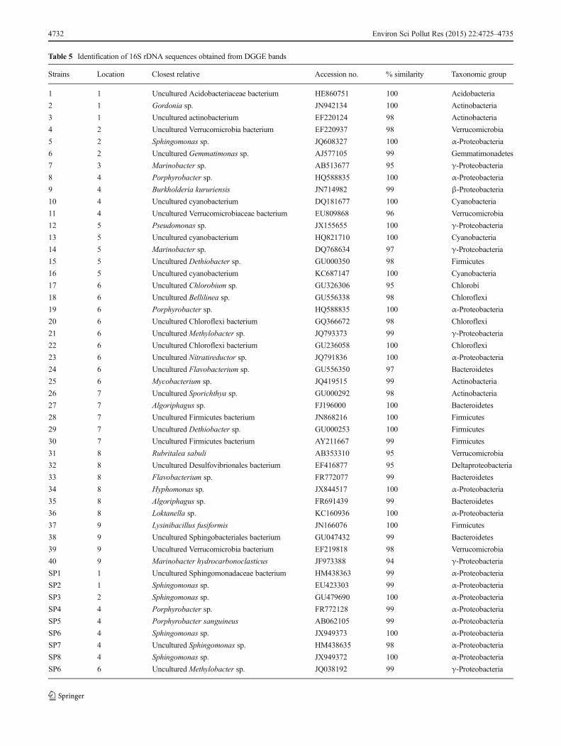
Table 5 Identification of 16S rDNA sequences obtained from DGGE bands
Strains Location Closest relative Accession no. % similarity Taxonomic group
1 1 Uncultured Acidobacteriaceae bacterium HE860751 100 Acidobacteria
2 1 Gordonia sp. JN942134 100 Actinobacteria
3 1 Uncultured actinobacterium EF220124 98 Actinobacteria
4 2 Uncultured Verrucomicrobia bacterium EF220937 98 Verrucomicrobia
5 2 Sphingomonas sp. JQ608327 100 α-Proteobacteria
6 2 Uncultured Gemmatimonas sp. AJ577105 99 Gemmatimonadetes
7 3 Marinobacter sp. AB513677 95 γ-Proteobacteria
8 4 Porphyrobacter sp. HQ588835 100 α-Proteobacteria
9 4 Burkholderia kururiensis JN714982 99 β-Proteobacteria
10 4 Uncultured cyanobacterium DQ181677 100 Cyanobacteria
11 4 Uncultured Verrucomicrobiaceae bacterium EU809868 96 Verrucomicrobia
12 5 Pseudomonas sp. JX155655 100 γ-Proteobacteria
13 5 Uncultured cyanobacterium HQ821710 100 Cyanobacteria
14 5 Marinobacter sp. DQ768634 97 γ-Proteobacteria
15 5 Uncultured Dethiobacter sp. GU000350 98 Firmicutes
16 5 Uncultured cyanobacterium KC687147 100 Cyanobacteria
17 6 Uncultured Chlorobium sp. GU326306 95 Chlorobi
18 6 Uncultured Bellilinea sp. GU556338 98 Chloroflexi
19 6 Porphyrobacter sp. HQ588835 100 α-Proteobacteria
20 6 Uncultured Chloroflexi bacterium GQ366672 98 Chloroflexi
21 6 Uncultured Methylobacter sp. JQ793373 99 γ-Proteobacteria
22 6 Uncultured Chloroflexi bacterium GU236058 100 Chloroflexi
23 6 Uncultured Nitratireductor sp. JQ791836 100 α-Proteobacteria
24 6 Uncultured Flavobacterium sp. GU556350 97 Bacteroidetes
25 6 Mycobacterium sp. JQ419515 99 Actinobacteria
26 7 Uncultured Sporichthya sp. GU000292 98 Actinobacteria
27 7 Algoriphagus sp. FJ196000 100 Bacteroidetes
28 7 Uncultured Firmicutes bacterium JN868216 100 Firmicutes
29 7 Uncultured Dethiobacter sp. GU000253 100 Firmicutes
30 7 Uncultured Firmicutes bacterium AY211667 99 Firmicutes
31 8 Rubritalea sabuli AB353310 95 Verrucomicrobia
32 8 Uncultured Desulfovibrionales bacterium EF416877 95 Deltaproteobacteria
33 8 Flavobacterium sp. FR772077 99 Bacteroidetes
34 8 Hyphomonas sp. JX844517 100 α-Proteobacteria
35 8 Algoriphagus sp. FR691439 99 Bacteroidetes
36 8 Loktanella sp. KC160936 100 α-Proteobacteria
37 9 Lysinibacillus fusiformis JN166076 100 Firmicutes
38 9 Uncultured Sphingobacteriales bacterium GU047432 99 Bacteroidetes
39 9 Uncultured Verrucomicrobia bacterium EF219818 98 Verrucomicrobia
40 9 Marinobacter hydrocarbonoclasticus JF973388 94 γ-Proteobacteria
SP1 1 Uncultured Sphingomonadaceae bacterium HM438363 99 α-Proteobacteria
SP2 1 Sphingomonas sp. EU423303 99 α-Proteobacteria
SP3 2 Sphingomonas sp. GU479690 100 α-Proteobacteria
SP4 4 Porphyrobacter sp. FR772128 99 α-Proteobacteria
SP5 4 Porphyrobacter sanguineus AB062105 99 α-Proteobacteria
SP6 4 Sphingomonas sp. JX949373 100 α-Proteobacteria
SP7 4 Uncultured Sphingomonas sp. HM438635 98 α-Proteobacteria
SP8 4 Sphingomonas sp. JX949372 100 α-Proteobacteria
SP6 6 Uncultured Methylobacter sp. JQ038192 99 γ-Proteobacteria
4732 Environ Sci Pollut Res (2015) 22:4725–4735

RHDα genes (Cébron et al. 2008; DeBruyn et al. 2007), andsome studies used real-time PCR to investigate polar ecosys-tems (Yergeau et al. 2007; Abell and Bowman 2005).
PAH-RHDα-GN genes related to naphthalene dioxygenasefrom B. glathei were found, and they comprised 70 % of thePAH-RHDα-GN library. The sequences contained only anidentity of 46 %; therefore, these clones are most likely toencode for novel dioxygenases involved in the degradation ofPAHs and may be important for degrading PAHs in Antarcticsoils and sediments. Moreover, sequences associated with thephnAc from B. sartisoli (45 % of identity) were found for23 % of the PAH-RHDα-GN library. B. sartisoli has beenisolated from a PAH-contaminated soil sample in a previousreport. This strain has been described as a versatile degrader oflowmolecular weight PAHs and is able to grow on PAHs suchas naphthalene, anthracene, and phenanthrene as a carbon andenergy source (Laurie and Lloyd-Jones 1999). In contrast, insome previous studies, the sequences obtained from theseprimers showed high identity with those reported so far.Jurelevicius et al. (2012a) used the same primer sets for thedetection of PAH-RHDα-coding genes from Gram-negativebacteria in contaminated soil samples from King GeorgeIsland in Antarctica. The sequences obtained shared highidentity with NagAc from Polaromonas naphthalenivoransCJ2 (97 %), PhnAc from Acidovorax sp. NA3 (95 %), andBurkholderia sp. Eh1-1 (96 %). Therefore, this informationtogether with our results suggests that in different regions ofthe Antarctica, hydrocarbon catabolic gene variants weredifferent.
Sequences associated with nidA3 fromMycobacterium sp.py143 and pdoA from Terrabacter sp. HH4 comprised 38 and28 % of the PAH-RHDα-GP library, respectively. NidA3fromMycobacterium has been correlated with the transforma-tion of aromatic hydrocarbon compounds, such as biphenyl,naphthalene, phenanthrene, anthracene, fluoranthene, pyrene,benz[a]anthracene, and benzo[a]pyrene (Kweon et al. 2010).Jurelevicius et al. (2012a) showed that the PdoA fromTerrabacter sp. HH4 was obtained in a diesel oil-contaminated soil sample that was collected adjacent to theBrazilian Antarctic Station Comandante Ferraz. Terrabactersp. HH4 can use fluoranthene for growth (Zhou et al. 2006).Moreover, sequences associated with nidA fromDiaphorobacter sp. KOTLB (a pyrene-degrading strain)(Klankeo et al. 2009) and pdoA2 from Mycobacterium sp.CH-2 (a pyrene-degrading strain) (Churchill et al. 2008) werealso found in the PAH-RHDα-GP library. The presence ofGram-positive bacteria that have the ability to degrade differ-ent PAH compounds in Antarctic soils could represent animportant tool for bioremediation processes.
In addition, extradiol dioxygenase gene variants were de-tected in several soil and sediment samples using primersxylE-F/xylE-R and bphC-F/bphC-R, which are specific forthe xylE and bphC genes, respectively. The bphC and xylE
genes encode 2,3-dihydroxybiphenyl-1,2-dioxygenase andcatechol 2,3-dioxygenase, respectively, catalyzing the ringcleavage reaction in the PAH degradation pathway. Cunliffeet al. (2006) monitored PAH metabolism of Sphingobiumyanoikuyae B1 by measuring bphC and xylE gene expressionusing qPCR. The finding of a hydrocarbon-degrading gene isa good indicator of the biodegradation potential of the indig-enous bacterial population in the environment. Nevertheless,further studies should be performed to analyze the PAH-RHDgene expression in Antarctic soils and sediments.
A possible reason for the presence of functional genesrelated to the degradation of aromatic hydrocarbons in thesamples where PAHs could not be detected in this study mightbe due to low PAH levels, either natural or anthropogenic,which are readily degraded by indigenous bacteria and thusbecome undetectable. In some reports, they showed the de-tection of genes for the degradation of aromatic compounds inthe uncontaminated areas and explained the possible reasonthat it could be related to the presence of naturally occurringPAHs (Flocco et al. 2009; Margesin et al. 2003).
As reported previously, different bacterial genera have beenidentified in Antarctic soils as PAH degraders, such asPseudomonas, Rhodococcus, and Sphingomonas (Panickeret al. 2010). In this study, the Sphingomonas community, asanalyzed by PCR-DGGE and PCR products, could be detect-ed in the samples from locations 1, 2, 4, and 6. The bacterialgenus Sphingomonas has been isolated from oil-contaminatedsoil samples collected from Scott Base, Antarctica, and it hasthe ability to degrade various PAHs, such as naphthalene,phenanthrene, and fluorene (Aislabie et al. 2000). As shownin Table 5, some of the bands were closely related toSphingomonas such as Porphyrobacter sp. Hiraishi et al.(2002) reported that Porphyrobacter sanguineus was able togrow on dibenzofuran and biphenyl as a carbon source. Fur-thermore, and interestingly, some of the dominant bands in theDGGE profiles of total bacteria that had similarity toSphingomonas sp. and Porphyrobacter sp. were found in thesamples from locations 2, 4, and 6.
Many different bacterial genera that are able to degradePAHs and have previously been isolated from the environ-ment belong to the genera Alcal igenes , Vibrio ,Mycobacterium, Comamonas, Arthrobacter, Burkholderia,and Flavobacterium (Zhang et al. 2011). In this study, dom-inant bands in the total bacterium DGGE profiles of eachsample were sequenced, which showed the presence of bac-terial genera that are commonly related to biodegradation. Forexample, sequences from Burkholderia sp., Pseudomonas sp.,Mycobacterium sp., and Flavobacterium sp. were found as thedominant DGGE bands in the samples from locations 4, 5, 6,and 8. Furthermore, the dominant band of location 1 wasidentical to the sequences from Gordonia sp. The genusGordonia has also been reported to utilize a variety of aliphat-ic, aromatic hydrocarbons or other pollutants in the
Environ Sci Pollut Res (2015) 22:4725–4735 4733
environment (Arensköter et al. 2004). The dominant band oflocations 3 and 5 was identical to the sequences fromMarinobacter sp., and the dominant band of location 9 wasiden t i ca l to the sequences f rom Marinobac terhydrocarbonoclasticus. Marinobacter is a widely distributedbacterium that has been isolated from the Antarctic environ-ment (Liu et al. 2012). Some Marinobacter strains, such asM. hydrocarbonoclasticus, were isolated from petroleumhydrocarbon-contaminated sediments, and it has the abilityto degrade several aliphatic components of crude oil andaromatic compounds, such as phenanthrene (Gauthier et al.1992).
Conclusions
In this study, the presence of indigenous bacteria related togenera that contain known hydrocarbon degraders and thepresence of hydrocarbon catabolic genes around the SyowaStation in Antarctica were first revealed. These results implythat these environments have the potential ability to degradehydrocarbons, and this information will be useful for thebioremediation of hydrocarbon contamination in Antarcticsoils and sediments. It would also be appropriate to carry outmineralization assays with radiolabelled PAHs to confirm insitu degradative activity.
Acknowledgments This work was supported by the National Instituteof Polar Research (Japan); L’Oreal (Thailand) Ltd.; Faculty of Science,Chulalongkorn University; and National Research University Project of theOffice of Commission for Higher Education and RatchadaphiseksomphotEndowment Fund, Chulalongkorn University—Climate Change Cluster(CC1043A). We also would like to thank all the members of the 51stJapanese Antarctic Research Expedition (JARE-51) for their field supportand assistance.
References
Abell GCJ, Bowman JP (2005) Ecological and biogeographic relation-ships of class Flavobacteria in the SouthernOcean. FEMSMicrobiolEcol 51:265–277
Aislabie J, Foght JM (2010) Response of polar soil bacterial communities tofuel spills. In: Bej AK, Aislabie J, Atlas RM (eds) The ecology,biodiversity and bioremediation potential of microorganisms in ex-tremely cold environments, Taylor & Francis, Florida, pp 215–230
Aislabie J, Foght J, Saul D (2000) Aromatic hydrocarbon-degrading bacteriafrom soil near Scott Base, Antarctica. Polar Biol 23:183–188
Aislabie JM, Balks MR, Foght JM, Waterhouse EJ (2004) Hydrocarbonspills on Antarctic soils: effects and management. Environ SciTechnol 38:1265–1274
Amann RI, Ludwig W, Schleifer KH (1995) Phylogenetic identificationand in situ detection of individual microbial cells without cultiva-tion. Microbiol Mol Biol Rev 59:143–149
Arensköter M, Broker D, Steinbüchel A (2004) Biology of metabolicallydiverse genus Gordonia. Appl Environ Microbiol 70:3195–3204
Cébron A, Norini MP, Beguiristain T, Leyval C (2008) Real-time PCRquantification of PAH-ring hydroxylating dioxygenase (PAH-RHDalpha) genes from Gram positive and Gram negative bacteriain soil and sediment samples. J Microbiol Meth 73:148–159
Churchill PF, Morgan AC, Kitchens E (2008) Characterization of apyrene-degrading Mycobacterium sp. strain CH-2. J Environ SciHealth B 43:698–706
Cunliffe M, Kawasaki A, Fellows E, Kertesz MA (2006) Effect of inoculumpretreatment on survival, activity and catabolic gene expression ofSphingobium yanoikuyae B1 in aged polycyclic aromatichydrocarbon-contaminated soil. FEMS Microbiol Ecol 58:364–372
Das R, Kazy SK (2014) Microbial diversity, community composition andmetabolic potential in hydrocarbon contaminated oily sludge: pros-pects for in situ bioremediation. Environ Sci Pollut Res. doi:10.1007/s1135601426402
DeBruyn JM, Chewning CS, Sayler GS (2007) Comparative quantitativeprevalence of Mycobacteria and functionally abundant nidA, nahAcand nagAc dioxygenase genes in coal tar contaminated sediments.Environ Sci Technol 41:5426–5432
Delille D, Coulon F (2008) Comparative mesocosm study of biostimula-tion efficiency in two different oil-amended sub-Antarctic soils.Microb Ecol 56:243–252
Fernández-Luqueño F, Valenzuela-Encinas C, Marsch R, Martínez-Suárez C, Vázquez-Núñez E, Dendooven L (2011) Microbial com-munities to mitigate contamination of PAHs in soil—possibilitiesand challenges: a review. Environ Sci Pollut Res 18:12–30
Flocco CG, Gomes NCM, CormackWM, Smalla K (2009) Occurrence anddiversity of naphthalene dioxygenase genes in soil microbial commu-nities from the Maritime Antarctic. Environ Microbiol 11:700–714
Gauthier MJ, Lafay B, Christen T, Fernandez L, AcquavivaM, Bonin PC,Betrand JC (1992) Marinobacter hydrocarbonoclasticus gen. nov.,sp. nov., a new, extremely halotolerant, hydrocarbon-degradingmarine bacterium. Int J Syst Bacteriol 43:568–576
Hiraishi A, Yonemitsu Y, Matsushita M, Shin YK, Kuraishi H, KawaharaK (2002) Characterization of Porphyrobacter sanguineus sp. nov.,an aerobic bacteriochlorophyll-containing bacterium capable ofdegrading biphenyl and dibenzofuran. Arch Microbiol 178:45–52
Junca H, Pieper DH (2004) Functional gene diversity analysis in BTEXcontaminated soils by means of PCR-SSCP DNA fingerprinting:comparative diversity assessment against bacterial isolates andPCR-DNA clone libraries. Environ Microbiol 6:95–110
Jurelevicius D, Alvarez VM, Peixoto R, Rosado AS, Seldin L (2012a)Bacterial polycyclic aromatic hydrocarbon ring-hydroxylatingdioxygenases (PAH-RHD) encoding genes in different soils fromKing George Bay, Antarctic Peninsula. Appl Soil Ecol 55:1–9
Jurelevicius D, Cotta SR, Peixoto R, Rosado AS, Seldin L (2012b)Distribution of alkane-degrading bacterial communities in soils fromKing George Island, Maritime Antarctic. Eur J Soil Biol 51:37–44
Klankeo P, Nopcharoenkul W, Pinyakong O (2009) Two novel pyrene-degrading Diaphorobacter sp. and Pseudomonas sp. isolated fromsoil. J Biosci Bioeng 108:488–495
Kweon O, Kim SJ, Freeman JP, Song J, Baek S, Cerniglia CE (2010)Substrate specificity and structural characteristics of the novel Rieskenon heme iron aromatic ring-hydroxylating oxygenases NidAB andNidA3B3 from Mycobacterium vanbaalenii PYR-1. mBio 1:1–11
Lau EV, Gan S, Ng HK (2010) Extraction techniques for polyaromatichydrocarbons in soils. Int J Anal Chem. doi:10.1155/2010/398381
Laurie AD, Lloyd-Jones G (1999) The phn genes of Burkholderia sp.strain RP007 constitute a divergent gene cluster for polycyclicaromatic hydrocarbon catabolism. J Bacteriol 181:531–540
Leys NM, Ryngaert A, Bastiaens L, Verstraete W, Springael D (2004)Occurrence and phylogenetic diversity of Sphingomonas strain insoils contaminated with polycyclic aromatic hydrocarbons. ApplEnviron Microbiol 70:1944–1955
Liu C, Chen CX, Zhang XY, Yu Y, Liu A, Li GW, Chen XL, Chen B,Zhou BC, Zhang YZ (2012) Marinobacter antarcticus sp. nov., a
4734 Environ Sci Pollut Res (2015) 22:4725–4735

halotolerant bacterium isolated from Antarctic intertidal sandy sed-iment. Int J Syst Evol Microbiol 62:1838–1844
Ma Y, Wang L, Shao Z (2006) Pseudomonas, the dominant polycyclicaromatic hydrocarbon-degrading bacteria isolated from Antarcticsoils and the role of large plasmids in horizontal gene transfer.Environ Microbiol 8:455–465
Marcos MS, Lozada M, Dionisi HM (2009) Aromatic hydrocarbondegradation genes from chronically polluted Subantarctic marinesediments. Lett Appl Microbiol 49:602–608
Margesin R, Labbé D, Schinner F, Greer CW, Whyte LG (2003)Characterization of hydrocarbon-degrading microbial populationsin contaminated and pristine alpine soils. Appl Environ Microbiol69:3085–3092
Muangchinda C, Pansri R, Wongwongsee W, Pinyakong O (2013)Assessment of polycyclic aromatic hydrocarbon biodegradationpotential in mangrove sediment from Don Hoi Lot, SamutSongkram Province, Thailand. J Appl Microbiol 114:1311–1324
Panicker G, Mojib N, Aislabie J, Bej AK (2010) Detection, expressionand quantitation of the biodegradative genes in Antarctic microor-ganisms using PCR. Antonie Van Leeuwenhoek 97:275–287
Peng RH, Xiong AS, Xue Y, Fu XY, Gao F, Zhao W, Tian YS, Yao QH(2008) Microbial biodegradation of polyaromatic hydrocarbons.FEMS Microbiol Rev 32:927–955
Powell SM, Bowman JP, Snape I, Stark JS (2003) Microbial communityvariation in pristine and polluted nearshore Antarctic sediments.FEMS Microbiol Ecol 45:135–145
Ruberto L, Vazquez SC, Mac Cormack WP (2003) Effectiveness of thenatural bacterial flora, biostimulation and bioaugmentation on thebioremediation of a hydrocarbon contaminated Antarctic soil. IntBiodeterior Biodegrad 52:115–125
Ruberto LAM, Vazquez SC, Curtosi A, Mestre MC, Pelletier E, MacCormack WP (2006) Phenanthrene biodegradation in soils using anAntarctic bacterial consortium. Biorem J 10:191–201
Sipila TP, Riisio H, Yrjala K (2006) Novel upper meta-pathway extradioldioxygenase gene diversity in polluted soil. FEMS Microbiol Ecol58:134–144
Suenaga H, Mizuta S, Miyazaki K (2009) The molecular basis foradaptive evolution in novel extradiol dioxygenases retrieved fromthe metagenome. FEMS Microbiol Ecol 69:472–480
US EPA Method 3500C (2007) Organic extraction and sample prepara-tion. Revision 3. February, 2007
US EPA Method 3540C (1996) Soxhlet extraction. Revision 3.December, 1996
US EPA Method 8310 (1986) Polyaromatic hydrocarbons. September,1986
Yergeau E, Arbour M, Brousseau R, Juck D, Lawrence JR, Masson L,Whyte LG, Greer CW (2009) Microarray and real-time PCR anal-yses of the responses of high-arctic soil bacteria to hydrocarbonpollution and bioremediation treatments. Appl Environ Microbiol75:6258–6267
Yergeau E, Bokhorst S, Huiskes AHL, Boschker HTS, Aerts R,Kowalchuk GA (2007) Size and structure of bacterial, fungal andnematode communities along an Antarctic environmental gradient.FEMS Microbiol Ecol 59:436–451
Zhang S, Wan R, Wang Q, Xie S (2011) Identification of anthracenedegraders in leachate-contaminated aquifer using stable isotopeprobing. Int Biodeterior Biodegrad 65:1224–1228
Zhou HW, Guo CL, Wong YS, Tam NFY (2006) Genetic diversity ofdioxygenase genes in PAH-degrading bacteria isolated from man-grove sediments. FEMS Microbiol Lett 262:148–157
Environ Sci Pollut Res (2015) 22:4725–4735 4735