Embed Size (px)
Citation preview
OCCASIONAL PAPE~S
OF
BERNICE P. BISHOP MUSEUM
HONOLULU, HAWAII
Volume XXII February 27, 1959 N4mber9
Acarina from Southeastern Polynesia-II (Oribatidae)lBy MAX SELLNICK
HOISDORF, NEAR HAMBURG, GERMANY
This is the second report on Acarina, or mites, collected by theMangarevan Expedition which was sent by Bishop Museum to exploreislands of southeastern Polynesia in 1934. The first report, coveringthe Mesostigmata, was published by Ivar Tragardh in 1951 (29).2The Tragardh report includes 14 species and 13 genera, of which 7genera and 11 species are described as new to science. To this may nowbe added the 31 species of Oribatidae which are reported upon in thispaper.
My report is based upon 324 specimens of Oribatidae collected bythe Mangarevan Expedition and includes 26 species and five varietiesin 21 genera. Of these, 14 species and one variety are described as newto science. In addition, Humerobates humeralis perkinsi Jacot is raisedto specific status.
For a detailed survey of the mite fauna of any locality, it is necessary that a careful and extensive use of the Berlese funnel be made.By the use of this funnel, the acarologist can obtain thousands of mitesfrom a suitable biotope, but much time must be spent making suchcollections. There was no acarologist on the Mangarevan Expedition,and the mites collected were taken as incidental collections by the entomologist, the botanists, and the malacologists. In fact, a number ofspecimens were obtained from the empty shells of land mollusks whenthey were cleaned in the laboratory after the expedition. However,despite the lack of time and of facilities for specialized mite collectingtechniques, the results obtained are a good addition to our knowledge
1 Mangarevan Expedition publication 43.2 Numbers in parentheses refer to Bibliography p. 151.
110 Bernice P. Bishop Museum-Occasional Papers XXII, 9
of the acarid fauna of the Pacific islands. No oribatids had been previously known from the islands represented by the collections reportedupon here.
In addition to the Oribatidae, two species belonging to other suborders were found in the material studied. One of these belongs to theBdellidae, but the mount is so badly preserved that it is impossible todetermine the specimen. The other species is a bird mite, which Dr.
.Charles D. Radford of Manchester, England, was kind enough to identify as Alloptes crassipes (Canestrini), 1878.
Only two larger papers on the oribatid mites from Pacific islandshave been published, both by Jacot (13, 14). I have discovered severalof the species listed by Jacot in the Mangarevan Expedition collection.
Dr. Elwood C. Zimmerman, the entomologist on the MangarevanExpedition, assisted me in translating my original mannscript fromGerman into English and revised the manuscript. I am also indebtedto Dr. G. O. Evans of the British Museum (Natural History) for hishelp.
All collections reported here were made by the Mangarevan Expedition in 1934, and the types of the new species are deposited inBishop Museum. Oribatidae were collected on the islands of Tahiti,Moorea, Huahine, Raiatea, Tahaa, Borabora, Rapa, Marotiri, Raivavae, Tubuai, Rurutu, Rimatara, Maria, and Flint. It is of interest thatthe expedition evidently found no oribatids on the several atoll islandsof the Tuamotus which they visited. No specimens were found on Pitcairn or nearby Henderson Island, although they probably occur there.
FAMILY CAMISIIDAE SELLNICK, 1929
Genus Nothrus C. L. Koch, 1836
Type N othrus palustris C. L. Koch, 1839.
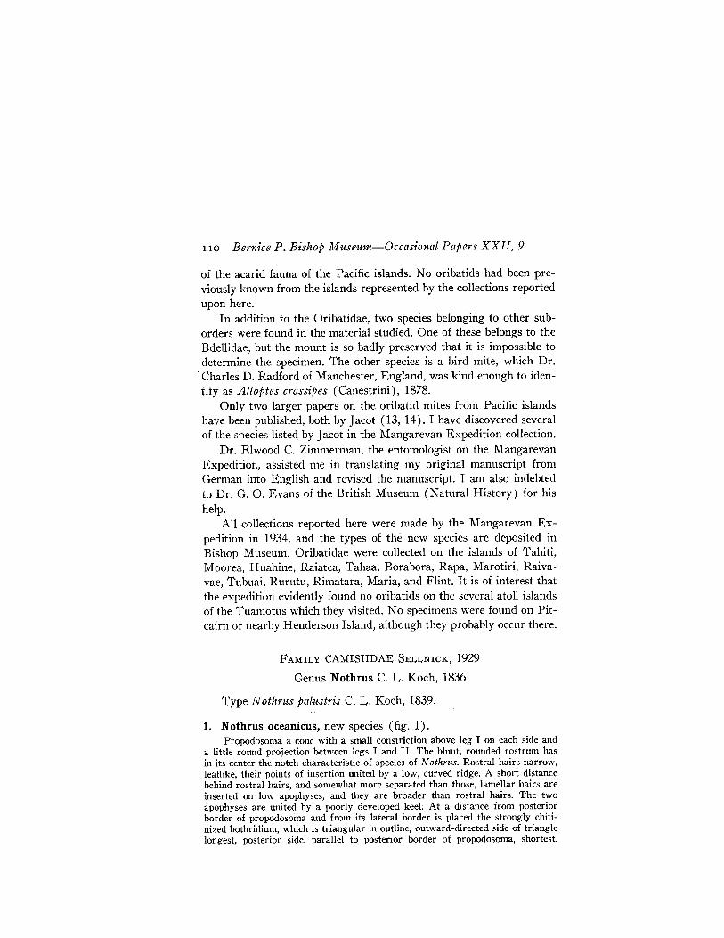
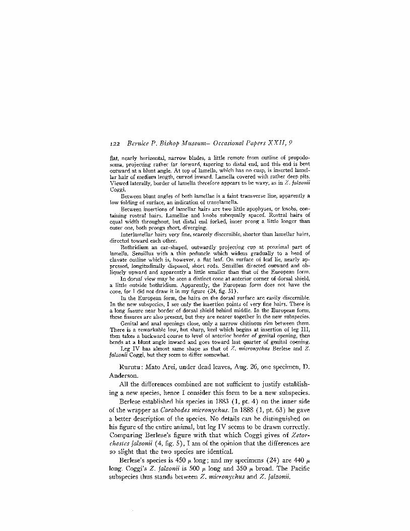
1. Nothrus oceanicus, new species (fig. 1).Propodosoma a cone with a small constriction above leg I on each side and
a little round projection between legs I and II. The blunt, rounded rostrum hasin its center the notch characteristic of species of N othms. Rostral hairs narrow,leaflike, their points of insertion united by a low, curved ridge. A short distancebehind rostral hairs, and somewhat more separated than those, lamellar hairs areinserted on low apophyses, and they are broader than rostral hairs. The twoapophyses are united by a poorly developed keeL At a distance from posteriorborder of propodosoma and from its lateral border is placed the strongly chitinized bothridium, which is triangular in outline, outward-directed side of triangleloilgest, posterior side, parallel to posterior border of propodosoma, shortest.
Sellnick~Acarina from Southeastern Polynesia III
Sensillus, directed outward and a little upward, is a bristle tapering gradually toits distal end, somewhat longer than one-half width of propodosoma, and with afew fine cilia on distal part. Interlamellar hair, at a level with sensillus, is 32 p.distant from bothridium. This hair is broken off, but it may be of same size aslamellar hairs. Behind bothridium and between it and anterior border of hysterosoma is a sickle-shaped chitinization, the arc opened inward. Except part behind
~\~
\[2
\
FIGURE l.-Nothrus oceanicus, dorsal side.
bothridium, whole surface of propodosoma covered with pits, diameter 12-16 p.;bottoms of pits appear finely granulated, spaces between pits form a network ofridges. There is a regular longitudinal depression between and in front of bothridiae.
Width of anterior border of hysterosoma 288 p.. Body widest behind middle.Anterior border projects slightly, and is slightly longitudinally sulcate. Wholesurface covered with distinct, but not perfectly equal, pits, delimited by a net of
112 Bernice P. Bishop Museum--Occasional Papers XXII, 9
more strongly chitinized ridges. Close to inner side of marginal hairs is a longitudinal stripe of smaller and considerably less distinct pits, situated where marginof dorsal shield is bent upward. Flat knobs touch each other on margin. Posteriorborder of body slightly convex. Outer corners of posterior border well markedby low indentations in front and behind last marginal hair (F2). Whole posteriorpart of dorsal plate between outer corners a trapezium, sloping backward. Anterior side of trapezium shorter than posterior border of body.
Hairs of dorsal shield leaflike, each with a longitudinal rib from which smalllateral ribs project obliquely forward. Margins of these leaves smooth or provided with little notches.
Marginal row of hairs on each side of dorsal shield has bristles C3, D3, E2,and F2. Distance from C3 to D3 is much greater than distances from D3 to E2and from E2 to F2. There are two median rows: CI, DI, D2, EI, and Fl. Thedistances from CI to Dl and from El to Fl are greater than from Dl to D2and D2 to El. The distances from Cl to Cl and from D1 to D 1 are equal; fromD2 to D2 and from El to El are greater. The space between Fl and FI equalsthat between D2 and D2. Between Cl and C3, hair C2, a little smaller than theothers, is nearer to CI than to C3. At outer posterior corner of body is hair Kl.On posterior margin is hair PNI ; and somewhat higher on dorsal surface, nearerto KI than to PNl, PN2 is inserted. Under posterior rounded corner of body ishair PN3, which is smaller than hairs on dorsal shield. All other hairs on ventralside simple, short bristles. Epimera I to IV on each side of venter fused. Also,epimera I and II of one side fused with those of other side. Between epimeraIII and IV of one side and III and IV of other side is a deep notch.
Tarsi with three distinct claws, middle one much stronger than lateral ones,which resemble curved hairs. Most segments with low pits and narrow leaflikehairs. Many hairs on tarsi simple and smooth.
Holotype, slide 001.Raivavae: Ahuoivi Point, on dead leaves, Aug. 9, one specimen,
Cooke, Kondo, and D. Anderson. Type locality.This new species resembles N othrus biciliatus C. L. Koch, but sev
eral characters separate it from the German species. N. oceanicus (afemale) is 765 p.long and 405 p. broad. N. biciliatus is 924 p. long and450 p. broad.
Genus Phyllonothrus, new genus
This genus is similar to the genus Camisia, but lacks any real bothridium. The hairs on the dorsal shield are all pick-shaped; on the legs,mostly small blades. A single nymph composes the collection.
Type Phyllonothrus runcifer, new species.
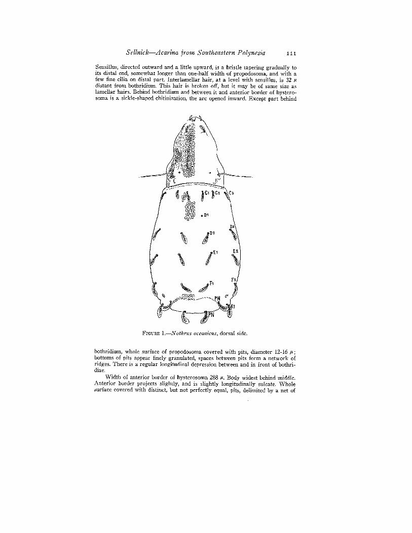
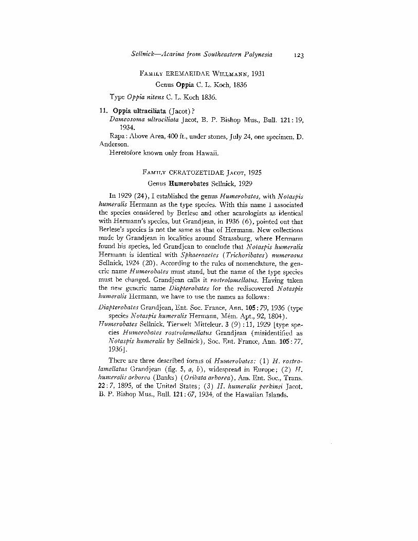
2. Phyllonothrus runcifer, new species (fig. 2).Length 50S iL, width 200 iL. Whitish yellow in color.Propodosoma posteriorly smaller than anterior edge of hysterosoma, sides
somewhat converging toward top, with low indentations over leg I, but withrostrum truncate. Rostral hairs simple, short, curved. Behind rostral hairs, atborder of rostrum, are low apophyses for lamellar hairs, which are formed like
Sellnicl?-Acarina from Southeastern Polynesia 113
a double pickax (pointed at both ends and provided with a short handle in themiddle) and each inserted at top of apophysis. There seems to be a fold in thesurface of the propodosoma between the lamellar apophyses, thus forming a kindof translamella.
At a level with the middle of coxa I are insertions of interlamellar hairs,separated from each other as are lamellar hairs. (The hairs are broken off, butI believe that they were of the size of the lamellar hairs, considering their largepoints of insertion.) Behind and inside interlamellar hairs are two elongatedtrapeziform areas, flat indentations, which do not reach anterior border ofhysterosoma.
FIGURE 2.-Phyllonothrus runcifer: a, dorsal side; b, part of ventral side;c, leg II.
Outside interlamellar hairs a sharp angle is directed outward, limited anteriorly by a concave chitinous line, beginning at interlamellar hair, and an almoststraight lateral line which is directed backward and a little inward. Corner of theangle does not reach side of propodosoma. At its apex is inserted a simple, short,fine hair which I believe to be the sensillus. I see no bothridium.
Dorsum of hysterosoma almost parallel-sided in basal three-fourths of itslength, enlarging a little posteriorly; body constricted, forming a short part withright angles, and finally body narrowed again, forming a trapezium with astraight posterior border with blunt corners. This trapezoidal part seems to lieon a lower level than rest of dorsum.
There is a marginal row of six of above-described double pickax hairs, fourequidistant from each other on first three-fourths of border, one at corner of
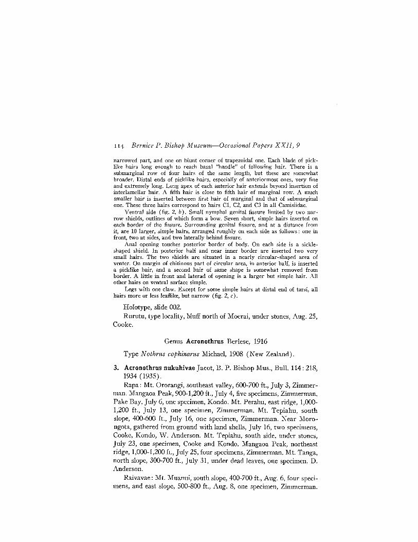
114 Bernice P. Bishop Museum-Occasional Papers XXII, 9
narrowed part, and one on blunt corner of trapezoidal one. Each blade of picklike hairs long enough to reach basal "handle" of following hair. There is asubmarginal row of four hairs of the same length, but these are somewhatbroader. Distal ends of picklike hairs, especially of anteriormost ones, very fineand extremely long. Long apex of each anterior hair extends beyond insertion ofinterlamellar hair. A fifth hair is close to fifth hair of marginal row. A muchsmaller hair is inserted between first hair of marginal and that of submarginalone. These three hairs correspond to hairs Cl, C2, and C3 in all Camisiidae.
Ventral side (fig. 2, b). Small nymphal genital fissure limited by two narrow shields, outlines of which form a bow. Seven short, simple hairs inserted oneach border of the fissure. Surrounding genital fissure, and at a distance fromit, are 10 larger, simple hairs, arranged roughly on each side as follows: one infront, two at sides, and two laterally behind fissure.
Anal opening touches posterior border of body. On each side is a sickleshaped shield. In posterior half and near inner border are inserted two verysmall hairs. The two shields are situated in a nearly circular-shaped area ofventer. On margin of chitinous part of circular area, in anterior half, is inserteda picklike hair, and a second hair of same shape is somewhat removed fromborder. A little in front and laterad of opening is a larger but simple hair. Allother hairs on ventral surface simple.
Legs with one claw. Except for some simple hairs at distal end of tarsi, allhairs more or less leaflike, but narrow (fig. 2, c).
Holotype, slide 002.Rurutu, type locality, bluff north of Moerai, under stones, Aug. 25,
Cooke.
Genus Acronothrus Berlese, 1916
Type N othrus cophinarus Michael, 1908 (New Zealand).
3. Acronothrus nukuhivae Jacot, B. P. Bishop Mus., Bull. 114: 218,1934 (1935).Rapa: Mt. Ororangi, southeast valley, 600-700 ft., July 3, Zimmer
man. Mangaoa Peak, 900-1,200 ft., July 4, five specimens, Zimmerman.Pake Bay, July 6, one specimen, Kondo. Mt. Perahu, east ridge, 1,0001,200 ft., July 13, one specimen, Zimmerman. Mt. Tepiahu, southslope, 400-600 ft., July 16, one specimen, Zimmerman. Near Morongota, gathered from ground with land shells, July 16, two specimens,Cooke, Kondo, W. Anderson. Mt. Tepiahu, south side, under stones,July 23, one specimen, Cooke and Kondo. Mangaoa Peak, northeastridge, 1,000-1,200 ft., July 25, four specimens, Zimmerman. Mt. Tanga,north slope, 300-700 ft., July 31, under dead leaves, one specimen, D.Anderson.
Raivavae: Mt. Muanui, south slope, 400-700 ft., Aug. 6, four specimens, and east slope, 500-800 ft., Aug. 8, one specimen, Zimmerman.
Sellnick-Acarina from Southeastern Polynesia 115
Tubuai: Mt. Taitaa [mapped Taita], northwest ridge, 1,200 ft.,Aug. 21, one specimen, and southwest ridge, 1,200 ft., Aug. 23, twospecimens, Zimmerman.
Rurutu: Mt. Teape, southwest slope, 1,000 ft., Sept. 2, one specimen, Zimmerman.
This species has been known heretofore only from the MarquesasIslands.
FAMILY NEOLIODIDAE WILLMANN, 1931
Genus Liodes von Heyden, 1826
Several present-day acarologists do not realize that the genus N eoliodes Berlese must be called Liodes von Heyden. Berlese was of theopinion that Liodes was preoccupied by· Latreille, 1796, for a genusof Coleoptera. But Latreille called his genus Leiodes. If this name islater changed to Liodes by coleopterologists, it is an unauthorizedchange which must be withdrawn. Liodes is the name of a genus ofOribatidae, with N otaspis theleproctus Hermann, 1804, as type. Jacotestablished in 1929 (9) the genus Udetaliodes with Liodes concentricusBanks as type. I stated my opinion regarding this in 1930 (25), and Ihave not changed it. Grandjean, 1936 (6), collecting in the neighborhood of Strassburg, rediscovered Hermann's species, but he does notmention Jacot's new genus. Graf Vitzthum, in 1943 (30), consideredthe name Udetaliodes to be identical with N eoliodes Berlese. The shortdiagnosis of the genus, as well as the figures, in Jacot's paper (9, p.31), demonstrates that his species belong to the genus Liodes and thatthe new name was superfluous.
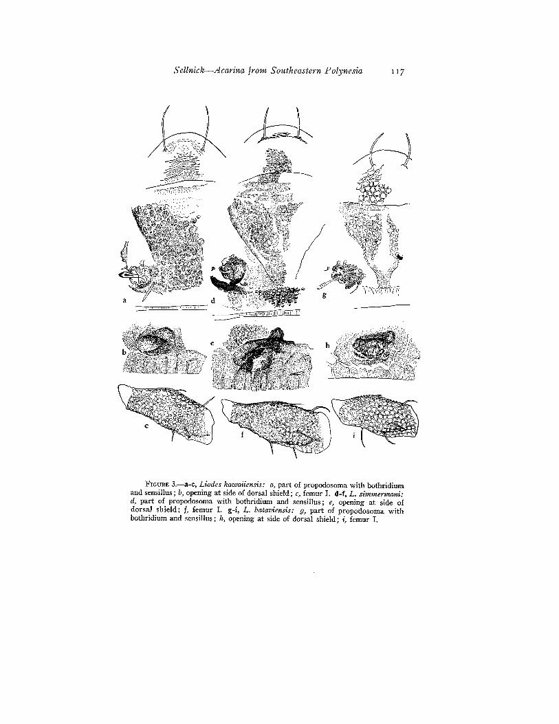
There are three different species of Liodes in the Mangarevan Expedition collection: Liodes batamensis (Sellnick), 1925, Liodes hawaiiensis (Jacot), 1929, and Liodes zimmermani, new species.
In Jacot's key to the species of Liodes found in the Pacific islands(9), he divides them into two groups: species which have one longitudinal keel in the middle of the anterior part of the dorsal shield andspecies which have three, five, or seven parallel keels in the same place.The nymphal exuviae which always remain on the dorsum also havethese lines. The species Liodes zimmermani has no keel. If we wish toput the species into Jacot's key, we must establish a third group. Theanterior part of the dorsal shield of Liodes zimmermani is d~lJ.sely
covered with low knobs which nearly touch each other, and the; intervals between the knobs form a kind of polygonal reticulation. .
116 Bernice P. Bishop Museum-Occasional Papers XXII, 9
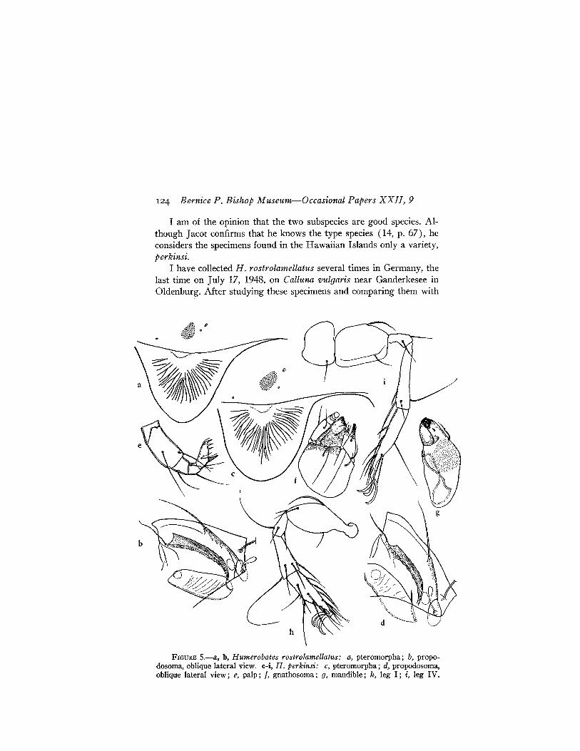
Figure 3, b (hawaiiensis), 3, e (zimmermani) , and 3, h (bataviensis) show an opening (of a gland?) on the dorsal shield near itslateral border. I mentioned this opening in 1925 (22, p. 463). Apparently Jacot, who has described many new species, has paid no attentionto such foramina. In studies made of specimens in toto, the openingsare not visible, or they are very difficult to find. It is necessary to dissect the animals to see them. (See figures 3, b, e, and h.)
There are very distinct differences between femur I in the variousspecies, as is noted in the following descriptions.
4. Liodes hawaiiensis (Jacot). (Figure 3, a-c.)Udetaliodes hawaiiensis Jacot, Am. Microscopical Soc., Trans. 48:
31, 1929 (with figures).Only distal half or a little more of surface of femur I is covered with pits.
Pits relatively distinct, but network of lines between them less developed. Anterior dorsal hairs straight, inclined a little forward. Posterior dorsal hair insertedin middle of dorsal edge, inclined forward, and bowed. Only one short,erect,straight hair about middle of lower edge of femur. Lateral hair inserted in frontof femur and near ventral edge.
Slides 007-012.Rapa: Mt. Ororangi, southeast valley, 600-700 ft., July 3, two
specimens, Zimmerman. Mangaoa Peak, northeast ridge, 1,000-1,200ft., July 25, one specimen, Zimmerman.
Tubuai: Mt. Taita, northwest ridge, 1,200 ft., Aug. 21, 33 specimens, Zimmerman.
Tahiti: Mt. Aorai Trail, 3,500-4,500 ft., on M etrosideros, Sept. 13,one specimen, Zimmerman.
Previously known only from the Hawaiian Islands.
5. Liodes zimmermani, new species (fig. 3, d-f).Femur I is larger than on other species. Only part of anterior half of the
segment pitted, but pits not so distinctly limited as in the other two species.Surface of proximal half with more or less distinct transverse ridges covering it.Anterior dorsal hair curved; posterior one, situated at middle of dorsal edge,rather straight and nearly appressed. Lower edge of femur with two short,straight hairs, posterior one of which is inserted in middle. Both are inclined alittle forward. Lateral hair, inserted in a line with anterior one on lower edge,is straight and a little longer than lower hairs.
Largest specimen 1,530 p, long and 990 p, broad.
Type, slides 003 a-g., paratypes, slides 004-006.Marotiri: Southeast Islet (type locality), July 22, 12 specimens,
Zimmerman and Fosberg.
Sellnick-Acarina from Southeastern Polynesia 117
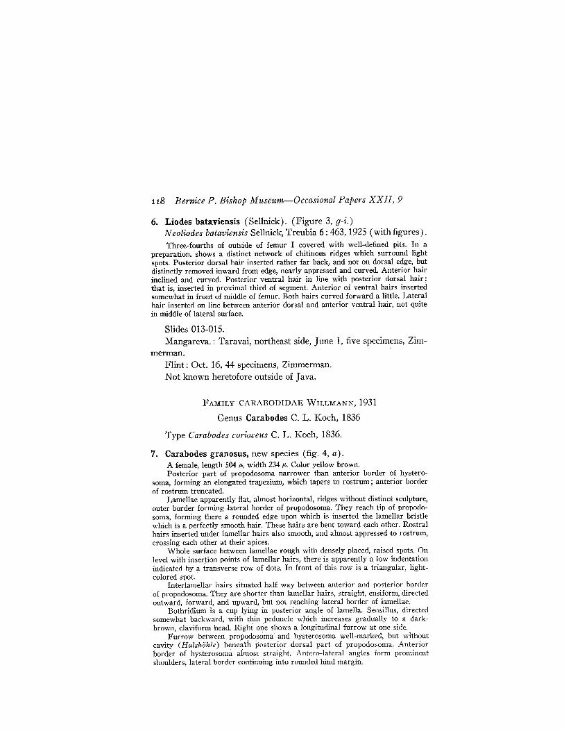
FIGURE 3.-a-c, Liodes hawaiiensis: a, part of propodosoma with bothridiumand sensillus; b, opening at side of dorsal shield; c, femur 1. d-f, L. zimmermani:d, part of propodosoma with bothridium and sensillus; e, opening at side ofdorsal shield; j, femur 1. g-i, L. bataviensis: g, part of propodosoma withbothridium and sensillus; h, opening at side of dorsal shield; i, femur I.
118 Hernice P. Bishop Museum-Occasional Papers XXII, 9
6. Liodes bata;viensis (Sellnick). (Figure 3, g-i.)N eoliodes bataviensis Sellnick, Treubia 6: 463, 1925 (with figures).Three-fourths of outside of femur I covered with well-defined pits. In a
preparation, shows a distinct network of chitinous ridges which surround lightspots. Posterior dorsal hair inserted rather far back, and not on dorsal edge, butdistinctly removed inward from edge, nearly appressed and curved. Anterior hairinclined and curved. Posterior ventral hair in line with posterior dorsal hair;that is, inserted in proximal third of segment. Anterior of ventral hairs insertedsomewhat in front of middle of femur. Both hairs curved forward a little. Lateralhair inserted on line between anterior dorsal and anterior ventral hair, not quitein middle of lateral surface.
Slides 013-015.Mangareva.: Taravai, northeast side, June 1, five specimens, Zim
merman.Flint: Oct. 16, 44 specimens, Zimmerman.Not known heretofore outside of Java.
FAMILY CARABODIDAE WILLMANN, 1931
Genus Carabodes C. L. Koch, 1836
Type Carabodes coriaceus C. L. Koch, 1836.
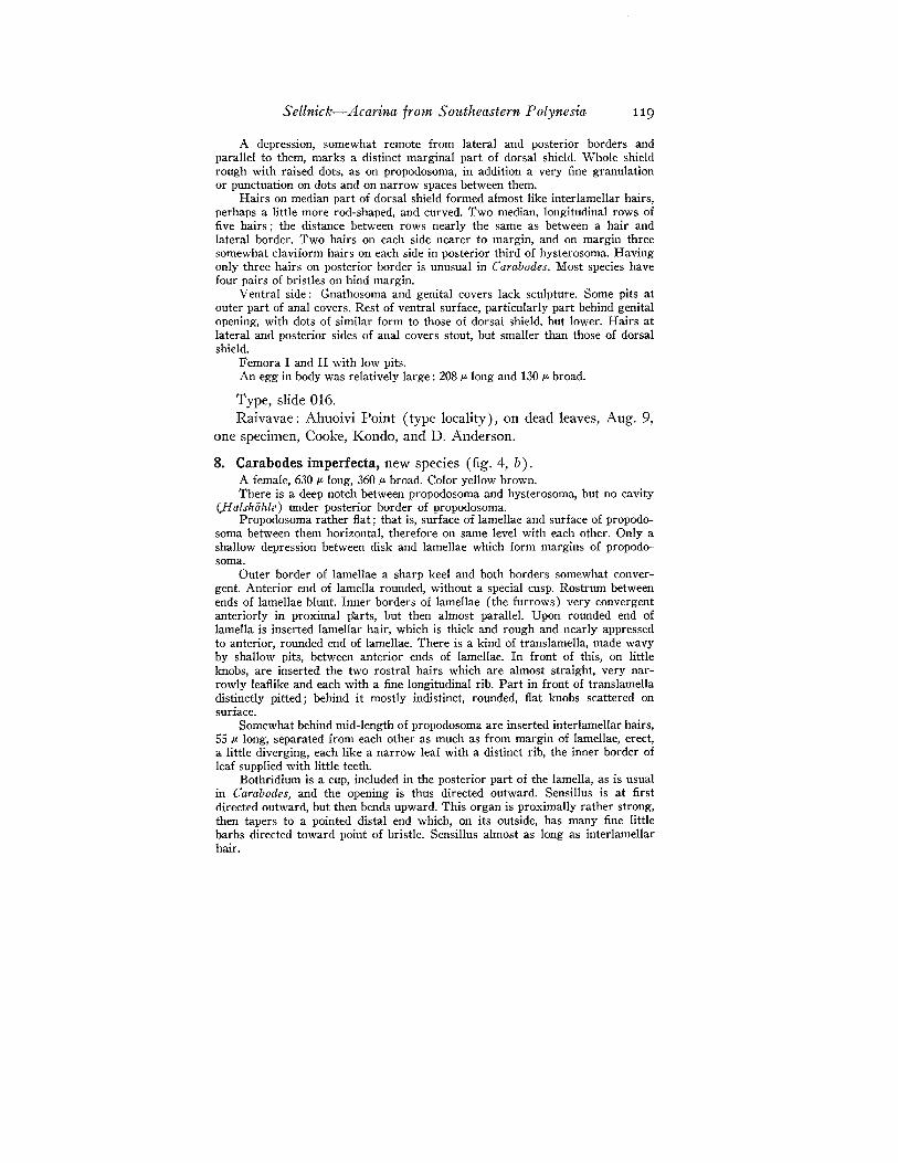
7. Carabodes granosus, new species (fig. 4, a).A female, length 504 iL, width 234 IJ-. Color yellow brown.Posterior part of propodosoma narrower than anterior border of hystero
soma, forming an elongated trapezium, which tapers to rostrum; anterior borderof rostrum truncated.
Lamellae apparently fiat, almost horizontal, ridges without distinct sculpture,outer border forming lateral border of propodosoma. They reach tip of propodosoma, forming there a rounded edge upon which is inserted the lamellar bristlewhich is a perfectly smooth hair. These hairs are bent toward each other. Rostralhairs inserted under lamellar hairs also smooth, and almost appressed to rostrum,crossing each other at their apices.
Whole surface between lamellae rough with densely placed, raised spots. Onlevel with insertion points of lamellar hairs, there is apparently a low indentationindicated by a transverse row of dots. In front of this row is a triangular, lightcolored spot.
Interlamellar hairs situated half way between anterior and posterior borderof propodosoma. They are shorter than lamellar hairs, straight, ensiform, directedoutward, forward, and upward, but not reaching lateral border of lamellae.
Bothridium is a cup lying in posterior angle of lamella. Sensillus, directedsomewhat backward, with thin peduncle which increases gradually to a darkbrown, claviform head. Right one shows a longitudinal furrow at one side.
Furrow between, propodosoma and hysterosoma well-marked, but withoutcavity (Halshohle) beneath posterior. dorsal part of propodosoma. Anteriorborder of hysterosoma aimost straight.' Al1tero-Iateral angles form prominentshoulders, lateral border continuing into rounded hind margin.
Sellnick-Acarina from Southeastern Polynesia 119
A depression, somewhat remote from lateral and posterior borders andparallel to them, marks a distinct marginal part of dorsal shield. Whole shieldrough with raised dots, as on propodosoma, in addition a very fine granulationor punctuation on dots and on narrow spaces between them.
Hairs on median part of dorsal shield formed almost like interlamellar hairs,perhaps a little more rod-shaped, and curved. Two median, longitudinal rows offive hairs; the distance between rows nearly the same as between a hair andlateral border. Two hairs on each side nearer to margin, and on margin threesomewhat c1aviform hairs on each side in posterior third of hysterosoma. Havingonly three hairs on posterior border is unusual in Carabodes. Most species havefour pairs of bristles on hind margin.
Ventral side: Gnathosoma and genital covers lack sculpture. Some pits atouter part of anal covers. Rest of ventral surface, particularly part behind genitalopening, with dots of similar form to those of dorsal shield, but lower. Hairs atlateral and posterior sides of anal covers stout, but smaller than those of dorsalshield.
Femora I and II with low pits.An egg in body was relatively large: 208 p- long and 130 p- broad.
Type, slide 016.Raivavae: Ahuoivi Point (type locality), on dead leaves, Aug. 9,
one specimen, Cooke, Kondo, and D. Anderson.
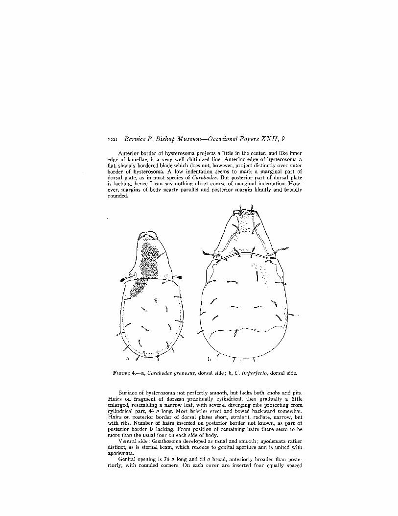
8. Carabodes imperfecta, new species (fig. 4, b).A female, 630 p- long, 360 j.£ broad. Color yellow brown.There is a deep notch between propodosoma and hysterosoma, but no cavity
()falshohle) under posterior border of propodosoma.Propodosoma rather flat; that is, surface of lamellae and surface of propodo
soma between them horizontal, therefore on same level with each other. Only ashallow depression between disk and lamellae which form margins of propodosoma.
Outer border of lamellae a sharp keel and both borders somewhat convergent. Anterior end of lamella rounded, without a special cusp. Rostrum betweenends of lamellae blunt. Inner borders of lamellae (the furrows) very convergentanteriorly in proximal parts, but then almost parallel. Upon rounded end oflamella is inserted lamellar hair, which is thick and rough and nearly appressedto anterior, rounded end of lamellae. There is a kind of translamella, made wavyby shallow pits, between anterior ends of lamellae. In front of this, on littleknobs, are inserted the two rostral hairs which are almost straight, very narrowly leaflike and each with a fine longitudinal rib. Part in front of translamelladistinctly pitted; behind it mostly indistinct, rounded, flat knobs scattered onsurface.
Somewhat behind mid-length of propodosoma are inserted interlamellar hairs,55 j.£ long, separated from each other as much as from margin of lamellae, erect,a little diverging, each like a narrow leaf with a distinct rib, the inner border ofleaf supplied with little teeth.
Bothridium is a cup, included in the posterior part of the lamella, as is usualin Carabodes, and the opening is thus directed outward. Sensillus is at firstdirected outward, but then bends upward. This organ is proximally rather strong,then tapers to a pointed distal end which, on its outside, has many fine littlebarbs directed toward point of bristle. Sensillus almost as long as interlamellarhair.
120 Bernice P. Bishop Museum-Occasional Papers XXII, 9
Anterior border of hysterosoma projects a little in the center, and like inneredge of lamellae, is a very well chitinized line. Anterior edge of hysterosoma aflat, sharply bordered blade which does not, however, project distinctly over outerborder of hysterosoma. A low indentation seems to mark a marginal part ofdorsal plate, as in most species of Carabodes. But posterior part of dorsal plateis lacking, hence I can say nothing about course of marginal indentation. However, margins of body nearly parallel and posterior margin bluntly and broadlyrounded.
"':: ..
b
/' ~\~
;-1~~I:1
FIGURE 4.-a, Carabodes granoslts, dorsal side; b, C. imperfecta, dorsal side.
Surface of hysterosoma not perfectly smooth, but lacks both knobs and pits.Hairs on fragment of dorsum proximally cylindrical, then gradually a littleenlarged, resembling a narrow leaf, with several diverging ribs projecting fromcylindrical part, 44 }Jo long. Most bristles erect and bowed backward somewhat.Hairs on posterior border of dorsal plates short, straight, radiate, narrow, butwith ribs. Number of hairs inserted on posterior border not known, as part ofposterior border is lacking. From position of remaining hairs there seem to bemore than the usual four on each side of body.
Ventral side: Gnathosoma developed as usual and smooth; apodemata ratherdistinct, as is sternal beam, which reaches to genital aperture and is united withapodemata.
Genital opening is 76 }Jo long and 68 }Jo broad, anteriorly broader than posteriorly, with rounded corners. On each cover are inserted four equally spaced
Sellnick-Acarina from Southeastern Polynesia 121
hairs in a longitudinal row nearer genital fissure than outer border. Hairs relatively long, 38 JL.
Anal aperture 88 JL long, broadest part of equal width; broader posteriorlythan anteriorly. Two hairs on each cover, stouter than those of genital coversand inserted near anal fissure, anterior one a little in front of middle, posteriorone approaching posterior margin. At sides of anal opening are inserted the following bristles: the first a little in front of cover, and 28 ,.. from it; second at alevel with half the length of cover, 28 JL from it; third behind posterior third ofopening and close to it. All hairs stouter than those of ventral side, but not asstout as those of posterior margin of body.
Genital opening 96 JL from anal opening, space between them covered withlow knobs which can only be seen in lateral aspect. Pair of hairs near genitalopening fine and fully as long as those on genital covers. Distance between thesehairs as great as that of anterior pair of anal covers, 100 JL.
Only one claw on tarsus. At' outside of genu I and II is inserted a swordlikehair which is 33 JL long and has a distinct longitudinal rib. No pits on femora Iand II.
Type, slide 017.Rurutu: Mato Naa (type locality), Aug. 30, one speCimen, Fos
berg.This species is related to the European Carabodes marginatus
(Michael). The sensillus has nearly the same form. But the sculptureof the dorsal surface is not the same, and the rostral and dorsal hairsof each species are individually distinct.
FAMILY HERMANNIELLIDAE GRANDJEAN, 1934
Genus Hermanniella Berlese, 1908
Type H ermannia granulata Nicolet, 1855.
9. Hermanniel1a punctulata columbiana Berlese, Redia 6: 362, 1910.Rimatara: northwest of Aurau, Sept. 5, one specimen, D. Anderson
and Y. Kondo.Heretofore recorded from British Columbia.
FAMILY ZETORCHESTIDAE MICHAEl., 1898
, Genus Zetorchestes Berlese, 1888
Type Carabodes micronychus Berlese, 1883.
10. Zetorchestes micronychus pacmcus, new subspecies.Female, 468 JL long and 360 JL wide.This new subspecies has the same somewhat globular body as typical species.
Border between propodosoma and hysterosoma a mere punctuated line. Lamellae
122 Bernice P. Bishop Museum-Occasional Papers XXII, 9
flat, nearly horizontal, narrow blades, a little remote from outline of propodosoma, projecting rather far forward, tapering to distal end, and this end is bentoutward at a blunt angle. At top of lamella, which has no cusp, is inserted lamellar hair of medium length, curved inward. Lamella covered with rather deep pits.Viewed laterally, border of lamella therefore appears to be wavy, as in Z. falzoniiCoggi.
Between blunt angles of both lamellae is a faint transverse line, apparently alow folding of surface, an indication of translamella.
Between insertions of lamellar hairs are two little apophyses, or knobs, containing rostral hairs. Lamellae and knobs subequally spaced. Rostral hairs ofequal width throughout, but distal end forked, inner prong a little longer thanouter one, both prongs short, diverging.
Interlamellar hairs very fine, scarcely discernible, shorter than lamellar hairs,directed toward each other.
Bothridium an ear-shaped, outwardly projecting cup at proximal part oflamella. Sensillus with a thin peduncle which widens gradually to a head ofclavate outline which is, however, a flat leaf. On surface of leaf lie, nearly appressed, longitudinally disposed, short rods. Sensillus directed outward and obliquely upward and apparently a little smaller than that of the European form.
In dorsal view may be seen a distinct cone at anterior corner of dorsal shield,a little outside bothridium. Apparently, the European form does not have thecone, for I did not draw it in my figure (24, fig. 51).
In the European form, the hairs on the dorsal surface are easily discernible.In the new subspecies, I see only the insertion points of very fine hairs. There isa long fissure near border of dorsal shield behind middle. In the European form,these fissures are also present, but they are nearer together in the new subspecies.
Genital and anal openings close, only a narrow chitinous rim between them.There is a remarkable low, but sharp, keel which begins at insertion of leg III,then takes a backward course to level of anterior border of genital opening, thenbends at a blunt angle inward and goes toward last quarter of genital opening.
Leg IV has almost same shape as that of Z. micronychus Berlese and Z.falzonii Coggi, but they seem to differ somewhat.
Rurutu: Mato Arei, under dead leaves, Aug. 26, one specimen, D.Anderson.
All the differences combined are not sufficient to justify establishing a new species, hence I consider this form to be a new subspecies.
BerIese established his species in 1883 (1, pt. 4) on the inner sideof the wrapper as Carabodes nticronychus. In 1888 (1, pt. 63) he gavea better description of the species. No details can be distinguished onhis figure of the entire animal, but leg IV seems to be drawn correctly.Comparing Berlese's figure with that which Coggi gives of Zetorchestes falzonii (4, fig. 5), I am of the opinion that the differences areso slight that the two species are identical.
Berlese's species is 450 p. long; and my specimens (24) are 440 p.
long. Coggi's Z. falzonii is 500 p. long and 350 p. broad. The Pacificsubspecies thus stands between Z. nticronychus and Z. jalzonii.
Sellnick-Acarina from Southeastern Polynesia 123
FAMILY EREMAEIDAE WILLMANN, 1931
Genus Oppia C. L. Koch, 1836
Type Oppia nitens C. L. Koch 1836.
11. Oppia ultraciliata (Jacot) ?Dameosoma ultraciliata Jacot, B. P. Bishop Mus., Bull. 121: 19,
1934.Rapa: Above Area, 400 ft., under stones, July 24, one specimen, D.
Anderson.Heretofore known only from Hawaii.
FAMILY CERATOZETIDAE JACOT, 1925
Genus Humerobates Sellnick, 1929
In 1929 (24), I established the genus H umerobates, with N otaspishumeralis Hermann as the type species. With this name I associatedthe species considered by Berlese and other acarologists as identicalwith Hermann's species, but Grandjean, in 1936 (6), pointed out thatBerlese's species is not the same as that of Hermann. New collectionsmade by Grandjean in localities around Strassburg, where Hermannfound his species, led Grandjean to conclude that N otaspis humeralisHermann is identical with Sphaerozetes (Trichoribates) numerosusSellnick, 1924 (20). According to the rules of nomenclature, the generic name Humerobates must stand, but the name of the type speciesmust be changed. Grandjean calls it rostrolamellatus. Having takenthe new generic name Diapterobates for the rediscovered N otaspishumeralis Hermann, we have to use the names as follows:
Diapterobates Grandjean, Ent. Soc. France, Ann. 105: 79, 1936 (typespecies N otaspis humeralis Hermann, Mem. Apt., 92, 1804).
Hwrnerobates Sellnick, Tierwelt Mitteleur. 3 (9) : 11, 1929 [type species Humerobates rostrolamellatus Grandjean (misidentified asN otaspis humeralis by Sellnick), Soc. Ent. France, Ann. 105: 77,1936] .
There are three described forms of Humerobates: (1) H. rostrolamellatus Grandjean (fig. 5, a, b), widespread in Europe; (2) H.humeralis arborea (Banks) (Oribata arborea) , Am. Ent. Soc., Trans.22: 7,1895, of the United States; (3) H. humeralis perkinsi Jacot,B. P. Bishop Mus., Bull. 121: 67, 1934, of the Hawaiian Islands.
124 Bernice P. Bishop Museum-Occasional Papers XXII, 9
I am of the opinion that the two subspecies are good species. Although Jacot confirms that he knows the type species (14, p. 67), heconsiders the specimens found in the Hawaiian Islands only a variety,perkinsi.
I have collected H. rostrolamellatus several times in Germany, thelast time on July 17, 1948, on Calluna vulgaris near Ganderkesee inOldenburg. After studying these specimens and comparing them with
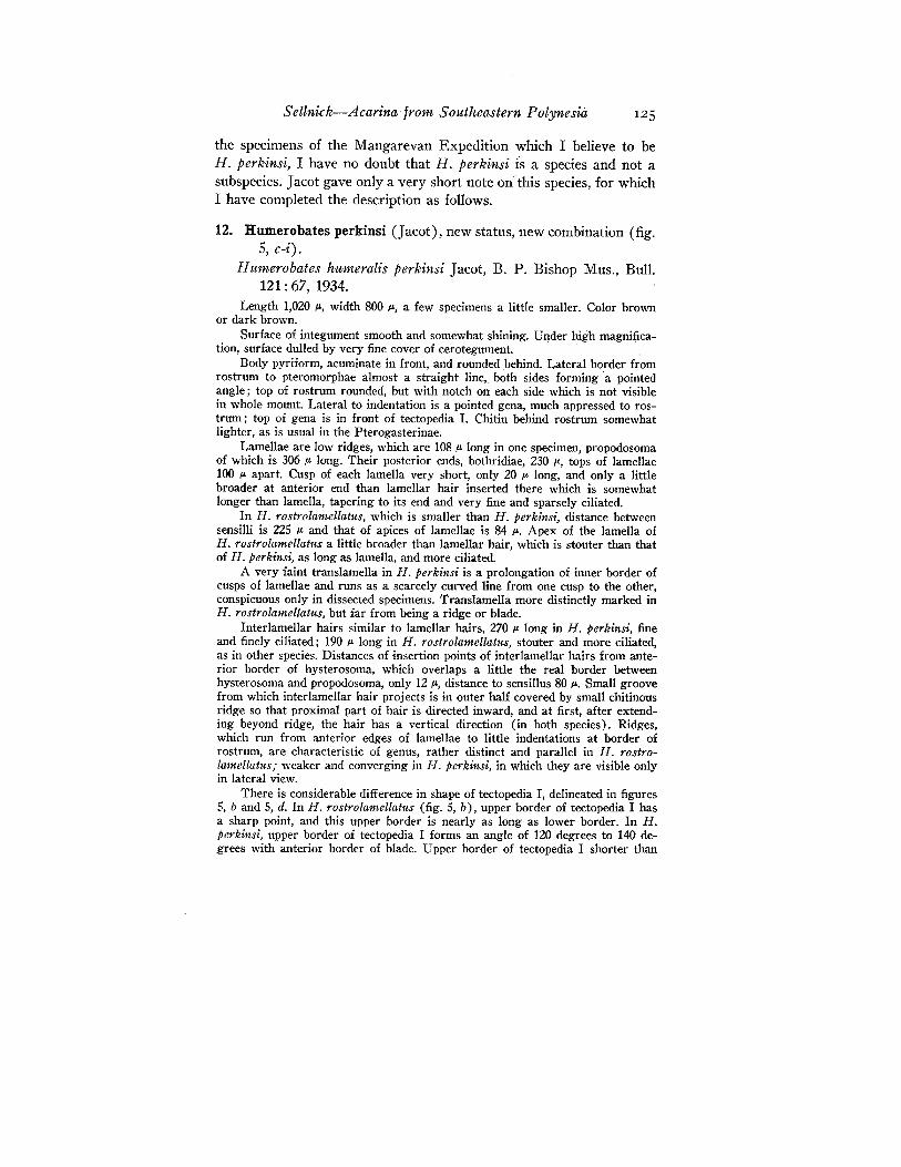
FIGURE 5.-a, b, Humerobates rostrolamellatus: a, pteromorpha; b, propodosoma, oblique lateral view. c-i, H. perkinsi: c, pteromorpha; d, propodosoma,oblique lateral view; e, palp; f, gnathosoma; g, mandible; h, leg I; i, leg IV.
Sellnick-Acarina from SOlttheastern Polynesia 125
the specimens of the Mangarevan Expedition which I believe to beH. perkinsi, I have no doubt that H. perkinsi is a species and not asubspecies. Jacot gave only a very short note on"this species, for whichI have completed the description as follows.
12. Humerobates perkinsi (Jacot), new status, new combination (fig.5, c-i).
Humerobates humeralis perkinsi Jacot, B. P. Bishop Mus., Bull.121: 67, 1934.
Length 1,020 iL, width 800 iJ-, a few specimens a little smaller. Color brownor dark brown.
Surface of integument smooth and somewhat shining. Under high magnifjcation, surface dulled by very fine cover of cerotegument.
Body pyriform, acuminate in front, and rounded behind. Lateral border fromrostrum to pteromorphae almost a straight line, both sides forming a pointedangle; top of rostrum rounded, but with notch on each side which is not visiblein whole mount. Lateral to indentation is a pointed gena, much appressed to rostrum; top of gena is in front of tectopedia 1. Chitin behind rostrum somewhatlighter, as is usual in the Pterogasterinae.
Lamellae are low ridges, which are 108 iJ- long in one specimen, propodosomaof which is 306 iJ- long. Their posterior ends, bothridiae, 230 JL, tops of lamellae100 JL apart. Cusp of each lamella very short, only 20 JL long, and only a littlebroader at anterior end than lamellar hair inserted there which is somewhatlonger than lamella, tapering to its end and very fine and sparsely ciliated.
In H. rostrolamellatus, which is smaller than H. perkinsi, distance betweensensilli is 225 JL and that of apices of lamellae is 84 JL. Apex of the lamella ofH. rostrolamellatlls a little broader than lamellar hair, which is stouter than thatof H. perkinsi, as long as lamella, and more ciliated.
A very faint translamella in H. perkinsi is a prolongation of inner border ofcusps of lamellae and runs as a scarcely curved line from one cusp to the other,conspicuous only in dissected specimens. Translamella more distinctly marked inH. rostrolamellatus, but far from being a ridge or blade.
Interlamellar hairs similar to lamellar hairs, 270 iJ- long in H. perkinsi, fineand finely ciliated; 190 iL long in H. rostrolamellatrts, stouter and more ciliated,as in other species. Distances of insertion points of interlamellar hairs from anterior border of hysterosoma, which overlaps a little the real border betweenhysterosoma and propodosoma, only 12 iJ-, distance to sensillus 80 JL. Small groovefrom which interlamellar hair projects is in outer half covered by small chitinousridge so that proximal part of hair is directed inward, and at first, after extending beyond ridge, the hair has a vertical direction (in both species). Ridges,which run from anterior edges of lamellae to little indentations at border ofrostrum, are characteristic of genus, rather distinct and parallel in H. rostrolamellatus; weaker and converging in H. pcrkinsi, in which they are visible onlyin lateral view.
There is considerable difference in shape of tectopedia I, delineated in figures5, band 5, d. In H. rostrolamellatlts (fig. 5, b), upper border of tectopedia I hasa sharp point, and this upper border is nearly as long as lower border. In H.perkinsi, upper border of tectopedia I forms an angle of 120 degrees to 140 degrees with anterior border of blade. Upper border of tectopedia I shorter than
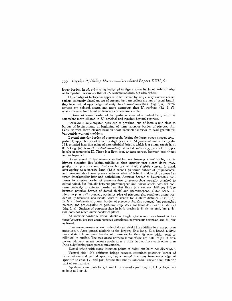
126 Bernice P. Bishop Museum-Occasional Papers XXII, 9
lower border. In H. arborea, as indicated by figure given by Jacot, anterior edgeof tectopedia I resembles that of H. rostrolamellatlls, but also differs.
Upper edge of tectopedia appears to be formed by single very narrow archedrodlets, obliquely placed on top of one another. As rodlets are not of equal length,they terminate at upper edge unevenly. In H. rostrolamellatus (fig. 5, b), terminations are pointed, sharp, and more numerous than H. perkinsi (fig. 5, d),where three to four blunt or truncate corners are visible.
In front of lower border of tectopedia is inserted a rostral hair, which issomewhat more ciliated in H. perkinsi and reaches beyond rostrum.
Bothridium an elongated open cup at proximal end of lamella and close toborder of hysterosoma, at beginning of inner anterior border of pteromorpha.Sensillus with short, clavate head on short peduncle; interior of head granulated,but outside without markings.
Beyond anterior border of pteromorpha begins the large, spoon-shaped tectopedia II; upper border of which is slightly curved. At proximal end of tectopediaII is situated insertion point of exobothridial bristle, which is a stout, rough hair,80 JL long (65 JL in H. rostrolamellatus), directed anteriorly, parallel to upperborder of tectopedia II. There is a light spot, an area porosa, between bothridiumand tectopedia I.
Dorsal shield of hysterosoma arched but not forming a real globe, for itshighest elevation lies behind middle so that anterior part slopes down moregently· than posterior one. Anterior border of shield slightly convex forward,overlapping as a narrow band (12 JL broad) posterior border of propodosoma,and covering short area porosa anterior situated behind middle of distance between interlamellar hair and bothridium. Anterior border of hysterosoma continues to anterior border of pteromorphae. Pteromorphae movably attached todorsal shield, but fine slit between pteromorphae and dorsal shield does not continue perfectly to anteriorporder, so that there is a narrow chitinous bridgebetween anterior border of dorsal shield and pteromorphae. Outer border ofpteromorphae well rounded; posterior edge of pteromorpha continues along border of hysterosoma and bends down to venter for a short distance (fig. 5, c).In H. rostrolamellatus, outer border of pteromorpha also rounded, but somewhatpointed, and prolongation of posterior edge does not bend downward at its end(fig. 5, a). Surface of pteromorphae in both species is finely striated, but striation does not reach outer border of plates.
At anterior border of dorsal shield is a light spot which is as broad as distance between the two areae porosae anteriores, converging posteriad and as longas broad.
Four areae porosae on each side of dorsal shield (in addition to areae porosaeanteriores). Area porosa adalaris is the largest, 60 JL long, 32 JL broad, a littlemore distant from inner border of pteromorpha than its own width, oval orelliptical in outline. The two areae porosae mesonoticae not half length of areaporosa adalaris. Areae porosae posteriores a little farther from each other thanfrom neighboring area porosa mesonotica.
Dorsal shield with many insertion points of hairs, but hairs not discernible.Ventral side: No chitinous bridge between chitinized posterior border of
camerostome and genital aperture, but a curved line runs from outer edge ofaperture to coxa IV, and part behind this line is somewhat darker than anteriorpart of ventral side.
Apodemata are dark bars, I and II of almost equal length; III perhaps halfas long as I or II.
Sellnick-Acarina from Southeastern Polynesia 127
Genital opening a rounded trapezium, anterior border broader than posterior{me, with seven fine hairs on each cover.
Ami! opening, twice length of genital opening, much larger, broadly oval,anterior part more pointed, with usual two hairs widely· separated.
Hairs on ventral plate of moderate length (SO /L), slender on sternal part,stouter behind anal opening.
There is an area porosa postanalis close to posterior border of ventral platewhich is discernible only if posterior part is seen from behind. This area is 16 /Llong and 100 JL broad, as broad as one of the covers of anal opening. H. rostrolameliatlts bas this area too. Thus, it seems to be a generic character. In 1923(19), I discovered this area in the Galumninae. It exists, as far as I know, ina number of Galumninae, but not in Neoribates and Stictozetes. It is extraordinary that Hltmerobates and Trihumerozetes, new genus, also have this area.
Tarsi terminated by trihomohamate ungues, outer hooks longer and lessbent at distal half than inner one. The three species have some differences as toarmature of legs, especially in leg IV. Distal end of tibia IV with four bristles.Hair on dorsal side of segment not exactly length of segment, very fine, andlacks ciliation. Two ciliated bristles on ventral side. In H. arborea, Jacot drawsboth bristles as equal in length; in H. rostrolamelia/lts, distal of these two bristlesis one and one-half times as long as the other; in H. perki1!si, distal bristle isthree times as long as the other. On ventral side of tarsus are numerous barbedbristles, and there are also differences between these bristles in the three forms.
Figure 5, e shows the palp; 5, f, the gnathosoma; and 5, g, themandible of H. perkinsi.
Slides 018-020.Rapa: Mt. Ororangi, southeast valley, 600-700 ft., July 3, one
specimen, Zimmerman. Mangaoa Peak, 900-1,200 ft., July 4, twospecimens, Zimmerman.
Marotiri: Southeast Islet, July 22, 72 specimens, Zimmerman andFosberg.
Tahiti: Mt. Aorai Trail, 3,500-4,500 ft., on M etrosideros, Sept. 13,one specimen, Zimmerman.
Heretofore known only from the Hawaiian Islands.
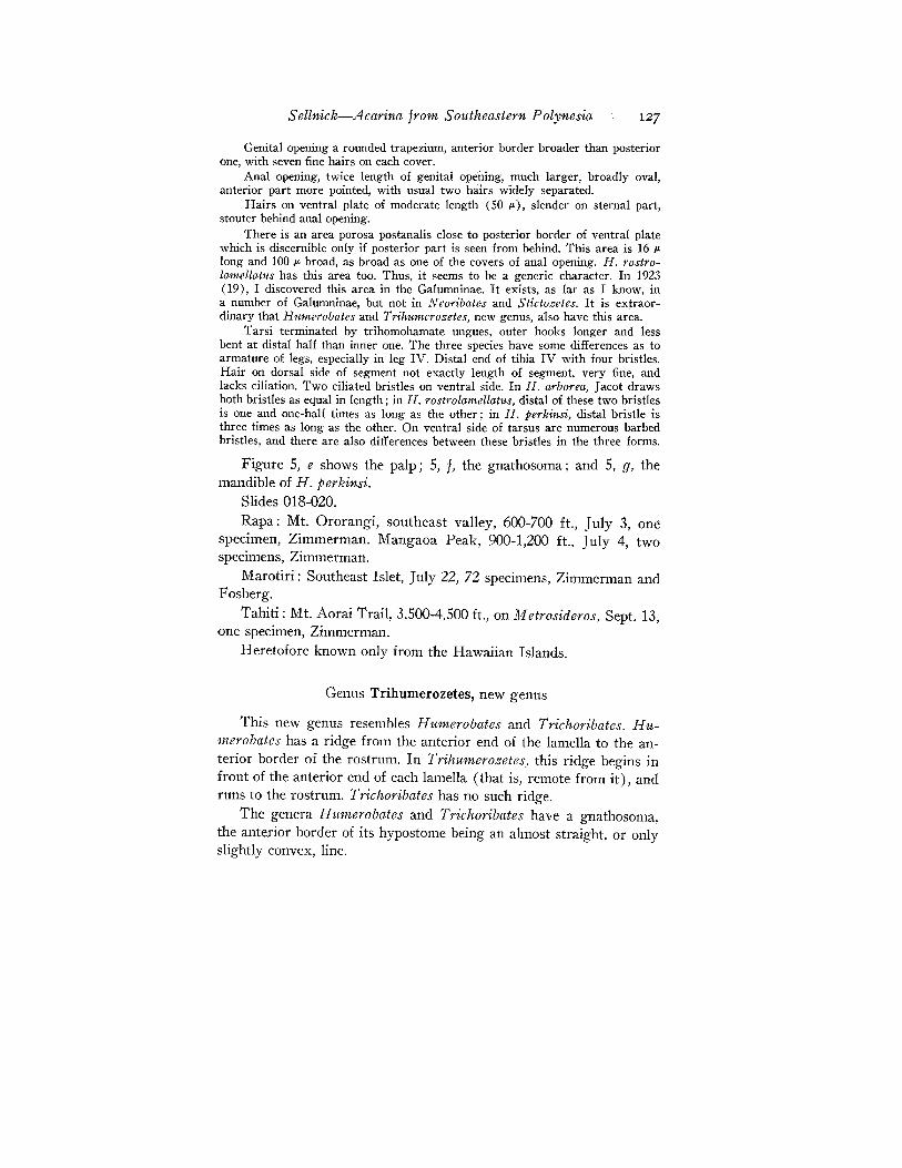
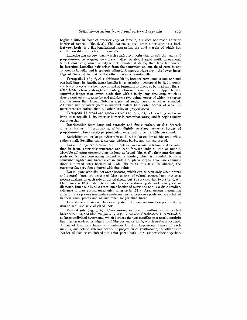
Genus Trihumerozetes, new genus
This new genus resembles Humerobates and Trichoribates. Humerobates has a ridge from the anterior end of the lamella to the anterior border of the rostrum. In Trihu,merozetes, this ridge begins infront of the anterior end of each lamella (that is, remote from it), andruns to the rostrum. Trichoribates has no such ridge.
The genera Humerobates and Trichoribates have a gnathosoma,the anterior border of its hypostome being an almost straight, or onlyslightly convex, line.
128 Bernice P. Bishop Museum-Occasional Papers XXII, 9
In Trihumerozetes, the anterior border of the hypostome has oneach anterior corner a remarkable projection, similar to a knob, orhorn (fig. 6, f). I know of no genus of Oribatidae with a hypostomeformed in such a manner.
Type Trihumerozetes cornutus, new species.
13. Trihumerozetes cornutus, new species (fig. 6).Length 790 1Jo, width 560 1Jo. Color of unmounted animal black; pteromorphae
brown.Dorsal side of body vcry highly arched.Propodosoma makes up nearly a third of body. Rostrum bluntly rounded, and
with a little impression on each side at anterior edge. A longitudinal carina
c
FIGURE 6.-Trihumero::etes cornutus: a, dorsal side; b, ventral side; c,area porosa postanalis; d, pteromorpha; e, propodosoma; f, gnathosoma; g,genu and tibia I; h, leg IV.
Sellnick-Acarina from Southeastern Pol)mesia 129
begins a little in front of anterior edge of lamella, but does not reach anteriorborder of rostrum (fig. 6, e). This carina, as seen from outer side, is a keel.Between keels, is a flat longitudinal impression, the hind margin of which hasa little nose-like projection in its middle.
Lamellae are narrow keels which reach from bothridiae to half the length ofpropodosoma, converging toward each other, of almost equal width throughout,with a short cusp which is only a little broader at its top than lamellar hair atits insertion. Lamellar hair arises from the somewhat oblique tip. of cusp, is notas long as lamella, and is sparsely ciliated. A narrow ridge from the lower inneredge of one cusp to that of the other marks a translamella.
Tectopedia I (fig. 6, e) a chitinous blade, broader than lamella and one andone-half times its length, hence lamella is remarkably overtopped by it. Its upperand lower borders are bent downward at beginning in front of bothridium; thereafter, blade is nearly straight and enlarges toward its anterior end. Upper bordersomewhat longer than lower; blade thus with a fairly long, free cusp, which isdeeply notched at its anterior end and forms two points, upper of which is shorterand narrower than lower. Notch is a pointed angle, base of which is, rounded.At inner side of lower point is inserted rostral hair, outer border of which ismore strongly barbed than all other hairs of propodosoma.
Tectopedia II broad and spoon-shaped (fig. 6, a, b), not reaching as far infront as tectopedia 1. Its anterior border is somewhat wavy, and it begins underpteromorpha.
Interlamellar hairs long and sparsely and finely barbed, arising beneathanterior border of hysterosoma, which slightly overlaps posterior border ofpropodosoma. Hairs nearly perpendicular, only distally bent a little backward.
Bothridium rather large, oviform in outline, but flat on dorsal side, and orificerather small. Sensillus short, clavate, without barbs, and not sculptured.
Dorsum of hysterosoma oviform in outline, well-rounded behind and broaderthan in front, anteriorly truncated and bent forward only a little at middle.Movable adhering pteromorphae as long as broad (fig. 6, d), their anterior andposterior borders converging toward outer border, which is rounded. From asomewhat lighter and broad area in middle of pteromorpha arise fine channelsdirected toward outer borders of blade, like roots of a tree. In addition, thepteromorpha very finely dotted with fine points.
Dorsal plate with distinct areae porosae, which can be seen only when dorsaland ventral plates are separated. Most species of related genera have one areaporosa adalaris on each side of dorsal shield, but T. cornutus has two (fig. 6, a).Outer area is 18 I-' distant from outer border of dorsal plate and is as great indiameter. Inner one is 55 I-' from inner border of outer one and is a little smaller.Distance to area porosa mesonotica anterior is 125 1-'. Area porosa mesonoticaanterior, area porosa mesonotica posterior, and area porosa posterior are situatedin their usual places and all are much longer than broad.
I could see no hairs on the dorsal plate; but there are insertion points at theusual places, and several gland spots.
Ventral side (fig. 6, b): Camerostome oviform in outline and somewhathroader behind, and hind margin only slightly convex. Gnathosome is remarkable,as large undivided hypostome, which borders the two maxillae in a nearly straightline, has on each outer edge a toothlike corner, or knob, which projects forward.A pair of fine, long hairs is in anterior third of hypostome. Hairs on eachmaxilla, one behind anterior border of projection of gnathosome, the other nearborder of darker chitinized posterior part; both hairs rather close together.
130 Bernice P. Bishop Museum-Occasional Papers XXII, 9
A real sternal beam is lacking, but there are two rather clear apodemata onboth sides. Hairs on sternal part shown in figure 6, b.
Genital aperture has rounded trapezoidal aspect frequently found in Oribatidae, broader in front than behind, each cover with six hairs, anterior ones longerthan posterior.
Anal aperture large, its own length distant from genital aperture, not thesame distance from hind margin of ventral plate, which cannot be seen frombelow, because this part is almost perpendicular. Each anal cover has usual twohairs, the hair in front small, near anterior border, and in middle of plate,posterior one longer and near anal fissure. Behind each posterior edge of aperture are two hairs, as long as posterior bristles of anal covers.
A special character of the species is an area porosa postanalis, which is foundin only a few genera of Oribatidae, which is close to posterior border of ventralplate behind anal aperture and can only be seen if specimen is dissected (fig. 6, c).
Tarsi of all legs have three claws, middle one thicker than outer ones. Thereis a very long hair on dorsal side of genu and tibia I and II (fig. 6, g). On lowerpart of front edge of each genu of both legs is a sharp chitinous point or a bladedirected toward tibia and overlapping posterior end of this segment. Femur IVdilated on ventral edge into a flat keel, anterior corner of which is rounded,posterior corner rectangular. Even ventral edge of coxa IV ·is a keel (fig. 6, h).On the other hand, dorsal edge of tibia and tarsus IV is a very sharply flattenedborder. Hairs of legs have fine, sharp points.
Slides of the dissected holotype 021, a-f.Rapa: Mt. Perahu (type locality), east ridge, 1,400-1,700 ft., one
specimen. There was no collector or date mentioned on the label in thetube, but in comparison with other labels, it is probable that the dateis July 28 and the collector Zimmerman.
FAMILY GALUM~nDAEGRAND]EAN, 1936
Genus Notogalumna, new genus
No border between propodosoma and hysterosoma. Hind margin of hysterosoma a straight line, and posterior lateral margins of body straight to pteromorphae. Posterior margin and posterior lateral margin form an angle of morethan 90 degrees; comer of angle rounded.
Areae porosae adalares exceedingly remote from pteromorphae. One largearea porosa mesonotica and one area porosa posterior on each side.
Type N otogalumna praetiosa, new species.
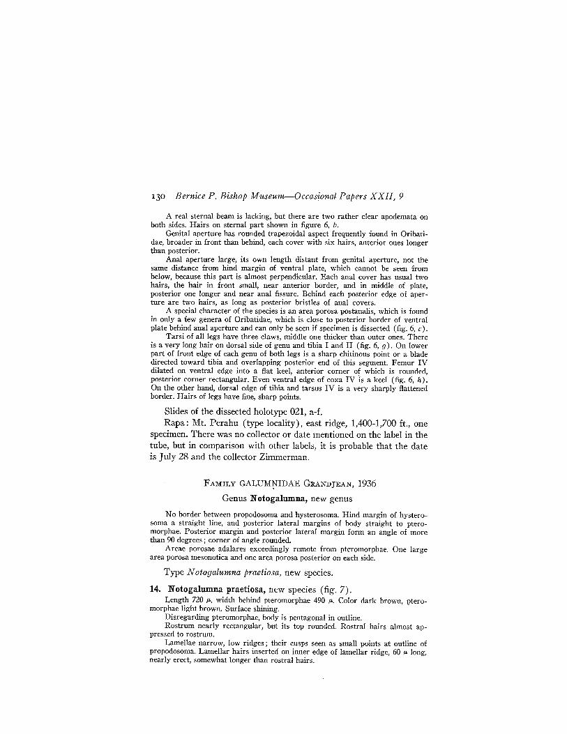
14. Notogalumna praetiosa, new species (fig. 7).Length 720 flo, width behind pteromorphae 490 flo. Color dark brown, ptero
morphae light brown. Surface shining.Disregarding pteromorphae, body is pentagonal in outline.Rostrum nearly rectangular, but its top rounded. Rostral hairs almost ap
pressed to rostrum.Lamellae narrow, low ridges; their cusps seen as small points at outline of
propodosoma. Lamellar hairs inserted on inner edge of lamellar ridge, 60 flo long,nearly erect, somewhat longer than rostral hairs.
Sellnick-Acarina from Southeastern Polynesia 131
Interlamellar hairs finer than lamellar hairs, and inserted near inner borderof beginning of lamellar ridges, 40 fL long, bent downward and almost appressedto surface of propodosoma.
Bothridium a short cup. Sensillus a slender hair, tapering and pointingtoward its distal end, directed backward and a little outward, 100 p. long.
A narrow, short ridge running parallel to lamellar ridge outside of lamellatoward insertion of leg I is half as long as lamella. I believe that this ridge mustbe tectopedia 1.
m··:"" ~" ".....-
FIGURE 7.-Notogalltmna praetiosa: a, dorsal side; b, pteromorpha; C, ventral side.
There is no border between propodosoma and hysterosoma. But there seemsto be a very short slit near the inside of bothridium; perhaps this is the indication of a front margin on hysterosoma. In front of this slit is the very narrowand short area porosa anterior. Both slit and area can be seen only under highmagnification in dissected specimens. This area must not be confused with thetwo dark spots behind interlamellar hairs, which apparently are thickenedchitinous parts in interior of body (attachment area of mandibular retractor?).(See figure 7, a.)
Areae porosae adalares short, elliptical, 40 fL in diameter, but' form not constant. Some specimens have one area rounded, the other with corners. Distance ofthe area from inner edge of pteromorpha is 120 fL, three times the diameter ofarea. This position, far from the pteromorphae, is remarkable and strange in thePterogasterinae. Distance between the two areae is 128 fL, the same distancebetween areae porosae adalares and area porosa mesonotica. There is only onearea porosa mesonotica, which is very large, 162 fL in total length, and formed
132 Bernice P. Bishop Museum-Occasional Papers XXII, 9
like a little sock, the foot part of which is anterior, the heel directed outward.Area porosa posterior covers posterior corner of body, is triangular in shape,and is almost as long as area porosa mesonotica. Outward from latter are several fissures. There are a number of insertion points of hairs, but I could notdiscern any hairs.
Pteromorphae movable, attached to edge of dorsal shield. Their shape issimilar to that of Z etes. A very fine venation issues from light spots in pteromorphae (fig. 7, b).
Ventral side (fig. 7, c) shows the parallel edges of real sternal plate; inline with genital opening, edges bend outward and end at posterior corner ofpteromorphae.
First apodema a narrow bar, almost horizontal; both of these apodemataunited by a distinct transverse line. Apodemall as long as I; it is at first directedobliquely backward, then bends toward anterior corner of genital opening, butdoes not reach it. Apodema III a little more than half as long as II, horizontalat first, then bent backward, ending in a small thickening. Several light spotson sternal part and behind apodema III. .
Genital opening is far forward and a little broader in front than behind,rounded, laterally straight, posterior border straight. Two hairs inserted parallelto and near anterior border of each cover, and three hairs longitudinally inserted,parallel to side and placed more toward outer border of cover. A point nearposterior border of each cover may be insertion point of a hair.
Anal opening is, as usual, anteriorly narrower than posteriorly, of equallength and width, twice its own length from genital opening, half its length fromposterior border of body. Apparently three hairs on each cover, one near anteriorborder, two longitudinally inserted near posterior end of cover.
Following hairs discernible on ventral side of body: one in front of centerof apodema I; a second behind outer corner of apodemall and outside brim atmargin of ventral plate; a third between knob on apodema III and genital opening; a fourth behind knob and somewhat outward; a fifth behind genital opening(as far from each other and from opening as width of latter) ; in front of outercorner of anal opening is sixth hair and behind posterior corner of opening andobliquely inserted are seventh and eighth bristles. At outside of anterior quarterof anal opening and close to it is a longer fissure.
No area porosa postanalis behind anal opening, but a number of light, smallspots, perhaps muscle scars.
Tarsi with three claws of almost equal size. On ventral side of tarsi twobristles, slightly toothed on ventral face.
Slides of allotype 022, a-d; slide of paratype 023.Raivavae: Mt. Turivao (type locality), south slope, Aug. 11, two
specimens, W. Anderson.Raiatea: Toahiva Valley, Oct. 7, one specimen, D. Anderson.
15. Zetes bryani marquesi Jacot, B. P. Bishop Mus., Bull. 114: 231,1934 (1935).Slides 024, a-d.Rapa: Mt. Tepiahu, south slope, 400-600 ft., July 20, eight speci
mens, Zimmerman.. Teutu, on dead leaves, July 27, two specimens,
Sellnick-Acarina from Southeastern Polynesia 133
Kondo. Mt. Tanga, north slope, 300-700 ft., July 31, one specimen, D.Anderson.
Flint: Oct. 16, 10 specimens, Kondo and D. Anderson.Known elsewhere only from the Marquesas.
16. Galumna hawaiiensis Jacot, B. P. Bishop Mus., Bull. 121: 77,1934.Slides 025, a-b.Rapa: Near Ahurei cemetery, under stones, July 28, two speci
mens, W. Anderson. Mt. Tanga, north slope, July 31, 300-700 ft.,under dead leaves, one specimen, D. Anderson.
Tahiti: Mt. Aorai Trail, 3,500 ft., Sept. 12, one specimen, Zimmerman.
Known heretofore from the Hawaiian Islands.
17. Galumna hawaiiensis marquesana Jacot, B. P. Bishop Mus., Bull.114: 232, 1934 (1935).Slides 026, a-b.Rapa: Mt. Tepiahu, south slope, 400-600 ft., July 20, one speci
men, Zimmerman.
FAMILY ORIPODIDAE JACOT, 1925
Genus Anoripoda, new genus
Very similar to Oripoda Banks and Pergande, 1904, because pteromorphaeare formed as in that genus. Bothridium entirely covered by anterior border ofpteromorphae, and sensiIIus partially covered. In Oripoda, anterior border ofrostrum is gently and broadly rounded; in Anoripoda center of rostrum projectslike a narrow but rounded nose, and on each side of this projection is a pro·tuberance of equal length, but narrower, at head of which is inserted rostral hair.
Type Anoripoda nasalis, new species.
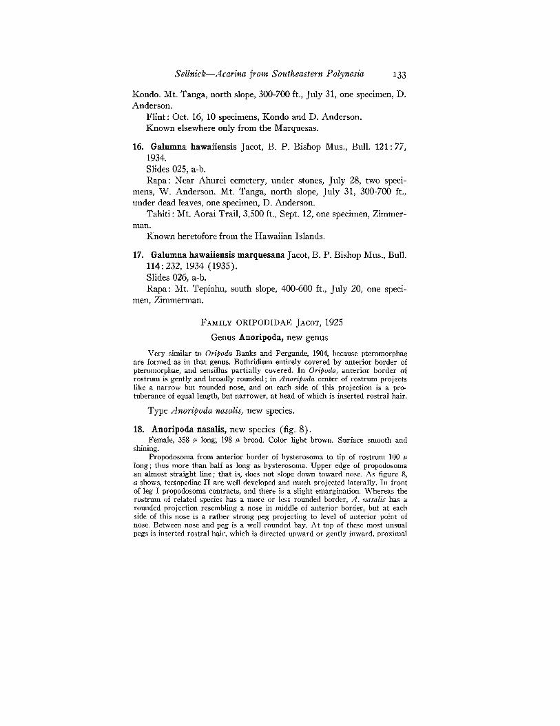
18. Anoripoda nasalis, new species (fig. 8).Female, 358 p, long, 198 p. broad. Color light brown. Surface smooth and
shining.Propodosoma from anterior border of hysterosoma to tip of rostrum 100 p.
long; thus more than half as long as hysterosoma. Upper edge of propodosomaan almost straight line; that is, does not slope down toward nose. As figure 8,a shows, tectopediae II are well developed and much projected laterally. In frontof leg I propodosoma contracts, and there is a slight emargination. Whereas therostrum of related species has a more or less rounded border, A. nasalis has arounded projection resembling a nose in middle of anterior border, but at eachside of this nose is a rather strong peg projecting to level of anterior point ofnose. Between nose and peg is a well rounded bay. At top of these most unsualpegs is inserted rostral hair, which is directed upward or gently inward, proximal
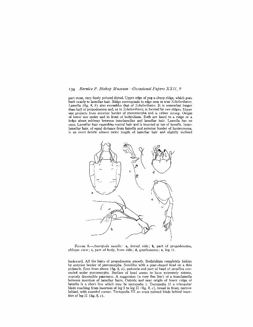
134 Bernice P. Bishop Museum-Occasional Papers XXII, 9
part stout, very finely pointed distad. Upper edge of peg a sharp ridge, which goesback nearly to lamellar hair. Ridge corresponds to edge seen in true Scheloribates.Lamella (fig. 8, b) also resembles that of S cheloribates. It is somewhat longerthan half of propodosoma and, as in Scheloribates, is formed by two ridges. Upperone projects from anterior border of pteromorpha and is rather strong. Originof lower one under and in front of bothridium. Both are fused to a ridge or aledge about midway between interlamellar and lamellar hair. Lamella has nocusp. Lamellar hair resembles rostral hair and is inserted at top of lamella. Interlamellar hair, of equal distance from lamella and anterior border of hysterosoma,is an erect bristle almost twice length of lamellar hair and slightly inclined
a ./~.
FIGURE 8.-Anoripoda nasalis: a, dorsal side; b, part of propodosoma,oblique view; c, part of body, from side; d, gnathosoma; e, leg II.
backward. All the hairs of propodosoma smooth. Bothridium completely hiddenby anterior border of pteromorpha. Sensillus with a pear-shaped head on a thinpeduncle. Seen from above (fig. 8, a), peduncle and part of head of sensillus concealed under pteromorpha. Surface of head seems to have extremcly minute,scarcely discernible punctures. A suggestion (a very fine line) of a translamellabetween insertion of lamellar hairs. Outside and near origin of lower ridge oflamella is a short line which may be tectopedia I. Tectopedia II a triangularblade reaching from insertion of leg I to leg II (fig. 8, c), broad in front, narrowbehind, with rounded corner. Tectopedia III an erect pointed blade behind insertion of leg II (fig. 8, c).
Sellnick-Acarina from Southeastern Polynesia 135
Suture between propodosoma and hysterosoma very distinct, perfectly-straight, and bent posteriorly only at outer ends. Suture crosses surface of bodywell separated from anterior border of pteromorphae. Edge of lamellae andanterior edge of pteromorpha form a rounded excavation, leaving head of sensillusvisible. Pteromorphae drawn like a narrow chitinous band around posterior partof body; similar to those of Oripoda, but still more distinct. No suture betweendorsal shield and pteromorphae. Dorsal shield appears to reach to outer borderof pteromorphae; but through integument, one can see real border of body.
Hairs on dorsal shield thin but distinctly discernible, a lateral row of fourhairs and a mesal one of three. Kear excavation behind sensillus is an areaporosa, and in front of second hair of inner row, a long transverse fissure. Possibly there are more areae, but I could not discover them.
Nose of rostrum forms a tectal plate for anterior ends of mandibles. Lowerborder of nose is anterior border of camerostome. Gnathosoma has common formin Oribatidae. Plate covers posterior part of camerostome, maxillae foremost.Palpus with five segments, if we consider a very narrow chitinous piece aspalpcoxa. There are three short, stout, little thorns in one row on the border ofpalptarsus and over them a short, thick bristle (fig. 8, d).
Mandibles normal; the movable segment with five stout dark teeth; fixedsegment with four teeth.
A few chitinous lines directed obliquely backward could be called apodemata.Apodemall goes to anterior border of genital aperture, III goes to the posteriorborder, and IV is very short and almost horizontal.
Genital opening situated a little in front of middle of ventral side of body;it is small, anteriorly a little narrower than posteriorly, corners rounded. Analaperture lies at posterior margin of ventral side, is twice as long as broad, andone and one-half times its own length distant from genital opening. Rather longhairs on each anal cover, and two others close to aperture on venter.
Tarsi trihomohamate. Tarsus of all legs rather short. Femur I without bladeon ventral edge. Femur II with a not very broad but distinct blade, with roundedanterior corner. Femora III and IV with a kind of keel, but no blade.
Holotype, slides 027, a-d.Raiatea: Tetaro Islet (type locality), Oct. 4, one specimen, Cooke
and Kondo.
FAMILY ORIBATULIDAE JACOT, 1929
Genus Scheloribates Berlese, 1908
Type: Zetes latipes C. L. Koch, 1841.
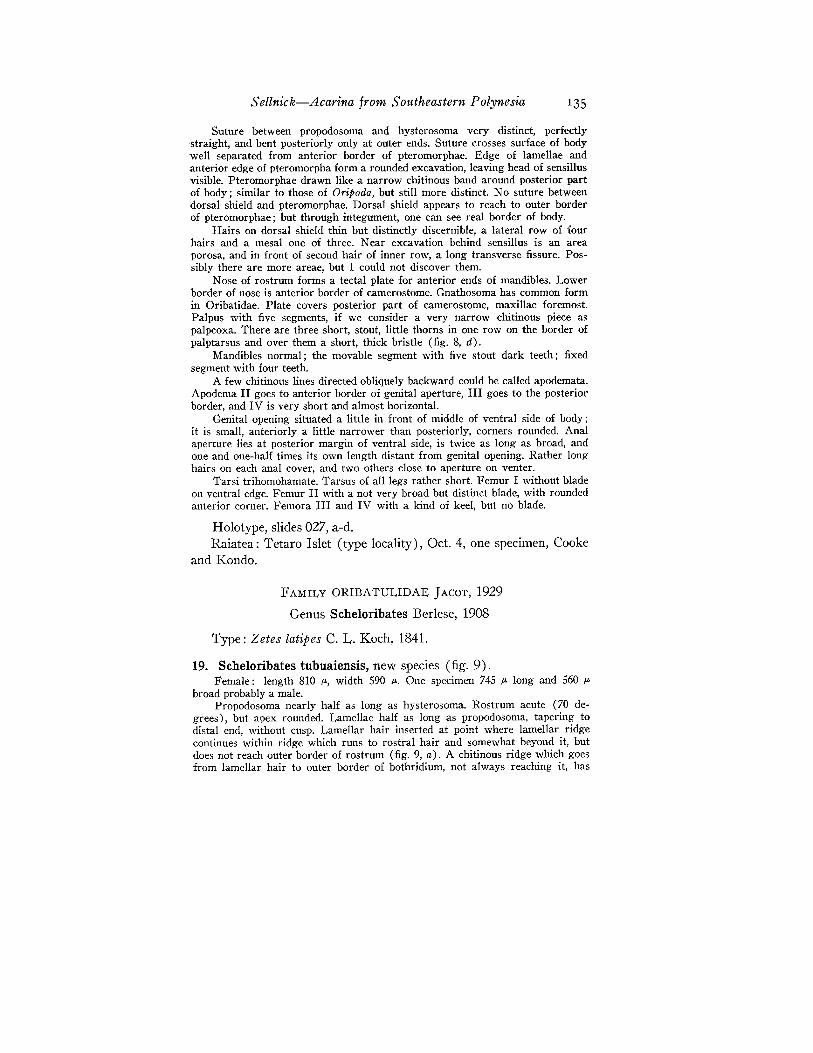
19. Scheloribates tubuaiensis, new species (fig. 9).Female: length 810 P-, width 590 P-. One specimen 745 P- long and 560 P
broad probably a male.Propodosoma nearly half as long as hysterosoma. Rostrum acute (70 de
grees), but apex rounded. Lamellae half as long as propodosoma, tapering todistal end, without cusp. Lamellar hair inserted at point where lamellar ridgecontinues within ridge which runs to rostral hair and somewhat beyond it, butdoes not reach outer border of rostrum (fig. 9, a). A chitinous ridge which goesfrom lamellar hair to outer border of bothridium, not always reaching it, has
136 Bernice P. Bishop Museum--Occasional Papers XXII, 9
been called tectopedia I by several acarologists. A short line at inside of tip oflamella marks beginning of a translamella.
Interlamellar hair is longer than lamellar hair, erect, sometimes bent backward. All hairs of propodosoma simple, not barbed.
Orifice of bothridium a chitinous ring with a pointed angle directed outward, and a blade-like chitinous scale which partly overlaps opening. Scale plainlyvisible in dorsal view. Sensillus with penduncle bent in S-form, shorter thanclaviform head; head bent upward or somewhat backward beyond anterior borderof pteromorpha; it has no hairs or aciculation.
Tectopedia II a spoon-shaped blade.
).1
FIGURE 9.-Scheloribates tubuaiensis: a, dorsal side; b, ventral side; e,part of propodosoma, laterally; d, leg I; e, leg II; f, leg IV.
Anterior margin of hysterosoma slightly convex. Dorsal plate polished. Noreal areae porosae. Along border of shield, and somewhat remote from it, areone or two rows of small muscle scars. Close to these rows, and on inner side ofthem, are on each side of dorsal shield four elongated spots which may be calledfissures, which are longitudinal slits; behind them, in inner part of chitinization,is an indistinct cavity. Position of fissures corresponds to those of areae porosaein other Pterogasterinae. A similarly formed fissure lies on a level with anteriormesonotical one, as far or nearly as far from it as anterior mesonotical fissure
Sellnick-Acarina from Southeastern Polynesia 137
is from second mesonotical fissure. In chitinization of dorsal shield are two other,transverse, slits, one in front of anterior mesonotica, the second close outsideposterior fissure.
I saw seven pairs of insertion points of hairs on hysterosoma, each withnerve channel into interior of dorsal shield, but could see no hairs.
Figure 9, b shows ventral side of body, legs, and mouthparts omitted. A verydistinct medial sternal rib extends from posterior border of genital opening.Apodemata, which are more or less distinct, are united with sternal rib. Hairson sternal part minute.
Genital opening very small compared with anal opening. Distance betweenthe two openings a little greater than length of anal opening. On each cover ofgenital opening are four setae, each anal opening cover with two hairs.
Femur II (fig. 9, e) with distinct broad blade on ventral edge, this bladewith two excavations in front of usual hair. Thus three teeth are formed on edgeof blade; however, one specimen has only two teeth. On femur IV is a verydistinctly projecting acute tooth at distal edge of ventral blade (fig. 9, f).
Holotype, slides 028, a-f; specimens from Rurutu, slides 029, a-e.Tubuai: Tapapatauai Islet, under trash, Aug. 19, one specimen,
Kondo. Mt. Taita (type locality), under dead leaves, Aug. 20, onespecimen, D. Anderson. North of Araua, under dead Pandanus leaves,Aug. 22, one specimen, W. Anderson.
Rurutu: north of Avera, near shore, Aug. 31, one specimen, Kondoand D. Anderson.
Only two species of the genus Scheloribates have the dimensions oftubuaiensis. In S. decumanus Berlese (2) from South America, thelamellae run dose to the sides of the propodosoma and are scarcelyvisible. The other species, Protoribates (Scheloribates) longilamellatusBerlese (3), has a sensillus "very long, very narrow, scarcely thickenedtowards distal end, outer edge barbed." That species has been discovered in Africa and Java. These details easily distinguish the newspecies from the two Berlese species.
20. Scheloribates praeincisus Berlese, Redia 6: 384, 1910.Slide 031.Rurutu: west of Moerai, Aug. 24, one specimen, Cooke.Borabora: Vaitape Village, 100-200 ft., Oct. 3, one specimen, Zim-
merman, Kondo, and D. Anderson.Known heretofore from Java and Sumatra.
21. Scheloribates praeincisus interruptus Berlese, Redia 12: 315,1916.Rurutu: Mato Naa, on dead banana leaf, Aug. 24, one specimen,
D. Anderson.Recorded heretofore from Java and Sumatra.
138 Bernice P. Bishop Museum-Occasional Papers XXII, 9
22. Scheloribates indica (Oudemans).Murcia indica Oudemans, Ent. Berichten Nederl. 4: 192-200, 1915.Slide 030.Meetia: south slope, May 12, one specimen, Cooke and D. Ander
son.
23. Scheloribates muiri Jacot, B. P. Bishop Mus., Bull. 121: 53, 1934.Tubuai: Taita, northeast ridge, on decayed leaves, Aug. 20, one
specimen, Kondo and D. Anderson. North of Araua, Aug. 22, onespecimen, W. Anderson.
Tahiti: Arihiri, Pare, Mar. 16, three specimens, Zimmerman.Previously known in the Hawaiian Islands.
24. Scheloribates fimbriatus Sig Thor, Zool. Anzeiger 88: 196, 1930.Raivavae: north of Ahuoivi, under trash, Aug. 9, one specimen,
Cooke, Kondo, and D. Anderson.Known previously from Turkestan, but subspecies recorded in the
Hawaiian Islands.
Genus Styloribates Jacot, 1934
Type Styloribates pectinatus Jacot, 1934.
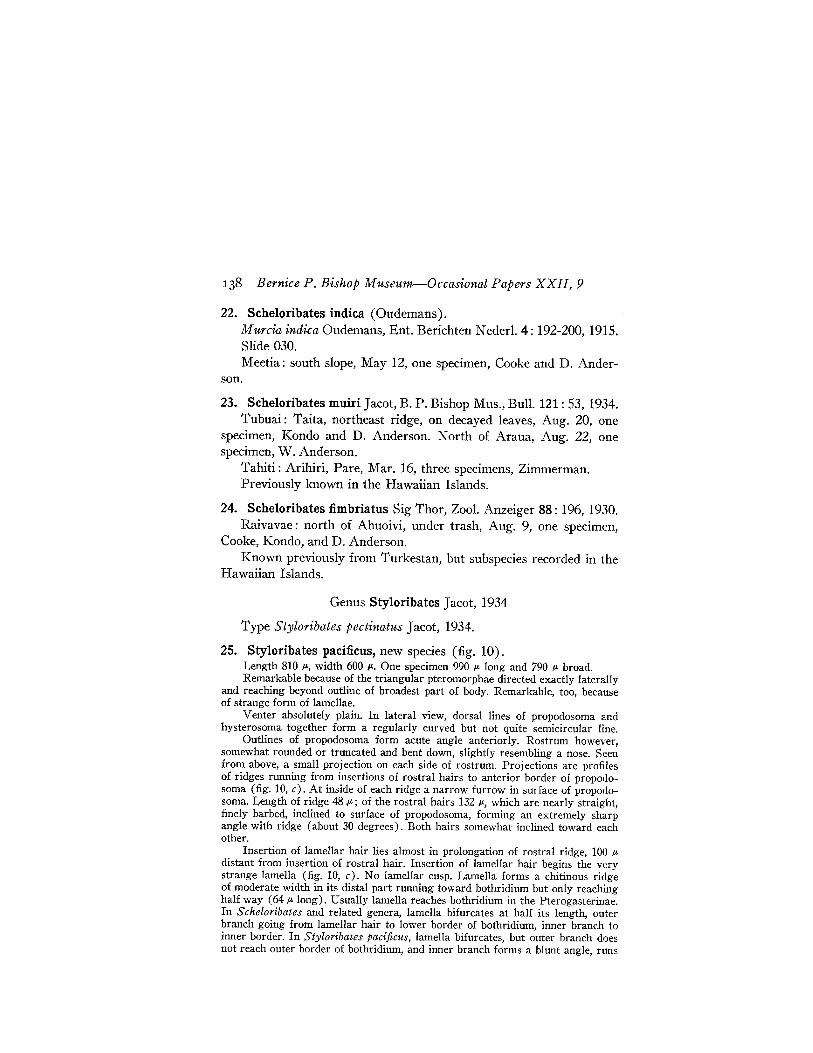
25. Styloribates paci:ficus, new species (fig. 10).Length 810 Jl-, width 600 Jl-. One specimen 990 Jl- long and 790 Jl- broad.Remarkable because of the triangular pteromorphae directed exactly laterally
and reaching beyond outline of broadest part of body. Remarkable, too, becauseof strange form of lamellae.
Venter absolutely plain. In lateral view, dorsal lines of propodosoma andhysterosoma together form a regularly curved but not quite semicircular line.
Outlines of propodosoma form acute angle anteriorly. Rostrum however,somewhat rounded or truncated and bent down, slightly resembling a nose. Seenfrom above, a small projection on each side of rostrum. Projections are profilesof ridges running from insertions of rostral hairs to anterior border of propodosoma (fig. 10, c). At inside of each ridge a narrow furrow in surface of propodosoma. Length of ridge 48,u.; of the rostral hairs 132 Jl-, which are nearly straight,finely barbed, inclined to surface of propodosoma, forming an extremely sharpangle with ridge (about 30 degrees). Both hairs somewhat inclined toward eachother.
Insertion of lamellar hair lies almost in prolongation of rostral ridge, 100 Jldistant from insertion of rostral hair. Insertion of lamellar hair begins the verystrange lamella (fig. 10, c). No lamellar cusp. Lamella forms a chitinous ridgeof moderate width in its distal part running toward bothridium but only reachinghalf way (64 Jl-Iong). Usually lamella reaches bothridium in the Pterogasterinae.In Scheloribates and related genera, lamella bifurcates at half its length, outerbranch going from lamellar hair to lower border of bothridium, inner branch toinner border. In Styloribates pacifiws, lamella bifurcates, but outer branch doesnot reach outer border of bothridium, and inner branch forms a blunt angle, runs
c
Sellnick-Acarina from Southeastern Polynesia 139
as a more or less sharp ridge to insertion of interlamellar hair, and beyond thisto anterior border of hysterosoma to a conspicuous knob of chitin. This directiontoward inner branch of lamella quite unusual in the Pterogasterinae.
There is no translamella.Lamellar hair 200 p. long, very fine in its distal half, sparsely ciliated and
inclined toward surface (at about 40 degrees).Interlamellar hair, 240 p. long, erect and sometimes bent a little backward.
Interlamellar hairs 152 p. apart.Bothridium with elliptical opening, surrounded by a chitinous ring which is
posteriorly somewhat broader than at inner side. Sensillus with a clubbed headon a curved peduncle. Head rounded at top and without hairs or other markingson surface.
A small area porosa outside point of bifurcation of lamella, and in front ofarea some spots surrounded by darker chitin. No true tectopedia I.
~.q[...,.;"
FIGURE 10.-St.vloribates pacifiws: a, dorsal side; b, ventral side; c,propodosoma, oblique lateral view; d, palp; e, gland of dorsal side; f, leg I;g, leg II; h, leg IV.
140 Bernice P. B'ishop Museum-Occasional Papers XXII, 9
A very fine exobothridial hair is inserted between bothridium and insertionof leg II.
Anterior border of hysterosoma nearly straight, more or less distinctlymarked. Border continues to anterior margin of pteromorphae. Pteromorphaetriangular, outer edge rounded, directed exactly laterally, and without a separation between them and dorsal plate of hysterosoma; often a slight radial striationon their surfaces.
Notogaster has no true areae porosae. One or two rows of small, clear spotsrun parallel to outer border of dorsal shield from one pteromorpha to otheraround body; these spots are places of affixation of muscles. Somewhat higher isa row of dark elongate chitinizations, the first at a level with middle of pteromorpha, five or six behind it. As far as visible, there is a short and very fineslit in cuticle of dorsal shield (fig. 10, e), from which a number of fine parallelchannels run into body to a cavity which is surrounded by wrinkled chitin andmay be a gland. Between some chitinizations are fine punctures, apparentlyinsertion points of hairs, but even under high magnification, I saw no hairs.
Ventral side of body (fig. 10, b) with camerostome oval in outline. Its concave hind margin continues with a ridge to outer edge of body, and this partsomewhat prolonged beyond outline of propodosoma. In front of ridge is arounded, deep furrow. Prolonged part of propodosoma, which may be calledtectopedia II, rounded on outside and with single curved hair in middle. Behindtectopedia II is a sharply pointed projection, tectopedia III. On posterior sideis a bristle.
A chitinous bridge extends between chitinized hind border of camerostomeand genital opening, which is surrounded by chitinization. (See apodemata andventral hairs in figure 10, b.) Genital opening small and rounded in outline.Four fine hairs on each cover. Anal opening quadrangular in outline with roundededges, four times as large as genital opening. Distance between openings morethan twice length of genital opening.
Terminal segment of palp formed as in species described by Jacot in 1934(14, pI. 10, fig. 101), Styloribates pectinatus, but it seems as if style on uppershort projection of the new species is double and that on the lower and longerprojection are three spines instead of two hairs (fig. 10, d).
Legs illustrated in figure 10, I-h, shown from inside, disclose porose spotson segments. Tarsi triheterodactyle, middle claw the strongest.
Holotype, slides 032, a-f; paratypes, slides 033, a-e.Rapa: near Morongota (type locality), July 16, 18 specimens gath
ered from ground with land shells, Cooke, Kondo, and D. Anderson.
FAMILY HAPLOZETIDAE GRANDJEAN, 1936
Genus Protoribates Berlese, 1908
Type Oribates monodactylus Haller, 1884.
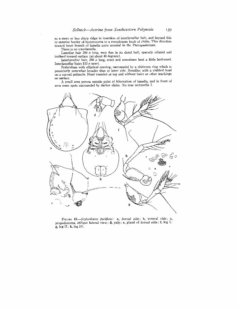
26. Protoribates boraboraensis, new species (fig. 11, a).Length 450-468 IJ., width 324-342 IJ.. Color light brown. Surface smooth.Propodosoma pointed in front, but rostrum blunt. A remarkable character
of this species is absence of a suture between propodosoma and hysterosoma.
Sellnick-Acarina from Southeastern Polynesia 141
Anterior end of pteromorpha, at level of bothridium, bends obliquely inwardand forward and ends behind insertion of interlamellar hair. In other species ofrelated genera, this line confluent with suture between propodosoma and hysterosoma behind interlamellar hair. Interlamellar hair long, strongly developed proximally, tapering toward distal end. A poorly developed ridge runs from interlamellar hair to insertion of lamellar hair, not quite reaching it. Lamellar hairinserted in anterior end of a distinct keel, shorter than interlamellar hair. Veryfine keel goes from insertion of lamellar hair to rostral hair, which is shorterthan lamellar hair and is bent inward. Only this hair is barbed on its outer side.No translamella present. Sensillus a clavigerous bristle, only club of whichprojects beyond border of pteromorphae. Behind interlamellar hair is a faint areaporosa.
Hysterosoma posteriorly well rounded, with usual areae porosae, adalaresof which are a little larger than the others; ·also very fine hairs found on dorsalsurface.
Pteromorphae of medium size; their anterior and lateral borders nearlyforming a right angle.
Apodemata strongly developed. Sternum present, but not so distinct asapodemata.
Genital and anal openings far apart, nearly as distant as length of bothapertures combined.
Tarsi with one claw.
Holotype, slide 035; paratypes, slides 036-038.
,, ,
i \!/
(/.. :t......J
.•_-_ - ..,_.:._ ."\ ,/
) /, I
; '\
t \c --.\
':.. .,0"-.. ...!-:
a
FIGURE n.-a, Protoribates boraboraensis, half of dorsal side. b, C, Phenopelops rapaensis: b, dorsal side; c, prQpodosoma.
142 Bernice P. Bishop Museum-Occasional Papers XXII, 9
Raivavae: near Ahuoivi Point, under trash, Aug. 9, one specimen,Cooke, Kondo, Zimmerman, and D. Anderson.
Borabora: Vaitape Village, 100-200 ft., 17 specimens, Kondo, Zimmerman, and D. Anderson. Cliff back of Vaitape (type locality), 350ft., Oct. 13, one specimen, D. Anderson.
Tahaa: Haamene Valley, north ridge, 400-1,100 ft., under stonesand dead leaves, Oct. 10, two specimens, Zimmerman.
FAMILY PELOPIDAE EWING, 1917
Genus Phenopelops Petrunkevitch, 1955
The name Pelops (C. L. Koch, 1836) is preoccupied by PelopsGistl, 1834, a genus of Coleoptera. Petrunkevitch (27) has proposedcalling this oribatid genus Phenopelops on the same type species,Pelops hirsutus C. L. Koch, 1844 (16, pt. 38).
A new species of the genus in the Mangarevan Expedition collection follows:
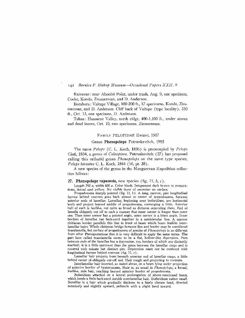
27. Phenopelops rapaensis, new species (fig. 11, b, c).Length 792 1Jo, width 630 1Jo. Color black. Integument dark brown in prepara
tions, dorsal spot yellow. No visible layer of secretion on surface.Propodosoma sharply pointed (fig. 11, b). A long, narrow, pale longitudinal
furrow behind rostrum goes back almost to center of propodosoma, betweenanterior ends of lamellae. Lamellae, beginning near bothridium, are horizontalkeels and project beyond middle of propodosoma, converging a little. Anteriorhalf of each is leaflike, not quite as broad as distance separating them. End oflamella obliquely cut off in such a manner that inner corner is longer than outerone. Thus inner corner has a pointed angle, outer corner is a blunt angle. Innerborders of lamellae run backward together in a semicircular line. A narrowchitinous border parallels this line in front of beam which bears leaflike interlamellar hairs. Whole chitinous bridge between line and border may be consideredtranslamella, but surface of propodosoma of species of Phenopelops is so differentfrom other Pterogasterinae that it is very difficult to apply the same terms. Thepart here called translamella seems to be a fiat, hollow-like depression. Partbetween ends of the lamellae has a depression, too, borders of which are distinctlymarked; it is a little narrower than the space between the lamellar cusps and iscovered with minute but distinct pits. Depression must not be confused withlongitudinal furrow behind rostrum (fig. 11, c).
Lamellar hair projects from beneath anterior end of lamellar CllSpS, a littlebehind center of obliquely cut-off end. Hair rough and projecting to rostrum.
Interlamellar hair inserted, as stated above, on a beam lying under projectionof anterior border of hysterosoma. Hair is, as usual in Phenopelops, a broad,leaflike, pale hair, reaching beyond anterior border of propodosoma.
Bothridium attached on a lateral prolongation of above-mentioned beam,which bends a little backward outside interlamellar hair. Bothridium rather small.Sensillus is a hair which gradually thickens to a fairly clavate head, directedanteriorly and slightly upward, peduncle with a slight bend inward.
Sellnick-Acarina from Sou,theastern Polynesia 143
Tectopedia I has a sharp point which projects nearly to anterior border ofpropodosoma, and rostral hair seems to be inserted at inner end of it. TectopediaII a broad, spoonlike leaf with an impression at outer side, behind which is akind of transverse ridge. Surface of this tectopedia distinctly pitted.
Outline of hysterosoma oviform, posterior border bluntly rounded, anteriorend with converging sides, bluntly pointed, and, as in all species of Phenopelops,with a median projection which overhangs beam containing insertions of interlamellar hairs (fig. 11, b).
Pteromorphae of normal shape; that is, their anterior border straight, lowerone rounded. They are movably attached at dorsal shield. At a level with centeris a long, elliptical yellow spot on dorsal shield.
Inner row of dorsal hairs almost straight, but not equally spaced: J3 to J3a little smaller than J2 to J2; J2 to J3 a little less than J2 to Jl ; Jl insertedbehind border of yellow spot. S1 and S2 are, as usual, inserted near pteromorphae,and S2 in a line with J1. J3 and S4 not convergent. Hairs of row on posteriorborder of body, Rl to R3, of medium length, radiately projecting backward. Allhairs of dorsal surface simple, not thick and not covered with secretions; hair S2longest.
Areae porosae barely points. In addition to areae porosae, are two light arealike spots, placed close together, on right side of dorsal shield. On left side, onlyone larger light spot with only one point directed outward. As there is only onespecimen in the collection, it is impossible to decide which is the normal area.A small, indistinct spot between S3 and S4.
Ventral surface without any remarkable characters.Tibia II with toothed spine on dorsal side.
Holotype, slides 039, a-e.Rapa: Teutu (type locality), on dead leaves, July 27, one speCI
men, Kondo.This is the first species of Phenopelops reported from the Pacific
islands. There is no species in the countries surrounding the oceanwhich agrees with A. rapaensis.
FAMILY PHTHIRACARIDAE PERTY, 1841
SUBFAMILY EUPHTHIRACARINAE JACOT, 1930
The subfamily Euphthiracarinae was established in 1930 by Jacot(10). In his opinion, the genera Oribotritia Jacot, Indotritia Jacot,Pseudotritia Willmann, and Euphthiracarus Ewing belong to this subfamily. Toward the end of his paper, Jacot gives a key to the generaof the subfamily. Oribotritia and Indotritia have, along the ventralchitinization, a pair of genital shields, or plates, and behind them apair of anal shields. At each side of these usually long and narrowshields is a shield as long as the two together. The part near the genitalshield is called the adgenital shield, the part near the anal plate, theadanal shield. In Pseudotritia and Euphthiracarus all four shields are
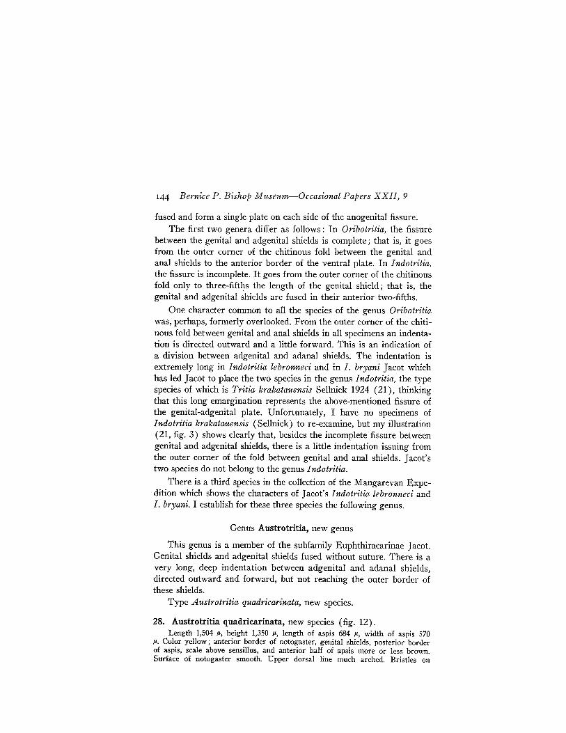
144 Bernice P. Bishop Museum-Occasional Papers XXII, 9
fused and form a single plate on each side of the anogenital fissure.The first two genera differ as follows: In Oribotritia, the fissure
between the genital and adgenital shields is complete; that is, it goesfrom the outer corner of the chitinous fold between the genital andanal shields to the anterior border of the ventral plate. In Indotritia,the fissure is incomplete. It goes from the outer corner of the chitinousfold only to three-fift~s the length of the genital shield; that is, thegenital and adgenital shields are fused in their anterior two-fifths.
One character common to all the species of the genus Oribotritiawas, perhaps, formerly overlooked. From the outer corner of the chitinous fold between genital and anal shields in all specimens an indentation is directed outward and a little forward. This is an indication ofa division between adgenital and adanal shields. The indentation isextremely long in Indotritia lebronneci and in I. bryani Jacot which
. has led Jacot to place the two species in the genus Indotritia, the typespecies of which is Tritia krakatauensis Sellnick 1924 (21), thinkingthat this long emargination represents the above-mentioned fissure ofthe genital-adgenital plate. Unfortunately, I have no specimens ofIndotritia krakatauensis (Sellnick) to re-examine, but my illustration(21, fig. 3) shows clearly that, besides the incomplete fissure betweengenital and adgenital shields, there is a little indentation issuing fromthe outer corner of the fold between genital and anal shields. Jacot'stwo species do not belong to the genus Indotritia.
There is a third species in the collection of the Mangarevan Expedition which shows the characters of Jacot's Indotritia lebronneci andI. bryani. I establish for these three species the following genus.
Genus Austrotritia, new genus
This genus is a member of the subfamily Euphthiracarinae Jacot.Genital shields and adgenital shields fused without suture. There is avery long, deep indentation between adgenital and adanal shields,directed outward and forward, but not reaching the outer border ofthese shields.
Type Austrotritia quadricarinata, new species.
28. Austrotritia quadricarinata, new species (fig. 12).Length 1,504 iL, height 1,350 iL, length of aspis 684 iL, width of aspis 570
iL. Color yellow; anterior border of notogaster, genital shields, posterior borderof aspis, scale above sensillus, and anterior half of apsis more or less brown.Surface of notogaster smooth. Upper dorsal line much arched. Bristles 011
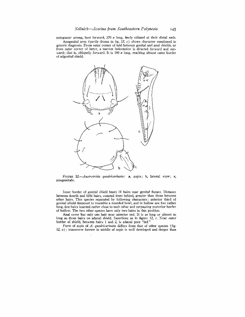
Sellnick-Acarina from Southeastern Polynesia 145
notogaster strong, bent forward, 270 ,.. long, finely ciliated at their distal ends.Anogenital area (partly drawn in fig. 12, c) shows character mentioned in
generic diagnosis. From outer corner of fold between genital and anal shields, orfrom outer corner of latter, a narrow indentation is directed forward and outward; that is, obliquely forward. It is 160 ,.. long, reaching almost outer borderof adgenital shield.
FIGURE 12.-Austrotritia quadricarinata: a, aspis ; b, lateral view; c,anogenitale.
Inner border of genital shield bears 11 hairs near genital fissure. Distancebetween fourth and fifth hairs, counted from behind, greater than those betweenother hairs. This species separated by following characters: anterior third ofgenital shield deepened to resemble a rounded bowl, and in holloware five ratherlong, fine hairs inserted rather close to each other and surpassing posterior borderof hollow. The two other species have only two hairs in this position.
Anal cover has only one hair near anterior end. It is as long or almost aslong as three hairs on adanal shield. Insertions as in figure 12, c. ::-Jear outerborder of shield, between hairs 1 and 2, is adanal pore "iad."
Form of aspis of A. quadricarinata differs from that of other species (fig.12, a) ; transverse furrow in middle of aspis is well developed and deeper than
146 Bernice P. Bishop Museum-Occasional Papers XXII, 9
in Indotritia bryani. Seen from above, part behind middle is light yellow, exceptposterior border, which is darkly chitinized. Anterior part of aspis, however,brown. This part, which strongly slopes down forward, has four longitudinalridges, outer ridges longer than inner ones. At inner side of outer ridge is insertedrostral hair, 120 p. long, directed forward and a little upward and parallel. Upperhairs, inserted in a line with bothridium, broken but, judging from remainingstumps, must have been long. Distance between hairs less than distance fromthem to an outer bristle, which is inserted in front of upper hairs. Outer bristleappressed to surface of aspis, 180 p. long, and directed forward. Posterior borderof aspis somewhat convex. From its outer posterior corner projects a scale, asquamose chitinous projection, which is situated above bothridium. Sensillus asimple bristle, tapering distally, 200 p. long and directed outward. Bothridiumhas usual snail-shell form. Distinct line extends from scale to lower border ofaspis near anterior border. A chitinous peg, a projection from each corner ofposterior border of aspis, may be a place for contractyle muscles to move aspis.
Gnathosoma resembles that of Oribotritia.Three claws on tarsi, central one stronger than others.
Holotype, slides 040, a-e.Rapa: Mt. Perahu (type locality), 1,9OO£t., July 21, one specimen,
Fosberg.
REVISED KEY TO GENERA OF EUPHTHIRACARINAE
1. On each side of anogenital fissure is only one shield which reachesfrom anterior border to posterior border of venter. At pointwhere genital fissure continues to anal one, about middle oflength of ventral shield, is a dark .chitinous triangle, sharp pointof which is directed backward. A longitudinal serpentlike fissuregoes through this triangle which is a mechanism to lock ano-genital fissure m m •• m m no •••••••no 2
On each side of anogenital fissure posteriorly is a narrow analshield and on its outer side an adanal shield. Genital shieldeither completely separated from adgenital, or partially or completely fused with it. At point where genital fissure continues toanal fissure, somewhat in front of middle of length of ventralshield, is a narrow, triangular, transverse fold of posterior partof genital shield which overhangs anterior border of anal plates.Outside outer corners of this fold is a more or less deep inden-tation toward adgenital-adanal shield m nom..no 3
2(1). Surface of notogaster only very finely dotted, nearly smooth. Aspisslightly flattened in front. Central rib from posterior border ofaspis shorL u no Pseudotritia Will-mann, 1920 [type: Tritia (Pseudotritia) monodactyla Willmann, 1920)
Surface of notogaster extremely distinctly sculptured, mostly withpits. Aspis large, arched in anterior part. Central rib from posterior border of aspis reaches nearly to middle of aspis......... m ....
Euphthiracarus Ewing 1917 [type: Phthiracants flavus Ewing, 1908)
3(1). Indentation from outer corner of fold between genital and analshields very long, going obliquely forward nearly to outer border
Sellnick-Acarina from Sontheastern Polynesia 147
of adgcnital-adanal shield; genital shield fused with adgenitalshield and without a suturc...__ ..._.............. ... AustrotritiaSellnick, 1959 [type: Austrotritia quadricarinata Sellnick, 1959]
Indentation from outer corner of fold bctween genital and analshield short and often only indicated.. ......._........; .. .. ... 4
4(3). Longitudinal fissure between genitale and adgenitale goes fromouter corner of fold between genital and anal shields to anteriorborder of ventral area. Both shields completely separated............Oribotritia Jacot, 1924 [type: Hoplophora decumana C. L. Koch, 1836]
Longitudinal fissure between genital and adgenital shield goes forward to three-fifths of length of genital shield, parallel to genitalfissure. Anterior two-fifths of shields fused and without a suture..........._Indotritia Jacot, 1928 [type: Tritia krakataltensis Sellnick, 1923]
In addition to Austrotritia quadricarinata, the following Euphthiracarinae are in the collection of the Mangarevan Expedition.
29. Austrotritia lebronneci (Jacot).Indotritia lebronneci Jacot, B. P. Bishop Mus., Bull. 114: 234,
1934 (1935).
Slides 041, a-c; 042; 043, a-b.Rapa: Maitua, July 2, one specimen, Cooke and D. Anderson.
Teutu, on dead leaves, July 27, one specimen, Kondo. Mt. Tanga,north slope, 300-700 ft., under dead leaves, July 31, one specimen, D.Anderson. Kopenaena Valley, July 24, one specimen, Kondo.
Raivavae: Ahuoivi Point, on dead leaves, Aug. 9, three specimens,Cooke, Kondo, and D. Anderson. Hoauatua Islet, Aug. 11, one specimen, Cooke, Zimmerman, Sam Wight, and Ernest Lyons. Mt. Turivao, south slope, Aug. 11, one specimen, W. Anderson.
Tubuai: Tapapatauai Islet, under trash, Aug. 19, one specimen,Kondo. Near Araua, under dead Pandanus leaves, Aug. 22, one specimen, W. Anderson. North of Araua, Aug. 22, one specimen, W. Anderson.
Rurutu: Mato Arei, under trash on ground, Aug. 27, two specimens, D. Anderson and Kondo. Mato Naa, Aug. 30, one specimen,Fosberg. North of Avera, near shore, Aug. 31, one specimen, Kondoand D. Anderson. Northwest of Avera, Sept. 1, one specimen, Kondoand D. Anderson. Mato Arapia, under stones, Sept. 2, Cooke, Kondo,and D. Anderson. Mato Arapia, south of Avera, Sept. 2, five specimens, Cooke, Kondo, and D. Anderson.
Maria: northeast islet, 5 ft., Sept. 6, one specimen, Zimmerman.Tahiti: Arihiri, Pare, Mar. 16, one specimen, Zimmerman.
148 Bernice P. Bishop Museum-Occasional Papers XXII, 9
Raiatea: Tetaro Islet, Oct. 4, one specimen, Zimmerman, Kondo,and Cooke. Toahiva Valley, in trash on ground, Oct. 7, one specimen,C. Gessler.
Flint: 5-8 ft., Oct. 16, one specimen, D. Anderson.It is of interest that almost all of these specimens were collected
incidental to the collection of terrestrial mollusks.Known previously from the Marquesas.
30. Pseudotritia loricata (Rathke) Entomologiske Jagttage1ser, INSkrivter af Naturhandl. Selskabet 5 (1) : 191-200, 1799.
Trombidium loricatus Rathke, 1799.Rurutu: north of Avera, near shore, Aug. 31, one specimen, Kondo
and D. Anderson.Tahiti: Arihiri, Pare, Mar. 16, three specimens, Zimmerman.Widespread over Europe and Canada.
Genus Hoplophorella Berlese, 1923
Type H oploderma cucullatun~ Ewing, 1909.There are always five bristles on the anal plates of the Phthira
carini. Jacot, 1933 (12), believes the concentration of these bristlesalong the median edge to be a definite trend in the subfamily. InH oplophthiracarus there are only two bristles along the median edge.Phthiracarus and H oplophorella have three bristles. Steganacarus, Tropacarus, and Atropacarus have four bristles. In H oplophorello, thethree bristles are close together on the median edge; therefore, the species represented by the present collection belongs to this genus.
31. Hoplophorella singularis, new species (fig. 13).Length of hysterosoma 630 fL, width 360 fL, height 360 fl. Length of aspis
295 IL, width 198 IL. Color yellow, except for collar of notogaster and posteriorborder of aspis which are light brown. Surface of notogaster evenly covered withshallow pits; without distinct borders.
Body rather long, as broad as high, well rounded posteriorly.On each side of notogaster 15 stout bristles distinctly barbed, or with fine,
sharp points, in their distal half. Part of hair within chitinized skin strangelyformed (fig. 13, e).
Anal plates high, rounded, sculptured, but not so regularly as notogaster.Three long, smooth hairs close to inner edge of each plate, equally spaced. (Ibelieve that the excrement slips out of the anal opening along these elastic hairs.)At a level with space between anteriormost and following bristles, a smoothbristle, which is longer than those on inner edge, is inserted near middle ofcover (fig. 13, f). Farther in front of these hairs, at an equal distance fromedges of cover, is a shorter stout and barbed bristle similar to those on notogaster.
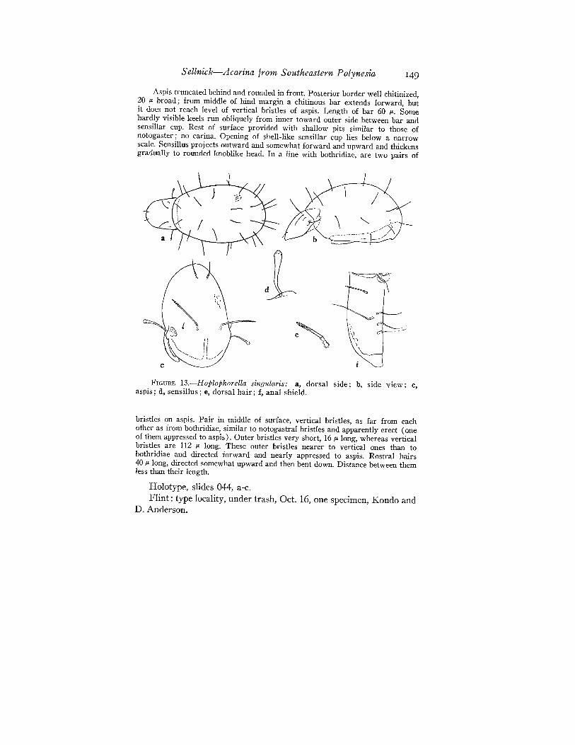
Sellnick-Acarina from Southeastern Polynesia 149
Aspis truncated behind and rounded in front. Posterior border well chitinized,20 J.' broad; from middle of hind margin a chitinous bar extends forward, butit does not reach level of vertical bristles of aspis. Length of bar 60 11-. Somehardly visible keels run obliquely from inner toward outer side between bar andsensillar cup. Rest of surface provided with shallow pits similar to those ofnotogaster; no carina. Opening of shell-like sensillar cup lies below a narrowscale. Sensillus projects outward and somewhat forward and upward and thickensgradually to rounded knoblike head. In a line with bothridiae, are two pairs of
FIGURE 13.-Hoplophorella singltlaris: a, dorsal side; b, side view; c,aspis; d, sensillus; e, dorsal hair; f, anal shield.
bristles on aspis. Pair in middle of surface, vertical bristles, as far from eachother as from bothridiae, similar to notogastral bristles and apparently erect (oneofthem appressed to aspis). Outer bristles very short, 16 11- long, whereas verticalbristles are 112 11- long. These outer bristles nearer to vertical ones than tobothridiae and directed forward and nearly appressed to aspis. Rostral hairs40 11- long, directed somewhat upward and then bent down. Distance between themless than their length.
Holotype, slides 044, a-c.Flint: type locality, under trash, Oct. 16, one specimen, Kondo and
D. Anderson.

Sellnick-Acarina from Southeastern Polynesia 151
BIBLIOGRAPHY
1. BERLESE, ANTONIO, Acari, Myriapoda, et Scorpiones hucusque in Italia re-perta, pts. 1-101, Padova, 1882-1903.
2. BERLEsE, ANTONIO, Elenco di genera e specie ,nuove di Acari: Redia 5, 1908.
3. BERLEsE, ANTONIO, Centuria terza di Acari nuovi: Redia 12, 1916.
4. CocGI, A., Descrizione di specie nuove di Oribatidi italiani . . .: Soc. Ent.Ital., Boll. 30, 1898.
5. GRANDJEAN, F. A., Observations sur les Oribates: Mus. Hist. Nat. Paris,Bull. II, 5 (6), 1933.
6. GRANDJEAN, F. A., Les Oribates de Jean-Frederic Hermann et de son pere:Soc. Ent. France, Ann. 105, 1936.
7. HEYDEN, C. H. G. VON, Versuch einer systematischen Einteilung derAcariden: Isis (von Oken) 18, 1826.
8. JACOT, A. P., New oribatoid mites: Psyche 35, 1928.
9. JACOT, A. P., Concerning the genus Neoliodes (Oribatoidea-Acarina): Am.Microscopical Soc., Trans. 48, 1929.
10. JACOT, A. P., Oribatid mites of the subfamily Phthiracarinae of the northeastern United States: Boston Soc. Nat. Hist., Proc. 39, 1930.
11. JACOT, A. P., A common arboreal moss mite, Humerobates humeralis: Boston Soc. Nat. Hist., Occ. Papers 5, 1931.
12. JACOT, A. P., Phthiracarid mites of Florida: Elisha Mitchell Sci. Soc., Jour.48, 1933.
13. JACOT, A. P., Some Tyrogtyphina (Sarcoptiformes) of the MarquesasIslands: B. P. Bishop Mus., Bull. 114, article 17, 1934 (1935).
14. JACOT, A. P., Some Hawaiian Oribatoidea (Acarina): B. P. Bishop Mus.,Bull. 121, 1934.
15. JACOT, A. P., Some new western North Carolina moss mites: Ent. Soc.Washington, Proc. 40, 1938.
16. KocH, C. L., Deutschlands Crustaceen, Myriapoden und Arachniden, Regensburg, 1836-1844.
17. OUDEMANS, A. C., Notizen iiber Acari, 25 Reihe: Archiv Naturgesch. A,Jahrg. 82, 6, 1917.
18. SELLNICK, MAX, Die mir bekannten Arten der Gattung Tritia: Acari 3, 1923.
19. SELLNICK, MAX, Oribatideos brasileiros: Arch. Mus. Rio de Janeiro 24,1923.
20. SELLNICK, MAX, Zur Kenntnis der estliindischen Moorfauna, Oribatiden:Naturf.-Gesell. Dorpat, S. Ber. 33, 1924.
21. SELLNICK, MAX, Oribatiden der Insel Krakatau: Treubia 5, 1924.
22. SELLNICK, MAX, Javanische Oribatiden: Treubia 6, 1925.
23. SELLNICK, MAX, Fauna sumatrensis: Suppl. Ent. 11, 1925.
152 Bernice P. Bishop Museum-Occasional Papers XXII, 9
24. SELLNICK, MAX, Oribatei: Tierwe1t Mitte1eur. 3 (9), 1929.
25. SELLNICK, MAX, Eine neue brasilianische Neoliodes-Art und Bemerkungenilber die Gattung Neoliodes: Zool. Anzeiger 89, 1930.
26. SELLNICK, MAX, Oribatiden aus dem Karst.: Zoo!' Jahrb. Syst. 63, 1932.
27. STORMER, L., PETRUNKEVITCH, A., and HEDGEPETH, I. \V., Treatise on invertebrate paleontology, directed and edited by R. C. Moore, Arthropoda(P) : 98, 1955. (Published by the Geological Society of America and Universityof Kansas Press.)
28. THOR, SIG, Einige Acarina, besonders Hydracarina aus Turkestan: Zool.Anzeiger 88, 1930. .
29. TRAGARDH, IVAR, Acarina collected by the Mangarevan Expedition to SouthEastern Polynesia in 1934 ... : Arkiv for Zoologi II, 4 (1): 45-90, 1951(1952).
30. VITZTHUM, H. GRAF, Acarina, in Bronn's K!. und Ord. des Tierreichs 5 (4),1940-1943.