Embed Size (px)
Citation preview
The University of Manchester Research
Acute D3 Antagonist GSK598809 Selectively EnhancesNeural Response During Monetary Reward Anticipation inDrug and Alcohol DependenceDOI:10.1038/npp.2016.289
Document VersionAccepted author manuscript
Link to publication record in Manchester Research Explorer
Citation for published version (APA):Murphy, A., Nestor, L. J., McGonigle, J., Paterson, L., Boyapati, V., Ersche, K. D., Flechais, R., Kuchibatla, S.,Metastasio, A., Orban, C., Passetti, F., Reed, L., Smith, D., Suckling, J., Taylor, E., Robbins, T. W., Lingford-Hughes, A., Nutt, D. J., Deakin, J. F., & Elliott, R. (2017). Acute D3 Antagonist GSK598809 Selectively EnhancesNeural Response During Monetary Reward Anticipation in Drug and Alcohol Dependence.Neuropsychopharmacology. https://doi.org/10.1038/npp.2016.289Published in:Neuropsychopharmacology
Citing this paperPlease note that where the full-text provided on Manchester Research Explorer is the Author Accepted Manuscriptor Proof version this may differ from the final Published version. If citing, it is advised that you check and use thepublisher's definitive version.
General rightsCopyright and moral rights for the publications made accessible in the Research Explorer are retained by theauthors and/or other copyright owners and it is a condition of accessing publications that users recognise andabide by the legal requirements associated with these rights.
Takedown policyIf you believe that this document breaches copyright please refer to the University of Manchester’s TakedownProcedures [http://man.ac.uk/04Y6Bo] or contact [email protected] providingrelevant details, so we can investigate your claim.
Download date:08. Aug. 2020
Accepted Article Preview: Published ahead of advance online publication
Neuropsychopharmacologywww.neuropsychopharmacology.org
Methylphenidate modifies the motion of the circadian clock
Lamotrigine in mood disorders and cocaine dependence
Cortical glutamate in postpartum depression
Acute D3 Antagonist GSK598809 Selectively Enhances Neural
Response During Monetary Reward Anticipation in Drug and
Alcohol Dependence
Anna Murphy, Liam J Nestor, John McGonigle, LouisePaterson, Venkataramana Boyapati, Karen D Ersche, RemyFlechais, Shankar Kuchibatla, Antonio Metastasio, CsabaOrban, Filippo Passetti, Laurence Reed, Dana Smith, JohnSuckling, Eleanor Taylor, Trevor W Robbins, Anne Lingford-Hughes, David J Nutt, John FW Deakin, Rebecca Elliott,ICCAM Platform
Cite this article as: Anna Murphy, Liam J Nestor, John McGonigle, Louise
Paterson, Venkataramana Boyapati, Karen D Ersche, Remy Flechais, Shankar
Kuchibatla, Antonio Metastasio, Csaba Orban, Filippo Passetti, Laurence Reed,
Dana Smith, John Suckling, Eleanor Taylor, Trevor W Robbins, Anne Lingford-
Hughes, David J Nutt, John FW Deakin, Rebecca Elliott, ICCAM Platform,
Acute D3 Antagonist GSK598809 Selectively Enhances Neural Response During
Monetary Reward Anticipation in Drug and Alcohol Dependence, Neuropsycho-
pharmacology accepted article preview 2 January 2017; doi: 10.1038/npp.2016.289.
This is a PDF file of an unedited peer-reviewed manuscript that has been accepted
for publication. NPG are providing this early version of the manuscript as a service
to our customers. The manuscript will undergo copyediting, typesetting and a proof
review before it is published in its final form. Please note that during the production
process errors may be discovered which could affect the content, and all legal
disclaimers apply.
Received 18 August 2016; revised 9 December 2016; accepted 19 December 2016;Accepted article preview online 2 January 2017
© 2017 Macmillan Publishers Limited. All rights reserved.
Title: Acute D3 antagonist GSK598809 selectively enhances
neural response during monetary reward anticipation in
drug and alcohol dependence
Running title: GSK598809 effects on reward and inhibitory control processes
Anna Murphy1, Liam J Nestor
2,4, John McGonigle
2, Louise Paterson
2, Venkataramana Boyapati
1, Karen D
Ersche3,4
Remy Flechais2,
Shankar Kuchibatla1, Antonio Metastasio
1, Csaba Orban
2, Filippo Passetti
2,
Laurence
Reed2, Dana Smith
3,4, John Suckling
4, Eleanor Taylor
1, Trevor W Robbins
34, Anne Lingford-Hughes
2, David J
Nutt2, John FW Deakin
1 and Rebecca Elliott
1 ICCAM Platform*
1Neuroscience and Psychiatry Unit, University of Manchester;
2Centre for Neuropsychopharmacology, Division
of Brain Sciences, Imperial College London; 3Department of Psychology, University of Cambridge;
4Department
of Psychiatry, University of Cambridge
Word Count
Manuscript 3935
Abstract 234
Figures:3
Tables:1
Corresponding Author:
Anna Murphy
Neuroscience and Psychiatry Unit
G.708 Stopford Building
University of Manchester
Oxford Road, Manchester,UK, M13 9PT
Email: [email protected]
Tel:+44 (0)161 275 7764, Fax: +44 (0)161 275 7428
© 2017 Macmillan Publishers Limited. All rights reserved.
Abstract
Evidence suggests that disturbances in neurobiological mechanisms of reward and inhibitory control
maintain addiction and provoke relapse during abstinence. Abnormalities within the dopamine
system may contribute to these disturbances and pharmacologically targeting the D3 dopamine
receptor (DRD3) is therefore of significant clinical interest. We used functional magnetic resonance
imaging to investigate the acute effects of the DRD3 antagonist GSK598809 on anticipatory reward
processing, using the monetary incentive delay task (MIDT), and response inhibition using the go/no-
go task (GNGT). A double-blind, placebo-controlled, crossover-design approach was used in
abstinent alcohol dependent, abstinent poly-drug dependent and healthy control volunteers.
For the MIDT, there was evidence of blunted ventral striatal response to reward in the poly-drug
dependent group under placebo. GSK598809 normalised ventral striatal reward response and
enhanced response in the DRD3 rich regions of the ventral pallidum and substantia nigra.
Exploratory investigations suggested that the effects of GSK598809 were mainly driven by those
with primary dependence on alcohol but not opiates. Taken together, these findings suggest that
GSK598809 may remediate reward deficits in substance dependence.
For the GNGT, enhanced response in the inferior frontal cortex of the poly-drug group was found.
However, there were no effects of GSK598809 on the neural network underlying response inhibition,
nor were there any behavioural drug effects on response inhibition.
Conclusion: GSK598809 modulated the neural network underlying reward anticipation but not
response inhibition suggesting DRD3 antagonists may restore reward deficits in addiction.
© 2017 Macmillan Publishers Limited. All rights reserved.
Introduction
Evidence suggests dysregulation of neurobiological networks involved in reward processing and
inhibitory control contributes to the risk and maintenance of addiction and relapse during
abstinence. Disturbances in reward functioning involve hypo-responsivity to non-drug reward
which is associated with increased craving, drug use and brain response to drug-related stimuli
(Blum et al, 2000; Lubman et al, 2009; Wrase et al, 2007). Failures of impulse control across a range
of domains have been a consistent finding in addiction and are associated with relapse (Taylor et al,
2016). fMRI studies implicate reduced recruitment of lateral and medial prefrontal regions in
impaired impulse control (Forman et al, 2004; Kaufman et al, 2003), although enhanced recruitment
has been found in those who have successfully achieved abstinence from cocaine (Connolly et al,
2012).
Abnormalities in reward and impulse control may be effects of blunted dopamine signalling in
addiction (Trifilieff and Martinez, 2014). Reductions in dopamine release and receptor density are
associated with increased drug use and craving and may precede the development of addiction
(Casey et al, 2014; Heinz et al, 2004). Dopamine plays a pivotal role in reward-related behaviours,
mediating reward learning (Schultz, 1998) and ‘incentive salience’ of reward stimuli (Robinson and
Berridge, 1993). Deficits in dopamine neurotransmission may impair impulse control, as low striatal
D2/D3 binding is associated with increased impulsivity in rodents and humans (Clark et al, 2012;
Ghahremani et al, 2012). Drugs increasing extracellular dopamine improve response inhibition in
cocaine and alcohol dependence (Garrison and Potenza, 2014) . These lines of evidence suggest
increasing brain dopamine may be a useful therapeutic strategy for addiction (Nutt et al, 2015).
The D3 receptor (DRD3) is preferentially expressed within ventral striatal and limbic brain regions
involved in reward processing. In- vitro studies demonstrate the highest density of DRD3s within the
ventral striatum of the human brain (Gurevich and Joyce, 1999) whereas in-vivo human positron
emission tomography (PET) studies demonstrated maximal DRD3 density within the ventral
© 2017 Macmillan Publishers Limited. All rights reserved.
pallidum, followed by the substantia nigra and ventral striatum, with lower levels in thalamus and
dorsal striatum (Tziortzi et al, 2011). Exposure to drugs of abuse results in upregulation of DRD3 in
rodent models of addiction (Le Foll and Di Ciano, 2015). Upregulated DRD3 density has been found
within the ventral striatum and substantia nigra in a post-mortem study of cocaine dependence
(Staley and Mash, 1996), while upregulated nigral DRD3s correlated positively with impulsivity and
risky decision making in stimulant dependence (Boileau et al, 2012; Payer et al, 2014). Trends for
upregulated ventral pallidal DRD3 have been found in both alcohol dependence and heavy stimulant
use (Boileau et al, 2012; Erritzoe et al, 2014). Consequently, there is much interest in this receptor as
a target for drug therapy.
DRD3 antagonists have shown promise in preclinical studies, reducing self-administration, cue-
induced drug-seeking, and conditioned place preference (Heidbreder and Newman, 2010). In clinical
populations, the novel DRD3 antagonist GSK598809 transiently reduced craving in nicotine
dependence (Mugnaini et al, 2013) and attentional bias for food cues in low restrained eaters
(Nathan et al, 2012). The exact mechanism by which DRD3 antagonists achieve these effects is
currently unknown, although there is evidence the DRD3 is an autoreceptor controlling the synthesis
and release of dopamine (Diaz et al, 2000; Zapata and Shippenberg, 2002). Blockade of DRD3 with
GSK598809 may therefore increase extra-synaptic dopamine.
The ICCAM Platform study is a multicentre research study that aimed to 1) identify brain networks
underlying addiction to alcohol, cocaine and opiates and relapse vulnerability and 2) identify
potential new treatments for addiction based upon their ability to modulate these networks. Here,
we present the effects of GSK598809 on networks underlying the anticipation of reward, using the
monetary incentive delay task (Knutson et al, 2000) and response inhibition using the go/no-go task
(Garavan et al, 2002). We hypothesised that reward and inhibitory control disturbances would be
found in abstinent drug dependent individuals and that GSK598809 would mitigate such
disturbances.
© 2017 Macmillan Publishers Limited. All rights reserved.
Patients and Methods
Participants
Participants were recruited as part of the ICCAM multi-centre study. Detailed description of
recruitment and participant characteristics are described elsewhere (Paterson et al. 2015). Briefly,
substance dependent individuals were recruited according to the following criteria: aged 20-64
years, meeting DSM-IV (1994)criteria for current or prior alcohol, cocaine or opiate dependence,
abstinent for at least 4 weeks, free from any current primary axis-1 mental health disorder, no
history of severe enduring mental illness, no psychoactive medications, no serious physical health
problems, no neurological disease and no contraindications for MRI scanning. Healthy controls (HC)
were recruited according to the same criteria except they no had current, or history of dependence
to any drug except nicotine.
88 participants completed both placebo and GSK598809 sessions: a healthy control group (n=35), an
abstinent alcohol dependent (AD) (n=20) and an abstinent poly-drug dependent (PD) group (n=33).
Five people were excluded from the MID analysis, and 14 from the GNG analysis leaving final Ns of
83 and 74 respectively (see supplementary materials for details).
The HC group was matched with the AD and PD groups for age, sex, smoking status and handedness
and additionally with the AD group, but not the PD group, for IQ and years of education (see Tables
S1 and S2). AD and PD groups differed significantly for age for the MID analysis, with a trend for a
difference for the GNG analysis (p=0.06).
© 2017 Macmillan Publishers Limited. All rights reserved.
Procedures and tasks
Procedures are described in detail elsewhere (Paterson et al, 2015). Briefly, the ICCAM study
involved 5 separate scanning sessions (one screening, including fMRI scanning to familiarise
participants with the scanner environment and tasks, and four drug testing sessions). Placebo and
GSK598809 (60mg) were administered in a double-blind manner with a cross-over design. Due to
concerns over study dropout and loss of placebo data, a weighted randomization was used with the
placebo session administered in study session 2 or 3 whereas GSK598809 was administered in
session 4 or 5 (with the other two sessions testing other drugs as part of the ICCAM platform;
Paterson et al, 2015). The mean (SD) inter-session interval between placebo and GSK598809
sessions was 34.39 days (40.91) (MID task) and 36.15 days (42.72) (GNG task), with no difference
between groups; MID (F(2,80)=0.25, p=0.78), GNG (F(2,71) = 0.03, p=0,98).
Scans occurred 2 hours after administration of drug or placebo and tasks were practised before
scanning. All participants had an alcohol breathalyser reading of 0.0%. Participants were urine
screened. A positive test for cannabis was allowed due to its long half-life provided there was no use
in the previous week. Participants tested negative for all other drugs with two allowed exceptions
(see supplementary materials).
Tasks
Both the MID task and GNG tasks are described in detail within the supplementary materials.
The MID task was modified from Knutson et al. 2000 and was designed to probe reward sensitivity.
Participants could win or lose money (or neither win nor lose) depending upon how quickly they
reacted to a target stimulus that was predicted by a win, loss or neutral cue. The task was designed
such that win accuracy would be 66% and £10 would be won at each session.
© 2017 Macmillan Publishers Limited. All rights reserved.
The GNG task was an event-related task adapted from Garavan et al. 2002, consisting of a series of
letter Xs and letter Ys. Participants were instructed to respond as fast as they could to each letter (go
trial) except when the letter was the same as the previous letter (no-go trial).
Analysis of Behavioural data
For the MID task, reward-neutral reaction time (RT) was analysed. For the GNG task, percentage
accuracy for Go trials, Nogo trials and RT for Go trials were analysed. All analyses used mixed
ANOVAs with drug session as the within-subject factor and group as the between-subject factor. Age
was included as a mean-adjusted covariate in all analyses.
Analysis of fMRI data
Details of data acquisition and pre-processing are in supplementary materials.
For the MID task, analysis focused on the ‘cue and anticipation’ phase and was modelled as a mini-
block beginning at the cue onset and ending at the onset of the target stimulus (see supplementary
materials for details). The contrast of interest is the average of the ‘reward cue anticipation’
compared with ‘neutral cue anticipation’ across both runs.
For the GNG task successful inhibitions of nogo trials (“stops”) and unsuccessful nogos (“errors”)
were modelled against an implicit baseline of go trials. Stops that were preceded by a go trial that
also did not have a response were considered “fake inhibitions” and were modelled separately as
conditions of no-interest. The task was powered to look at “stops” rather than “errors” therefore
only the “stops>go” contrast is explored further.
Realignment parameters and movement outliers (scan to scan displacement of >3mm) were added
to the models as nuisance regressors.
A region of interest (ROI) approach was used. ROIs of the ventral striatum (VS), ventral pallidum (VP)
and substantia nigra (SN) were chosen for the MID task due to their key roles in reward processing
© 2017 Macmillan Publishers Limited. All rights reserved.
(Haber and Knutson et al. 2010) and evidence of abnormalities within these regions in addiction.
Additionally, as reviewed above, these regions are particularly rich in DRD3s and therefore are
potential targets for GSK598809 effects. For the GNG task, bilateral inferior frontal gyri (IFG) and
anterior cingulate cortex (ACC) were chosen due to their key role in motor inhibition (Aron et al,
2003) and abnormal recruitment in addiction. Although not key ROIs for this task, exploratory
investigations were also carried out with the DRD3 rich regions of VS, VP and SN.
Mean reward-neutral (MID) and stop-go (GNG) contrast estimates were extracted from the relevant
ROIs for each participant and entered into mixed ANOVAs (see supplementary materials for details).
A Bonferroni correction for the 3 regions investigated for each task was applied, with significance set
at p<0.017. Additional exploratory whole-brain investigations were carried out using a voxel-wise
threshold of p<0.05, Family Wise Error Corrected. (See figure 1 for ROIs and supplementary
materials for further details).
Correlational analyses were additionally carried out to investigate relationships between ROI brain
response and performance; reward and impulsivity variables; ROI brain response and duration of
abstinence; and drug effects on ROI response and performance (see supplementary materials for
details). Twenty nine analyses were carried out therefore Bonferroni-corrected significance was set
at p<0.0017.
© 2017 Macmillan Publishers Limited. All rights reserved.
Results
MID Behavioural
There were no significant drug or group effects or interactions for MID performance (see figure S1).
MID fMRI
Effect of Task
The reward-neutral contrast for each group for both the placebo and GSK598809 conditions
revealed a highly significant network of activation, including the VP, VS and SN, in line with previous
studies (Knutson et al 2000). See figure S3.
Effects of group and drug: whole brain analysis
Mixed ANOVAs demonstrated a significant main effect of drug within the left ventral pallidum,
caudate and cerebellum (Table 1 and Figure S5). These effects appear to be driven by increased
reward-neutral anticipation response in the GSK598809 session compared to the placebo session, in
particular for the AD group and to a lesser extent the PD group. However these apparent
interactions were not significant.
A significant effect of drug was also observed in the right middle frontal gyrus (corresponding to the
dorsolateral prefrontal cortex, DLPFC, see table 1 and figure S3), which was driven by a significant
group by drug session interaction. GSK598809 increases reward-neutral BOLD response to a greater
degree in the AD group compared to both the HC and the PD groups.
© 2017 Macmillan Publishers Limited. All rights reserved.
ROI analysis
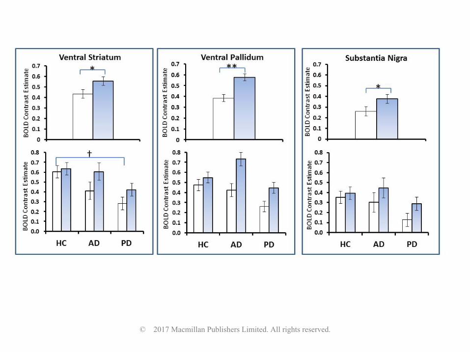
Mixed ANOVAs demonstrated a main effect of drug within the ventral striatum p=0.005, ventral
pallidum (p<0.001) and substantia nigra (p=0.009) (Table 1). These effects are due to increased
reward-neutral BOLD response in the GSK598809 session compared to the placebo session (Figure
2). Whilst these effects appear to be mainly driven by the dependent groups in each ROI, only a
trend for a drug by group interaction was found, and only in the ventral pallidum (p=0.041, Table 1).
Post-hoc paired t-tests revealed a significant effect of GSK598809 on ventral pallidum reward-
neutral BOLD response within the AD group (p<0.001) and PD group (p=0.003) but not the HC group
(p=0.145).
No significant main effects of group were found although trends for ventral striatum and substantia
nigra response were found (p = 0.048 and 0.042 respectively). Figure 2 suggests that these effects
appear to be driven by blunting occurring within the placebo condition of the PD group. Exploratory
post-hoc investigations carried out with the placebo session data only demonstrated a significant
main effect of group in the VS, F(2,79)=5.03,p=0.009, PD<HC) and a trend for a significant blunting in
the SN that just fell short of Bonferroni corrected significance, (F(2,79)=5.03,p=0.022, PD<HC). Post
hoc tests revealed no difference or trends between AD and HC or AD and PD. No group effects or
trends emerged for corresponding analysis of the GSK598809 session.
Additional exploratory investigations were carried out within the ROIs, separating the groups by
primary drug of dependence (see supplementary materials). This suggested that drug effects were
driven by participants with a primary alcohol but not opiate dependence (see figure S7).
Investigations into primary cocaine dependence were not carried out due to small numbers.
© 2017 Macmillan Publishers Limited. All rights reserved.
GNG Behavioural
There were no significant drug or group effects or interactions for GNG performance (see figure S1).
GNG fMRI - Effect of Task
The stops>go contrast for each group for both the placebo and GSK598809 conditions revealed a
highly significant network of activation, in line with previous studies using this task (Garavan et al,
2002). See figure S4.
Effects of group and drug: whole-brain analysis
Whole-brain analyses using mixed ANOVAs revealed a significant group effect within the left
cerebral peduncle region of the midbrain due to increased activation in the AD group (figure S6). No
drug effects or interactions were found.
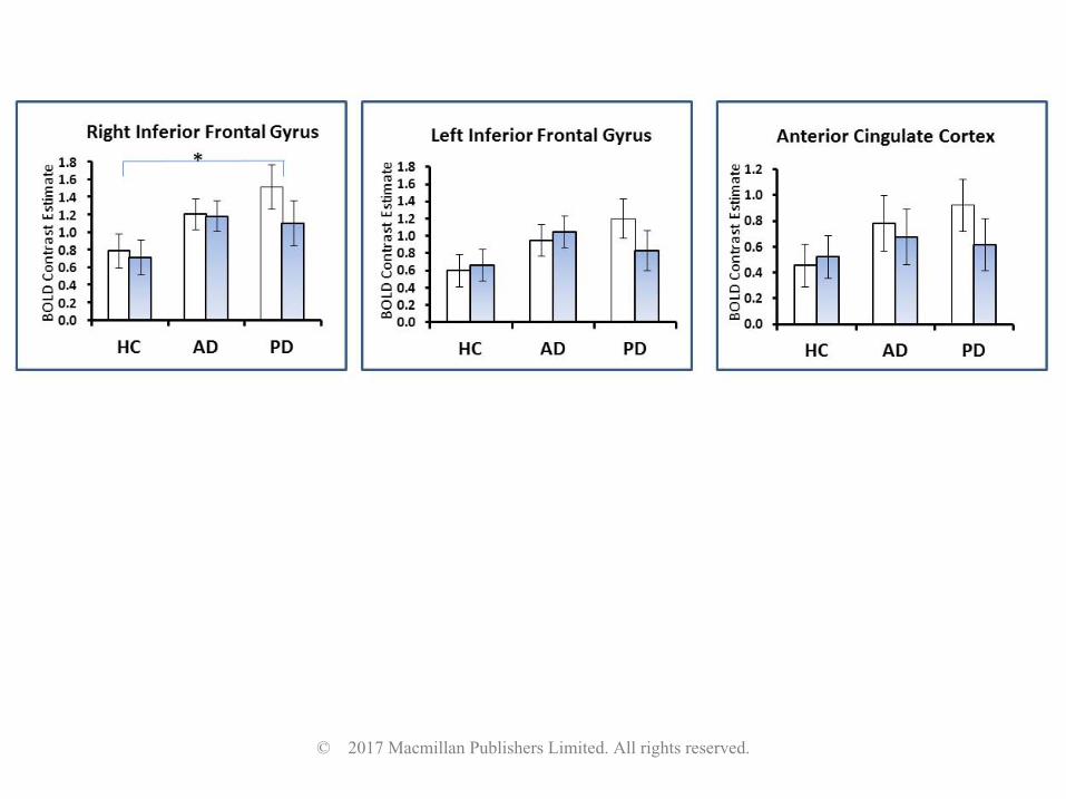
ROI analysis
There was no effect of GSK598809 in any of the ROIs. A main effect of group was found in the right
IFG (F(2,)=4.71, p=0.012), and at trend level in the left IFG (p=0.024), driven by hyper-activation in
the PD compared with the HC group (p=0.016). See figure 4.
Exploratory analysis revealed no significant effects of GSK598809 in DRD3 rich regions (VS, VP, SN).
Correlational Analyses
Ventral striatal ROI activation during the placebo condition for the MID task, in all participants,
correlated with impulsivity measured by the Barratt Impulsivity Scale (BIS) (Patton et al, 1995) (r= -
0.368, p=0.001) but not the reward responsivity subscale of the BISBAS (Carver and White, 1994)
(Table S4). To rule out the possibility of this negative association being primarily driven by group
© 2017 Macmillan Publishers Limited. All rights reserved.
differences, an additional correlation was carried out within the healthy control group only, which
supported this negative relationship (r= -0.372, p = 0.033). No correlations were found with any of
the GNG ROIs (Table S3 and S4). No relationships were found between the MID and GNG task ROI
activation or fMRI and questionnaire behavioural measures (Table S4) or with duration of abstinence
(Table S5). No relationship was found between the effect of GSK598809 on ventral pallidum
activation and MID performance (Table S6).
Discussion
This study aimed to investigate the effects of the selective DRD3 antagonist GSK598809 on networks
involved in the anticipation of reward and response inhibition. The main findings were that
GSK598809 significantly increased reward-neutral reward anticipatory responses for the MID task
but had no effects on brain response during response inhibition.
Group effects were found for both tasks, although in opposite directions. Blunting of the ventral
striatum in the PD group, and DLPFC response in the AD group, was found for the MID task under
placebo. Enhanced inferior frontal response in the PD group and enhanced midbrain response in the
AD group was found for the GNG task. There were no effects of either drug or group on task
performance, therefore all reported differences in BOLD response occur in the context of normal
performance.
MID
Deficits in ventral striatal reward-related signalling are hypothesised to confer vulnerability to
addiction and relapse (Trifilieff and Martinez, 2014; Blum et al 2000). We found blunted ventral
striatal response in the PD group, and an overall negative correlation between ventral striatum
response and impulsivity. Reduced MID ventral striatum response associated with increased
impulsivity has been previously demonstrated in alcohol dependence (Beck et al, 2009; Wrase et al,
© 2017 Macmillan Publishers Limited. All rights reserved.
2007), therefore our finding adds to a growing literature suggesting that deficits in ventral striatal
reward signalling confer vulnerability to addiction and relapse (Trifilieff and Martinez, 2014; Blum et
al 2000). In contrast to the studies above, we did not find significant blunting in the AD group. This
could suggest recovery with long-term abstinence in our cohort; however we did not find a
significant association between length of abstinence and ventral striatal response in the AD group,
perhaps due to small sample size.
There was a general restorative or enhancing effect of GSK598809 on reward anticipatory BOLD
response.. This finding is in line with evidence suggesting that D3 receptors may act as
autoreceptors, inhibiting dopamine synthesis and release (Diaz et al, 2000; Zapata et al, 2002). It has
been hypothesised that tonic extracellular dopamine may inhibit phasic reward dopamine signalling
via action on autoreceptors (Grace, 1991). This may explain the opposing effects on MID reward
anticipatory BOLD response of GSK598809 and amphetamine, which releases dopamine.
Amphetamine increases tonic dopamine levels but decreases reward anticipatory BOLD response. In
contrast, by blocking D3 autoreceptors, GSK598809 may enhance phasic reward dopamine signalling
(Sokoloff et al, 2006) and reward anticipatory BOLD response. This reward enhancing effect of
dopamine autoreceptors is supported by a recent study using low doses of the D2/D3 antagonist
amisulpride (chosen to result in preferential autoreceptor receptor blockade) which was found to
increase MID reward responsivity in depressed participants (Admon et al, 2016).
Despite these BOLD enhancing effects of GSK598809, no behavioural effects were found. This may
reflect ceiling effects as the behavioural requirements of the MIDT are very simple - reaction times
were unimpaired in the placebo condition suggesting that participants were already performing at
maximum capacity for a speeded motor response. Furthermore, 60mg of GSK598809 results in only
partial D3 blockade (Erritzoe et al, 2014), therefore behavioural effects may emerge with increasing
dose.
© 2017 Macmillan Publishers Limited. All rights reserved.
Although no significant drug by group interactions were found, the effects of GSK598809 appeared
to be largely driven by effects within the abstinent drug dependent groups rather than controls.
(especially within the DRD3 rich ventral pallidum where a strong trend to an interaction was
observed). These patterns may be due to upregulation of DRD3s in stimulant and alcohol
dependence (Boileau et al, 2012; Erritzoe et al, 2014; Payer et al, 2014; Staley et al, 1996) and
evidence of enhanced dopamine autoreceptor actions in response to chronic exposure to alcohol
(Siciliano et al, 2016). Although requiring replication in a larger sample, exploratory investigations
raised the intriguing possibility that GSK598809 may be relatively ineffective in opiate dependence.
At the time of writing, we were unable to find any published studies on DRD3 density in human
opiate addicts. However, in contrast to the increases in DRD3 expression in rodents after alcohol and
cocaine exposure (Le Foll and Di Ciano 2015), a very recent study demonstrated that heroin
exposure decreases DRD3 expression (Zhu et al, 2016), possibly explaining the apparent reduced
efficacy of GSK598809 in those with a primary opiate dependence.
In addition to effects within DRD3 rich regions, whole brain analyses revealed effects within the
caudate, cerebellum and DLPFC. In-vivo PET studies do not report high DRD3 levels within these
regions. An enhanced MIDT reward anticipatory caudate response was also found with DRD3
receptor blocking doses of amisulpride (Admon et al, 2016). There is evidence supporting the
caudate, lateral prefrontal regions and cerebellum to be important regions mediating the integration
of motivation with goal directed behaviour (Harsay et al, 2011; Schutter, 2013; Watanabe and
Sakagami, 2007). Enhanced activation within these regions may therefore be a downstream
consequence of GSK598809’s effects on the ventral striatum and ventral pallidum, regions critically
involved in incentive motivation (Haber and Knutson, 2010; Smith et al, 2009).
.
© 2017 Macmillan Publishers Limited. All rights reserved.
Together, these findings suggest hypo-functioning of reward signalling in substance dependence.
Evidence suggests, in alcohol and stimulant dependence, that this may be caused, in part, by
excessive D3 autoreceptor inhibition of dopamine systems. GSK898809 may ameliorate reward
deficits by disinhibiting these systems.
GNG
In contrast to GNG studies demonstrating prefrontal hypo-activation and impaired response
inhibition in current dependence, we found hyper-activation of lateral prefrontal regions (PD group),
or midbrain (AD group) together with unimpaired behavioural performance. Our results are
consistent with two other studies in cocaine and alcohol abstinence (Bell et al, 2014; Connolly et al,
2012). They perhaps reflect recovery of prefrontal structure and function in successful abstinence
due to cessation of drug use and cognitive control practices (Garavan et al, 2013). Whether this
hyper-activation, which is also found in relatives of alcohol patients, is protective for relapse or an
addiction vulnerability marker is unknown. However fMRI hyper-activation during cognitive tasks has
been reported in detoxified alcohol dependent participants who subsequently abstained, but not in
those who relapsed (Charlet et al, 2014), supporting the notion that prefrontal hyper-activation may
be protective.
Despite previous studies suggesting a link between over-expression of DRD3s and increased
impulsivity as measured by questionnaire (Boileau et al, 2012; Payer et al, 2014), we found no
evidence to suggest GSK598809 modulated performance or neuronal response of the network
underlying response inhibition. This may be due to the multifaceted nature of impulsivity, with
DRD3s affecting some impulsivity measures but not response inhibition as measured here. We found
no association between a questionnaire measure of impulsivity and GNG brain response or
performance. Furthermore, whilst mood-related impulsivity correlates more strongly with D2/D3
© 2017 Macmillan Publishers Limited. All rights reserved.
binding in the ventral rather than dorsal striatum in pathological gamblers (Clark et al 2012),
response inhibition correlates with receptor binding in the dorsal (where DRD3 density is low) but
not the ventral striatum (Ghahremani et al, 2012). These findings suggest different impulsivity
measures have different underlying neuro-circuitry. Prefrontal regions are important for response
inhibition (Aron et al, 2003; Garavan et al, 2013), again regions where DRD3s are low. Low DRD3
density in regions implicated in response inhibition may explain the lack of a modulatory effect of
GSK598809 in our study. We additionally carried out exploratory investigations within the DRD3 rich
regions for the GNG task. However, no effects of GSK598809 modulations were found. These
findings suggest that D3 agents selectively modulate brain mechanisms of incentive motivation.
Limitations
The main limitation of the study was the introduction of an order confound, arising from (ultimately
groundless) concerns over study dropout. However, practise and habituation effects were minimised
by all participants having carried out the tasks in full within the scanner at screening (placebo session
was either the second or third task scanning session), and the tasks being practised outside the
scanner before each session. A post-hoc exploratory ROI analysis was carried out within the anterior
insula, a region considered to process salience and therefore likely to be sensitive to habituation
effects. Notably the anterior insula is reliably implicated in MID performance (in a meta-analysis
performed by our group) but is devoid of DRD3s. No effects of GSK598809 or interactions were
found within this region, supporting the suggestion that the effects seen were indeed drug effects
rather than non-specific habituation effects. Another limitation is the age difference between the
AD and PD groups (consistent with typical clinical presentation) therefore caution should be used
when interpreting AD vs PD differences.
© 2017 Macmillan Publishers Limited. All rights reserved.
Conclusion
GSK598809 enhances reward anticipatory BOLD response to non-drug rewards within abstinent
substance dependent groups, with strongest effects in those with a primary alcohol dependence.
These results have implications for considering D3 antagonism as a potential treatment for
normalising reward deficiencies in substance dependence.
Supplementary information is available at the Neuropsychopharmacology website
Funding and Disclosure
This article presents independent research funded by the Medical Research Council as part of their
addiction initiative (grant number G1000018). GlaxoSmithKline (GSK) supplied the GSK598809 drug
used in this study and funded the functional and structural MRI scans that took place at Imperial
College
David Nutt is an advisor to British National Formulary, MRC, GMC, Dept of Health, is President of the
European Brain Council, past President of the British Neuroscience Association and European College
of Neuropsychopharmacology, chair of DrugScience, is a member of the International Centre for
Science in Drug Policy, advisor to Swedish government on drug, alcohol and tobacco research, editor
of the Journal of Psychopharmacology, sits on advisory Boards at Actelion MSD, and Nalpharm, has
received speaking honoraria (in addition to above) from BMS/Otsuka, GSK, Lilly, Janssen, Servier, is a
member of the Lundbeck International Neuroscience Foundation, has received grants or clinical trial
payments from P1vital, MRC, NHS, Lundbeck, has share options with P1vital, has been expert
witness in a number of legal cases relating to psychotropic drugs, and has edited/written 30 books -
some purchased by pharma companies.
© 2017 Macmillan Publishers Limited. All rights reserved.
Trevor W Robbins has research grants with Eli Lilly and Lundbeck, has received royalties from
Cambridge Cognition (CANTAB), has received editorial honoraria from Springer Verlag, Elsevier,
Society for Neuroscience; has performed educational lectures for Merck, Sharpe and Dohme and
does consultancy work for Cambridge Cognition, Eli Lilly, Lundbeck, Teva and Shire Pharmaceuticals.
Bill Deakin currently advises or carries out research funded by Autifony, Sunovion, Lundbeck,
AstraZeneca and Servier. All payment is to the University of Manchester.
Anne Lingford-Hughes has received speaking honoraria and research support from Lundbeck and
research support from GSK .
Liam J Nestor was a Senior Research Scientist employed by GSK during ICCAM data collection.
All other authors declared no conflict of interest.
Acknowledgements
We wish to thank the Medical Research Council (MRC) for funding the study and GSK for supplying
the study drug and funding scans at London.
The research was carried out at the NIHR/Wellcome Trust Imperial Clinical Research Facility, the
NIHR/Wellcome Trust Cambridge Research Facility and Clinical Trials Unit at Salford Royal NHS
Foundation Trust, and is supported by the North West London, Eastern and Greater Manchester
NIHR Clinical Research Networks. The views expressed are those of the author(s) and not necessarily
those of the Medical Research Council, the NHS, the NIHR or the Department of Health.
We wish to thank Sanja Abbott for help with programming the fMRI tasks used at Cambridge and
research assistants Claire Whitelock, Heather Agyepong, Rania Christoforou and Natalie Cuzen for
their help with data collection, MR physicist Rex Newbould and MR technician, Jonathan Howard for
© 2017 Macmillan Publishers Limited. All rights reserved.
their assistance with MR acquisition and task set-up, Shane McKie for help with statistical analysis
and Martyn McFarquhar for help with task programming and statistical analysis.
Recruitment partners- We wish to thank our recruitment partners; Imperial College Healthcare NHS
Trust, Central and North West London NHS trust, Camden and Islington NHS trust, Cambridge
University Hospitals NHS Foundation Trust, Norfolk and Suffolk NHS Foundation Trust, Cambridge
and Peterborough NHS Foundation Trust, South Staffordshire and Shropshire NHS Foundation Trust,
Manchester Mental Health NHS and Social Care Trust, Greater Manchester West NHS Foundation
Trust, Pennine Care NHS Foundation Trust, Salford Royal NHS Foundation Trust, Addaction,
Foundation 66 and CRI (Crime Reduction Initiative).
*ICCAM Platform collaborators
David Nutt, Anne Lingford-Hughes, Louise Paterson, John McGonigle, Remy Flechais, Csaba Orban,
Bill Deakin, Rebecca Elliott, Anna Murphy, Eleanor Taylor, Trevor Robbins, Karen Ersche, John
Suckling, Dana Smith, Laurence Reed, Filippo Passetti, Luca Faravelli, David Erritzoe, Inge Mick,
Nicola Kalk, Adam Waldman, Liam Nestor, Shankar Kuchibatla, Venkataramana Boyapati, Antonio
Metastasio, Yetunde Faluyi, Emilio Fernandez-Egea, Sanja Abbott, Barbara Sahakian, Valerie Voon,
Ilan Rabiner.
© 2017 Macmillan Publishers Limited. All rights reserved.
References
Admon R, Kaiser RH, Dillon DG, Beltzer M, Goer F, Olson DP, et al (2016). Dopaminergic Enhancement of Striatal Response to Reward in Major Depression. The American journal of psychiatry: appiajp201616010111.
Aron AR, Fletcher PC, Bullmore ET, Sahakian BJ, Robbins TW (2003). Stop-signal inhibition disrupted by damage to right inferior frontal gyrus in humans. Nature neuroscience 6(2): 115-116.
Association AP (1994). Diagnostic and Statistical Manual of Mental Disorders - Fourth Edition (DSM-IV). Americal Psychiatric Association: Washington, DC.
Beck A, Schlagenhauf F, Wustenberg T, Hein J, Kienast T, Kahnt T, et al (2009). Ventral striatal activation during reward anticipation correlates with impulsivity in alcoholics. Biological psychiatry 66(8): 734-742.
Bell RP, Foxe JJ, Ross LA, Garavan H (2014). Intact inhibitory control processes in abstinent drug abusers (I): a functional neuroimaging study in former cocaine addicts. Neuropharmacology 82: 143-150.
Blum K, Braverman ER, Holder JM, Lubar JF, Monastra VJ, Miller D, et al (2000). Reward deficiency syndrome: a biogenetic model for the diagnosis and treatment of impulsive, addictive, and compulsive behaviors. Journal of psychoactive drugs 32 Suppl: i-iv, 1-112.
Boileau I, Payer D, Houle S, Behzadi A, Rusjan PM, Tong J, et al (2012). Higher binding of the dopamine D3 receptor-preferring ligand [11C]-(+)-propyl-hexahydro-naphtho-oxazin in methamphetamine polydrug users: a positron emission tomography study. The Journal of neuroscience : the official journal of the Society for Neuroscience 32(4): 1353-1359.
Carver CL, White TL (1994). Behavioral inhibition, behavioral activation, and affective responses to impending reward and punishment: The BIS/BAS Scales. Journal of personality and social psychology 67(2): 14.
Casey KF, Benkelfat C, Cherkasova MV, Baker GB, Dagher A, Leyton M (2014). Reduced dopamine response to amphetamine in subjects at ultra-high risk for addiction. Biological psychiatry 76(1): 23-30.
Charlet K, Beck A, Jorde A, Wimmer L, Vollstadt-Klein S, Gallinat J, et al (2014). Increased neural activity during high working memory load predicts low relapse risk in alcohol dependence. Addiction biology 19(3): 402-414.
© 2017 Macmillan Publishers Limited. All rights reserved.
Clark L, Stokes PR, Wu K, Michalczuk R, Benecke A, Watson BJ, et al (2012). Striatal dopamine D(2)/D(3) receptor binding in pathological gambling is correlated with mood-related impulsivity. NeuroImage 63(1): 40-46.
Connolly CG, Foxe JJ, Nierenberg J, Shpaner M, Garavan H (2012). The neurobiology of cognitive control in successful cocaine abstinence. Drug and alcohol dependence 121(1-2): 45-53.
Cousineau D, O’Brien F (2014). Error bars in within-subject designs: a comment on Baguley (2012). Behavior Research Methods 46(4): 1149-1151.
Diaz J, Pilon C, Le Foll B, Gros C, Triller A, Schwartz JC, et al (2000). Dopamine D3 receptors expressed by all mesencephalic dopamine neurons. The Journal of neuroscience : the official journal of the Society for Neuroscience 20(23): 8677-8684.
Erritzoe D, Tziortzi A, Bargiela D, Colasanti A, Searle GE, Gunn RN, et al (2014). In vivo imaging of cerebral dopamine D3 receptors in alcoholism. Neuropsychopharmacology : official publication of the American College of Neuropsychopharmacology 39(7): 1703-1712.
Forman SD, Dougherty GG, Casey BJ, Siegle GJ, Braver TS, Barch DM, et al (2004). Opiate addicts lack error-dependent activation of rostral anterior cingulate. Biological psychiatry 55(5): 531-537.
Garavan H, Brennan KL, Hester R, Whelan R (2013). The neurobiology of successful abstinence. Current opinion in neurobiology 23(4): 668-674.
Garavan H, Ross TJ, Murphy K, Roche RA, Stein EA (2002). Dissociable executive functions in the dynamic control of behavior: inhibition, error detection, and correction. NeuroImage 17(4): 1820-1829.
Garrison KA, Potenza MN (2014). Neuroimaging and biomarkers in addiction treatment. Current psychiatry reports 16(12): 513.
Ghahremani DG, Lee B, Robertson CL, Tabibnia G, Morgan AT, De Shetler N, et al (2012). Striatal dopamine D(2)/D(3) receptors mediate response inhibition and related activity in frontostriatal neural circuitry in humans. The Journal of neuroscience : the official journal of the Society for Neuroscience 32(21): 7316-7324.
Grace AA (1991). Regulation of spontaneous activity and oscillatory spike firing in rat midbrain dopamine neurons recorded in vitro. Synapse 7(3): 221-234.
Gurevich EV, Joyce JN (1999). Distribution of dopamine D3 receptor expressing neurons in the human forebrain: comparison with D2 receptor expressing neurons. Neuropsychopharmacology : official publication of the American College of Neuropsychopharmacology 20(1): 60-80.
© 2017 Macmillan Publishers Limited. All rights reserved.
Haber SN, Knutson B (2010). The reward circuit: linking primate anatomy and human imaging. Neuropsychopharmacology : official publication of the American College of Neuropsychopharmacology 35(1): 4-26.
Harsay HA, Cohen MX, Oosterhof NN, Forstmann BU, Mars RB, Ridderinkhof KR (2011). Functional connectivity of the striatum links motivation to action control in humans. The Journal of neuroscience : the official journal of the Society for Neuroscience 31(29): 10701-10711.
Heidbreder CA, Newman AH (2010). Current perspectives on selective dopamine D(3) receptor antagonists as pharmacotherapeutics for addictions and related disorders. Annals of the New York Academy of Sciences 1187: 4-34.
Heinz A, Siessmeier T, Wrase J, Hermann D, Klein S, Grusser SM, et al (2004). Correlation between dopamine D(2) receptors in the ventral striatum and central processing of alcohol cues and craving. The American journal of psychiatry 161(10): 1783-1789.
Kaufman JN, Ross TJ, Stein EA, Garavan H (2003). Cingulate hypoactivity in cocaine users during a GO-NOGO task as revealed by event-related functional magnetic resonance imaging. The Journal of neuroscience : the official journal of the Society for Neuroscience 23(21): 7839-7843.
Knutson B, Westdorp A, Kaiser E, Hommer D (2000). FMRI visualization of brain activity during a monetary incentive delay task. NeuroImage 12(1): 20-27.
Le Foll B, Di Ciano P (2015). Neuronal circuitry underlying the impact of D3 receptor ligands in drug addiction. European neuropsychopharmacology : the journal of the European College of Neuropsychopharmacology 25(9): 1401-1409.
Lubman DI, Yucel M, Kettle JW, Scaffidi A, Mackenzie T, Simmons JG, et al (2009). Responsiveness to drug cues and natural rewards in opiate addiction: associations with later heroin use. Archives of general psychiatry 66(2): 205-212.
Mugnaini M, Iavarone L, Cavallini P, Griffante C, Oliosi B, Savoia C, et al (2013). Occupancy of brain dopamine D3 receptors and drug craving: a translational approach. Neuropsychopharmacology : official publication of the American College of Neuropsychopharmacology 38(2): 302-312.
Nathan PJ, O'Neill BV, Mogg K, Bradley BP, Beaver J, Bani M, et al (2012). The effects of the dopamine D(3) receptor antagonist GSK598809 on attentional bias to palatable food cues in overweight and obese subjects. Int J Neuropsychopharmacol 15(2): 149-161.
Nutt DJ, Lingford-Hughes A, Erritzoe D, Stokes PR (2015). The dopamine theory of addiction: 40 years of highs and lows. Nature reviews Neuroscience 16(5): 305-312.
© 2017 Macmillan Publishers Limited. All rights reserved.
Paterson LM, Flechais RS, Murphy A, Reed LJ, Abbott S, Boyapati V, et al (2015). The Imperial College Cambridge Manchester (ICCAM) platform study: An experimental medicine platform for evaluating new drugs for relapse prevention in addiction. Part A: Study description. J Psychopharmacol 29(9): 943-960.
Patton JH, Stanford MS, Barratt ES (1995). Factor structure of the Barratt impulsiveness scale. Journal of clinical psychology 51(6): 768-774.
Payer DE, Behzadi A, Kish SJ, Houle S, Wilson AA, Rusjan PM, et al (2014). Heightened D3 dopamine receptor levels in cocaine dependence and contributions to the addiction behavioral phenotype: a positron emission tomography study with [11C]-+-PHNO. Neuropsychopharmacology : official publication of the American College of Neuropsychopharmacology 39(2): 311-318.
Robinson TE, Berridge KC (1993). The neural basis of drug craving: an incentive-sensitization theory of addiction. Brain research Brain research reviews 18(3): 247-291.
Schultz W (1998). The phasic reward signal of primate dopamine neurons. Adv Pharmacol 42: 686-690.
Schutter DJLG (2013). Human Cerebellum in Motivation and Emotion. In: Manto M, Schmahmann JD, Rossi F, Gruol DL, Koibuchi N (eds). Handbook of the Cerebellum and Cerebellar Disorders. Springer Netherlands: Dordrecht, pp 1771-1782.
Siciliano CA, Calipari ES, Yorgason JT, Lovinger DM, Mateo Y, Jimenez VA, et al (2016). Increased presynaptic regulation of dopamine neurotransmission in the nucleus accumbens core following chronic ethanol self-administration in female macaques. Psychopharmacology 233(8): 1435-1443.
Smith KS, Tindell AJ, Aldridge JW, Berridge KC (2009). Ventral pallidum roles in reward and motivation. Behavioural brain research 196(2): 155-167.
Sokoloff P, Diaz J, Le Foll B, Guillin O, Leriche L, Bezard E, et al (2006). The dopamine D3 receptor: a therapeutic target for the treatment of neuropsychiatric disorders. CNS & neurological disorders drug targets 5(1): 25-43.
Staley JK, Mash DC (1996). Adaptive increase in D3 dopamine receptors in the brain reward circuits of human cocaine fatalities. The Journal of neuroscience : the official journal of the Society for Neuroscience 16(19): 6100-6106.
Taylor EM, Murphy A, Boyapati V, Ersche KD, Flechais R, Kuchibatla S, et al (2016). Impulsivity in abstinent alcohol and polydrug dependence: a multidimensional approach. Psychopharmacology 233(8): 1487-1499.
© 2017 Macmillan Publishers Limited. All rights reserved.
Trifilieff P, Martinez D (2014). Blunted dopamine release as a biomarker for vulnerability for substance use disorders. Biological psychiatry 76(1): 4-5.
Tziortzi AC, Searle GE, Tzimopoulou S, Salinas C, Beaver JD, Jenkinson M, et al (2011). Imaging dopamine receptors in humans with [11C]-(+)-PHNO: dissection of D3 signal and anatomy. NeuroImage 54(1): 264-277.
Watanabe M, Sakagami M (2007). Integration of cognitive and motivational context information in the primate prefrontal cortex. Cereb Cortex 17 Suppl 1: i101-109.
Wrase J, Schlagenhauf F, Kienast T, Wustenberg T, Bermpohl F, Kahnt T, et al (2007). Dysfunction of reward processing correlates with alcohol craving in detoxified alcoholics. NeuroImage 35(2): 787-794.
Zapata A, Shippenberg TS (2002). D(3) receptor ligands modulate extracellular dopamine clearance in the nucleus accumbens. Journal of neurochemistry 81(5): 1035-1042.
Zhu Y, Wang Y, Lai J, Wei S, Zhang H, Yan P, et al (2016). Dopamine D1 and D3 Receptors Modulate Heroin-induced Cognitive Impairment through Opponent Actions in Mice. International Journal of Neuropsychopharmacology.
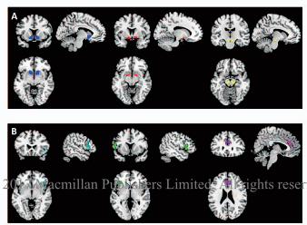
Figure1: Regions of Interest: A) ROIs for the MID task, left/blue (when in colour) shows the ventral
striatum, middle/red shows the ventral pallidum (both defined according to the guidelines of
Tziortzi et al. 2011), and right/yellow shows the substantia nigra. B) ROIs for the GNG task, left/cyan
shows the right inferior frontal gyrus, middle/green shows the left inferior frontal gyrus, and
right/purple shows the anterior cingulate.
Figure 2. ROI response during the MID task: mean reward-neutral anticipation BOLD contrast
estimate for both the placebo and the GSK598809 sessions. White bars represent the placebo
session whereas light grey/blue (when in colour) represent the GSK598809 session. Hisograms on
the top show the main effect of drug within each ROI (*= significant at p<0.01, **= significant at
p<0.001) whereas histopgrams below show the BOLD contrast estimates for the placebo and
GSK598809 sessions for each group separately (†=significant effect of group at p<0.01 in the placebo
© 2017 Macmillan Publishers Limited. All rights reserved.
condition only). . Error Bars indicate within-subject standard error of the mean (Cousineau and
O’Brien, 2014)) suitable for assessing drug rather than group effects.
Figure 3 . ROI response during the GNG task: mean ‘stops-go’ BOLD contrast estimate within each
ROI for the placebo (white bars) and the GSK598809 (grey or blue when in colour) sessions. Error
Bars indicate within-subject standard error of the mean (Cousineau and O’brien 2014). * = main
effect of group, significant at a Bonferroni corrected value of p< 0.017.
Table 1. Results from ROIs and whole-brain analyses for the MID (top, grey) and GNG task (bottom).
© 2017 Macmillan Publishers Limited. All rights reserved.
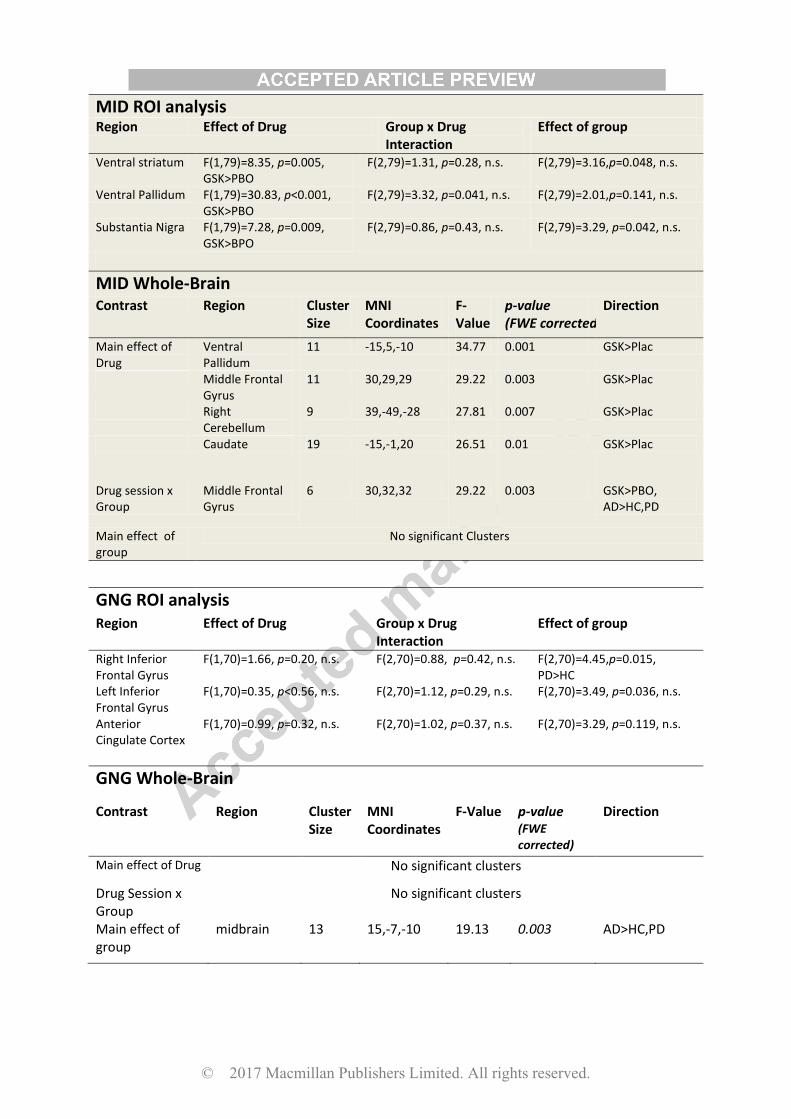
MID ROI analysis Region Effect of Drug Group x Drug
Interaction Effect of group
Ventral striatum F(1,79)=8.35, p=0.005, GSK>PBO
F(2,79)=1.31, p=0.28, n.s. F(2,79)=3.16,p=0.048, n.s.
Ventral Pallidum F(1,79)=30.83, p<0.001, GSK>PBO
F(2,79)=3.32, p=0.041, n.s. F(2,79)=2.01,p=0.141, n.s.
Substantia Nigra F(1,79)=7.28, p=0.009, GSK>BPO
F(2,79)=0.86, p=0.43, n.s. F(2,79)=3.29, p=0.042, n.s.
MID Whole-Brain
Contrast Region Cluster Size
MNI Coordinates
F-Value
p-value (FWE corrected)
Direction
Main effect of Drug
Ventral Pallidum
11 -15,5,-10 34.77 0.001 GSK>Plac
Middle Frontal Gyrus
11 30,29,29 29.22 0.003 GSK>Plac
Right Cerebellum
9 39,-49,-28 27.81 0.007 GSK>Plac
Caudate 19 -15,-1,20 26.51 0.01 GSK>Plac
Drug session x Group
Middle Frontal Gyrus
6 30,32,32 29.22 0.003 GSK>PBO, AD>HC,PD
Main effect of group
No significant Clusters
GNG ROI analysis Region Effect of Drug Group x Drug
Interaction Effect of group
Right Inferior Frontal Gyrus
F(1,70)=1.66, p=0.20, n.s. F(2,70)=0.88, p=0.42, n.s. F(2,70)=4.45,p=0.015, PD>HC
Left Inferior Frontal Gyrus
F(1,70)=0.35, p<0.56, n.s. F(2,70)=1.12, p=0.29, n.s. F(2,70)=3.49, p=0.036, n.s.
Anterior Cingulate Cortex
F(1,70)=0.99, p=0.32, n.s. F(2,70)=1.02, p=0.37, n.s. F(2,70)=3.29, p=0.119, n.s.
GNG Whole-Brain
Contrast Region Cluster Size
MNI Coordinates
F-Value p-value (FWE corrected)
Direction
Main effect of Drug No significant clusters
Drug Session x Group
No significant clusters
Main effect of group
midbrain 13 15,-7,-10 19.13 0.003 AD>HC,PD
© 2017 Macmillan Publishers Limited. All rights reserved.
© 2017 Macmillan Publishers Limited. All rights reserved.
© 2017 Macmillan Publishers Limited. All rights reserved.
© 2017 Macmillan Publishers Limited. All rights reserved.