Embed Size (px)
Citation preview
1
Activation of the proton pump, V-ATPase, triggers JNK-dependent cell invasion and
overgrowth in a Drosophila epithelium
Astrid G. Petzoldt1,2,5,6, Eva Maria Gleixner1,2,6, Arianna Fumagalli4, Thomas Vaccari4 and
Matias Simons1,2,3
1 Center for Systems Biology (ZBSA), University of Freiburg, Habsburgerstr. 49, 79104
Freiburg, Germany 2 Renal Division, University Hospital Freiburg, Hugstetter Str. 55, 79106 Freiburg, Germany 3 BIOSS Centre for Biological Signaling Studies, University of Freiburg, Germany 4 IFOM - FIRC Institute of Molecular Oncology, Via Adamello 16, 20139 Milan, Italy 5 Present address: Institut für Biologie / Genetik, Freie Universität Berlin, Takustraße 6,
14195 Berlin, Germany
6 These authors contributed equally Correspondance should be addressed to:
Matias Simons, Center for Systems Biology (ZBSA)/ Department of Medicine, University of
Freiburg, Habsburgerstr. 49, 79104 Freiburg, Germany, Tel.: +49-761-20397206, Fax: +49-
761-20397188, email: [email protected]
Thomas Vaccari, IFOM - FIRC Institute of Molecular Oncology, Via Adamello 16, 20139
Milan, Italy, Tel.: +39 02574303823, Fax.: +39 02574303231, email:
Running title: ATP6V1C1 induces cell transformation
Keywords: V-ATPase, endosomes, signaling, cell invasion, cell proliferation, apoptosis,
acidification
Dise
ase
Mod
els &
Mec
hani
sms
D
MM
Acce
pted
man
uscr
ipt
© 2012. Published by The Company of Biologists Ltd.This is an Open Access article distributed under the terms of the Creative Commons Attribution Non-Commercial Share Alike License(http://creativecommons.org/licenses/by-nc-sa/3.0), which permits unrestricted non-commercial use, distribution and reproduction inany medium provided that the original work is properly cited and all further distributions of the work or adaptation are subject to the same Creative Commons License terms.
http://dmm.biologists.org/lookup/doi/10.1242/dmm.010660Access the most recent version at DMM Advance Online Articles. Posted 18 January 2013 as doi: 10.1242/dmm.010660
http://dmm.biologists.org/lookup/doi/10.1242/dmm.010660Access the most recent version at First posted online on 18 January 2013 as 10.1242/dmm.010660
2
Summary
The C subunit of the vacuolar H+-ATPase or V-ATPase regulates the activity and assembly of
the proton pump at cellular membranes. It has been shown to be strongly upregulated in oral
squamous cell carcinoma, a highly metastatic epithelial cancer. In addition, increased V-
ATPase activity appears to correlate with invasiveness of cancer cells, but the underlying
mechanism is largely unknown. Using the Drosophila wing imaginal epithelium as an in vivo
model system, we demonstrate that overexpression of Vha44, the Drosophila orthologue of
the C subunit, causes a tumor-like tissue transformation in cells of the wing epithelium.
Overexpressing cells are excluded from the epithelium and acquire invasive properties while
displaying high apoptotic rates. Blocking apoptosis in these cells unmasks a strong
proliferation stimulus, leading to overgrowth. Furthermore, we show that excess Vha44
greatly increases acidification of endocytic compartments and interferes with endosomal
trafficking. As a result, cargoes such as GFP-Lamp1 and Notch accumulate in highly acidified
enlarged endolysosomal compartments. Consistent with previous reports on the endocytic
activation of Eiger/JNK signaling, we find that V-ATPase stimulation by Vha44 causes JNK
signaling activation while downmodulation of JNK signaling rescues the invasive phenotypes.
In summary, our in vivo-findings demonstrate that increased levels of V-ATPase C subunit
induce a Eiger/JNK-dependent cell transformation within an epithelial organ that recapitulates
early carcinoma stages.
Introduction
Extra- and intracellular pH is tightly regulated by a number of proton transport systems. A
prominent member is the vacuolar H+-ATPase or in short V-ATPase. The proton pump is
required for several cellular processes depending on the cell type and intracellular localization
(Hinton et al., 2009). Through the regulation of endolysosomal acidification, the V-ATPase
participates in protein degradation as well as in the control of endosomal trafficking and
sorting (Hurtado-Lorenzo et al., 2006). This function was recently also suggested to be
required in a number of signaling pathways important in development and cancer (Niehrs and
Boutros, 2010; Yan et al., 2009; Buechling et al., 2010; Cruciat et al., 2010; Hermle et al.,
2010; Vaccari et al., 2010; Hermle et al., 2013). In tumor cells, enhanced plasma membrane
Dise
ase
Mod
els &
Mec
hani
sms
D
MM
Acce
pted
man
uscr
ipt
3
activity of the V-ATPase and other proton transporter strongly correlates with increased cell
proliferation and invasive cell migration (Webb et al., 2011).
V-ATPases are large protein complexes that are organized into two domains: the
membrane-embedded V0 domain responsible for proton translocation, and the cytoplasmic V1
domain carrying out ATP hydrolysis. As a multi-subunit complex, the activity of the V-
ATPase depends on the reversible assembly of the complex as well as the dynamic expression
of V-ATPase subunits at different cellular membranes. Whereas most of the fifteen subunits
are essential structural and functional components, others function as regulatory subunits
controlling assembly and subcellular localization (Forgac, 2007). One important regulatory
subunit is the C subunit, termed ATP6V1C1 in mammals and Vha44 in Drosophila. It is the
only subunit that is released into the cytosol during disassembly of the proton pump (Kane,
2000; Merzendorfer et al., 2000). Its phosphorylation by Protein kinase A has been suggested
to promote (re-)assembly of the V0-V1 holoenzymes (Voss et al., 2007). Furthermore,
biochemical analysis has revealed that its presence stabilizes complex assembly and increases
pump activity (Puopolo et al., 1992).
Among the V-ATPase subunits, ATP6V1C1 is the most upregulated gene in oral
squamous cell carcinoma, a highly metastatic human cancer (Otero-Rey et al., 2008).
Expression is predominant at the periphery and invasive fronts of tumors (Garcia-Garcia et
al., 2012), suggesting an important role of the C subunit in invasive growth and migration
(Perez-Sayans et al., 2009). Other V-ATPase subunits, including the a3 and c subunits, have
also been implicated in cancer metastasis (Lu et al., 2005; Nishisho et al., 2011). Moreover, it
has been shown that plasma membrane activity of the V-ATPase is strongly increased in
highly metastatic breast cancer cells (Sennoune et al., 2004). The general conclusion of these
and other studies has been that the V-ATPase contributes to the reduced extracellular pH of
tumors (Robey et al., 2009; Webb et al., 2011). V-ATPase inhibitors have therefore been
tested in human cancer trials with the rationale to lower tumor acidity and to improve patient
outcome (Otero-Rey et al., 2008; Hinton et al., 2009; Fais, 2010; Webb et al., 2011). Most
experimental data concerning the role of V-ATPase subunits in tumorigenesis has been
obtained by using xenografts or cultured tumor cell lines separated from their tissue
microenvironment (Sennoune et al., 2004; Lu et al., 2005; Nishisho et al., 2011). The question
whether changes in cellular pH are cause or consequence of cancerous transformation
therefore largely remains unanswered. Furthermore, it is not known whether altered V-
ATPase function might impact cell behavior via the regulation of signaling pathways.
Dise
ase
Mod
els &
Mec
hani
sms
D
MM
Acce
pted
man
uscr
ipt
4
Here, we report on the oncogenic properties of the C subunit of the V-ATPase, Vha44,
in the fruit fly Drosophila melanogaster. Drosophila imaginal discs are excellent model
tissues to study the function of individual oncogenes and tumor suppressors but also the
cooperativity of genes in native cellular environments (Vidal et al., 2006; Halder and Mills,
2011). By contrast to xenografts or cultured cancer cell lines, the mutational load can be
precisely controlled, allowing the genetic requirements of cell transformation to be analyzed
within the tissue of origin. Moreover, because tumor cells are genetically produced,
mechanical disruption of cells and of the extracellular matrix is avoided. Using the wing disc
as a model epithelium, we show that the overexpression of Vha44 is sufficient to trigger
invasive cell behavior in the epithelial wing disc tissue. Our data suggest that ectopic Vha44
expression increases vesicular acidification, impairs endolysosomal degradation and induces a
TNF/Eiger- and JNK-dependent transformation program.
Results
Increased Vha44 expression causes cell invasiveness
Overexpression of Vha44 was targeted to the patched (ptc) expression domain of the
larval wing disc epithelium using the GAL4/UAS expression system. This domain constitutes
a narrow stripe at the anterior-posterior (AP) boundary, in which expression is highest at the
boundary-facing side of the stripe (Fig. 1A-E). Expression of both HA-tagged and untagged
Vha44 caused a striking unidirectional migration of the cells from the anterior ptc stripe into
the posterior compartment (Fig. 1B,D and S1A,B). Additional expression of mRFP confirmed
the identity of the invading cells as originating from the ptc stripe (Fig. 1B,B’). In control
discs, mRFP expression was always confined within the ptc stripe (Fig. 1A,C), consistent
with the role of the AP boundary in preventing mixing of cells (Landsberg et al., 2009). X-z
axis projection revealed that migration took place on the basal side of the wing epithelium
(Fig. 1D). These results suggest that cells close to the boundary are extruded basally and then
move towards the posterior compartment. Basal invasion into the neighbouring compartment
was also seen by Vha44 overexpression in other compartments of the wing disc such as the
dorsal part using apterous (ap)-GAL4 (Fig. S1C,D). Therefore, we conclude that Vha44
overexpression can trigger invasion in different regions of the disc epihelium.
Dise
ase
Mod
els &
Mec
hani
sms
D
MM
Acce
pted
man
uscr
ipt
5
Blocking apoptosis unmasks overgrowth caused by Vha44
Basal extrusion of epithelial cells in Drosophila tissue is often accompanied by
apoptosis (Vidal et al., 2006; Igaki et al., 2009; Marinari et al., 2012). Accordingly, we
observed that most of the extruded cells displayed pyknotic nuclei suggestive of apoptotic cell
death (not shown). Further investigation revealed that, unlike mRFP-expressing control cells,
a significant fraction of the invasive cells were positive for cleaved Caspase 3 (Cas3) as well
as TUNEL labeling (Fig.1F,G and S2A,B). Apoptosis was highest at the AP boundary and in
the invasive front. This suggests that cells were displaced into the basal extracellular matrix
(ECM) at the boundaries due to apoptosis and then migrated into the posterior compartment.
Consistent with previous results, we further verified that unlike Vha44 the induction of
apoptosis by overexpression of a pro-apoptotic protein is not sufficient to cause cell migration
(Fig. S2C,D; Vidal et al., 2006). This indicates that additional mechanisms are involved in the
invasive behavior.
Next, we co-expressed the baculovirus protein p35, which acts as a strong suppressor
of apoptosis by inhibiting Cas3 function (Hay et al., 1994). As a result, we observed no
apoptosis and basal exclusion, but nevertheless the ptc stripe appeared broadened and
irregular (Fig. 2A,B and S2E,F). Staining for phospho-histone 3 (PH3) revealed increased
proliferation in the ptc stripe in Vha44/p35- but not in mRFP/p35-expressing control cells
(Fig.2C,D). Expressing Vha44 in the ap or nubbin (nub) compartments, encompassing most
of the wing disc or wing pouch, respectively, led to overgrowth even in the absence of p35, as
shown by the increased size of the compartments and the protruding bulges at the wing disc
edges (Fig. 2F,G and Fig. S1C). We therefore conclude that Vha44 overexpression promotes
proliferation, an effect that is masked by strong apoptosis at the AP boundary.
Vha44 cooperates with oncogenic Ras
The phenotypes observed upon Vha44 overexpression are reminiscent of the cellular
transformation occurring during tumorigenesis. Thus, we next investigated whether ectopic
Vha44 expression activates oncogenic signaling pathways previously reported to mediate cell
invasion and overgrowth in the wing disc (Vidal et al., 2006; Singh et al., 2010). Silencing the
Src inhibitor Csk with a previously validated RNAi line did not significantly increase the
level of Vha44-induced migration (Vidal et al., 2006; Fig. S3A-D). Neither did we find a
significant activation of Abl in the Vha44-expressing cells (Fig. S3E). In contrast, the co-
overexpression of Vha44 and RasV12, the oncogenic form of Ras, led to a strong increase of
cell migration compared to overexpressing Vha44 and the control protein mRFP (Fig. 3B-D).
Dise
ase
Mod
els &
Mec
hani
sms
D
MM
Acce
pted
man
uscr
ipt
6
Co-expressing RasV12 and Vha44 also caused a synergistic effect with regard to overgrowth
(Fig. 3F-H). Since RasV12 expression alone did not lead to any invasion or overgrowth (Fig.
3A,E), these results indicate that Vha44 cooperates with oncogenic Ras in invasion and
overgrowth.
Overexpression of Vha44 lowers endolysosomal pH and alters V-ATPase subunit levels
To understand the cellular mechanism by which Vha44 overexpression induces this
complex tumor-like phenotype, we first examined the subcellular localization of
overexpressed Vha44 in wing discs. Consistent with the reported localization of V-ATPase
(Feng et al., 2009), endogenous and overexpressed Vha44 was mainly found in intracellular
organelles but also at the plasma membrane of epithelial wing cells (Fig. S4B-D). A
substantial fraction of these organelles were positive for the late endosomal marker Rab7 (Fig.
S4D). To investigate the effects of Vha44 overexpression on V-ATPase function, we applied
the acidotrophic fluorescent dye LysoTracker to isolated live wing discs overexpressing
Vha44 under ptc-GAL4. We observed a strong accumulation of LysoTracker in the ptc stripe
but not in the neighboring wild-type cells or in control cells (Figure 4A,B). This indicates that
organellar acidification is increased in Vha44-expressing cells. Blocking V-ATPase activity
with the specific inhibitor Concanamycin A resulted in the complete loss of the LysoTracker
labeling, indicating that the Vha44-mediated increase in organellar acidification is due to
enhanced V-ATPase function (Fig. 4C). In addition, we found that overexpression of another
V-ATPase subunit, Vha100-1, did not alter Lysotracker incorporation or cause invasion and
overgrowth phenotypes (Fig. S5A-C). In line with the proposed V-ATPase assembly function
for Vha44, these results suggest that unlike Vha100-1 Vha44 is sufficient to cause association
of V0 and V1 subcomplexes, thus, increasing the amount of functional holoenzymes within
membranes of intracellular organelles. Accordingly, using a GFP insertion in the genomic
locus of Vha16-1, we found enhanced levels of the V0 component Vha16-1-GFP upon Vha44
overexpression. (Figure 4D). By contrast, a GFP insertion in VhaSFD was downregulated
upon Vha44 overexpression (Figure 4E). The reason for this downregulation is unclear.
However, as VhaSFD (or H subunit) has been reported to inhibit ATP hydrolysis of the
unassembled V1 sector (Jefferies and Forgac, 2008; Diab et al., 2009), its reduction upon
Vha44 overexpression may be consistent with increased pump assembly.
To test whether increased proton transport is the cause of the observed tissue
alterations, we overexpressed the sodium-proton exchanger Nhe2. This multi-transmembrane
protein translocates protons across membranes in the same direction as the V-ATPase and has
Dise
ase
Mod
els &
Mec
hani
sms
D
MM
Acce
pted
man
uscr
ipt
7
been shown to localize to the plasma membrane and endosomes (D'Souza et al., 1998). We
found that Nhe2-YFP overexpression caused weaker but very similar phenotypes compared to
Vha44: invasion, apoptosis and overgrowth and increased organellar acidification, (Fig. S6A-
D). By co-staining with Avl and Rab7, we confirmed that a pool of overexpressed Nhe2-YFP
localized to early and late endosomal compartments (Fig. S6E,F). Taken together, these
results suggest that increased endolysosomal acidification may be sufficient for invasion and
overgrowth phenotypes.
Endolysosomal proteolysis is impaired in Vha44-expressing cells
V-ATPase-dependent acidification has been linked to endosomal sorting and
lysosomal degradation (Hurtado-Lorenzo et al., 2006). To study these membrane trafficking
processes, we first expressed a GFP-Lamp1 fusion construct under the ubiquitous tubulin
promoter. In Drosophila cells, the LAMP1-derived cytoplasmic tail is sufficient to target this
fusion protein from the Golgi to late endosomes and lysosomes, where hydrolases degrade
GFP (Rohrer et al., 1996; Pulipparacharuvil et al., 2005). Whereas GFP-Lamp1 was hardly
detectable in the neighboring tissue and in mRFP-expressing control cells (Figure 5A-C),
overexpression of Vha44 (with and without p35) caused a severe accumulation of GFP-
Lamp1 in the ptc stripe (Fig. 5B,C). GFP-Lamp1 partially co-localized in enlarged organelles
with the late endosomal marker Rab7 and Lysotracker (Fig. 5D,E), giving rise to a ring-like
structure with luminal GFP signal and Rab7 signal at the limiting membrane (inset in Fig.
5D). Generally, there seemed to be an irregular localization pattern for Rab7 in the Vha44-
expressing ptc stripe with accumulations in enlarged compartments (Fig. 5F,G). Together,
these data suggest that Vha44 overexpression impairs endolysosomal degradation leading to
cargo accumulation in enlarged late endosomes and/or lysosomes.
Notch accumulation in acidic compartments does not significantly alter Notch signaling in
Vha44-expressing cells
Endocytic activation and/or impaired degradation of signaling receptors have been
implicated in sustained tumor growth and invasion. In Drosophila, oncogenic Notch and c-jun
N-terminal kinase (JNK) signaling has been demonstrated to occur in endosomes (Vaccari et
al., 2008; Igaki et al., 2009; Vaccari and Bilder, 2005; Wilkin et al., 2008; Rodahl et al., 2009;
Hori et al., 2011). Therefore, we investigated the role of Notch and JNK signaling in Vha44-
induced transformation. Consistent with alterations of protein trafficking and degradation
upon ectopic Vha44 expression, we found an accumulation of the transmembrane receptor
Dise
ase
Mod
els &
Mec
hani
sms
D
MM
Acce
pted
man
uscr
ipt
8
Notch in the lumen of enlarged late endosomes (Fig. 5I,J). In addition, we found a significant
co-localization of Notch-YFP with LysoTracker (Fig. 5M,N). To monitor directly whether
Notch is degraded in Vha44 expressing cells, we applied an antibody against the extracellular
domain of Notch (NECD) onto live wing discs. Notch was found at the cell surface at t=0
hours in Vha44-expressing cells (Fig. 5K). In contrast after 5 hours, the receptor accumulated
in intracellular compartments in Vha44-cells (Fig. 5L). These results indicate that, similar to
GFP-Lamp1, Notch accumulates in Vha44 overexpressing cells due to reduced
endolysosomal degradation.
Previous studies have shown that Notch activation requires V-ATPase activity (Yan et
al., 2009; Vaccari et al., 2010; Hermle et al., 2013). We therefore tested the status of Notch
signaling activation in cells overexpressing Vha44. We found that in Vha44-expressing cells
expression of the Notch signaling reporter E(Spl)mβeta-lacZ was well detectable in areas in
which Notch signaling is normally low, such as the presumptive vein regions of the disc.
However, it was not as high as in peak signaling portions of the disc, such as the presumptive
wing margin (Fig. S7A-D). Accordingly, overexpression of Vha44 resulted in botch Notch
gain- and loss-of-function phenotypes in adult wings (Fig. S7G-J). Finally, quantitative real-
time PCR (qPCR) demonstrated no significant change in E(Spl)mβeta expression in wing
discs expressing Vha44 with nub-GAL4 (Fig. S7K). This suggests that despite Notch
accumulation in acidified organelles, there is no major alteration of Notch pathway activation
in Vha44-overexpressing cells compared to wild-type cells.
Eiger/JNK signaling is required for Vha44-dependent invasion and overgrowth
In contrast to Notch signaling, the qPCR revealed increased expression of the JNK
signaling inhibitor and target gene puckered (puc; Fig. S7K). We therefore monitored JNK
activity in the Vha44-expressing cells by immunostaining for the activated phosphorylated
form of JNK (pJNK) and by assessing puc expression using the transcriptional reporter puc-
lacZ. Both, pJNK and puc-lacZ levels were increased in Vha44-expressing cells but not in
mRFP-expressing control cells (Fig. 6A,B and S8A,B), indicating that JNK signaling is
activated. Similar results were obtained for Nhe2 (Fig. S6B). Accordingly, we found that the
expression of HepCA, the activated form of JNKK kinase, is sufficient to drive invasion (Fig.
S8D). A further readout for upregulated JNK activity is increased expression of Matrix
Metalloproteinase-1 (MMP1). MMPs are essential downstream targets of JNK that control
invasive migration via basal membrane degradation (Uhlirova and Bohmann, 2006; Vidal et
al., 2006). Consistent with JNK activation, we found increased MMP1 levels basally in the
Dise
ase
Mod
els &
Mec
hani
sms
D
MM
Acce
pted
man
uscr
ipt
9
domain of Vha44 but not mRFP expression (Fig. 6C,D). We also observed a clear loss of the
basal membrane components collagen IV and laminin (Fig. 6E and S8F). These effects were
abolished by the co-expression of p35 (Fig. 6F), indicating that the degradation of the basal
membrane is linked to apoptosis and basal extrusion of cells. Taken together, these results
suggest that excess Vha44 leads to JNK activation, resulting in apoptosis as well as MMP1
upregulation and ECM degradation, which may be a prerequisite for basal extrusion and
invasion.
To test whether inhibition of JNK signaling is not only necessary but also sufficient to
drive invasive behavior, we co-expressed Puc and Vha44. Inhibition of JNK signaling rescued
apoptosis as well as the migration and proliferation phenotypes (Fig. 6G,G’). A similar
suppression of apoptosis and proliferation was observed when co-expressing Basket (Bsk)-
DN, a dominant-negative form of the Drosophila orthologue of JNK, with ap-GAL4 (Fig.
6H,H’). Consistent with these results, we found that the activation of JNK signaling resulting
from expression of puc-lacZ, a hypomorphic allele of the inhibitor Puc, increased the invasion
phenotype (Fig. 6B, compare with 6D). Finally, we tested the contribution of Eiger, the only
Drosophila orthologue of the TNF-alpha ligand and potent activator of JNK signaling (Igaki
et al., 2002; Moreno et al., 2002), by expressing Vha44 in a mutant eiger background. Loss of
both eiger copies strongly suppressed apoptosis, invasion and overgrowth of Vha44-
expressing cells (Fig. 6I, J). Together, these results demonstrate that Eiger/JNK signaling is
both necessary and sufficient for Vha44-dependent invasion and overgrowth (for a model see
Fig. 7).
Discussion
Excess Vha44 leads to tumorigenesis in the wing disc
Increasing evidence suggests that pH alterations significantly affect cancer features
including sustained growth, tissue invasion, metastatic potential and chemoresistance (Stock
and Schwab, 2009; Webb et al., 2011). Whether the effect of dysregulated pH on cancer
progression is a cause or consequence for the development of these tumor traits has been
difficult to address using traditional approaches such as xenografts or cultured cancer cell
lines. In addition, because pH alterations ultimately affect multiple cellular processes, it is
unclear what process is required for tissue transformation.
Dise
ase
Mod
els &
Mec
hani
sms
D
MM
Acce
pted
man
uscr
ipt
10
Drosophila imaginal discs are excellent model tissues to study the impact of
transforming events in the native epithelium (Vidal et al., 2006; Halder and Mills, 2011).
Here, we have shown that overexpression of the V-ATPase subunit Vha44 in different
compartments of the epithelial wing disc is sufficient to induce tumor-like transformations.
Cells overexpressing Vha44 overproliferate giving rise to protruding cell masses. In addition,
they display increased apoptotic rates and basal invasion, which is facilitated by ECM
degradation. Both overgrowth and invasion are increased by oncogenic Ras and inhibited by
downregulation of Eiger/JNK signaling, demostrating that the latter pathway is a key driver of
cellular transformation in our tumor model.
Enhanced V-ATPase activity alters endosomal trafficking
How does Vha44 induce such a complex tumor-like phenotype? In line with the
proposed role of the C subunit as an assembly and disassembly regulator (Puopolo et al.,
1992; Huss et al., 2011), we have found that Vha44 overexpression leads to enhanced uptake
of LysoTracker in wing epithelial cells. This effect is reversed by pharmacological V-ATPase
inhibition and mimicked by overexpression of a different proton exchanger, suggesting that
Vha44-overexpressing cells possess increased proton pump assembly and activity at
intracellular membranes. While the overexpression of another V1 subunit also increased
vacuolar acidity in yeast (Hughes and Gottschling, 2012), the overexpression of a
transmembrane V0 subunit, Vha100-1, did not lead to additional LysoTracker uptake. This
suggests that excess cytosolic V1 components may be more effective in enhancing V-ATPase
assembly and, thus, activity.
We expected that increased pump activity would result in more lysosomal degradation.
In contrast, we found the opposite. GFP-Lamp1 and Notch accumulated in enlarged late
endosomal compartments in which Notch degradation is impaired. A similar combination of
increased acidification and decreased proteolysis has been observed in cells deficient for the
lysosomal transporter Spinster (Rong et al., 2011). How endosomal sorting and transport
becomes blocked upon increased acidification is currently unclear. An interesting possibility
is that changes in luminal pH might alter the binding properties of proteins and lipids at the
cytosolic side of the endosomes (Hurtado-Lorenzo et al., 2006). Indeed, in mammalian cells,
the acidification by the V-ATPase was shown to promote transport towards lysosomes by
recruiting the GTPase ARF6 (and possibly Rab7 as suggested here) to early endosomes
(Hurtado-Lorenzo et al., 2006). Therefore, excessive acidification may alter endosomal
maturation and cargo progress towards functional lysosomes.
Dise
ase
Mod
els &
Mec
hani
sms
D
MM
Acce
pted
man
uscr
ipt
11
How could endosomal trapping of signaling receptors promote cellular transformation?
Our results are in accordance with previous findings demonstrating that the inhibition
of lysosome formation in dor, car, tsg101 and vps25 mutants can cause tumor growth and
invasion in the fly (Moberg et al., 2005; Thompson et al., 2005; Vaccari and Bilder, 2005; Chi
et al., 2010). These mutations block the maturation of late endosomes to lysosomes at
different levels, similarly causing signaling receptor accumulation. Furthermore, the
progression of vps25-dependent tumors was shown to be suppressed by mutants for V-
ATPase subunits, suggesting that oncogenic signaling requires low intraluminal pH (Vaccari
et al., 2010). It is therefore possible that a decreased luminal pH might promote clustering or
conformational changes of receptors that alter recruitment of cytosolic signal transducers. The
activation of Eiger/JNK signaling, the key pathway in our tumor model, has also been linked
to endosomes (Igaki et al., 2009). However, at this point it is unclear to which extent acidity
contributes to the activation of Eiger, its receptor Wengen and/or the phosphorylation of JNK
(Igaki et al., 2011). For the canonical Wnt (or Wingless (Wg)) pathway, it has been shown
that the receptor complex, consisting of Frizzled and LRP6, can directly bind to a V-ATPase
subunit and that phosphorylation of LRP6, a prime event in the signaling cascade, occurs
upon entry into acidic endosomes (Buechling et al., 2010; Cruciat et al., 2010; Hermle et al.,
2010). In the case of the Notch receptor, acidification may promote the intramembraneous S3
cleavage by presenilins (Vaccari et al., 2010). Alternatively, the reduced proteolysis rate in a
disturbed endolysosomal pathway might increase the time of transit of receptors in signaling-
competent endosomes, leading to sustained signaling. Further investigation is therefore
needed to elucidate the precise mechanisms by which signaling receptors and their
downstream transducers, particularly within the JNK signaling pathway, become activated in
acidified compartments.
The epithelial environment influences the transformation process
The induction of cellular transformation in a patch of cells within an epithelium, as
described here, elicits a myriad of cell-autonomous and non-cell-autonomous responses.
Epithelial neighbors and recruited inflammatory cells release both proliferative and apoptotic
stimuli that strongly influence tumor growth (Cordero et al., 2010). Transformed cells that
become apoptotic may further stimulate proliferation of the surviving ones by releasing
growth-promoting morphogens such as Wg and Dpp (Huh et al., 2004; Perez-Garijo et al.,
2004; Ryoo et al., 2004; Morata et al., 2011). Accordingly, we observed that Wg strongly
accumulates in parts of the Vha44-expressing tissue and that Wg signaling is moderately
Dise
ase
Mod
els &
Mec
hani
sms
D
MM
Acce
pted
man
uscr
ipt
12
upregulated upon Vha44 overexpression (Fig. S7F,K). Increased proliferation at tissue
boundaries is particularly interesting since mechanical strains at the boundary can lead to
overcrowding, which has recently been shown to cause delamination and apoptosis (Marinari
et al., 2012). These effects possibly play a role when Vha44 is expressed in the ptc domain.
Here, cells become strongly apoptotic and are basally excluded close to the boundary. Upon
inhibition of apoptosis, the proliferation effect induced by Vha44 becomes visible that may be
further supported by the release of growth factors from the transformed cells themselves or
from the tissue environment. Beyond developmental boundaries, such as the AP boundary, it
is conceivable that in situ-carcinomas can generate similar border effects (Vidal et al., 2006;
Vidal et al., 2010).
Vha44-induced tumorigenesis depends on Ras and JNK signaling
A major advantage of Drosophila for studying cancer-related processes is the
introduction of multiple genetic alterations. Genetic interaction experiments are very useful
for assessing cooperativity of oncogenic pathways. Such studies have highlighted the
importance of Ras signaling in eliciting cancerous transformation upon defects in epithelial
polarity and membrane trafficking (Pagliarini and Xu, 2003; Chi et al., 2010). Although
poorly understood, the JNK signaling pathway seems to play a central role in this process
(Igaki et al., 2006; Uhlirova and Bohmann, 2006; Leong et al., 2009; Wu et al., 2010). One
example is the RasV12-induced shift of the JNK activating ligand, Eiger/TNF-alpha, from a
tumor suppressor into a tumor promoter (Cordero et al., 2010). Whether the increase of
invasion and overgrowth by RasV12 in our tumor model involves JNK signaling and whether
the activation of JNK by Vha44 requires Ras remains to be further explored. It also needs to
be determined how Eiger/JNK inhibition rescues the pathological aspects of our model. The
inhibition of apoptosis may be the prime reason for the rescue, but more direct influences on
the endolysosomal or associated pathways, such as autophagy, cannot be excluded at this
stage (Wu et al., 2009).
Conclusions
Here, we introduce a novel tumor model in Drosophila based on V-ATPase subunit C
overexpression. We describe the induction of cellular transformation in the native epithelium
with high similarity to human cancers. Genetic dissection of our model revealed increased
endosomal acidification, disrupted endolysosomal trafficking and degradation as well as a
requirement for JNK signaling. All these effects have previously not been associated with V-
Dise
ase
Mod
els &
Mec
hani
sms
D
MM
Acce
pted
man
uscr
ipt

13
ATPase overexpression in tumors. A recent study by Vidal and colleagues showed that
sections of human squamous cell carcinoma boundaries exhibited similarities with the wing
disc invasion model, including the upregulation of MMPs (Vidal et al., 2010). Due to the
elevated levels of ATP6V1C1 in oral squamous cell carcinomas (Otero-Rey et al., 2008), our
study suggests that clinical cancer trials involving V-ATPase inhibitors could be expanded to
include this type of tumor (Fais, 2010). As demonstrated by the genetic interaction with Ras
signaling, our model may also be used for studying cooperativity of oncogenic pathways and
for the evaluation of combinatorial therapies.
Dise
ase
Mod
els &
Mec
hani
sms
D
MM
Acce
pted
man
uscr
ipt
14
Material and Methods
Fly stocks
For Vha44 expression in the wing disc, we used ptc-Gal4, dpp-Gal4, nub-GAL4 (all
Bloomington) or ap-Gal4 (a gift of M.Mlodzik) drivers. Controls for UAS-Vha44-HA or
UAS-Vha44 were UAS-myr-mRFP or UAS-GFP (both Bloomington). Apoptosis inhibition
was performed by co-expression of UAS-p35 (Bloomington). As JNK reporter we used puc-
lacZ (E69), which also acts as a mild JNK activator. The basal membrane was visualized with
viking-GFP (Flytrap). Other flystocks were: egr3 (a gift of M.Vidal), UAS-Nhe2-YFP
(Simons et al., 2009), UAS-puc2A, UAS-Bsk-DN (both a gift of S.Noselli), E(Spl)β -lacZ (a
gift of E.Lai) and UAS-Vha100-1 (a gift of R.Hiesinger). Flies were raised and crossed at
25°C, except for p35 or ap-GAL4 crosses at 29°C. For the generation of the UAS-Vha44 and
the UAS-Vha44-HA fly strains we cloned Vha44 with the following primers: F: 5’ CAC CAT
GAT GTC GGA ATA CT 3’ and R: 5’ TTA GAC CTT GGC CTG CTC CA 3’ (with STOP
codon for Vha44) or R: 5’ GAC CTT GGC CTG CTC CAC CA 3’ (without STOP codon for
Vha44-HA). The amplicon was first inserted into the pENTRTM/SD/D-TOPO® Gateway
vector (Invitrogen) and then cloned into the pUASg-attB or pUASg-HA-attB destination
vectors (kindly provided by K. Basler). Transgenesis was performed by Bestgene, Inc.
Antibody and dye stainings
Wing discs were dissected from L3 larvae in PBS and immunostained according to standard
procedures (Hermle et al., 2010). Antibodies and dyes used in this study were: mouse anti-
Dlg (1:25), rat anti-DE-cadherin (1:25), mouse anti-Notch (NICD) (1:50), mouse anti-Notch
(NECD) (1:50), mouse anti-Wg (1:50), mouse anti-MMP1 (1:25), mouse anti-b-gal (1:25; all
from DSHB), rat anti-HA (1:200; from Roche), rabbit anti-cleaved Caspase 3 (1:200; from
Cell signaling), rabbit anti-Laminin (1:25; from Abcam), rabbit anti-Rab7 (1:1000; kindly
provided by A. Nakamura), rabbit anti-Avl (1:250; kindly provided by D. Bilder), guinea pig
anti-v100 (1:2000; kindly provided by R. Hiesinger), mouse anti-pJNK (G7) (1:50, from
Santa Cruz); rabbit anti-pAbl[pY412] (1:100, from Invitrogen). A polyclonal antibody against
Vha44 was raised in guinea pig against the following peptide: QIGQIDGDLKTKSQA
(Eurogentec). For F-actin and nuclei visualization, AlexaFluor 488- and 555-Phalloidin
(1:1000; from Invitrogen), HOE33342 (1:1000; from Invitrogen) were used, respectively. For
TUNEL staining, wing discs were dissected in PBS, fixed in 4% PFA and permeabilized. The
Fluorescein In Situ Cell Death Detection Kit (Roche) was used according to manual
Dise
ase
Mod
els &
Mec
hani
sms
D
MM
Acce
pted
man
uscr
ipt
15
instructions. To assess intravesicular acidification, dissected L3 larval discs were incubated
for 2 min at RT with LysoTracker green DND26 or red DND99 (1 µM; Invitrogen) in PBS,
then mounted and immediately analyzed. V-ATPase activity was blocked with Concanamycin
A (1 µM; Sigma) treatment for 3h at RT in PBS. Notch internalization assays were performed
as described (Vaccari and Bilder, 2005). Discs were analyzed by Zeiss LSM 510 laser
confocal microscopy. Images were processed with Adobe Photoshop and ImageJ software.
For quantification, the area of the Cas3 or mRFP positive cells was determined and
normalized to the area of the ptc stripe. The mean pixel intensities of the immunoreactive
Rab7 signal were measured in square areas of equal size in Vha44-expressing and
neighboring wild-type tissue.
Western Blotting
15 wing discs for each genotype were dissected and immediately lysed in cold RIPA buffer.
The whole disc extract was loaded on 12% SDS-Gel Page and Western Blot was performed
following standard procedures. The Vha44 antibody was used at 1:500. Other antibodies were
anti-β-tubulin (1:1000; from DSHB) and HRP-conjugated anti-guinea pig and anti-mouse
(1:5000; Santa Cruz and DAKO, respectively).
Quantitative RT-PCR
Total RNA from wing imaginal discs expressing (40 discs per sample) was extracted using
TRIZOL Reagent (Invitrogen) and RNeasy Mini Kit (Qiagen) according to the manufacturer's
protocol. Concentration and purity was determined by measuring optical density at 260 and
280 nm using a Nanodrop spectrophotometer. 500 μg of total RNA was reverse transcribed
using a SuperScript VILO cDNA Synthesis kit (Invitrogen) according to the manufacturer's
protocol. 5 ng of cDNA was amplified (in triplicate) in a reaction volume of 15 uL containing
the following reagents: 7.5ul of TaqMan PCR Mastermix 2x No UNG (Applied Biosystems,
Foster City, CA), 0.75 ul of TaqMan Gene expression assay 20x (Applied Biosystems, Foster
City, CA). 300nM of primers and 100nM of Roche probes were used for each sample. RT-
PCR was carried out on the ABI/Prism 7900 HT Sequence Detector System (Applied
Biosystems), using a pre-PCR step of 10 min at 95°C, followed by 40 cycles of 15 s at 95°C
and 60 s at 60°C.
The following primers were used: F: 5’ GAG TGC CTG ACC CAG GAG 3’ and R: 5’ CGG
TCA GCT CCA GGA TGT 3’(for E(spl)mβ), F: 5’ GTC ACA CCA ATC AGT GGA G 3’
and R: CGA GCA GCC GGA TTC TAT TA 3’ (for fz3), F: 5’ GCC ACA TCA GAA CAT
Dise
ase
Mod
els &
Mec
hani
sms
D
MM
Acce
pted
man
uscr
ipt
16
CAA GC 3’(for puc), F: 5’ CGG ATC GAT ATG CTA AGC TGT 3’ and R: 5’ CGA CGC
ACT CTG TTG TCG 3’ (for rpL32-RA).
Data were analyzed using GraphPad Prism 5.0d (GraphPad Software). Values were
normalized by the amount of rpl32 in each sample. Statistical analysis was performed using
the Student’s t-test.
Dise
ase
Mod
els &
Mec
hani
sms
D
MM
Acce
pted
man
uscr
ipt
17
Acknowledgements:
We thank Susanne Helmstädter for excellent technical support. We thank G. Pyrowolakis, M.
Mlodzik, M. Vidal, T. Xu, S. Noselli, R. Rousset, Flytrap, the Bloomington Stock Center, and
the VDRC for fly strains. We thank the DHSB for antibodies, as well as A. Nakamura and D.
Bilder for antibodies. We acknowledge Roland Nitschke from the Life Imaging Center
Freiburg for help with confocal microscopy. We thank M. Vidal and M. Norman for helpful
suggestions and the critical reading of the manuscript. AGP, EMG and MS are supported by
an Emmy-Noether grant SI1303/2-1 by the Deutsche Forschungsgemeinschaft and by the
Bundesministerium für Bildung und Forschung (Gerontosys-NephAge). AF and TV are
supported by a new unit start-up grant from Associazione Italiana Ricerca contro il Cancro.
Dise
ase
Mod
els &
Mec
hani
sms
D
MM
Acce
pted
man
uscr
ipt
18
Translational Impact box:
Clinical issue: Cancer is the leading cause of death in the Western world, and results from
uncontrolled growth and tissue invasion. Dysregulated pH is an emerging cancer hallmark, as
many cancers show a reversed pH gradient with a high intracellular and low extracellular pH.
The gradient facilitates cancer progression by promoting changes in metabolism,
proliferation, migration and invasion. On the molecular level, several proton transport
systems, including the V-ATPase, have been reported to show upregulated activity in cancer
cells. Moreover, new evidence suggests that organellar acidification is also important for
oncogenic signaling processes. However, it is not clear whether this upregulation is cause or
consequence of oncogenic transformation. To study this in more detail, cancer models are
needed that recapitulate transformation in the native tissue.
Results: In this paper, the authors show that the overexpression of a V-ATPase subunit,
Vha44 (or ATP6V1C1 in mammals), is sufficient to trigger overgrowth and invasion of
epithelial cells in the Drosophila wing disc. This subunit is strongly upregulated in oral
squamous cell carcinoma in humans. Central to the in vivo-transformation process is the JNK
signaling pathway, as all Vha44-induced phenotypes can be blocked by JNK inhibition. By
contrast, the phenotype is enhanced by co-expression of an oncogenic form of Ras. Ectopic
Vha44 increases the activity of the V-ATPase in intracellular organelles and causes severe
defects in endolysosomal degradation. As JNK signaling has been shown to be activated at
endosomes, the endolysosomal defects may be central to the oncogenic transformation.
Implications and future directions: Signaling out of endosomes is an emerging theme in
signal transduction biology. Many developmental signaling pathways, including Notch, Wnt
and JNK, require acidified organelles for proper signaling. Conclusions from this work are
that for oncogenesis the endosomal acidification via the V-ATPase is more important than
previously anticipated. The quest for inhibitors of the V-ATPase suitable for cancer treatment
should therefore include molecules that are able to target the endosomal pool of the V-
ATPase. Small-molecule inhibitors against endosomal V-ATPases might be particularly
useful for tumors that depend on the synergistic activation of Notch, Wnt and JNK signaling.
Screening for genetic modifiers as well as testing of combinatorial therapies could be
performed using the Drosophila model established in this paper. The main advantage of this
model is that oncogenic transformation is induced genetically within in the native epithelium.
Dise
ase
Mod
els &
Mec
hani
sms
D
MM
Acce
pted
man
uscr
ipt
19
References:
Buechling, T., Bartscherer, K., Ohkawara, B., Chaudhary, V., Spirohn, K., Niehrs, C. and Boutros, M. (2010). Wnt/Frizzled signaling requires dPRR, the Drosophila homolog of the prorenin receptor. Curr Biol 20, 1263-1268. Chi, C., Zhu, H., Han, M., Zhuang, Y., Wu, X. and Xu, T. (2010). Disruption of lysosome function promotes tumor growth and metastasis in Drosophila. J Biol Chem 285, 21817-21823. Cordero, J. B., Macagno, J. P., Stefanatos, R. K., Strathdee, K. E., Cagan, R. L. and Vidal, M. (2010). Oncogenic Ras diverts a host TNF tumor suppressor activity into tumor promoter. Dev Cell 18, 999-1011. Cruciat, C. M., Ohkawara, B., Acebron, S. P., Karaulanov, E., Reinhard, C., Ingelfinger, D., Boutros, M. and Niehrs, C. (2010). Requirement of prorenin receptor and vacuolar H+-ATPase-mediated acidification for Wnt signaling. Science 327, 459-463. D'Souza, S., Garcia-Cabado, A., Yu, F., Teter, K., Lukacs, G., Skorecki, K., Moore, H. P., Orlowski, J. and Grinstein, S. (1998). The epithelial sodium-hydrogen antiporter Na+/H+ exchanger 3 accumulates and is functional in recycling endosomes. J Biol Chem 273, 2035-2043. Diab, H., Ohira, M., Liu, M., Cobb, E. and Kane, P. M. (2009). Subunit interactions and requirements for inhibition of the yeast V1-ATPase. J Biol Chem 284, 13316-13325. Fais, S. (2010). Proton pump inhibitor-induced tumour cell death by inhibition of a detoxification mechanism. J Intern Med 267, 515-525. Feng, S., Deng, L., Chen, W., Shao, J., Xu, G. and Li, Y. P. (2009). Atp6v1c1 is an essential component of the osteoclast proton pump and in F-actin ring formation in osteoclasts. Biochem J 417, 195-203. Forgac, M. (2007). Vacuolar ATPases: rotary proton pumps in physiology and pathophysiology. Nat Rev Mol Cell Biol 8, 917-929. Garcia-Garcia, A., Perez-Sayans Garcia, M., Rodriguez, M. J., Antunez-Lopez, J., Barros-Angueira, F., Somoza-Martin, M., Gandara-Rey, J. M. and Aguirre-Urizar, J. M. (2012). Immunohistochemical localization of C1 subunit of V-ATPase (ATPase C1) in oral squamous cell cancer and normal oral mucosa. Biotech Histochem 87, 133-139. Halder, G. and Mills, G. B. (2011). Drosophila in cancer research: to boldly go where no one has gone before. Oncogene 30, 4063-4066. Hay, B. A., Wolff, T. and Rubin, G. M. (1994). Expression of baculovirus P35 prevents cell death in Drosophila. Development 120, 2121-2129. Hermle, T., Saltukoglu, D., Grunewald, J., Walz, G. and Simons, M. (2010). Regulation of Frizzled-dependent planar polarity signaling by a V-ATPase subunit. Curr Biol 20, 1269-1276. Hermle, T., Guida, M. C., Beck, S., Helmstadter, S. and Simons, M. (2013). Drosophila ATP6AP2/VhaPRR functions both as a novel planar cell polarity core protein and a regulator of endosomal trafficking. EMBO J advance online publication. Hinton, A., Bond, S. and Forgac, M. (2009). V-ATPase functions in normal and disease processes. Pflugers Arch 457, 589-598. Hori, K., Sen, A., Kirchhausen, T. and Artavanis-Tsakonas, S. (2011). Synergy between the ESCRT-III complex and Deltex defines a ligand-independent Notch signal. J Cell Biol 195, 1005-1015. Hughes, A. L. and Gottschling, D. E. (2012). An early age increase in vacuolar pH limits mitochondrial function and lifespan in yeast. Nature.
Dise
ase
Mod
els &
Mec
hani
sms
D
MM
Acce
pted
man
uscr
ipt
20
Huh, J. R., Guo, M. and Hay, B. A. (2004). Compensatory proliferation induced by cell death in the Drosophila wing disc requires activity of the apical cell death caspase Dronc in a nonapoptotic role. Curr Biol 14, 1262-1266. Hurtado-Lorenzo, A., Skinner, M., El Annan, J., Futai, M., Sun-Wada, G. H., Bourgoin, S., Casanova, J., Wildeman, A., Bechoua, S., Ausiello, D. A. et al. (2006). V-ATPase interacts with ARNO and Arf6 in early endosomes and regulates the protein degradative pathway. Nat Cell Biol 8, 124-136. Huss, M., Vitavska, O., Albertmelcher, A., Bockelmann, S., Nardmann, C., Tabke, K., Tiburcy, F. and Wieczorek, H. (2011). Vacuolar H(+)-ATPases: intra- and intermolecular interactions. Eur J Cell Biol 90, 688-695. Igaki, T., Pagliarini, R. A. and Xu, T. (2006). Loss of cell polarity drives tumor growth and invasion through JNK activation in Drosophila. Curr Biol 16, 1139-1146. Igaki, T., Pastor-Pareja, J. C., Aonuma, H., Miura, M. and Xu, T. (2009). Intrinsic tumor suppression and epithelial maintenance by endocytic activation of Eiger/TNF signaling in Drosophila. Dev Cell 16, 458-465. Igaki, T., Kanda, H., Okano, H., Xu, T. and Miura, M. (2011). Eiger and wengen: the Drosophila orthologs of TNF/TNFR. Adv Exp Med Biol 691, 45-50. Igaki, T., Kanda, H., Yamamoto-Goto, Y., Kanuka, H., Kuranaga, E., Aigaki, T. and Miura, M. (2002). Eiger, a TNF superfamily ligand that triggers the Drosophila JNK pathway. EMBO J 21, 3009-3018. Jefferies, K. C. and Forgac, M. (2008). Subunit H of the vacuolar (H+) ATPase inhibits ATP hydrolysis by the free V1 domain by interaction with the rotary subunit F. J Biol Chem 283, 4512-4519. Kane, P. M. (2000). Regulation of V-ATPases by reversible disassembly. FEBS Lett 469, 137-141. Landsberg, K. P., Farhadifar, R., Ranft, J., Umetsu, D., Widmann, T. J., Bittig, T., Said, A., Julicher, F. and Dahmann, C. (2009). Increased cell bond tension governs cell sorting at the Drosophila anteroposterior compartment boundary. Curr Biol 19, 1950-1955. Leong, G. R., Goulding, K. R., Amin, N., Richardson, H. E. and Brumby, A. M. (2009). Scribble mutants promote aPKC and JNK-dependent epithelial neoplasia independently of Crumbs. BMC Biol 7, 62. Lu, X., Qin, W., Li, J., Tan, N., Pan, D., Zhang, H., Xie, L., Yao, G., Shu, H., Yao, M. et al. (2005). The growth and metastasis of human hepatocellular carcinoma xenografts are inhibited by small interfering RNA targeting to the subunit ATP6L of proton pump. Cancer Res 65, 6843-6849. Marinari, E., Mehonic, A., Curran, S., Gale, J., Duke, T. and Baum, B. (2012). Live-cell delamination counterbalances epithelial growth to limit tissue overcrowding. Nature 484, 542-545. Merzendorfer, H., Reineke, S., Zhao, X. F., Jacobmeier, B., Harvey, W. R. and Wieczorek, H. (2000). The multigene family of the tobacco hornworm V-ATPase: novel subunits a, C, D, H, and putative isoforms. Biochim Biophys Acta 1467, 369-379. Moberg, K. H., Schelble, S., Burdick, S. K. and Hariharan, I. K. (2005). Mutations in erupted, the Drosophila ortholog of mammalian tumor susceptibility gene 101, elicit non-cell-autonomous overgrowth. Dev Cell 9, 699-710. Morata, G., Shlevkov, E. and Perez-Garijo, A. (2011). Mitogenic signaling from apoptotic cells in Drosophila. Dev Growth Differ 53, 168-176. Moreno, E., Yan, M. and Basler, K. (2002). Evolution of TNF signaling mechanisms: JNK-dependent apoptosis triggered by Eiger, the Drosophila homolog of the TNF superfamily. Curr Biol 12, 1263-1268. Niehrs, C. and Boutros, M. (2010). Trafficking, acidification, and growth factor signaling. Sci Signal 3, pe26.
Dise
ase
Mod
els &
Mec
hani
sms
D
MM
Acce
pted
man
uscr
ipt
21
Nishisho, T., Hata, K., Nakanishi, M., Morita, Y., Sun-Wada, G. H., Wada, Y., Yasui, N. and Yoneda, T. (2011). The a3 isoform vacuolar type H-ATPase promotes distant metastasis in the mouse B16 melanoma cells. Mol Cancer Res 9, 845-855. Otero-Rey, E. M., Somoza-Martin, M., Barros-Angueira, F. and Garcia-Garcia, A. (2008). Intracellular pH regulation in oral squamous cell carcinoma is mediated by increased V-ATPase activity via over-expression of the ATP6V1C1 gene. Oral Oncol 44, 193-199. Pagliarini, R. A. and Xu, T. (2003). A genetic screen in Drosophila for metastatic behavior. Science 302, 1227-1231. Perez-Garijo, A., Martin, F. A. and Morata, G. (2004). Caspase inhibition during apoptosis causes abnormal signalling and developmental aberrations in Drosophila. Development 131, 5591-5598. Perez-Sayans, M., Garcia-Garcia, A., Reboiras-Lopez, M. D. and Gandara-Vila, P. (2009). Role of V-ATPases in solid tumors: importance of the subunit C (Review). Int J Oncol 34, 1513-1520. Pulipparacharuvil, S., Akbar, M. A., Ray, S., Sevrioukov, E. A., Haberman, A. S., Rohrer, J. and Kramer, H. (2005). Drosophila Vps16A is required for trafficking to lysosomes and biogenesis of pigment granules. J Cell Sci 118, 3663-3673. Puopolo, K., Sczekan, M., Magner, R. and Forgac, M. (1992). The 40-kDa subunit enhances but is not required for activity of the coated vesicle proton pump. J Biol Chem 267, 5171-5176. Robey, I. F., Baggett, B. K., Kirkpatrick, N. D., Roe, D. J., Dosescu, J., Sloane, B. F., Hashim, A. I., Morse, D. L., Raghunand, N., Gatenby, R. A. et al. (2009). Bicarbonate increases tumor pH and inhibits spontaneous metastases. Cancer Res 69, 2260-2268. Rodahl, L. M., Haglund, K., Sem-Jacobsen, C., Wendler, F., Vincent, J. P., Lindmo, K., Rusten, T. E. and Stenmark, H. (2009). Disruption of Vps4 and JNK function in Drosophila causes tumour growth. PLoS One 4, e4354. Rohrer, J., Schweizer, A., Russell, D. and Kornfeld, S. (1996). The targeting of Lamp1 to lysosomes is dependent on the spacing of its cytoplasmic tail tyrosine sorting motif relative to the membrane. J Cell Biol 132, 565-576. Rong, Y., McPhee, C. K., Deng, S., Huang, L., Chen, L., Liu, M., Tracy, K., Baehrecke, E. H., Yu, L. and Lenardo, M. J. (2011). Spinster is required for autophagic lysosome reformation and mTOR reactivation following starvation. Proc Natl Acad Sci U S A 108, 7826-7831. Ryoo, H. D., Gorenc, T. and Steller, H. (2004). Apoptotic cells can induce compensatory cell proliferation through the JNK and the Wingless signaling pathways. Dev Cell 7, 491-501. Sennoune, S. R., Bakunts, K., Martinez, G. M., Chua-Tuan, J. L., Kebir, Y., Attaya, M. N. and Martinez-Zaguilan, R. (2004). Vacuolar H+-ATPase in human breast cancer cells with distinct metastatic potential: distribution and functional activity. Am J Physiol Cell Physiol 286, C1443-1452. Simons, M., Gault, W. J., Gotthardt, D., Rohatgi, R., Klein, T. J., Shao, Y., Lee, H. J., Wu, A. L., Fang, Y., Satlin, L. M. et al. (2009). Electrochemical cues regulate assembly of the Frizzled/Dishevelled complex at the plasma membrane during planar epithelial polarization. Nat Cell Biol 11, 286-294. Singh, J., Aaronson, S. A. and Mlodzik, M. (2010). Drosophila Abelson kinase mediates cell invasion and proliferation through two distinct MAPK pathways. Oncogene 29, 4033-4045. Stock, C. and Schwab, A. (2009). Protons make tumor cells move like clockwork. Pflugers Arch 458, 981-992. Thompson, B. J., Mathieu, J., Sung, H. H., Loeser, E., Rorth, P. and Cohen, S. M. (2005). Tumor suppressor properties of the ESCRT-II complex component Vps25 in Drosophila. Dev Cell 9, 711-720.
Dise
ase
Mod
els &
Mec
hani
sms
D
MM
Acce
pted
man
uscr
ipt
22
Uhlirova, M. and Bohmann, D. (2006). JNK- and Fos-regulated Mmp1 expression cooperates with Ras to induce invasive tumors in Drosophila. EMBO J 25, 5294-5304. Vaccari, T. and Bilder, D. (2005). The Drosophila tumor suppressor vps25 prevents nonautonomous overproliferation by regulating notch trafficking. Dev Cell 9, 687-698. Vaccari, T., Lu, H., Kanwar, R., Fortini, M. E. and Bilder, D. (2008). Endosomal entry regulates Notch receptor activation in Drosophila melanogaster. J Cell Biol 180, 755-762. Vaccari, T., Duchi, S., Cortese, K., Tacchetti, C. and Bilder, D. (2010). The vacuolar ATPase is required for physiological as well as pathological activation of the Notch receptor. Development 137, 1825-1832. Vidal, M., Larson, D. E. and Cagan, R. L. (2006). Csk-deficient boundary cells are eliminated from normal Drosophila epithelia by exclusion, migration, and apoptosis. Dev Cell 10, 33-44. Vidal, M., Salavaggione, L., Ylagan, L., Wilkins, M., Watson, M., Weilbaecher, K. and Cagan, R. (2010). A role for the epithelial microenvironment at tumor boundaries: evidence from Drosophila and human squamous cell carcinomas. Am J Pathol 176, 3007-3014. Webb, B. A., Chimenti, M., Jacobson, M. P. and Barber, D. L. (2011). Dysregulated pH: a perfect storm for cancer progression. Nat Rev Cancer 11, 671-677. Wilkin, M., Tongngok, P., Gensch, N., Clemence, S., Motoki, M., Yamada, K., Hori, K., Taniguchi-Kanai, M., Franklin, E., Matsuno, K. et al. (2008). Drosophila HOPS and AP-3 complex genes are required for a Deltex-regulated activation of notch in the endosomal trafficking pathway. Dev Cell 15, 762-772. Wu, H., Wang, M. C. and Bohmann, D. (2009). JNK protects Drosophila from oxidative stress by trancriptionally activating autophagy. Mech Dev 126, 624-637. Wu, M., Pastor-Pareja, J. C. and Xu, T. (2010). Interaction between Ras(V12) and scribbled clones induces tumour growth and invasion. Nature 463, 545-548. Yan, Y., Denef, N. and Schupbach, T. (2009). The vacuolar proton pump, V-ATPase, is required for notch signaling and endosomal trafficking in Drosophila. Dev Cell 17, 387-402.
Dise
ase
Mod
els &
Mec
hani
sms
D
MM
Acce
pted
man
uscr
ipt
23
Figure legends
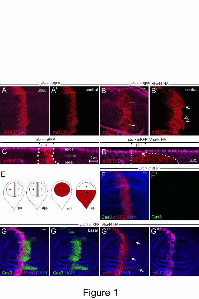
Figure 1: Vha44 overexpression causes invasive migration and apoptosis at the AP boundary
of the wing disc. (A,B,) ptc-GAL4 is used to drive expression of mRFP (A,A’) and
mRFP/Vha44-HA (B,B’) in an anterior stripe at the boundary to the posterior compartment.
Expression is highest close to the boundary. Staining for Discs large (Dlg) visualizes the
outline of individual cells and the whole tissue. Invasion is most prominent at dorsal and
ventral parts of the wing pouch (arrowhead indicates single cells migrating, arrow cell clusters
migrating in B’). (C,D) Invading cells (below dotted line) move at the basal side of the
epithelium as seen in the x-z projection. Dlg is missing right next to the boundary, most likely
due to basal extrusion and loss of apico-basal polarity (arrow in D). (E) Schematic drawings
of the wing disc and the respective expression domains for the GAL4 drivers used throughout
this study in red. (F,G) Whereas mRFP expression does not induce Cas3 reactivity (F,F’), co-
expression of Vha44-HA leads to Cas3-positive cells in the ptc stripe (G’). As demonstrated
by the mRFP signal (G’’), Vha44-expressing cells are of anterior identity. Note that the HA
tag is hardly visible in the invasive Cas3- and mRFP-positive front due to the apoptotic
cleavage (G’’’; see also Fig. S1 for further clarification). It therefore can be used to mark the
original ptc sripe. In images with higher exposure the HA signal is visible also in the invasive
front (see also Fig. S1A).
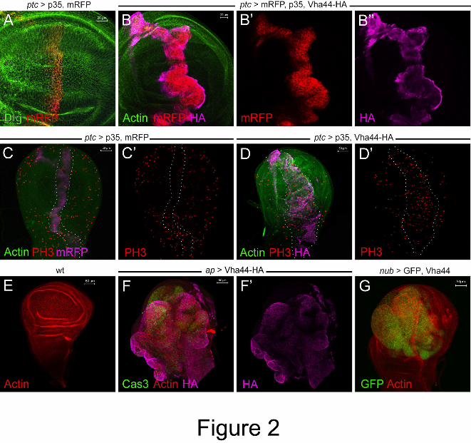
Figure 2: Vha44 overexpression causes overgrowth. (A,B) While the co-expression of p35
and mRFP does not lead to any overt phenotypes or apoptosis (A), additional expression of
Vha44-HA causes an expansion of the ptc stripe (B,B’,B’’). (C,D) Phospho-Histone 3 (PH3)
staining reveals an increased proliferation rate in ptc domain (marked by dotted lines in D and
D’), but not in the control ptc stripe expressing p35 and mRFP (marked by dotted lines in C
and C’). (E-G) Ap-GAL4- (F,F’) and nub-GAL4-mediated (G) overexpression of Vha44
causes overgrowth in the dorsal compartment and wing pouch, respectively. Cas3-positive
cells are also detectable (F). For comparison, a wild-type disc stained with Phalloidin is
shown (E).
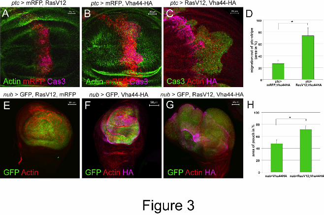
Figure 3: Vha44-induced invasion and overgrowth is enhanced by RasV12. (A-C)
RasV12/mRFP co-expression does not lead to significant invasion phenotypes (A). Co-
overexpression of the oncogenic form of Ras, RasV12, and Vha44 (C) increases the extent of
migration seen in Vha44/mRFP co-expression (B). (D) Quantified are the ratios (in %)
Dise
ase
Mod
els &
Mec
hani
sms
D
MM
Acce
pted
man
uscr
ipt
24
between the areas of invaded Cas3-positive cells and the areas of the original ptc stripe (n=9;
data represent the mean +/- s.d., t-test (p<0.01)). (E-H) The expression of mRFP and RasV12
with nub-GAL4 does not lead to overgrowth. Vha44/mFRP overexpression under nub-GAL4
causes overgrowth (F), which is significantly increased in the presence of RasV12 (G). (H)
Quantified are disc diameters (n=6; data represent the mean +/- s.d., t-test (p<0.0001).
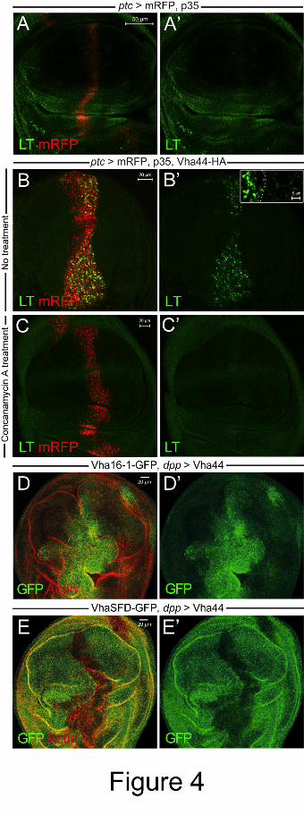
Figure 4: Vha44 expression increases organellar acidity and alters V-ATPase subunit levels.
(A-C) While LysoTracker (LT) uptake is normal in ptc stripes expressing mRFP and p35
(A,A’), LT accumulated strongly within intracellular puncta in cells overexpressing Vha44-
HA, mRFP and p35 (B,B’ and inset in B’; cells marked with mRFP in B). To reduce dye
uptake due to removal of apoptotic cells by acidic phagosomes of hemocytes or neighboring
cells, the anti-apoptotic p35 was co-expressed in these experiments. LT uptake by cells
outside the ptc stripe was also seen, but is hardly detectable at the laser intensity shown in B
and B’. (C) Pre-incubation with the V-ATPase inhibitor Concanamycin A abolishes all LT
signal in the ptc stripe. (D,D’) Endogenous Vha16 (tagged with GFP) expression is enhanced
in cells overexpressing Vha44 with dpp-GAL4. (E,E’) By contrast, VhaSFD-GFP expression
is decreased. Note that when Vha44 expression is targeted to the dpp domain, then there is
also overgrowth, because there are less boundary effects compared to the ptc domain (see
also Fig. 1E).
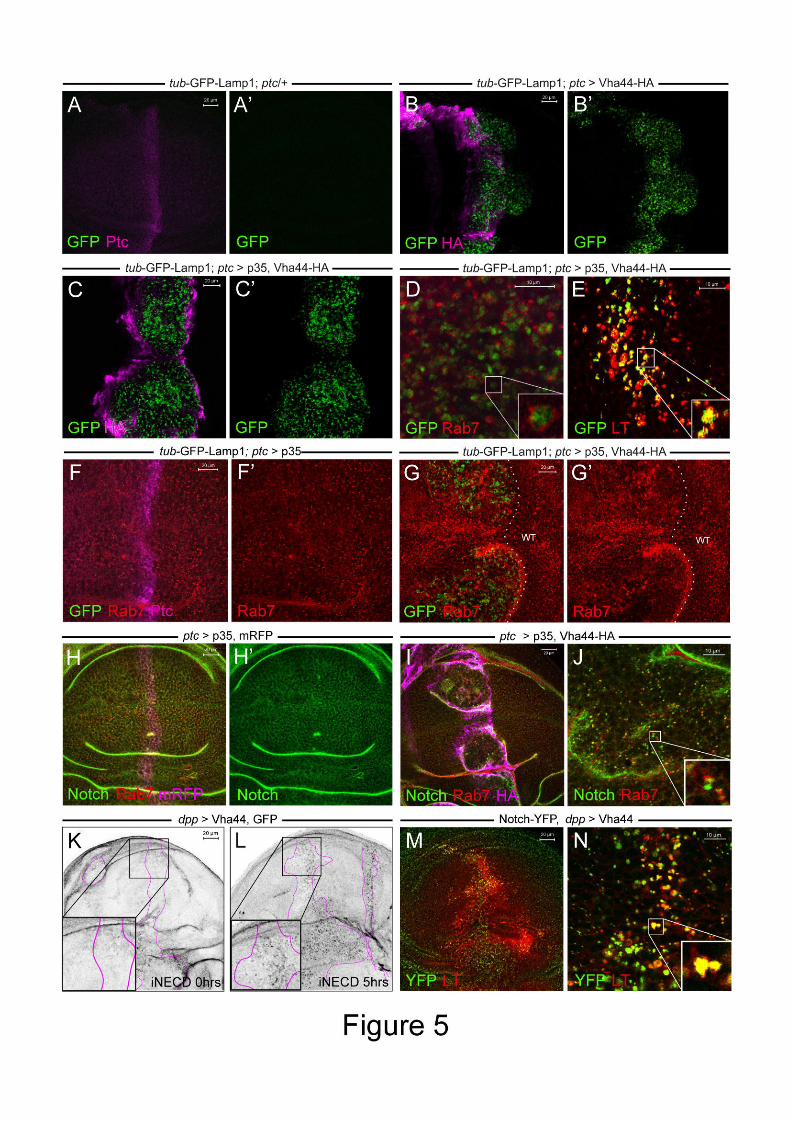
Figure 5: Endolysosomal degradation is impaired in Vha44-expressing cells. (A-G) Luminal
GFP is directed from the Golgi via early and late endosomes to the lysosomes for degradation
in all panels to assess defects in endolysosomal degradation. (A,B) GFP-Lamp1 (ubiquitously
expressed with a tub promoter) is hardly detectable in control discs (A,A’) and outside the ptc
stripe (B,B’), but accumulates in Vha44-expressing cells (B,B’). (C,C’) The accumulation
also occurs upon p35 co-expression. (D,E) Accumulated GFP-Lamp1 partially co-localizes
with the late endosomal marker Rab7 (D) and LysoTracker (E) in enlarged endosomes of the
p35/Vha44-HA-expressing ptc stripe. The inset in D shows that, whereas Rab7 localizes to
the cytosolic side of enlarged “ring” endosomes, the GFP in GFP-Lamp1 is luminal.
LysoTracker, on the other hand, shows a significant overlap with the GFP signal (inset in E).
(F) Rab7 looks normal in p35-overexpressing cells. (G) Upon Vha44-HA overexpression,
Rab7 becomes irregular and accumulates in enlarged compartments (ptc stripe is marked here
by GFP-Lamp1 accumulation). Note also that the ptc stripe is broadened and abnormally
folded due to p35 co-expression. (H) Notch localizes to membranes and intracellular puncta in
Dise
ase
Mod
els &
Mec
hani
sms
D
MM
Acce
pted
man
uscr
ipt
25
a control wing disc expressing p35 and mRFP. (I) In Vha44-HA expressing cells, Notch
accumulates in enlarged intracellular puncta. A higher magnification in I’ shows co-
localization of Notch and Rab7. The inset highlights one enlarged compartment positive for
both Notch and Rab7. (K,L) A Notch antibody targeting the extracellular domain of Notch
was applied to live wing discs. Compared to initial labeling (t=0 hrs in K) internalized anti-
Notch (iNECD) accumulates in the Vha44 expression domain (marked by pink lines based on
GFP co-expression) after culturing for 5hrs. In neighbouring wild-type tissue iNECD signal is
lost because of lysosomal degradation (L). This suggests that Notch degradation is impaired
in Vha44-expressing cells. (M,N) A pool of Notch-YFP that accumulates in Vha44 expressing
cell is present in acidified LysoTracker-positive compartments. The inset in N shows overlap
of Notch-YFP and Lysotracker in enlarged compartments.
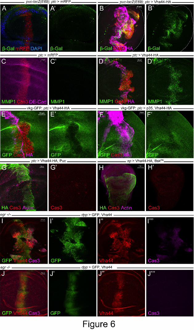
Figure 6: Vha44-mediated transformation requires the JNK signaling pathway. (A,B) The
puc-lacZ reporter is strongly activated in Vha44-expressing cells when stained with anti-β-gal
(B,B’). As the puc (E69)-lacZ insertion partly disrupts gene function of the JNK inhibitor puc,
invasion and overgrowth is enhanced in this genetic background (compare to previous
examples of Vha44-induced invasion or panel D). No β-gal staining is detected upon
expression of mRFP alone (A,A’). (C,D) Ptc>Vha44-HA (D,D’), but not mRFP (C,C’),
strongly upregulates MMP1 expression. (E) The gene trap viking (vkg)-GFP visualizes
collagen IV and was used to monitor ECM degradation in the ptc stripe expressing Vha44-HA
(E’). (F,F’) Vkg-GFP is unaffected upon p35 co-expression, which abolishes Cas3-positive
cells (F). Strong Cas3 reactivity is shown without p35 (E). (G) Co-expression of the negative
regulator Puc suppressed Vha44-induced apoptosis and cell invasion. Unlike p35 (in Figure
2B), overgrowth is also not seen. (H) The dominant-negative form of JNK, Bsk-DN, also
suppresses overgrowth and apoptosis of ap>Vha44. (I,J) Vha44 expression using dpp-GAL4
also leads to overgrowth and migration out of the dpp expression domain (I,I’). Removing
two copies of the eiger gene completely suppresses overgrowth, migration and apoptosis (J-
J’’’).
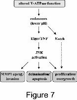
Figure 7: Model for the proposed effects of increased vesicular V-ATPase activity. Lower pH
in endosomes and defective lysosomal degradation leads to trapping of Notch and,
presumably, to increased JNK signaling via its ligand Eiger. Downstream are various
processes important for cellular transformation in the native epithelium. The Notch receptor
also accumulates intracellularly in Vha44-expressing cells. However, Notch signaling is not
Dise
ase
Mod
els &
Mec
hani
sms
D
MM
Acce
pted
man
uscr
ipt
26
significantly activated. Therefore, the contribution of Notch to the transformation phenotypes
is less prominent than for JNK signaling (dotted arrow).
Dise
ase
Mod
els &
Mec
hani
sms
D
MM
Acce
pted
man
uscr
ipt