Embed Size (px)
Citation preview

ACCF/AHA Pocket GuidelineNovember 2011
Management of Patients With
Peripheral Artery Disease(Lower Extremity, Renal, Mesenteric, and Abdominal Aortic)
Adapted from the 2005 ACCF/AHA Guideline and the 2011 ACCF/AHA Focused Update
Developed in Collaboration With the Society for Cardiovascular Angiography and Interventions, Society of Interventional Radiology, Society for Vascular Medicine, and Society for Vascular Surgery

B
© 2011 by the American College of Cardiology Foundation
and the American Heart Association, Inc.
The following material was adapted from the 2011 ACCF/AHA
focused update of the guideline for the management of
patients with peripheral artery disease Circulation 2011;
124:2020-2045 and the 2005 ACC/AHA guidelines for the
management of patients with peripheral arterial disease
(lower extremity, renal, mesenteric, and abdominal aortic)
Circulation 2006; 113:e463-e654. This pocket guideline is
available on the World Wide Web sites of the American
College of Cardiology (cardiosource.org) and the American
Heart Association (my.americanheart.org).
For copies of this document, please contact Elsevier Inc. Reprint
Department, e-mail: [email protected]; phone: 212-633-3813;
fax: 212-633-3820.
Permissions: Multiple copies, modification, alteration, enhancement,
and/or distribution of this document are not permitted without
the express permission of the American College of Cardiology
Foundation. Please contact Elsevier’s permission department at

Contents
1. Introduction ...................................................................................................3
2. Patient History and Physical Examination: Fundamental Principles ............................................................................................. 6
3. Evaluation and Treatment of Patients With, or at Risk for, PAD ........................................................................................9
4. Lower Extremity Arterial Disease .........................................................11A. Claudication ....................................................................................................11B. Critical Limb Ischemia (UPDATED) ................................................................25C. Acute Limb Ischemia ......................................................................................28D. Surveillance for Patients After Lower Extremity Revascularization .............30E. Ankle-Brachial Index, Toe-Brachial Index, and Segmental
Pressure Examination (UPDATED) .................................................................31F. Smoking Cessation (UPDATED) ......................................................................33G. Antithrombotic and Antiplatelet Therapy (UPDATED) ..................................33
5. Renal Arterial Disease ..............................................................................35A. Clinical Indications .........................................................................................35B. Diagnostic Methods ........................................................................................38C. Indications for Revascularization of Patients with
Hemodynamically Significant RAS .................................................................39D. Treatment Methods: Medical, Endovascular, and Surgical ...........................42
6. Mesenteric Arterial Disease....................................................................45A. Acute Intestinal Ischemia ...............................................................................45B. Acute Nonocclusive Intestinal Ischemia ........................................................46C. Chronic Intestinal Ischemia ............................................................................48
7. Aneurysms of the Abdominal Aorta, Its Branch Vessels, and the Lower Extremities ......................................................................49
A. Abdominal Aortic Aneurysms ........................................................................49B. Management Overview of Prevention of
Aortic Aneurysm Rupture (UPDATED) ...........................................................53C. Visceral Arterial Aneurysms ...........................................................................55D. Lower Extremity Arterial Aneurysms .............................................................57E. Femoral Artery Pseudoaneurysms .................................................................59
Lower Extrem
ityR
enalM
esentericA
bdominal

2

3
1. Introduction
This pocket guide provides rapid prompts for appropriate patient management, which is outlined in much greater detail in the full-text guidelines. It is not intended as a replacement for understanding the caveats and rationales that are stated carefully in the full-text guidelines. Users should consult the full-text guideline for more information.
The term peripheral artery disease (PAD) broadly encompass the vascular diseases caused primarily by atherosclerosis and thromboembolic pathophysiologic processes that alter the normal structure and function of the aorta, its visceral arterial branches, and the arteries of the lower extremity. PAD is the preferred clinical term and should be used to denote stenotic, occlusive and aneurysmal diseases of the aorta and its branch arteries, exclusive of the coronary arteries.
The scope of these pocket guidelines (updated for 2011) is limited to disorders of the lower extremity arteries, renal and mesenteric arteries, and disorders of the abdominal aorta. The purpose of these guidelines is to 1) aid in the recognition, diagnosis, and treatment of PAD of the lower extremities, and 2) highlight the prevalence, impact on quality-of-life, cardiovascular ischemic risk, and increased risk of critical limb ischemia (CLI) associated with PAD. Inasmuch as the burden of PAD is widespread, these guidelines are intended to assist all clinicians who might provide care for such patients, including primary care clinicians, vascular and cardiovascular specialists, trainees in the primary care and vascular specialties, as well as nurses, physical therapists, and rehabilitative personnel.
All recommendations provided in this document follow the format of previous American College of Cardiology Foundation/American Heart Association guidelines (Table 1). Recommendations that remain unchanged used the Class of Recommendation/Level of Evidence table from the 2005 guideline.

4
Table 1. Applying Classification of Recommendations and Level of Evidence†
LeveL AMultiple populations evaluated*
Data derived from multiple randomized clinical trials or meta-analyses
LeveL BLimited populations evaluated*Data derived from a single randomized trial or nonrandomized studies
LeveL CVery limited populations evaluated*
Only consensus opinion of experts, case studies, or standard of care
CLAss IBenefit >>> Risk
Procedure/Treatment shOuLD be performed/ administered
n Recommendation that procedure or treatment is useful/effective
n sufficient evidence from multiple randomized trials or meta-analyses
n Recommendation that procedure or treatment is useful/effective
n Evidence from single randomized trial or nonrandomized studies
n Recommendation that procedure or treatment is useful/effective
n Only expert opinion, case studies, or standard of care
CLAss IIaBenefit >> Risk
Additional studies with focused objectives needed
IT Is REasOnabLE to perform procedure/ administer treatment
n Recommendation in favor of treatment or procedure being useful/effective
n some conflicting evidence from multiple randomized trials or meta-analyses
n Recommendation in favor of treatment or procedure being useful/effective
n some conflicting evidence from single randomized trial or nonrandomized studies
n Recommendation in favor of treatment or procedure being useful/effective
n Only diverging expert opinion, case studies, or standard of care
should
is recommended
is indicated
is useful/effective/beneficial
suggested phrases for writing recommendations
is reasonable
can be useful/effective/beneficial
is probably recommended or indicated
S I z E o F T R E A T M E n T E F F E C T
Es
tim
At
E o
F C
Er
tA
iNt
y (
Pr
EC
isio
N)
oF
tr
EA
tm
EN
t E
FF
EC
t
treatment/strategy A is recommended/indicated in preference to treatment B
treatment A should be chosen over treatment B
Comparative effectiveness phrases†
treatment/strategy A is probably recommended/indicated in preference to treatment B
it is reasonable to choose treatment A over treatment B

5
*A recommendation with Level of
Evidence B or C does not imply that
the recommendation is weak. Many
important clinical questions
addressed in the guidelines do not
lend themselves to clinical trials.
Although randomized trials are
unavailable, there may be a very
clear clinical consensus that a
particular test or therapy is useful
or effective.
*Data available from clinical trials or
registries about the usefulness/
efficacy in different subpopulations
such as sex, age, history of diabetes,
history of prior myocardial infarction,
history of heart failure, and prior
aspirin use.
†For comparative effectiveness
recommendations (Class I and IIa;
Level of Evidence A and B only),
studies that support the use of
comparator verbs should involve
direct comparisons of the treatments
or strategies being evaluated.
Class IIb
Benefit ≥ RiskAdditional studies with broad objectives needed; additional registry data would be helpful
Procedure/Treatment May bE COnsIDERED
n Recommendation’s usefulness/efficacy less well established
n Greater conflicting evidence from multiple randomized trials or meta-analyses
n Recommendation’s usefulness/efficacy less well established
n Greater conflicting evidence from single randomized trial or nonrandomized studies
n Recommendation’s usefulness/efficacy less well established
n Only diverging expert opinion, case studies, or standard-of-care
Class III No Benefitor Class III Harm Procedure/ Test Treatment
COR III: not no Provenno benefit helpful benefit
COR III: Excess Cost harmfulharm w/o benefit to Patients or harmful
n Recommendation that procedure or treatment is not useful/effective and may be harmful
n sufficient evidence from multiple randomized trials or meta-analyses
n Recommendation that procedure or treatment is not useful/effective and may be harmful
n Evidence from single randomized trial or nonrandomized studies
n Recommendation that procedure or treatment is not useful/effective and may be harmful
n Only expert opinion, case studies, or standard-of-care
may/might be considered
may/might be reasonable
usefulness/effectiveness is unknown/unclear/uncertain or not well established
COR III: COR III:No Benefit Harm
is not potentially recommended harmful
is not indicated causes harm
should not be associated with performed/ excess morbidity/ administered/ mortality other
is not useful/ should not be
beneficial/
performed/
effective administered/
done
6
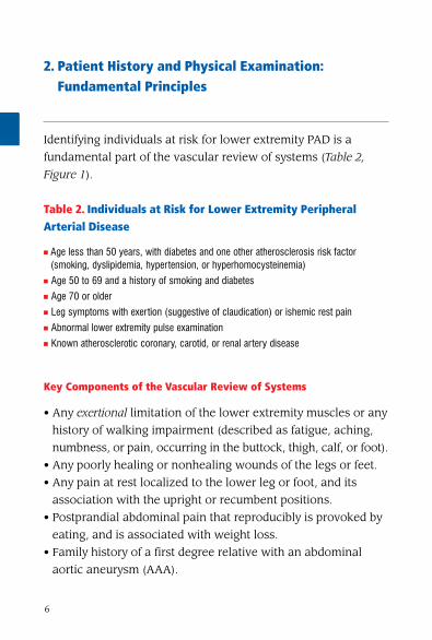
2. Patient History and Physical Examination: Fundamental Principles
Identifying individuals at risk for lower extremity PAD is a
fundamental part of the vascular review of systems (Table 2,
Figure 1).
Table 2. Individuals at Risk for Lower Extremity Peripheral Arterial Disease
n Age less than 50 years, with diabetes and one other atherosclerosis risk factor (smoking, dyslipidemia, hypertension, or hyperhomocysteinemia)
n Age 50 to 69 and a history of smoking and diabetesn Age 70 or oldern Leg symptoms with exertion (suggestive of claudication) or ishemic rest painn Abnormal lower extremity pulse examinationn Known atherosclerotic coronary, carotid, or renal artery disease
Key Components of the Vascular Review of Systems
• Any exertional limitation of the lower extremity muscles or any
history of walking impairment (described as fatigue, aching,
numbness, or pain, occurring in the buttock, thigh, calf, or foot).
• Any poorly healing or nonhealing wounds of the legs or feet.
• Any pain at rest localized to the lower leg or foot, and its
association with the upright or recumbent positions.
• Postprandial abdominal pain that reproducibly is provoked by
eating, and is associated with weight loss.
• Family history of a first degree relative with an abdominal
aortic aneurysm (AAA).

7
Individuals at Risk for Lower Extremity PaD:• Age less than 50 years with diabetes and one other atherosclerosis risk factor
(smoking, dyslipidemia, hypertension, or hyperhomocysteinemia)• Age 50 to 69 years and history of smoking or diabetes
• Age 70 years and older• Leg symptoms with exertion (suggestive of claudication) or ischemic rest pain
• Abnormal lower extremity pulse examination• Known atherosclerotic coronary, carotid, or renal arterial disease
Obtain history of walking impairment and/or limb ischemic symptoms: • Obtain a vascular review of symptoms:
• Leg discomfort with exertion • Leg pain at rest; nonhealing wound; gangrene
Sudden onset ischemic leg
symptoms or signs of acute limb ischemia:
The five “Ps”†
See Figure 2, Diagnosis and Treatment of
Asymptomatic PAD and Atypical
Leg Pain
See Figure 5, Diagnosis and Treatment of Critical Limb
Ischemia
See Figures 6 and 7,
Diagnosis and Treatment of Acute
Limb Ischemia
No leg pain
“Atypical” leg pain*
See Figure 2, Diagnosis and Treatment of
Asymptomatic PAD and Atypical
Leg Pain
* “Atypical” leg pain is defined by lower extremity discomfort that is exertional, but that does not consistently resolve with rest, consistently limit exercise at a reproducible distance, or meet all “Rose questionnaire” criteria.
† The five “Ps” are defined by the clinical symptoms and signs that suggest potential limb jeopardy: pain, pulselessness, pallor, paresthesias, and paralysis (with polar being a sixth “P”).
PAD indicates peripheral arterial disease.
Classic claudication symptoms:
Exertional fatigue, discomfort, or frank pain localized to leg muscle groups that
consistently resolves with rest
• Ischemic leg pain at rest
• Nonhealing wound • Gangrene
Perform a resting ankle-brachial index measurement
See Figures 3 and 4, Diagnosis and Treatment of
Claudication
Figure 1. Steps Toward the Diagnosis of PAD

8
Key Components of the Vascular Physical Examination
• Measurement of blood pressure in both arms and notation of
any inter-arm asymmetry.
• Palpation of the carotid pulses, and notation of the carotid
upstroke and amplitude, and presence of bruits.
• Auscultation of the abdomen and flank for bruits.
• Palpation of the abdomen and notation of the presence of the
aortic pulsation and its maximal diameter.
• Palpation of pulses at the brachial, radial, ulnar, femoral,
popliteal, dorsalis pedis, and posterior tibial sites. Perform
Allen’s test when knowledge of hand perfusion is needed.
• Ausculation of both femoral arteries for the presence of bruits
• Pulse intensity should be assessed and should be recorded
numerically as follows:
− 0, absent
− 1, diminished
− 2, normal
− 3, bounding
• The shoes and socks should be removed, the feet inspected, the
color, temperature, and integrity of the skin and intertriginous
areas evaluated, and presence of ulcerations recorded.
• Additional findings suggestive of severe PAD, including distal
hair loss, trophic skin changes, and hypertrophic nails, should
be sought and recorded.
3. Evaluation and Treatment of Patients With, or at Risk for, PAD
The noninvasive vascular laboratory provides a powerful set of
tools that can objectively assess the status of lower extremity
arterial disease and facilitate the creation of a therapeutic plan.

9
Although there are many diagnostic vascular tests available, the
clinical presentation of each patient can usually be linked to
specific and efficient testing strategies (Table 3).
Table 3. Typical noninvasive Vascular Laboratory Tests for Lower Extremity PAD Patients by Clinical Presentation
Clinical Presentation noninvasive Vascular Test
Asymptomatic lower extremity PAD ABI
Claudication ABI, PVR, or segmental pressures Duplex ultrasound
Exercise test with ABI to assess functional status
Possible pseudoclaudication Exercise test with ABI
Postoperative vein graft follow-up Duplex ultrasound
Femoral pseudoaneurysm; iliac or popliteal aneurysm
Duplex ultrasound
Suspected aortic aneurysm; serial AAA follow-up
Abdominal ultrasound, CTA, or MRA
Candidate for revascularization Duplex ultrasound, MRA, or CTA
AAA indicates abdominal aortic aneurysm; ABI, ankle-brachial index; CTA, computed tomography angiography; MRA, magnetic resonance angiography; PAD, peripheral artery disease and PVR, pulmonary vascular resistance.
Recommendations for Evaluation and Treatment of Individuals at Risk for PAD or With Asymptomatic PAD
Class I 1. A history of walking impairment, claudication,
ischemic rest pain, and/or nonhealing wounds is
recommended as a required component of a
standard review of systems for adults 50 years and
older who have atherosclerosis risk factors, or for
adults 70 years and older. (Level of Evidence: C)

10
Figure 2. Diagnosis and Treatment of Asymptomatic PAD and Atypical Leg Pain
ABI 0.91 to 1. 30 (borderline & normal) ABI ≤0.90
(abnormal)ABI >1.30 (abnormal)
Perform a resting ABI index measurement
normal post-exercise ankle-brachial index:
No PAD
Evaluate other causes of leg symptoms†
Decreased post-exercise ABI
Measure ABI after exercise test
normal results:No PAD
Abnormal results
Confirmation of PAD diagnosis
Risk factor normalization:Immediate smoking cessation
Treat hypertension: JNC-7 guidelinesTreat lipids: NCEP ATP-III guidelines
Treat diabetes mellitus: HbA1c <7%‡
Pharmacological Risk Reduction:Antiplatelet therapy (ACE-inhibition§; Class IIb, LOE C)
Pulse volume recording Toe-brachial index
(Duplex ultrasonography*)
Individual at risk of PaD (no leg symptoms or atypical leg symptoms):Consider use of the Walking Impairment Questionnaire
* Duplex ultrasonography should generally be reserved for use in symptomatic patients in whom anatomic diagnostic data is required for care. †Other causes of leg pain may include: lumbar disk disease, sciatica, radiculapthy; muscle strain; neuropathy; compartment syndrome. ‡It is not yet proven that treatment of diabetes mellitus will significantly reduce PAD-specific (limb ischemic) endpoints. Primary treatment of diabetes mellitus should be continued according to established guidelines. §The benefit of ACE inhibition in individuals without claudication has not been specifically documented in prospective clinical trials, but has been extrapolated from other “at risk” populations. ACE indicates angiotensin-converting enzyme; ABI, ankle-brachial index; HgbA1c, hemoglobin A1c; JNC-7, Seventh Report of the Joint National Committee on Prevention, Detection, Evaluation, and Treatment of High Blood Pressure; LOE, level of evidence; NCEP ATP III, National Cholesterol Education Program Adult Treatment Panel III; PAD, peripheral arterial disease. Adapted from Hiatt WR. Medical treatment of peripheral arterial disease and claudication. N Engl J Med 2001;344:1608–1621. Copyright © 2001 Massachusetts Medical Society. All rights reserved.

11
Lower Extrem
ity
2. Individuals with asymptomatic lower extremity
PAD should be identified by examination and/or
measurement of the ankle-brachial index (ABI, see
Figure 2) in order to offer therapeutic interventions
known to diminish their increased risk of MI, stroke,
and death. (Level of Evidence: B)
3. Smoking cessation, lipid lowering, diabetes and
hypertension treatment according to current
national treatment guidelines is recommended for
individuals with asymptomatic lower extremity PAD.
(Level of Evidence: B)
4. Antiplatelet therapy is indicated for individuals
with asymptomatic lower extremity PAD to reduce
the risk of adverse cardiovascular ischemic events.
(Level of Evidence: C)
4. Lower Extremity Arterial Disease
A. Claudication
Claudication is defined as fatigue, discomfort, or pain that
occurs in specific limb muscle groups during effort due to
exercise-induced ischemia (Figures 3 and 4).
General Management of Patients With Claudication
Class I 1. Patients with symptoms of intermittent
claudication should undergo a vascular physical
examination, including measurement of the ABI.
(Level of Evidence: B)

12
Low
er E
xtre
mit
y
2. In patients with symptoms of intermittent
claudication, the ABI should be measured post-exercise
if the resting index is normal. (Level of Evidence: B)
3. Before undergoing an evaluation for
revascularization, patients with intermittent
claudication should have significant functional
impairment with a reasonable likelihood of
symptomatic improvement and absence of other
disease that would comparably limit exercise even
if the claudication was improved (e.g., angina, heart
failure, chronic respiratory disease, orthopedic
limitations). (Level of Evidence: C)
4. Cilostazol (100 mg orally 2 times per day) is
indicated as an effective therapy to improve symptoms
and increase walking distance in patients with lower
extremity PAD and intermittent claudication (in the
absence of heart failure). (Level of Evidence: A)
5. A therapeutic trial of cilostazol should be considered
in all patients with lifestyle limiting claudication (in
the absence of heart failure). (Level of Evidence: A)
Class IIb 1. Pentoxifylline (400 mg 3 times per day) may be
considered as second line alternative therapy to
cilostazol to improve walking distance in patients
with intermittent claudication. (Level of Evidence: A)
2. The clinical effectiveness of pentoxifylline as
therapy for claudication is marginal and not well
established. (Level of Evidence: C)

13
3. The effectiveness of L-arginine for patients with
intermittent claudication is not well established.
(Level of Evidence: B)
4. The effectiveness of propionyl-L-carnitine or
ginkgo biloba as therapy to improve walking
distance in patients with intermittent claudication is
not well established. (Level of Evidence: B)
Class III 1.Oral vasodilator prostaglandins such as beraprost
and iloprost are not effective medications to walking
distance in patients with intermittent claudication.
(Level of Evidence: A)
2.Vitamin E is not recommended as a treatment for
patients with intermittent claudication. (Level of
Evidence: C)
3.Chelation (e.g., Ethylenediaminetetraacetic acid) is
not indicated for treatment of intermittent claudication
and may have harmful adverse effects. (Level of
Evidence: A)
The key elements of a therapeutic claudication exercise program
for patients with claudication are summarized in Table 4, page 19 .
For diagnosis and treatment of critical and acute limb ischemia,
see Figures 5, 6 and 7.
Lower Extrem
ity

14
Figure 3. Diagnosis of Claudication and Systemic Risk Treatment
* It is not yet proven that treatment of diabetes mellitus will significantly reduce PAD-specific (limb ischemic) end points. Primary treatment of diabetes mellitus should be continued according to established guidelines. †The benefit of ACE inhibition in individuals without claudication has not been specifically documented in prospective clinical trials but has been extrapolated from other at-risk populations.
ABI indicates ankle-brachial index; ACE, angiotensin-converting enzyme; HgbA1c, hemoglobin A1c; JNC-7, Seventh Report of the Joint National Committee on Prevention, Detection, Evaluation, and Treatment of High Blood Pressure; LOE, level of evidence; NCEP ATP III, National Cholesterol Education Program Adult Treatment Panel III; PAD, peripheral arterial disease; TBI, toe-brachial index.
Document pulse examination
ABI
AB <0.90
Confirmed PAD diagnosis
Risk factor normalization: Immediate smoking cessation
Treat hypertension: JNC-7 guidelines Treat lipids: NCEP ATP III guidelines
Treat diabetes mellitus: HbA1c <7%*
Pharmacological risk reduction: Antiplatelet therapy
(ACE inhibition†; Class IIa)
Go to Figure 4, Treatment of Claudication
Exercise ABI (TBI, segmental pressure, or duplex ultrasound
examination)
ABI >0.90
Classic Claudication symptoms:Muscle fatigue, cramping, or pain that reproducibly begins
during exercise and that promptly resolves with rest
Chart document the history of walking impairment (pain-free and total walking distance) and specific lifestyle limitations
Abnormal Results
Normal Results
No PAD or consider arterial
entrapment syndromes
Low
er E
xtre
mit
y

15
Figure 4. Treatment of Claudication
* Inflow disease should be suspected in individuals with gluteal or thigh claudication and femoral pulse diminution or bruit, and should be confirmed by noninvasive vascular laboratory diagnostic evidence of aorto-iliac stenoses.
† Outflow disease represents femoropopliteal and infrapopliteal stenoses, (the presence of occlusive lesions in the lower extremity arterial tree below the inguinal ligament from the common fermoral artery to the pedal vessels).
No significant functional disability
• No claudication treatment required.
• Follow-up visits at least annually to monitor for development of leg, coronary, or cerebrovascular ischemic symptoms.
Lifestyle limiting symptoms
Supervised exercise program
Pharmacological therapy:
Cilostazol (Pentoxifylline)
3 month trial 3 month trial
Preprogram and postprogram
exercise testing for efficacy
Clinical improvement:
Follow-up visits at least annually
significant disability despite medical therapy and/or inflow
endovascular therapy, with documentation of outflow† PAD,
with favorable procedural anatomy and procedural risk-benefit ratio
Evaluation for additional endovascular or surgical revascularization
Further anatomic definition by more extensive
noninvasive or angiographic diagnostic techniques
Endovascular therapy (or surgical bypass per
anatomy)
Lifestyle-limiting symptoms with evidence of inflow disease*
Confirmed PAD Diagnosis
Lower Extrem
ity

16
Figure 5. Diagnosis and Treatment of Critical Limb Ischemia
* Based on patient comorbidities. †Based on anatomy or lack of conduit. ‡Risk factor normalization: immediate smoking cessation, treat hypertension per the seventh report of the Joint National Committee on Prevention, Detection, Evaluation, and Treatment of High Blood Pressure guidelines; treat lipids per National Cholesterol Education Program Adult Treatment Panel III guidelines; treat diabetes mellitus (HgbA1c [hemoglobin A1c] <7%; Class IIa). It is not yet proven that treatment of diabetes mellitus will significantly reduce PAD-specific (limb ischemic) endpoints. Primary treatment of diabetes mellitus should be continued according to established guidelines.
ABI indicates ankle-brachial index; CTA, computed tomographic angiography; ECG, electrocardiogram; MRA, magnetic resonance angiography; PAD, peripheral arterial disease; PVR, pulse volume recording; TBI, toe-brachial index; TEE, transesophageal echocardiography; US, ultrasonography.
Systemic antibiotics if skin ulceration and limb infection are present
Obtain prompt vascular specialist consultation: Diagnostic testing strategy
Creation of therapeutic intervention plan
Patient is a candidate for revascularization
Patient not a candidate for revascularization*
Medical therapy or amputation
(when necessary)
Define limb arterial anatomy Assess clinical and objective severity of ischemia
Imaging of relevant arterial circulation (noninvasive and angiographic)
Revascularization possible (see treatment text, with application of thrombolytic, endovascular, and surgical therapies)
Written instructions for self-surveillance
Ongoing vascular surveillance (see text)‡
Revascularization not possible†: medical therapy; amputation
(when necessary)
No or minimal atherosclerotic arterial
occlusive disease
Consider atheroembolism,
thromboembolism, or phlegmasia cerulea
dolens
Evaluation of source (ECG or Holter monitor; TEE; and/or abdominal US, MRA, or CTA); or
venous duplex
Chronic symptoms: Ischemic rest pain, gangrene, nonhealing woundIschemic etiology must be established promptly: By examination and objective vascular studies
Implication: Impending limb loss
history and physical examination: Document lower-extremity pulses
Document presence of ulcers or infection
Assess factors that may contribute to limb risk: diabetes, neuropathy, chronic renal failure, infection
ABI, TBI, or duplex US
severe lower extremity PaD documented: ABI <0.4; flat PVR waveform; absent pedal flow
Low
er E
xtre
mit
y

17
ABI indicates ankle-brachial index; CTA, computed tomographic angiography; ECG, electrocardiogram; MRA, magnetic resonance angiography; PAD, peripheral arterial disease; PVR, pulse volume recording; TBI, toe-brachial index; TEE, transesophageal echocardiography; US, ultrasonography. Adapted from J Vasc Surg, 26, Rutherford RB, Baker JD, Ernst C, et al., Recommended standards for reports dealing with lower extremity ischemia: revised version, 517–38, Copyright 1997, with permission from Elsevier.
Lower Extrem
ity
Figure 6. Diagnosis of Acute Limb Ischemia
Rapid or sudden decrease in limb perfusion threatens tissue viability
History and physical examination; determine time of onset of symptoms
Emergent assessment of severity of ischemia: Loss of pulses
Loss of motor and sensory functionVascular laboratory assessment
No or minimal PAD
Consider atheroembolism,
thromboembolism, or phlegmasia cerulea dolens
Evaluation of source (ECG or Holter monitor; TEE; and/or abdominal
ultrasound, MRA, or CTA); or venous duplex
severe PaD documented:• ABI <0.4
• Flat PVR waveform• Absent pedal flow
Go to Figure 7, Treatment of
Acute Limb Ischemia
ABI, TBI, or duplex US

18
* Inflow disease should be suspected in individuals with gluteal or thigh claudication and femoral pulse diminution or bruit and should be confirmed by noninvasive vascular laboratory diagnostic evidence of aortoiliac stenoses. †Outflow disease represents femoropopliteal and infrapopliteal stenoses (the presence of occlusive lesions in the lower extremity arterial tree below the inguinal ligament from the common femoral artery to the pedal vessels).
ABI indicates ankle-brachial index; PAD, peripheral arterial disease; PVR, pulse volume recording; US, ultrasonography.
Adapted from J Vasc Surg, 26, Rutherford RB, Baker JD, Ernst C, et al., Recommended standards for reports dealing with lower extremity ischemia: revised version, 517–38, Copyright 1997, with permission from Elsevier.
Low
er E
xtre
mit
y
Figure 7. Treatment of Claudication
Viable limb • Not immediately
threatened • No sensory loss
• No muscle weakness • Audible arterial and
venous US
salvageable limb: threatened marginally (reversible ischemia)
• Salvageable if promptly treated
• Minimal (toes) or no sensory loss
• No muscle weakness Inaudible (often)
arterial Doppler signals • Audible venous Doppler signals
salvageable limb: threatened immediately
(reversible ischemia) • Salvageable with immediate
revascularization • Sensory loss more than toes,
associated with rest pain • Mild to moderate muscle weakness
• Inaudible (usually) arterial Doppler signals
• Audible venous Doppler signals
Guides to treatment:• Site and extent of occlusion • Embolus versus thrombus • Native artery versus bypass graft • Duration of ischemia
• Patient comorbidities • Contraindications to thrombolysis or surgery
Revascularization: Thrombolysis, endovascular, surgical
nonviable limb (irreversible ischemia) • Major tissue loss or
permanent nerve damage inevitable
• Profound, anesthetic sensory loss
• Profound paralysis (rigor)
• Inaudible arterial Doppler signals
• Inaudible venous Doppler signals
Amputation
severe PaD documented:ABI <0.4; flat PVR waveform; absent pedal flow
Immediate anticoagulation:Unfractionated heparin or low molecular weight heparin
Obtain prompt vascular specialist consultation: Diagnostic testing strategy
Creation of therapeutic intervention plan
assess etiology: • Embolic (cardiac, aortic, infrainguinal sources)
• Progressive PAD and in situ thrombosis (prior claudication history) • Leg bypass graft thrombosis • Arterial trauma
• Popliteal cyst or entrapment • Phlegmasia cerulea dolens • Ergotism • Hypercoagulable state

19
Table 4. Key Elements of a Therapeutic Claudication Exercise Training Program (Lower Extremity PAD Rehabilitation)
PRIMaRy CLInICIan ROLE
n Establish the PAD diagnosis using the ABI measurement or other objective vascular laboratory evaluations
n Determine that claudication is the major symptom limiting exercisen Discuss risk/benefit of claudication therapeutic alternatives, including pharmacological,
percutaneous, and surgical interventionsn Initiate systemic atherosclerosis risk modificationn Perform treadmill stress testing n Provide formal referral to a claudication exercise rehabilitation program
EXERCIsE GuIDELInEs FOR CLauDICaTIOn
n Warm-up and cool-down period of 5 to 10 minutes each
Types of Exercisen Treadmill and track walking are the most effective exercise for claudication n Resistance training has conferred benefit to individuals with other forms of
cardiovascular disease, and its use, as tolerated, for general fitness is complementary to but not a substitute for walking
Intensityn The initial workload of the treadmill is set to a speed and grade that elicit claudication
symptoms within 3 to 5 minutesn Patients walk at this workload until they achieve claudication of moderate severity,
which is then followed by a brief period of standing or sitting rest to permit symptoms to resolve
Durationn The exercise-rest-exercise pattern should be repeated throughout the exercise sessionn The initial duration will usually include 35 minutes of intermittent walking and should
be increased by 5 minutes each session until 50 minutes of intermittent walking can be accomplished
Frequencyn Treadmill or track walking 3 to 5 times per week
Lower Extrem
ity
20
ROLE OF DIRECT suPERVIsIOn
n As patients improve their walking ability, the exercise workload should be increased by modifying the treadmill grade or speed (or both) to ensure that there is always the stimulus of claudication pain during the workout
n As patients increase their walking ability, there is the possibility that cardiac signs and symptoms may appear (e.g., dysrhythmia, angina, or ST-segment depression). These events should prompt physician re-evaluation
Endovascular Treatment of Claudication
Class I 1. Endovascular procedures are indicated for
individuals with a vocational or lifestyle disability due
to intermittent claudication when clinical features
suggest a reasonable likelihood of symptomatic
improvement with endovascular intervention and (a)
there has been an inadequate response to exercise or
pharmacological therapy and/or (b) there is a very
favorable benefit/risk ratio (e.g., focal aorto-iliac
occlusive disease). (Level of Evidence: A)
2. Endovascular intervention is recommended as the
preferred revascularization technique for TransAtlantic
Inter-Society Consensus type A (see Tables 5 and 6
and Figure 8) iliac and femoropopliteal arterial
lesions. (Level of Evidence: B)
3. Translesional pressure gradients (with and
without vasodilation) should be obtained to evaluate
* These general guidelines should be individualized and based on the results of treadmill stress testing and the clinical status of the patient. A full discussion of the exercise precautions for persons with concomitant diseases can be found elsewhere for diabetes.
ABI indicates ankle-brachial index; PAD, peripheral arterial disease.
Adapted with permission from Stewart KJ, Hiatt WR, Regensteiner JG, Hirsch AT. Medical progress: exercise training for claudication. N Engl J Med 2002;347:1941–51. Copyright © 2002 Massachusetts Medical Society. All Rights Reserved.
Low
er E
xtre
mit
y

21
the significance of angiographic iliac arterial
stenoses of 50% to 75% diameter prior to
intervention. (Level of Evidence: C)
Class IIa 1. Stents (and other adjunctive techniques such as
lasers, cutting balloons, atherectomy devices, and
thermal devices) can be useful in the femoral,
popliteal, and tibial arteries as salvage therapy for a
suboptimal or failed result from balloon dilation
(e.g., persistent translesional gradient, residual
diameter stenosis >50%, or flow limiting dissection).
(Level of Evidence: C)
Class IIb 1. The effectiveness of stents, atherectomy, cutting
balloons, thermal devices, and lasers for the treatment
of femoral-popliteal arterial lesions (except to
salvage a suboptimal result from balloon dilation) is
not well established. (Level of Evidence: A)
2. The effectiveness of uncoated/uncovered stents,
atherectomy, cutting balloons, thermal devices, and
lasers for the treatment of infrapopliteal lesions
(except to salvage a suboptimal result from balloon
dilation) is not well established. (Level of Evidence: C)
Class III 1. Endovascular intervention is not indicated if there
is no significant pressure gradient across a stenosis
despite flow augmentation with vasodilators. (Level of
Evidence: C)
Lower Extrem
ity

22
2. Primary stent placement is not recommended in
the femoral, popliteal, or tibial arteries. (Level of
Evidence: C)
3. Endovascular intervention is not indicated as
prophylactic therapy in an asymptomatic patient
with lower extremity PAD. (Level of Evidence: C)
TasC type a iliac lesions: 1. Single stenosis <3 cm of the CIA or EIA (unilateral/bilateral)
TasC type b iliac lesions: 2. Single stenosis 3 to 10 cm in length, not extending into the CFA
3. Total of 2 stenoses <5 cm long in the CIA and/or EIA and not extending into the CFA
4. Unilateral CIA occlusion
TasC type C iliac lesions: 5. Bilateral 5- to 10-cm-long stenosis of the CIA and/or EIA, not extending into the CFA
6. Unilateral EIA occlusion not extending into the CFA 7. Unilateral EIA stenosis extending into the CFA 8. Bilateral CIA occlusion
TasC type D iliac lesions: 9. Diffuse, multiple unilateral stenoses involving the CIA, EIA, and CFA (usually >10 cm long)
10. Unilateral occlusion involving both the CIA and EIA 11. Bilateral EIA occlusions 12. Diffuse disease involving the aorta and both
iliac arteries 13. Iliac stenoses in a patient with an abdominal aortic
aneurysm or other lesion requiring aortic or iliac surgery
Table 5. Morphological Stratification of Iliac Lesions
Endovascular procedure is the treatment of choice for type A lesions, and surgery is the procedure of choice for type D lesions. CFA indicates common femoral artery; CIA, common iliac artery; EIA, external iliac artery; TASC, TransAtlantic Inter-Society Consensus.
Adapted from J Vasc Surg, 31, Dormandy JA, Rutherford RB, for the TransAtlantic Inter-Society Consensus (TASC) Working Group, Management of peripheral arterial disease (PAD), S1–S296, Copyright 2000, with permission from Elsevier.
Low
er E
xtre
mit
y

23
TasC type a femoropopliteal lesions: 1. Single stenosis <3 cm of the superficial femoral artery or popliteal artery
TasC type b femoropopliteal lesions: 2. Single stenosis 3 to 10 cm in length, not involving the distal popliteal artery
3. Heavily calcified stenoses up to 3 cm in length 4. Multiple lesions, each <3 cm (stenoses
or occlusions) 5. Single or multiple lesions in the absence of
continuous tibial runoff to improve inflow for distal surgical bypass
TasC type C femoropopliteal lesions: 6. Single stenosis or occlusion longer than 5 cm 7. Multiple stenoses or occlusions, each
3 to 5 cm in length, with or without heavy calcification
TasC type D femoropopliteal lesions: 8. Complete common femoral artery or superficial femoral artery occlusions or complete popliteal and proximal trifurcation occlusions
Table 6. Morphological Stratification of Femoropopliteal Lesions
Endovascular procedure is the treatment of choice for type A lesions, and surgery is the procedure of choice for type D lesions. More evidence is needed to make firm recommendations about the best treatment for type B and C lesions.
TASC indicates TransAtlantic Inter-Society Consensus.
Adapted from J Vasc Surg, 31, Dormandy JA, Rutherford RB, for the TransAtlantic Inter-Society Consensus (TASC) Working Group, Management of peripheral arterial disease (PAD), S1–S296, Copyright 2000, with permission from Elsevier.
Lower Extrem
ity

24
Low
er E
xtre
mit
y
Figure 8. Summary of Preferred options in Interventional Management of Iliac Lesions
Reprinted from J Vasc Surg, 31, Dormandy JA, Rutherford RB, for the TransAtlantic Inter-Society Consensus (TASC) Working Group, Management of peripheral arterial disease (PAD), S1–S296, Copyright 2000, with permission from Elsevier.
3–10 cm3–5 cm
5–10 cm5–10 cm
3–5 cm
<3 cmType a Endovascular treatment of choice
Type C Currently, surgical treatment is more often used but insufficient evidence for recommendation
Type D Surgical treatment of choice
Type b Currently, endovascular treatment is more often used but insufficient evidence for recommendation
<3 cm

25
Lower Extrem
ity
Surgical Treatment of Claudication
Class I 1. Surgical interventions are indicated for individuals
with claudication symptoms who have a significant
functional disability that is vocational or lifestyle
limiting, who are unresponsive to exercise or
pharmacotherapy, and who have a reasonable
likelihood of symptomatic improvement. (Level of
Evidence: B)
2. A preoperative cardiovascular risk evaluation
should be undertaken in those patients with lower
extremity PAD in whom a major vascular surgical
intervention is planned. (Level of Evidence: B)
Class IIb 1. Because the presence of more aggressive
atherosclerotic occlusive disease is associated with
less durable results in patients younger than 50
years of age, the effectiveness of surgical
intervention in this population for intermittent
claudication is unclear. (Level of Evidence: B)
Class III 1. Surgical intervention is not indicated to prevent
progression to limb threatening ischemia in patients
with intermittent claudication. (Level of Evidence: B)
B. Critical Limb Ischemia (UPDATED)
CLI is defined as limb pain occurring at rest or impending limb
loss that is caused by severe compromise of blood flow to the

26
affected extremity. This includes patients with chronic ischemia
rest pain, ulcers, or gangrene attributable to objectivitely proven
arterial occlusive disease. See Figure 5 for the diagnosis and
treatment pathway for CLI.
Endovascular Treatment of CLI
Class I 1. For individuals with combined inflow and outflow
disease with CLI, inflow lesions should be addressed
first. (Level of Evidence: C)
2. For individuals with combined inflow and outflow
disease, in whom symptoms of CLI or infection
persist after inflow revascularization, an outflow
revascularization procedure should be performed.
(Level of Evidence: B)
3. If it is unclear whether hemodynamically
significant inflow disease exists, intraarterial pressure
measurements across suprainguinal lesions should
be measured before and after the administration of a
vasodilator. (Level of Evidence: C)
Class IIa 1. For patients with limb-threatening lower extremity
ischemia and an estimated life expectancy of 2 years
or less or in patients in whom an autogenous vein
conduit is not available, balloon angioplasty is
reasonable to perform when possible as the initial
procedure to improve distal blood flow. (Level of
Evidence: B)
2. For patients with limb-threatening ischemia and
an estimated life expectancy of more than 2 years,
Low
er E
xtre
mit
y

27
bypass surgery, when possible and when an
autogenous vein conduit is available, is reasonable
to perform as the initial treatment to improve distal
blood flow. (Level of Evidence: B)
Thrombolysis for Acute and Chronic Limb Ischemia
Class I 1. Catheter-based thrombolysis is an effective and
beneficial therapy and is indicated for patients with
acute limb ischemia of less than 14 days duration.
(Level of Evidence: A)
Class IIa 1. Mechanical thrombectomy devices can be used as
adjunctive therapy for acute limb ischemia due to
peripheral artery occlusion. (Level of Evidence: B)
Class IIb 1. Catheter-based thrombolysis or thrombectomy
may be considered for patients with acute limb
ischemia of more than 14 days duration. (Level of
Evidence: B)
Surgery for CLI
Class I 1. For individuals with combined inflow and outflow
disease with critical CLI, inflow lesions should be
addressed first. (Level of Evidence: B)
2. For individuals with combined inflow and outflow
disease, in whom symptoms of CLI or infection
persist after inflow revascularization, an outflow
Lower Extrem
ity

28
revascularization procedure should be performed.
(Level of Evidence: B)
3. Patients who have significant necrosis of the
weight-bearing portions of the foot (in ambulatory
patients), an uncorrectable flexion contracture,
paresis of the extremity, refractory ischemic rest
pain, sepsis, or a very limited life expectancy due to
comorbid conditions should be evaluated for
primary amputation. (Level of Evidence: C)
Class III 1. Surgical and endovascular intervention is not
indicated in patients with severe decrements in limb
perfusion (e.g., ABI <0.4) in the absence of clinical
symptoms of CLI. (Level of Evidence: C)
C. Acute Limb Ischemia
Acute limb ischemia is defined as a rapid or sudden decrease in
limb perfusion that threatens limb viability (see Figure 6). The
five “Ps” suggest limb jeopardy: pain, paralysis, paresthesias,
pulselessness, and pallor (with polar being a sixth “P”). See
Figure 7 for the acute limb ischemia treatment pathway.
Management of Patients With Acute Limb Ischemia
Class I 1. Patients with acute limb ischemia and a salvageable
extremity should undergo an emergency evaluation
that defines the anatomic level of occlusion and that
leads to prompt endovascular or surgical
revascularization. (Level of Evidence: B)
Low
er E
xtre
mit
y

29
Class III 1. Patients with acute limb ischemia and a nonviable
extremity should not undergo an evaluation to
define vascular anatomy or efforts to attempt
revascularization. (Level of Evidence: B)
D. Surveillance for Patients After Lower Extremity Revascularization
Patients who have undergone revascularization procedures
require long-term care and vascular follow-up to detect recurrence
of disease at revascularized sites, as well as development of
new disease at remote sites.
Class I 1. Long-term patency of infrainguinal bypass grafts
should be evaluated in a surveillance program (Table
7), which should include an interval vascular history,
resting ABIs, physical examination, and a duplex
ultrasound at regular intervals if venous conduit has
been used. (Level of Evidence: B)
2. Duplex ultrasound is recommended for routine
surveillance following femoral-popliteal or femoral-
tibial-pedal bypass using venous conduit. Minimum
surveillance intervals are approximately 3 months, 6
months, 12 months, and then yearly following graft
placement. (Level of Evidence: A)
Lower Extrem
ity

30
Class IIa 1. Long-term patency of infrainguinal bypass grafts
may be considered for evaluation in a surveillance
program, which may include exercise ABIs and other
arterial imaging studies at regular intervals. (Level of
Evidence: B)
2. Long-term patency of endovascular sites may be
evaluated in a surveillance program, which may
include exercise ABIs and other arterial imaging
studies at regular intervals. (Level of Evidence: B)
Modified from Dormandy JA, Rutherford RB. Management of peripheral artery disease (PAD). TASC Working Group. TransAtlantic Inter-Society Consensus (TASC). J Vasc Surg. 2000 Jan;31(1 pt 2):S1-S296.
Patients undergoing vein bypass graft placement in the lower extremity for the treatment of claudication or limb-threatening ischemia should be entered into a surveillance program. This program should consist of:n Interval history (new symptoms)n Vascular examination of the leg with palpation of proximal, graft, and outflow vessel pulsesn Periodic measurement of resting and, if possible, postexercise ABIsn Duplex scanning of the entire length of the graft, with calculation of peak systolic velocities and velocity ratios across all identified lesions
Surveillance programs should be performed in the immediate postoperative period and at regular intervals for at least 2 yearsn Femoral-popliteal and femoral-tibial venous conduit bypass at approximately 3, 6, and 12 months and annually
Table 7. Surveillance Program for Infrainguinal Vein Bypass Grafts
Low
er E
xtre
mit
y

31
E. Ankle-Brachial Index, Toe-Brachial Index, and Segmental Pressure Examination (UPDATED)
Class I 1. The resting ABI should be used to establish the
lower extremity PAD diagnosis in patients with
suspected lower extremity PAD, defined as individuals
with 1 or more of the following: exertional leg
symptoms, nonhealing wounds, age 65 years and
older, or 50 years and older with a history of
smoking or diabetes. (Level of Evidence: B)
2. The ABI should be measured in both legs in all
new patients with PAD of any severity to confirm the
diagnosis of lower extremity PAD and establish a
baseline. (Level of Evidence: B)
3. The toe-brachial index should be used to establish
the lower extremity PAD diagnosis in patients in
whom lower extremity PAD is clinically suspected
but in whom the ABI test is not reliable due to
noncompressible vessels (usually patients with long-
standing diabetes or advanced age). (Level of Evidence: B)
4. Leg segmental pressure measurements are useful
to establish the lower extremity PAD diagnosis
when anatomic localization of lower extremity PAD
is required to create a therapeutic plan. (Level of
Evidence: B)
5. ABI results should be uniformly reported with
noncompressible values defined as greater than 1.40,
normal values 1.00 to 1.40, borderline 0.91 to 0.99,
and abnormal 0.90 or less. (Level of Evidence: B)
Lower Extrem
ity

32
F. Smoking Cessation (UPDATED)
Class I 1. Patients who are smokers or former smokers
should be asked about status of tobacco use at every
visit. (Level of Evidence: B)
2. Patients should be assisted with counseling and
developing a plan for quitting that may include
pharmacotherapy and/or referral to a smoking
cessation program. (Level of Evidence: A)
3. Individuals with lower extremity PAD who smoke
cigarettes or use other forms of tobacco should be
advised by each of their clinicians to stop smoking
and offered behavioral and pharmacological
treatment. (Level of Evidence: C)
4. For all patients in the absence of contraindication,
1 or more of the following pharmacological therapies
should be offered: varenicline, bupropion, and
nicotine replacement therapy*. (Level of Evidence: A)* http://www.fda.gov/Safety/MedWatch/SafetyInformation/SafetyAlertsforHumanMedicalProducts/ucm259469.htm
Low
er E
xtre
mit
y

33
G. Antithrombotic and Antiplatelet Therapy (UPDATED)
Class I 1. Antiplatelet therapy is indicated to reduce the risk
of MI, stroke, and vascular death in individuals with
symptomatic atherosclerotic lower extremity PAD,
including those with intermittent claudication or CLI,
prior lower extremity revascularization (endovascular
or surgical), or prior amputation for lower extremity
ischemia. (Level of Evidence: A)
2. Aspirin, typically in daily doses of 75 to 325 mg, is
recommended as safe and effective antiplatelet
therapy to reduce the risk of MI, stroke, or vascular
death in individuals with symptomatic
atherosclerotic lower extremity PAD, including those
with intermittent claudication or CLI, prior lower-
extremity revascularization (endovascular or
surgical), or prior amputation for lower-extremity
ischemia. (Level of Evidence: B)
3. Clopidogrel (75 mg per day) is recommended as a
safe and effective alternative antiplatelet therapy to
aspirin to reduce the risk of MI, ischemic stroke, or
vascular death in individuals with symptomatic
atherosclerotic lower extremity PAD, including those
with intermittent claudication or CLI, prior lower
extremity revascularization (endovascular or surgical),
or prior amputation for lower extremity ischemia.
(Level of Evidence: B)
Lower Extrem
ity
34
Class IIa 1. Antiplatelet therapy can be useful to reduce the risk
of MI, stroke, or vascular death in asymptomatic
individuals with an ABI less than or equal to 0.90.
(Level of Evidence: C)
Class IIb 1. The usefulness of antiplatelet therapy to reduce the
risk of MI, stroke, or vascular death in asymptomatic
individuals with borderline abnormal ABI, defined as
0.91 to 0.99, is not well established. (Level of Evidence: A)
2. The combination of aspirin and clopidogrel may be
considered to reduce the risk of cardiovascular events
in patients with symptomatic atherosclerotic lower
extremity PAD, including those with intermittent
claudication or CLI, prior lower extremity
revascularization (endovascular or surgical), or prior
amputation for lower-extremity ischemia and who are
not at increased risk of bleeding and who are at high
perceived cardiovascular risk. (Level of Evidence: B)
Class III: 1. In the absence of any other proven indication for
no Benefit warfarin, its addition to antiplatelet therapy to reduce
the risk of adverse cardiovascular ischemic events in
individuals with atherosclerotic lower extremity PAD
is of no benefit and is potentially harmful due to
increased risk of major bleeding. (Level of Evidence: B)
Low
er E
xtre
mit
y

35
5. Renal Arterial Disease
Renal artery stensosis (RAS) is both a common and progressive
disease in patients with atherosclerosis and a relatively
uncommon cause of hypertension.
A. Clinical Indications
Class I 1. The performance of diagnostic studies to identify
clinically significant RAS is indicated in patients with:
• the onset of hypertension before the age of 30
years. (Level of Evidence: B)
• the onset of severe hypertension (as defined in
The Seventh Report of the Joint National Committee
on Prevention, Detection, Evaluation, and
Treatment of High Blood Pressure: the JNC 7 report)
after the age of 55 years. (Level of Evidence: B)
• the following characteristics: (Level of Evidence: C)
– accelerated hypertension (sudden and persistent
worsening of previously controlled hypertension);
– hypertension resistant to treatment (use of at
least 2 antihypertensive medications of different
classes, including a diuretic)
– malignant hypertension (with end-organ
damage, i.e., acute renal failure, congestive heart
failure, visual or neurological disturbance, and/or
advanced (grade III to IV) retinopathy).
• new azotemia or worsening renal function after
the administration of an angiotensin converting
Renal

36
enzyme (ACE) inhibitor or an angiotensin receptor
blocking agent. (Level of Evidence: B)
• an unexplained atrophic kidney or a discrepancy
in size between the 2 kidneys of greater than 1.5
cm. (Level of Evidence: B)
• sudden, unexplained pulmonary edema (especially
in azotemic patients). (Level of Evidence: B)
Class IIa 1. The performance of diagnostic studies to identify
clinically significant RAS is reasonable in patients with
unexplained renal dysfunction, including individuals
starting renal replacement therapy (dialysis or renal
transplantation). (Level of Evidence: B)
Class IIb 1. The performance of arteriography to identify
significant RAS may be reasonable in patients with
multivessel coronary artery disease and none of the
clinical clues (Figure 9) (at the time of coronary
angiography) or PAD (at the time of arteriography).
(Level of Evidence: B)
2. The performance of diagnostic studies to identify
clinically significant RAS may be reasonable in
patients with unexplained congestive heart failure or
refractory angina (see definition in Figure 9 footnote).
Ren
al

37
Renal
* For definition of hypertension, please see Chobanian AV, Bakris GL, Black HR, et al. The Seventh Report of the Joint National Committee on Prevention, Detection, Evaluation, and Treatment of High Blood Pressure: the JNC 7 report. JAMA 2003; 289:2560–72 † For example, atrophic kidney due to chronic pyleonephritis is not an indication for renal artery stenosis (RAS) evaluation.
ACE indicates angiotensin-converting enzyme; ARB, angiotensin receptor blocking agent; CT, computed tomography; LOE, level of evidence; MRA, magnetic resonance angiography.
Figure 9. Clinical Clues to the Diagnosis of Renal Artery Stenosis
1. Onset of hypertension before the age of 30 years or severe hypertension after the age of 55.* (Class I; LOE B)2. Accelerated, resistant, or malignant hypertension.* (Class I; LOE C)3. Development of new azotemia or worsening renal function after administration of an ACE inhibitor or ARB agent.
(Class I; LOE B)4. Unexplained atrophic kidney or size discrepancy between kidneys of greater than 1.5 cm.† (Class I; LOE B)5. Sudden, unexplained pulmonary edema. (Class I; LOE B)6. Unexplained renal dysfunction, including individuals starting renal replacement therapy. (Class IIa; LOE B)7. Multi-vessel coronary artery disease. (Class IIb; LOE B)8. Unexplained congestive heart failure. (Class IIb; LOE C)9. Refractory angina. (Class IIb; LOE C)
Diagnostic Studies
noninvasive Imaging• Duplex ultrasound
• Gadolinium enhanced MRA•CT angiography
Renal angiography (and hemodynamics)
See Treatment Algorithm
See Treatment Algorithm
See Treatment Algorithm
Negative noninvasive test result but high clinical
index of suspicionEvidence of RASEvidence of RAS
Evidence of RAS
Invasive ImagingAbdominal aortography to assess the renal arteries
during coronary and peripheral angiography

38
Ren
al
B. Diagnostic Methods
Class I 1. Duplex ultrasound sonography is recommended as
a screening test to establish the diagnosis of RAS.
(Level of Evidence: B)
2. Computed tomography angiography (in individuals
with normal renal function) is recommended as a
screening test to establish the diagnosis of RAS.
(Level of Evidence: B)
3. Magnetic resonance angiography is recommended
as a screening test to establish the diagnosis of RAS.
(Level of Evidence: B)
4. When the clinical index of suspicion is high and the
results of noninvasive tests are inconclusive, catheter
angiography is recommended as a diagnostic test to
establish the diagnosis of RAS. (Level of Evidence: B)
Class III 1. Captopril renal scintigraphy is not recommended as
a screening test to establish the diagnosis of RAS.
(Level of Evidence: C)
2. Selective renal vein renin measurements are not
recommended as a useful screening test to establish
the diagnosis of RAS. (Level of Evidence: B)
3. The plasma renin activity is not recommended as a
useful screening test to establish the diagnosis of RAS.
(Level of Evidence: B)
4. The captopril test (measurement of plasma renin
activity following captopril administration) is not

39
recommended as a useful screening test to establish
the diagnosis of RAS. (Level of Evidence: B)
C. Indications for Revascularization of Patients with Hemodynamically Significant RAS
A treatment algorithm based on the current evidence is
provided in Figure 10.
Asymptomatic Stenosis
Class IIb 1. Percutaneous revascularization may be considered
for treatment of an asymptomatic bilateral or a
solitary viable kidney with a hemodynamically
significant RAS. (Level of Evidence: C)
2. The usefulness of percutaneous revascularization
of an asymptomatic unilateral hemodynamically
significant RAS in a viable kidney is not well
established and is presently clinically unproven.
(Level of Evidence: C)
Hypertension
Class IIa 1. Percutaneous revascularization is reasonable for
patients with hemodynamically significant RAS and
accelerated hypertension; resistant hypertension;
malignant hypertension; hypertension with an
unexplained unilateral small kidney; and hypertension
with intolerance to medication. (Level of Evidence: B)
Renal

40
Figure 10. Indications for Renal Revascularization
Hemodynamically significant RAS with recurrent,
unexplained CHF or sudden, unexplained
pulmonary edema(see full-text
guideline, Section 3.5.2.4)
(Class I; LOE B)
RAS with: • Accelerated,
resistant, or malignant
hypertension• Hypertension with
unilateral small kidney
• Hypertension with medication intolerance
(Class IIa; LOE B)
Renal angioplasty/stent†
Atherosclerotic RAS
Stent use is indicated in patients who meet criteria
for intervention (see full-text guideline, Section 3.5.3)
(Class I; LOE B)
Fibromuscular dysplasia RAS
PTA (with “bailout” stent use) is indicated for
patients meeting criteria for intervention (see full-text guideline, Section 3.5.3)
(Class I; LOE B)
RAS and CRI with bilateral RAS or RAS to solitary functioning
kidney (see full-text
guideline, Section 3.5.2.3)
(Class IIa; LOE B)
RAS and unstable angina
(see full-text guideline, Section 3.5.2.4)
(Class IIa; LOE B)
Ren
al

41
Renal artery surgery†
Asymptomatic bilateral or solitary
viable* kidney with a hemodynamically significant RAS
(Class IIb; LOE C)
Asymptomatic unilateral
hemodynamically significant RAS in a viable* kidney
(Class IIb; LOE C)
RAS and CRI with unilateral
RAS (2 kidneys present)
(Class IIb; LOE C)
* Viable means kidney linear length greater than 7 cm. †It is recognized that renal artery surgery has proven efficacy in alleviating RAS due to atherosclerosis and fibromuscular dysplasia. Currently, however, its role is often reserved for individuals in whom less invasive percutaneous RAS interventions are not feasible.
CHF indicates congestive heart failure; CRI, chronic renal insufficiency; LOE, level of evidence, RAS, renal artery stenosis; PTA, percutaneous transluminal angioplasty.
Renal

42
Ren
al
Preservation of Renal Function
Class IIa 1. Percutaneous revascularization is reasonable for
patients with RAS and progressive chronic kidney
disease with bilateral RAS or a RAS to a solitary
functioning kidney. (Level of Evidence: B)
Class IIb 1. Percutaneous revascularization may be considered
for patients with RAS and chronic renal insufficiency
with unilateral RAS. (Level of Evidence: C)
Congestive Heart Failure and Unstable Angina
Class I 1. Percutaneous revascularization is indicated for
patients with hemodynamically significant RAS and
recurrent, unexplained congestive heart failure, or
sudden, unexplained pulmonary edema (see text).
(Level of Evidence: B)
Class IIa 1. Percutaneous revascularization is reasonable for
patients with hemodynamically significant RAS and
unstable angina (see text). (Level of Evidence: B)
D. Treatment Methods: Medical, Endovascular, and Surgical
Pharmacological Treatment of Individuals with RAS
Multiple studies have now shown that the ACE
inhibitors and calcium channel blockers are effective
in the treatment of hypertension in the presence of
RAS. Pharmacological treatment of hypertension to

therapeutic goals, with any class of effective
antihypertensive medication, should be considered
an essential component of medical care for such
individuals with RAS and hypertension.
Class I 1. ACE inhibitors are effective medications for
treatment of hypertension associated with RAS.
(Level of Evidence: A)
2. ARBs are effective medications for treatment of
hypertension associated with unilateral RAS. (Level of
Evidence: B)
3. Calcium channel blockers are effective medications
for treatment of hypertension associated with
unilateral RAS. (Level of Evidence: A)
4. Beta blockers are effective medications for
treatment of hypertension associated with RAS.
(Level of Evidence: A)
Catheter-Based Interventions for RAS
Class I 1. Renal stent placement is indicated for ostial
atherosclerotic RAS lesions that meet the clinical
criteria for intervention. (Level of Evidence: B)
2. Balloon angioplasty with “bail-out” stent placement
if necessary is recommended for fibromuscular
dysplasia lesions. (Level of Evidence: B)
43
Renal

44
Surgery for RAS
Class I 1. Vascular surgical reconstruction is indicated for
patients with:
• fibromuscular dysplastic renal artery stenosis with
clinical indications for interventions (same as
percutaneous transluminal angioplasty), especially
those exhibiting complex disease extending into the
segmental arteries and those having
macroaneurysms. (Level of Evidence: B)
• atherosclerotic RAS and clinical indications for
intervention, especially those with multiple small
renal arteries or early primary branching of the
main renal artery. (Level of Evidence: B)
• atherosclerotic RAS in combination with para-
renal aortic reconstructions (in treatment of aortic
aneurysms or severe aorto-iliac occlusive disease).
(Level of Evidence: C)Ren
al

45
6. Mesenteric Arterial Disease
Acute intestinal ischemia may occur due to thromboembolism,
a hypercoagulable state, arterial dissection, or nonocclusive low
flow states. Chronic intestinal ischemia is virtually always due
to arterial obstruction.
A. Acute Intestinal Ischemia
Diagnosis of Acute Intestinal Ischemia
Class I 1. Patients with acute abdominal pain out of proportion
to physical findings and who have a history of
cardiovascular disease should be suspected of having
acute intestinal ischemia. (Level of Evidence: B)
2. Patients who develop acute abdominal pain after
arterial interventions in which catheters traverse the
visceral aorta or any proximal arteries, or have
arrhythmias such as atrial fibrillation, or recent MIs,
should be suspected of having acute intestinal
ischemia. (Level of Evidence: C)
Class III 1. In contrast to chronic intestinal ischemia, duplex
sonography of the abdomen is not an appropriate
diagnostic tool for suspected acute intestinal
ischemia. (Level of Evidence: C)
Mesenteric

46
Mes
ente
ric
Surgical Treatment of Acute Intestinal Ischemia
Class I 1. Surgical treatment of acute obstructive intestinal
ischemia includes revascularization, resection of
necrotic bowel, and, when appropriate, a “second look”
operation 24 to 48 hours following the revascularization.
(Level of Evidence: B)
Endovascular Treatment of Acute Intestinal Ischemia
Class IIb 1. Percutaneous interventions (including transcatheter
lytic therapy, balloon angioplasty and/or stenting)
are appropriate in selected patients with acute
intestinal ischemia caused by arterial obstructions.
Patients so treated may still require laparotomy.
(Level of Evidence: C)
B. Acute nonocclusive Intestinal Ischemia
Class I 1. Nonocclusive intestinal ischemia should be
suspected in patients:
• with low flow states or shock, (especially
cardiogenic shock) who develop abdominal pain.
(Level of Evidence: B)
• receiving vasoconstrictor substances and
medications (e.g., cocaine, ergot, vasopressin,
norepinephrine, etc.) who develop abdominal
pain. (Level of Evidence: B)

47
Mesenteric
• who develop abdominal pain after coarctation
repair, or after surgical revascularization for
intestinal ischemia caused by arterial obstruction.
(Level of Evidence: B)
2. Arteriography is indicated in patients suspected of
nonocclusive intestinal ischemia whose condition
does not improve rapidly with treatment of their
underlying disease. (Level of Evidence: B)
3. Treatment of the underlying shock state is the
initial most important step in treatment of nonocclusive
intestinal ischemia. (Level of Evidence: C)
4. Laparotomy and resection of nonviable bowel is
indicated in patients with nonocclusive intestinal
ischemia who have persistent symptoms despite
treatment. (Level of Evidence: B)
Class IIa 1. Transcatheter administration of vasodilator
medications into the area of vasospasm is indicated
in patients with nonocclusive intestinal ischemia
who do not respond to systemic supportive treatment,
or in patients with intestinal ischemia due to
cocaine or ergot poisoning. (Level of Evidence: B)

48
Abd
omin
al
C. Chronic Intestinal Ischemia
Diagnosis of Chronic Intestinal Ischemia
Class I 1. Chronic intestinal ischemia should be suspected
in patients with abdominal pain and weight loss,
without other explanation, especially those with
cardiovascular disease. (Level of Evidence: B)
2. Duplex ultrasound, computed tomography
angiography, and gadolinium enhanced magnetic
resonance angiography are useful initial tests for
supporting the clinical diagnosis of chronic intestinal
ischemia. (Level of Evidence: B)
3. Diagnostic angiography, including lateral
aortography, should be obtained in patients suspected
of having chronic intestinal ischemia for whom
noninvasive imaging is unavailable or indeterminate.
(Level of Evidence: B)
Treatment of Chronic Intestinal Ischemia
Class I 1. Percutaneous endovascular treatment of intestinal
arterial stenosis is indicated in patients with chronic
intestinal ischemia. (Level of Evidence: B)
2. Surgical treatment of chronic intestinal ischemia
is indicated in patients with chronic intestinal
ischemia. (Level of Evidence: B)
49
Abdom
inal
Class IIb 1. Revascularization of asymptomatic intestinal
arterial obstructions may be considered for patients
undergoing aortic/renal artery surgery for other
indications. (Level of Evidence: B)
Class III 1. Surgical revascularization is not indicated for
patients with asymptomatic intestinal arterial
obstructions, except in patients undergoing aortic/
renal artery surgery for other indications. (Level of
Evidence: B)
7. Aneurysms of the Abdominal Aorta, Its Branch Vessels, and the Lower Extremities
Arterial aneurysms share many of the same atherosclerotic risk
factors and pose similar threats to life, limb, and vital organ
function as occlusive artery disease. The presence of most
common aneurysms can be suspected on the basis of an
attentive physical examination and subsequently confirmed by
noninvasive, widely available imaging studies.
A. Abdominal Aortic Aneurysms
In general, an AAA is considered to be present when the minimum
anteroposterior diameter of the aorta reaches 3.0 cm. Risk factors
for AAA include advancing age, family history (particularly for first
degree relatives), male gender, and tobacco use.

50
Abd
omin
al
Screening High-Risk Populations for AAAs
Class I 1. Men 60 years of age or older who are either the
siblings or offspring of patients with AAAs should
undergo physical examination and ultrasound
screening for detection of aortic aneurysms. (Level of
Evidence: B)
Class IIa 1. Men who are 65 to 75 years of age who have ever
smoked should undergo a physical examination and
one time ultrasound screening for detection of
AAAs. (Level of Evidence: B)
General Patient Maagement
Class I 1. In patients with AAAs, blood pressure and fasting
serum lipid values should be monitored and
controlled as recommended for patients with
atherosclerotic disease. (Level of Evidence: C)
2. Patients with aneurysms or a family history of
aneurysms should be advised to stop smoking and
be offered smoking cessation interventions, including
behavior modification, nicotine replacement, or
bupropion. (Level of Evidence: B)
3. In patients with the clinical triad of abdominal
and/or back pain, a pulsatile abdominal mass and
hypotension, immediate surgical evaluation is
indicated. (Level of Evidence: B)

51
Abdom
inal
4. In patients with symptomatic aortic aneurysms,
repair is indicated regardless of diameter. (Level of
Evidence: C)
5. Perioperative administration of beta-adrenergic
blocking agents, in the absence of contraindications,
is indicated to reduce the risk of adverse cardiac
events and mortality in patients with coronary artery
disease undergoing surgical repair of atherosclerotic
aortic aneurysms. (Level of Evidence: A)
Class IIb 1. Beta-adrenergic blocking agents may be considered
to reduce the rate of aneurysm expansion in patients
with aortic aneurysms. (Level of Evidence: B)
Treatment of AAAs
For an overview of the treatment and management of
AAAs, see Figure 11.
Class I 1. Patients with infrarenal or juxtarenal AAAs
measuring 5.5 cm or larger should undergo repair to
eliminate the risk of rupture. (Level of Evidence: B)
2. Patients with infrarenal or juxtarenal AAAs
measuring 4.0 to 5.4 cm in diameter should be
monitored by ultrasound or computer tomography
scans every 6 to 12 months to detect expansion.
(Level of Evidence: A)
3. Open repair of infrarenal AAAs and/or common
iliac aneurysms is indicated in patients who are good
or average surgical candidates. (Level of Evidence: B)

52
Abd
omin
al
4. For patients who have undergone endovascular
repair of infrarenal aortic and/or iliac aneurysms,
periodic long-term surveillance imaging should be
performed to monitor for an endoleak, document
shrinkage or stability of the excluded aneurysm sac,
and to determine the need for further intervention.
(Level of Evidence: B)
Class IIa 1. Repair can be beneficial in patients with infrarenal
or juxtarenal abdominal aortic aneurysms 5.0 to 5.4
cm in diameter. (Level of Evidence: B)
2. Repair is probably indicated in patients with
suprarenal or Type IV thoraco-abdominal aortic
aneurysms larger than 5.5 to 6.0 cm. (Level of
Evidence: B)
3. In patients with AAAs smaller than 4.0 cm in
diameter, monitoring by ultrasound examination every
2 to 3 years is reasonable. (Level of Evidence: B)
4. Endovascular repair of infrarenal aortic and/or
common iliac aneurysms is reasonable in patients at
high risk of complications from open operations
because of cardiopulmonary or other associated
diseases. (Level of Evidence: B)
Class IIb 1. Endovascular repair of infrarenal aortic and/or
common iliac aneurysms may be considered in
patients at low or average surgical risk. (Level of
Evidence: B)

53
Abdom
inal
Class III 1. Intervention is not recommended for asymptomatic
infrarenal or juxtarenal abdominal aortic aneurysms
if they measure less than 5.0 cm in diameter in men
or less than 4.5 cm in diameter in women. (Level of
Evidence: A)
B. Management overview of Prevention of Aortic Aneurysm Rupture (UPDATED)
Class I 1. Open or endovascular repair of infrarenal AAAs
and/or common iliac aneurysms is indicated in
patients who are good surgical candidates. (Level of
Evidence: A)
2. Periodic long-term surveillance imaging should be
performed to monitor for endoleak, confirm graft
position, document shrinkage or stability of the
excluded aneurysm sac, and determine the need for
further intervention in patients who have undergone
endovascular repair of infrarenal aortic and/or iliac
aneurysms. (Level of Evidence: A)
Class IIa 1. Open aneurysm repair is reasonable to perform in
patients who are good surgical candidates but who
cannot comply with the periodic long-term
surveillance required after endovascular repair.
(Level of Evidence: C)
Class IIb 1. Endovascular repair of infrarenal aortic aneurysms
in patients who are at high surgical or anesthetic

54
Abd
omin
al
Figure 11. Management of Abdominal Aortic Aneurysms
Abdominal Aortic Aneurysm
InfrarenalPararenal, suprarenal,
or Type IV thoraco-abdominal
Symptomatic intact
Asymptomatic
Smaller than 4 cm
Ultrasound scan every 1
to 2 years
Annual contrast
CT or MR scan
4 cm to 5.4 cm 4 cm to
5.4 cm
Ultrasound scan every 6
to 12 moContrast
CT or MR scan every 6 to 12 mo
Medical evaluation
Low or average risk
Low or average risk
Elective open repair
Elective open repair
Urgent open repair
Endograph repair if aortic
anatomy appropriate
Continued CT or MR
surveillance
Continued CT or MR
surveillance
Symptoms or growth spurt
Symptoms or growth spurt
High Risk
High Risk
4 cm to 5.4 cm Greater
>5.5 cm or growth spurt
Ruptured
Low or average risk
High risk
Urgent open repair
Medical evaluation
Smaller than 4 cm
Asymptomatic
4 cm to 5.4 cm
Symptomatic intact
CT indicates computed tomography; IV, intravenous; MR, magnetic resonance imaging; mo, month; y, year.
>5.5 cm or growth
spurt
Contrast CT or MR Scan

55
Abdom
inal
risk as determined by the presence of coexisting
severe cardiac, pulmonary, and/or renal disease is
of uncertain effectiveness. (Level of Evidence: B)
C. Visceral Arterial Aneurysms
Visceral artery aneurysms are insidious because they usually
cannot be detected by physical examination and may be
overlooked on radiographs or computed tomography/magnetic
resonance scanning. Approximately half present with rupture,
and the mortality rate is 25% or higher. Risk factors include portal
hypertension, prior liver transplantation, and multiparous women.
Class I 1. Open repair or catheter-based intervention is
indicated for visceral aneurysms measuring 2 cm in
diameter or larger in women of childbearing age
who are not pregnant and in patients of either
gender undergoing liver transplantation. (Level of
Evidence: B)
Class IIa 1. Open repair or catheter-based intervention is
probably indicated for visceral aneurysms 2 cm in
diameter or larger in women beyond childbearing
age and in men. (Level of Evidence: B)

56
Figure 12. Diagnostic and Treatment Algorithm for Popliteal Mass
Popliteal Mass
Duplex Scan
Screen for incidental
aortic aneurysm
Operate
Not vascular
Manage as per nonvascular diagnosis
CT or arteriogram for runoff
Adequate runoffSize >2 cm
Observe yearly
duplex scan
Catheter directed
thrombolysis
Vascular
Symptoms
Yes
Yes
No
No
CT indicates computed tomography.
YesNo
Abd
omin
al

57
Aneurysm
s
D. Lower Extremity Arterial Aneurysms
In general, lower extremity arterial aneurysms are considered to
be significant when the minimum diameter reaches 3.0 cm
(common femoral) to 2.0 (popliteal). The presence of a lower
extremity arterial aneurysm should lead to examination for the
presence of an AAA. (Figure 12). Unlike AAAs, the natural history
of extremity artery aneurysms is not one of expansion and rupture
but one of thromboembolism or thrombosis.
Class I 1. In patients with femoral or popliteal aneurysms,
ultrasound (or computed tomography, magnetic
resonance) imaging is recommended to exclude
contralateral femoral or popliteal aneurysms and
AAA. (Level of Evidence: B)
2. Patients with a palpable popliteal mass should
undergo an ultrasound examination to exclude
popliteal aneurysm. (Level of Evidence: B)
3. Patients with popliteal aneurysms 2.0 cm in
diameter or larger should undergo repair to reduce
the risk of thromboembolic complications and limb
loss. (Level of Evidence: B)
4. Patients with anastomotic pseudoaneurysms or
symptomatic femoral artery aneurysms should
undergo repair. (Level of Evidence: A)

58
Class IIa 1. Surveillance by annual ultrasound imaging is
suggested for patients with asymptomatic femoral
artery true aneurysms smaller than 3.0 cm in
diameter. (Level of Evidence: C)
2. In patients with acute ischemia and popliteal
artery aneurysms and absent runoff, catheter-
directed thrombolysis and/or mechanical
thrombectomy is suggested to restore distal runoff
and resolve emboli. (Level of Evidence: B)
3. In patients with asymptomatic enlargement of the
popliteal arteries twice the normal diameter for age
and gender, annual ultrasound monitoring is
reasonable. (Level of Evidence: C)
4. In patients with femoral or popliteal artery
aneurysms, administration of antiplatelet medication
may be beneficial. (Level of Evidence: C)
Ane
urys
ms

Aneurysm
s
59
E. Femoral Artery PseudoaneurysmsFemoral artery pseudoaneurysms may occur after blunt trauma,
access for catheter-based procedures, injury resulting from
puncture for drug abuse, or disruption of a previous suture line
(see Figure 13).
Catheter-Related Femoral Artery Pseudoaneurysms
Class I 1. Patients with suspected femoral pseudoaneurysms
should be evaluated by duplex ultrasonography.
(Level of Evidence: B)
2. Initial treatment with ultrasound-guided
compression or thrombin injection is recommended
in patients with large and/or symptomatic femoral
artery pseudoaneurysms. (Level of Evidence: B)
Class IIa 1. Surgical repair is reasonable in patients with
femoral artery pseudoaneurysms 2.0 cm in diameter
or larger that persist or recur after ultrasound-guided
compression or thrombin injection. (Level of Evidence: B)
2. Reevaluation by ultrasound 1 month after the
original injury can be useful in patients with
asymptomatic femoral artery pseudoaneurysms
smaller than 2.0 cm in diameter. (Level of Evidence: B)

AV indicates arteriovenous; mo, month.
Figure 13. Diagnostic and Treatment Algorithm for Femoral Pseudoanuerysm
Suspected catheter-related femoral pseudoaneurysm
Duplex scan confirms
pseudoaneurysm
Minor local discomfort
Skin Erosion AV fistula
Nerve compression Expanding Hematoma
Large and/or multichambered
Small(<2 cm)
Persistant pseudoaneurysm
Nonoperative intervention
Observe Duplex scan
Failed therapy
Operate
Ultrasound-guided manual compression
Ultrasound-guided thrombin injection
Observe Duplex scan
in 1 mo
Symptomatic pseudoaneurysm
Asymptomatic pseudoaneurysm
Ane
urys
ms


The ACCF/AHA would like to acknowledge and thank our volunteer writing committee members for their time and contributions in support of the missions of our organizations.2011 PAD WRITInG GRoUP MEMBERS
Joshua A. Beckman, MD, FACC, FAHALaura K. Findeiss, MD Jafar Golzarian, MD Heather L. Gornik, MD, FACC, FAHAJonathan L. Halperin, Md, FACC, FAHAAlan T. Hirsch, MD, FACC, Vice ChairMichael R. Jaff, DO, FACCSanjay Misra, MD, Vice ChairGregory L. Moneta, MD, FACSJeffrey W. Olin, DO, FACC, FAHA Thom W. Rooke, MD, FACC, ChairAnton N. Sidawy, MD, MPH, FACS, Vice ChairJames C. Stanley, MD, FACSChristopher J. White, MD, FACC, FAHA, FSCAIJohn V. White, MD, FACSR. Eugene Zierler, MD, FACS
2005 PAD WRITInG CoMMITTEE MEMBERS
Curtis W. Bakal, MD, MPH, FAHAMark A. Creager, MD, FACC, FAHAJonathan L. Halperin, MD, FACC, FAHAZiv J. Haskal, MD, FAHA, FSIR, Vice ChairNorman R. Hertzer, MD, FACS, Vice ChairLoren F. Hiratzka, MD, FACC, FAHA, FACSAlan T. Hirsch, MD, FACC, ChairWilliam R.C. Murphy, MD, FACC, FACSJeffrey W. Olin, DO, FACCKenneth A. Rosenfield, MD, FACCDavid Sacks, MD, FSIRJames C. Stanley, MD, FACSLloyd M. Taylor Jr, MD, FACSChristopher J. White, MD, FACC, FAHA, FSCAIJohn V. White, MD, FACSRodney A. White, MD, FACS
62


©2011, American College of Cardiology Foundation and American Heart Association, Inc. All rights reserved. Unauthorized use prohibited. 8/11DS5341
american heart association 1-800-AHA-USA1 heart.org
american stroke association A division of the American Heart Association 1-888-4-STROKE (1-888-478-7653). For more
information on life after stroke, ask for the stroke family “Warmline.” StrokeAssociation.org
national center 7272 Greenville Avenue • Dallas, TX 75231-4596
The American Heart Association is an Equal Opportunity Employer.