Embed Size (px)
Citation preview

Articles
Accommodation of an N-(Deoxyguanosin-8-yl)-2-acetylaminofluorene Adduct in theActive Site of Human DNA Polymerase ι: Hoogsteen or Watson-Crick Base
Pairing?†
Kerry Donny-Clark,‡ Robert Shapiro,§ and Suse Broyde*,‡
Departments of Biology and Chemistry, New York UniVersity, New York, New York 10003
ReceiVed July 8, 2008; ReVised Manuscript ReceiVed October 24, 2008
ABSTRACT: Bypass across DNA lesions by specialized polymerases is essential for maintenance of genomicstability. Human DNA polymerase ι (polι) is a bypass polymerase of the Y family. Crystal structures ofpolι suggest that Hoogsteen base pairing is employed to bypass minor groove DNA lesions, placing themon the spacious major groove side of the enzyme. Primer extension studies have shown that polι is alsocapable of error-free nucleotide incorporation opposite the bulky major groove adduct N-(deoxyguanosin-8-yl)-2-acetylaminofluorene (dG-AAF). We present molecular dynamics simulations and free energycalculations suggesting that Watson-Crick base pairing could be employed in polι for bypass of dG-AAF. In polι with Hoogsteen-paired dG-AAF the bulky AAF moiety would reside on the cramped minorgroove side of the template. The Hoogsteen-capable conformation distorts the active site, disruptinginteractions necessary for error-free incorporation of dC opposite the lesion. Watson-Crick pairing placesthe AAF rings on the spacious major groove side, similar to the position of minor groove adducts observedwith Hoogsteen pairing. Watson-Crick-paired structures show a well-ordered active site, with a nearreaction-ready ternary complex. Thus our results suggest that polι would utilize the same spacious regionfor lesion bypass of both major and minor groove adducts. Therefore, purine adducts with bulk on theminor groove side would use Hoogsteen pairing, while adducts with the bulky lesion on the major grooveside would utilize Watson-Crick base pairing as indicated by our MD simulations for dG-AAF. Thissuggests the possibility of an expanded role for polι in lesion bypass.
Cancer initiation by bulky chemical carcinogens can stemfrom mutations induced by DNA adducts in oncogenes ortumor suppressors which govern cell cycle control (1, 2).Such bulky adducts can stall replicative polymerases, leadingto a switch to lesion bypass polymerases (3-5). Human DNApolymerase ι (polι) is a lesion bypass polymerase of the Yfamily (Figure 1A) (6-8). Though evolutionarily related topolη (6, 9), polι exhibits several properties unique amongDNA polymerases characterized to date. Polι has both thelowest processivity of any known DNA polymerase as wellas the highest rate of misincorporation (8, 10, 11). Theproperties of polι misincorporation are also unique, in thatpolι exhibits differential fidelity dependent on the identity
of the templating base: templating dA has the highest fidelity,with a misincorporation rate of ∼10-4, while polι actuallyprefers to insert dG opposite dT instead of the correct partnerdA (10, 12). This preferential misinsertion on undamagedtemplate proved difficult to explain in structural or functionalterms until the crystal structure of a polι ternary complexbecame available (13).
Crystal structures of ternary complexes of this polymerasewith adenine or guanine as the templating bases reveal synpurines with Hoogsteen (HG) pairing (Figure 1C) in theactive site (13-15). These structures suggest that HG pairingwith protonated dCTP (dCTP+) opposite templating guanineis favored due to specific features of the polι active site. Forexample, residues in the active site (Gln59, Lys60, andLeu62) grip the sugar of the templating base and induce ashort C1′-C1′ distance in the nascent base pair thataccommodates a narrow (∼8.7 Å) HG (dG syn) pair but notthe wider (∼10.6 Å) Watson-Crick (WC) (dG anti) pairing(15). Related Y-family polymerases η, κ, and Dpo4, all ofwhich employ WC pairing in the active site, have less bulkyresidues in the equivalent position (16-18). Crystal structuresof polι ternary complexes show a preformed active site thatis open and exposed to solvent on the major groove side of
† This research is supported by NIH Grant CA75449 to S.B. andR.S. and by the National Science Foundation through TeraGridresources provided by the San Diego Supercomputer Center. Supportfor computational infrastructure and systems management was alsoprovided by NIH Grant CA28038 to S.B. and R.S. The content is solelythe responsibility of the authors and does not necessarily represent theofficial views of the National Cancer Institute or the National Institutesof Health.
* Corresponding author. Tel: (212) 998-8231. Fax: (212) 995-4015.E-mail: [email protected].
‡ Department of Biology, New York University.§ Department of Chemistry, New York University.
Biochemistry 2009, 48, 7–18 7
10.1021/bi801283d CCC: $40.75 2009 American Chemical SocietyPublished on Web 12/12/2008
the nascent double helix (13-15, 19). The polymerase isclose to the nascent base pair on the minor groove side,leaving little space between enzyme and the DNA. Thus,HG pairing is a novel and effective way for the polymeraseto move bulky minor groove adducts away from the crampedminor groove side of the nascent base pair through rotationof the glycosidic bond to the syn domain. This allows fornucleotide incorporation opposite damage that compromisesWC hydrogen bonding. For clarity we note that a majorgroove adduct would reside on the major groove side of theadducted base in normal B-DNA with WC base pairing,while a minor groove adduct would reside on the minorgroove side. Numerous primer extension experiments withminor groove lesions such as N2-dG adducts derived frombenzo[a]pyrene diol epoxide (20), γ-hydroxy-1,N2-propano-dG (21, 22), 3-methyladenine (23, 24) and N2-methyl, -ethyl,-isobutyl, and -benzyl dG (25) reveal polι’s capability forbypass of minor groove lesions. A ternary crystal structureof polι with the Watson-Crick edge obstructing lesion 1,N6-ethenodeoxyadenosine (19) as well as biochemical data withbase analogues incapable of WC hydrogen bonding (26, 27)also supports this hypothesis.
Polι, like other Y-family polymerases, does not usestringent steric selection or a dNTP-binding induced fit toselect the incoming dNTP (12, 15). Furthermore, a strongcase has been made for the primacy of hydrogen bonding inselecting a matching incoming nucleotide in polι (26, 27)and other Y-family polymerases (12, 28, 29). This elegantlyexplains the template-based differential fidelity of polι. Forexample, dA is able to form two hydrogen bonds with dT inboth syn (HG) and anti (WC) conformations; however, ifthe incoming dCTP is protonated as in the polι ternary crystal
structure (14), forming dCTP+, then two HG hydrogen bondscan be formed with the templating dG syn. Thus polι hashigher fidelity with templating dA, and lower with dG,because the A-T HG pair has two hydrogen bonds withoutthe energetic cost of protonation that the G-C HG pairrequires. When polι encounters a templating pyrimidine, theincorporation spectrum resembles that of an abasic site,which may indicate that the pyrimidine is somehow ejectedfrom the active site (15, 30).
2-Acetylaminofluorene (AAF) is a mutagenic and carci-nogenic aromatic amine first identified as a carcinogen duringtoxicity trials to determine its suitability as a pesticide (31).After metabolic activation, 2-acetylaminofluorene-derivedcarcinogens selectively react with dG to produce two majoradducts, N-(deoxyguanosin-8-yl)-2-aminofluorene (dG-AF)and N-(deoxyguanosin-8-yl)-2-acetylaminofluorene (dG-AAF) (Figure 2), as well as a minor N2-dG-AAF adduct;early work describing this field is reviewed in refs 32 and33. The present study focuses on dG-AAF. More recently,the dG-AAF adduct’s mutagenic properties have beencharacterized in a wide variety of in Vitro and in ViVocontexts (34-43). Other investigations have focused onstudies by NMR (44-48).
Primer extension studies with dG-AAF have revealed thatpolι predominantly incorporates dC opposite the lesion (49)in an error-free manner. In this experiment no extensionbeyond the lesion was observed, as is frequently the case inpolι lesion bypass (21, 30). Early work at the monomer levelsuggested that dG-AAF favors the syn domain due todifficulty in avoiding steric clashes between the acetyl groupon the AAF moiety and the damaged nucleotide, adopting aconformation with carcinogen-base stacking (50-52). NMR
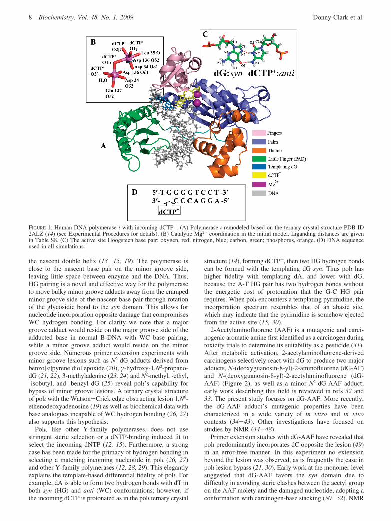
FIGURE 1: Human DNA polymerase ι with incoming dCTP+. (A) Polymerase ι remodeled based on the ternary crystal structure PDB ID2ALZ (14) (see Experimental Procedures for details). (B) Catalytic Mg2+ coordination in the initial model. Liganding distances are givenin Table S8. (C) The active site Hoogsteen base pair: oxygen, red; nitrogen, blue; carbon, green; phosphorus, orange. (D) DNA sequenceused in all simulations.
8 Biochemistry, Vol. 48, No. 1, 2009 Donny-Clark et al.

solution structures later showed that, as had been predicted,in double-stranded DNA dG-AAF predominantly adopts abase-displaced intercalated conformation in solution with asyn glycosidic torsion angle (44, 46), with some NMRevidence for an external conformation available as well (53).A base-displaced intercalated conformation displaces thetemplating dG, inconsistent with the error-free selection ofdCTP/dCTP+ opposite dG-AAF by polι. Therefore, the AAFfluorenyl rings must be placed on the major or minor grooveside of the nascent base pair, with anti or syn glycosidicbond conformations, respectively. While a syn conformationfor dG-AAF is consistent with HG pairing in polι, if the C8adduct is syn (HG), the bulky AAF fluorenyl rings wouldbe placed in a small, crowded region of the enzyme on theminor groove side of the template. With WC pairing, on theother hand, the AAF ring system would be well accom-modated in the large space on the major groove side.Recently, it has been shown through MD simulations thatdG-AAF can adopt the anti (WC) conformation with WCpairing through a modest alteration in sugar pucker in themodified nucleoside. This alleviates the steric hindrancebetween the acetyl and the adjacent sugar (54).
In the present study we investigated both WC- and HG-paired dG-AAF in polι using a molecular modeling andmolecular dynamics (MD) approach supplemented by po-tential of mean force (PMF) analysis. Our results show thatwith WC pairing the AAF adduct is well accommodated onthe major groove side of the growing double helix, withfrequent sampling of a near reaction-ready state. HG-pairedmodels led to severe distortion of the active site, and in nocase did they sample a near reaction-ready state. In addition,free energy calculations indicate a relatively small (∼5 kcal/mol) energy difference between the WC- and HG-paired dG-dCTP/dCTP+ in the active site of polι. These results suggestthat the dG-AAF adduct can be bypassed by polι utilizing aWC pairing scheme which places the bulky adduct on thespacious major groove side of the nascent base pair, similarto the accommodation of bulky minor groove lesions via HGbase pairing in the same space. Thus polι could play a rolein bypassing adducts on both the major and minor groovesides of a damaged base, adding to its versatility in humanlesion bypass.
EXPERIMENTAL PROCEDURES
Molecular Modeling of Initial Unmodified Control Struc-tures for MD. We used the polι ternary complex crystalstructure containing a dG-dCTP+ HG base pair (PDB (55)ID 2ALZ (14)) as the basis for our initial models. Missingloops containing residues 371-378 and residues 395-403were modeled with the program MODELER (56) on theModloop web server (57). The DNA sequence (Figure 1D)was taken from the crystal structure (14). While the crystalcontained three residues in the single-stranded 5′ overhang,the last two were not resolved and are not included in oursimulations. This structure was used as our HG unmodifiedcontrol simulation (Figure 1A). dCTP+ was modeled fromdCTP by protonation of N3. For the first Watson-Crickunmodified control simulation the glycosidic torsion angle� (Figure 2) on the templating dG was altered from thecrystal value of 64.9° (syn) to 254.1° (anti) which permittedWC pairing. A second unmodified WC structure was derivedfrom the WC-AAF2 trajectory’s most representative structure(see below). AAF atoms were deleted and replaced by ahydrogen atom, and the resulting WC unmodified structurewas subjected to our molecular dynamics protocol (seebelow).
Molecular Modeling of Initial dG-AAF Structures for MD.Structures from the last frame (10 ns) of the unmodifiedcontrol production dynamics simulations were used as theinitial structures for the dG-AAF simulations (HG controlfor HG-AAF, WC control for WC-AAF). A high-resolutionAAF structure (58) was obtained from the CambridgeStructural Database (59) (refcode ACAFLR) and bonded toC8 of the templating dG. Then the R′ and �′ linkage torsionangles (Figure 2) of the resultant dG-AAF were surveyedover their 360° range at 10° intervals starting at 5°, withglycosidic torsion � in the syn (HG) conformation (64.9°),as observed in the crystal structure (14) for a total of 1296structures created. These structures were evaluated for stericclashes with the bumpcheck utility of InsightII (AccelrysInc.). For these searches γ′ (Figure 2) was retained in theconformation of the AAF crystal structure (58), i.e. 340.7°.For the anti WC-AAF simulations the last frame (10 ns) ofthe WC control structure (� ) 192.7°) was used for a similarsearch of an additional 1296 structures. After these initialsearches were completed, R′, �′ combinations that exhibitedminimal close contacts were selected for further investigation.For the HG-AAF structures we identified two narrow regionsthat showed the least close contacts, one placing the fluorenylrings 3′ of the templating dG (HG-AAF1 and HG-AAF2)and one 5′ (HG-AAF3 and HG-AAF4). For the WC-AAFstructures, the AAF could be accommodated either in thebroad range of R′ 260° to 284° on the exposed major grooveside of the nascent base pair (WC-AAF1 and WC-AAF3)or in a narrow pocket in the little finger domain (WC-AAF2and WC-AAF4). For both WC-AAF and HG-AAF structureseach region could accommodate a pair of structures with thesame R′ torsion angles, but with the methylene bridge (C9in Figure 2) rotated ∼180° about the long axis of thefluorenyl rings, which is governed by �′. Finally, the R′, �′,and γ′ torsions of all structures were further adjusted tominimize close contacts (Figure S1, Table S1).
Force Field. The Cornell et al. force field (60) withmodifications (61, 62) and the PARM99 parameter set (63)
FIGURE 2: dG-AAF structure with torsion angles designated byarrows. Torsion angles at the linkage site are defined as follows:R′, N9sC8sNsC2; �′, C8sNsC2sC1; γ′, C8sNsCsC-(methyl); �, glycosidic torsion angle O4′sC1′sN9sC4, where O4′,C1′, N9, C4, and C8 are from dG and other atoms are from AAF.
Bypassing a Bulky Major Groove dG-AAF Adduct in Polι Biochemistry, Vol. 48, No. 1, 2009 9

were employed for all simulations. For the partial chargecalculations a syn dG-AAF model with no steric clashes wasmodeled using a generic B-DNA dG from the Biopolymermodule of InsightII (Accelrys Inc.) linked to a high-resolutionAAF structure (58) from the Cambridge Structural Database(59) (refcode ACAFLR). The structure had C2′ endoenvelope sugar pucker and � ) 34.2°, R′ ) 135°, �′ ) 83°,and γ′ ) 165°. Partial charges for the dG-AAF and fordCTP+ (Tables S2 and S3) were calculated with the HFmethod and the 6-31G* basis set (64) using Gaussian 03(65), and the restrained electrostatic potential fitting algorithmRESP (66, 67) was employed to fit the charge to each atomcenter. dCTP charges were taken from previous work (68).Parameters for atom types in dG-AAF and dCTP+ not foundin the PARM99 parameter set were taken from the GAFF(69) parameter set or developed by analogy to chemicallysimilar atom types in the PARM99 and GAFF parametersets. Parameters are provided in Table S4.
Molecular Dynamics Protocol. All minimizations and MDsimulations used the SANDER module of the AMBER 8.0software suite (70). The LEaP module of AMBER 8.0 (70)was used to add hydrogen atoms and neutralize the systemwith sufficient Na+ atoms to bring the net charge to zero.Hydrogen atoms of the solute (DNA, polymerase, andincoming dCTP/dCTP+) were minimized with implicitsolvent using a distance-dependent dielectric function of ε
) 4.0r (where r is the distance between an atom pair) for600 steps of steepest descent, followed by 600 steps ofconjugate gradient. The resulting ternary complex wasreoriented with SIMULAID (71) to minimize the numberof water molecules needed to solvate the system. A periodicTIP3P (72) rectangular water box with a buffer distance of10 Å between each wall and the closest solute atom in eachdirection was added with the LEaP module of AMBER. Boxdimensions were approximately 80 × 75 × 76 Å, with atotal of ∼14500 water molecules.
All systems employed the following equilibration and MDprotocols: (i) minimization of the counterions and solventmolecules for 2500 steps of steepest descent and 2500 stepsof conjugate gradient, with 50 kcal/mol restraints on thesolute atoms; (ii) 30 ps initial MD at 10 K with 25.0 kcal/mol restraints on the solute to allow the solvent to relax;(iii) heat-up from 10 to 310 K (37 °C) at constant volumeover 80 ps with 10 kcal/mol restraint on the solute; (iv) 20ps MD at constant volume and 310 K, with 10.0 kcal/molrestraints on the solute; (v) 30, 40, and 50 ps MD at 1 atmand 310 K with decreasing restraints of 10, 1, and 0.1 kcal/mol, respectively, on solute atoms and an increasing timeconstant of 1.0, 2.0, and 4.0 ps, respectively, for heat bathcoupling of the system; (vi) production MD was conductedat 1 atm, 310 K for 10 ns, with 1 ps coupling constants forboth pressure and temperature.
In all MD simulations, long-range electrostatic interactionswere treated with the particle mesh Ewald method (73, 74).A 9 Å cutoff was applied to the nonbonded Lennard-Jonesinteractions. The SHAKE algorithm (75) was applied toconstrain all bonds involving hydrogen atoms with relativegeometrical tolerance of 10-5 Å. The Berendsen couplingalgorithm (76) was used for temperature scaling. A 2 fs timestep was used, and the translational/rotational center-of-massmotion was removed every 0.5 ps (77).
Potential of Mean Force Calculations Thirty-six initialstructures were generated by taking the most representativestructure from the stable region of the HG control dynamicssimulations (6-10 ns; see Figure S2) and resetting � at 10°intervals from 5° to 355°. The most representative structurewas identified using the trajectory clustering function in theMOIL-VIEW (78) program with all residues within 8.0 Åof any atom in the nascent base pair selected for analysis.Initial structures were minimized and equilibrated as de-scribed below. MD simulations were then run for 200 ps,with a 30 kcal/mol harmonic restraint centered on the initialvalue of �, and � angle values in each 200 ps ensemble werecollected at 20 fs intervals. We performed three separate setsof 36 simulations with identical initial conditions anddifferent random initial velocity distributions. All � anglevalues from all three trials were used as input to a programdevised by A. Grossfield to implement the weighted histo-gram analysis method (WHAM) (available at http://membrane.urmc.rochester.edu/) for calculating potential of mean force(relative free energy).
Stability of MD Simulations. All simulations showedreasonable stability in the active site after 4 ns and stabilityin the whole enzyme after ∼6 ns. This was evaluated byinspecting the rmsd of the enzyme as compared to the initialstate after equilibration over time (Figure S2). Therefore,all analyses presented here were performed on the final 4 nsof the trajectories.
RESULTS
In Vitro primer extension data for polι containing a dG-AAF lesion as the templating base revealed that polι verystrongly selects the correct partner, dC, opposite the lesion(49). Thus we investigated structures with incoming dCTP/dCTP+ in order to determine the features that led toincorporation of the correct nucleotide opposite the adduct.We were specifically interested in whether the damaged baseassumes a syn (HG) or anti (WC) conformation in the activesite of the polymerase. A syn (HG) conformation placesthe bulky adduct on the cramped minor groove side of thegrowing double helix, while an anti (WC) conformationplaces the AAF lesion on the spacious major groove side.We performed 11 molecular dynamics simulations: threeunmodified controls, one with undamaged syn (HG) and twowith undamaged anti (WC) dG as the template, and eightdG-AAF damaged template simulations: four syn (HG) andfour anti (WC). Initial R′ and �′ torsion angles (Figure 2,Table S1, Figure S1) for the adduct were selected throughan extensive conformational search of 2592 structures asdetailed in the Experimental Procedures. We performed 10ns of production MD for each structure, with the final 4 nsused for analysis.
Criteria for a Near Reaction-Ready State. Using the well-organized active site of a high-resolution pol� crystalstructure containing two Mg2+ ions and a primer terminal 3′OH (79) and the results of a quantum mechanical molecularmodeling study (80) as our guides, we utilize the followingstructural criteria for a near reaction-ready state: (1) aPR-O3′ distance of less than 3.5 Å, (2) an O3′-PR-O3Rangle of attack of 170 ( 10°, and (3) reasonable coordinationof the two catalytic Mg2+ ions (Figure 1B) (discussed below).Furthermore, as a reasonably stringent criterion for a near
10 Biochemistry, Vol. 48, No. 1, 2009 Donny-Clark et al.

reaction-ready active site we evaluated the percentage ofstructures in our trajectories that satisfy all three of the abovecriteria simultaneously and term that the near reaction-readyoccupancy (Table 1).
We also considered the extent of hydrogen bondingbetween the incoming dCTP/dCTP+ and the templating basein order to determine if proper HG or WC hydrogen bondingwas taking place (Table S5). In addition, we analyzedhydrogen bonding between the nascent base pair and theenzyme, using the unmodified control HG simulation as ourbenchmark (Tables S6 and S7). Our criteria for hydrogenbonding are a donor and acceptor heavy atom distance of<3.4 Å and a donor-hydrogen-acceptor angle of 180° (55°. Finally, we have used WHAM (81) to derive the freeenergy profile around the glycosidic bond for the HGunmodified control simulation to gain energetic insight intothe overall cost of forming an anti structure in the activesite.
WC and HG Unmodified Control Simulations Share SomeCommon Features in the ActiVe Site. Both HG and WCunmodified control simulations were performed and analyzedas described above. The most representative active sitestructure was identified using the trajectory clustering func-tion in the MOIL-VIEW (78) program with all residueswithin 8.0 Å of any atom of the nascent base pair selectedfor analysis. The fingers domain of the enzyme shows thehighest degree of stability, with a fingers domain backbonermsd of <1 Å between the most representative active sitestructure of any of the unmodified control simulations andthe crystal structure. All of the active site hydrogen bondsbetween the incoming nucleotide and the enzyme observedin the crystal are maintained in the HG and both WCunmodified control simulations, with the exception of thehydrogen bond between Tyr68 and the incoming dCTP/dCTP+, which is replaced by two hydrogen bonds with waterin all three simulations (Table S6). In other respects theunmodified control simulations differ, as described below.
Hoogsteen Unmodified Control Is Faithful to the CrystalStructure. All ternary complex crystal structures of polι todate show a HG pair formed between the incoming dNTPand the templating base (13-15, 19). In our unmodifiedcontrol simulation the syn (HG) conformation of the tem-plating base is maintained throughout the HG unmodifiedcontrol simulation (Figure S3). The structural features ofthe active site are similar to the crystal, with a HG compatibleC1′-C1′ distance of 8.8 ( 0.2 Å (Figure S4). Good basestacking is observed between the incoming dCTP+ and theprimer terminus (Figure S5). The Mg2+ ions are wellcoordinated (discussed below) (Figures S6 and S7, Table S8),though the inter-Mg2+ distance does increase somewhat, from3.4 to 4.1 ( 0.1 Å. Overall near reaction-ready occupancyfor the HG unmodified control is 51% (Table 1), a figurethat is not surprising given polι’s extremely slow rate ofincorporation (8, 10). Hydrogen bonding in the nascent base
pair is good, with both HG hydrogen bonds present at >99%occupancy (Table 1, Table S5). Other features of the activesite resemble the ternary crystal structure (14), with the closeC1′-C1′ distance enforced by the tight coordination of theincoming dCTP+ via numerous hydrogen bonds (Table S6)and other polar interactions coupled with the stabilizationof the templating base’s sugar by Gln59, Lys60, and Leu62(Figure 3A). Lys60 forms a hydrogen bond with the freephosphates in the templating dG backbone, contributing tothe HG-compatible C1′-C1′ distance (Figure 3A, Table S7).Ser307, on the other hand, forms a hydrogen bond with thedT 5′ overhang phosphate group, as opposed to hydrogenbonding with the dG backbone phosphate as in the crystalstructure, due to a shift in the position of the mobile littlefinger domain (12). This change in hydrogen bonding doesnot appear to impact the organization of the nascent basepair or the active site as a whole.
Watson-Crick Unmodified Control Loses Important Con-tacts with the Polymerase. In order to evaluate the feasibilityof accommodating a WC pair in the active site of polι, weperformed two simulations: (1) WC unmodified simulation1, where the initial structure was identical to the HGunmodified control, except that the templating base wasinitiated with an anti (WC) conformation (Table S1), and(2) WC unmodified simulation 2, a WC unmodified simula-tion using the most representative structure from the WC-AAF2 simulation, with the AAF moiety replaced by ahydrogen atom, as the initial state; WC-AAF2 showed thehighest reaction-ready occupancy of the WC-AAF simula-tions. This simulation was suggested by a reviewer.
In both simulations the incoming dCTP was unprotonatedso as to allow three WC hydrogen bonds to form. Thesesimulations performed surprisingly well, both attaining astandard WC C1′-C1′ distance of 10.6 ( 0.2 Å (82) whichwas then maintained throughout the stable region of thetrajectory (Figure S4). For both simulations base stackingbetween the nascent pair and the previously incorporated pairis not disrupted (Figure 3C, Figure S5). Mg2+ coordinationdoes not differ significantly from the HG unmodified controlin either case (Figures S6 and S7, Table S8), and the inter-Mg2+ distance is 3.9 ( 0.1 Å in both simulations. Bothsimulations have two Watson-Crick hydrogen bonds thatare present at >99% occupancy, with the third at ∼86%occupancy in WC unmodified 1 and ∼98% in WC unmodi-fied 2 (Table S5). In addition, a hydrogen bond betweenGln59 and N2 of the templating dG is formed in bothsimulations, with ∼98% occupancy in WC unmodified 1 and∼77% occupancy in WC unmodified 2 (Table S7). Thishydrogen bond is not possible with the syn template in theHG pair. Moreover, the near reaction-ready occupancy is82% for WC unmodified 1 and 83% for WC unmodified 2(Table 1).
We sought to define the structural rearrangements associ-ated with forming such a near reaction-ready Watson-Crick
Table 1: Near Reaction-Ready Occupancy and Number of Hydrogen Bonds Formed between the Incoming dCTP(WC)/dCTP+(HG) and the TemplatingdG (Control)/dG-AAF (Damaged)
structuresHG
controlWC
control 1WC
control 2HG-
AAF1HG-
AAF2HG-
AAF3HG-
AAF4WC-
AAF1WC-
AAF2WC-
AAF3WC-
AAF4
near reaction-readyoccupancy (%)
51 82 83 0 0 0 0 76 75 0.4 70
no. of hydrogen bonds 2 3 3 0 0 0 1 2 3 3 3
Bypassing a Bulky Major Groove dG-AAF Adduct in Polι Biochemistry, Vol. 48, No. 1, 2009 11

pair in the unmodified control simulations. In both cases thehydrogen bonds between the incoming dCTP and thepolymerase are similar to the HG unmodified control (TableS6). However, in order to stabilize a wider C1′-C1′ distance,the dT 5′ to the templating dG moves toward the majorgroove side of the nascent base pair, pulling the DNAbackbone away from the polymerase. In WC unmodifiedcontrol 1 the single-stranded overhang stacks with a histidine(His354) in the little finger domain, pulling the C1′ carbonof the templating dG ∼2 Å away from the active site (Figure3C). In WC unmodified control 2 the 5′ OH of the overhanghydrogen bonds with N7 of the templating dG. Since theincoming dCTP remains stably positioned relative to thefingers domain of the protein in both cases, the movementof the overhang and backbone away from the confines ofthe polymerase allows the C1′-C1′ distance to increase to∼10.6 Å, appropriate for a WC pair (82) (Figure S4), andthe sugar pucker is a B-DNA standard C2′ endo (82) (TableS9). In both cases the DNA template backbone also losesthe hydrogen bond with Ser307 found in the HG unmodifiedcontrol simulation due to the rearrangement of the backbone.This hydrogen bond is replaced with a hydrogen bond toArg347 in the little finger domain in WC control 1 but is
lost entirely in WC control 2 (Figure 3C, Table S7). Thismajor groove position of the single-stranded base 5′ to thetemplate is not seen in any of the other simulations, andneither of these structures would be feasible if the 5′overhang were longer (Figure 3C) due to a dearth of roomon the major groove side of the duplex. This implies thatsuch a WC permissive conformation in the enzyme withunmodified DNA is likely to be disfavored during replication,consistent with the crystallographic observations to date.
Hoogsteen AAF Simulations Show a Variety of DistortionsThat PreVent AchieVement of a Near Reaction-Ready State.Of the 1296 initial models of dG-AAF with HG pairing thatwe screened (see Experimental Procedures and SupportingInformation) we selected four with minimal close contactsbetween the adduct and the enzyme for further study. HG-AAF MD simulations differed in the combined initial valuesof the torsion angles R′, �′, and γ′ and, therefore, theplacement of the lesion relative to the DNA and polymerase(Figure S1, Table S1). Two initial structures placed the AAFrings 3′ to the templating dG (HG-AAF1 and HG-AAF2),and two placed the AAF rings 5′ to the templating dG (HG-AAF3 and HG-AAF4) (Figure S1). Both members of eachpair of structures place the fluorenyl rings in the same region
FIGURE 3: Active site organization and hydrogen bonds formed by the templating dG/dG-AAF. Polι is in green, duplex DNA is in gray, andkey active site features are colored by atom: oxygen, red; nitrogen, blue; carbon, green; phosphorus, purple. Stereoviews are available inSupporting Information, Figure S11. (A) Unmodified HG control simulation. Two HG hydrogen bonds are formed between the incomingdCTP+ and the template. Hydrogen bonds between Ser307 and the terminal dT O5′ and between Lys60 and a backbone oxygen from thepreviously incorporated dG (labeled as OP) stabilize the base position. Note the stacking of the dT single-stranded overhang with Tyr61.(B) HG-AAF4. This structure was selected as the least distorted HG-AAF structure (Table 1). Note the absence of hydrogen bondingbetween the templating dG-AAF and the enzyme and the single hydrogen bond between dG-AAF and dCTP+. (C) Unmodified WC controlsimulation. Three WC hydrogen bonds are formed between dG and dCTP. The hydrogen bond between Ser307 and the terminal O5′ isreplaced by a hydrogen bond between Arg347 and a backbone oxygen from the templating dG, and the 5′ dT overhang stacks with His354instead of Tyr61. Lys60 forms a hydrogen bond with a backbone oxygen from the previously incorporated dG (labeled as OP) as in the HGcontrol. The dG sugar ring is pulled away from residues Gln59, Lys60, and Leu62. A novel hydrogen bond not possible in the HG conformationis formed between Gln59 and N2 of the templating dG. (D) WC-AAF2. The active site is stabilized by hydrogen bonds from a templatingdG backbone oxygen to Lys60 and from a backbone oxygen on the previously incorporated dG (labeled OP) to Ser307, as well as the novelhydrogen bond between Gln59 and N2 of the templating dG observed in the WC control. Three WC hydrogen bonds are formed in thenascent base pair, and the 5′ dT overhang stacks with Tyr61 as in the HG control.
12 Biochemistry, Vol. 48, No. 1, 2009 Donny-Clark et al.

relative to dG, but the methylene bridge (C9 in Figure 2) isrotated ∼180° around the long axis of the fluorenyl rings inthe pair members (Figure S1). After 10 ns of simulation theactive site is severely distorted in all four cases, with nocommon motifs for placement of the AAF (Figure 3B,Figures S8 and S9). Hydrogen bonding between the incomingdCTP+ and templating dG-AAF is poor or absent (Table 1,Table S5), PR-O3′ distances are long (Figure S4), and basestacking between the incoming dCTP+ and the primerterminus is often disrupted (Figure S5). While hydrogenbonds between the incoming dCTP+ and the enzyme aremostly maintained (Table S6), the interactions betweenthe templating base and the enzyme are lost or altered inthe majority of the HG dG-AAF simulations (Figure 3B,Table S7). Most importantly, the HG dG-AAF structures,without exception, have a near reaction-ready occupancy of0% (Table 1), strongly suggesting that incorporation of theincoming dCTP+ would be disfavored.
The HG dG-AAF structures attempt to accommodate thelesion in different ways. The bulky hydrophobic fluorenylmoiety stacks with the cytosine ring of the incoming dCTP+
in the HG-AAF1, HG-AAF2, and HG-AAF4 simulations(Figures S8 and S9). In the HG-AAF2 and HG-AAF4 casesthis causes the cytosine ring to rotate ∼90° around theglycosidic bond, disrupting hydrogen bonding with the dG-AAF (Figure 3B). Accommodation of the lesion has theadditional effect of displacing the modified guanine fromthe double helix in all trials save HG-AAF3 (Figures S8 andS9), resulting in increased C1′-C1′ distances (Figure S4).The primer terminus disengages from the active site, in thecase of HG-AAF2 moving as far as 11 Å away from theincoming dCTP+ PR (Figure S4). The acetyl moiety of dG-AAF is placed 3′ to the dG-AAF when there is significantdisplacement of the base and otherwise is placed 5′ of theguanine rings (Figures S8 and S9).
Watson-Crick AAF Simulations Accommodate the LesionWell with Little Distortion of the ActiVe Site. When dG-AAFwas modeled with an anti (WC) glycosidic torsion angle,four structures were again selected as initial models for MDsimulation from a scan of 1296 possible conformations (seeExperimental Procedures and Supporting Information) (TableS1, Figure S1). In all four cases the active site is stable andwell ordered after MD simulation (Figure 3D, Figures S8and S9). As seen in Table 1, there are three hydrogen bondsbetween the templating base and dCTP in all simulations;their occupancies range from 59% to 100% (Table S5). Basestacking between the incoming dCTP and the primerterminus is well maintained (Figure S5), and the C1′-C1′distance is near the typical WC value of 10.6 Å (Figure S4).The WC-AAF1, WC-AAF2, and WC-AAF3 simulationsproduce near reaction-ready occupancies (involving goodPR-O3′ distance (Figure S4), Mg2+ coordination (FiguresS6 and S7, Table S8), and in-line attack angle (Figure S3,Table S9) of >70%. However, the WC-AAF3 simulationshowed only 0.4% occupancy (Table 1) due to an overlongPR-O3′ distance (Figure S4) resulting from a hydrogen bondbetween Tyr39 and O3′ of dCTP (Figure S10, Table S6).This hydrogen bond pulls the dCTP PR away from O3′ ofthe primer terminus, and it is not observed in other WC-AAF simulations (Table S6).
The dG-AAF does not significantly disrupt the active sitein these simulations. Analysis of the four trials reveals two
motifs for placement of the fluorenyl rings and acetyl groupof the dG-AAF. WC-AAF1 and WC-AAF3 simulations hadthe fluorenyl rings placed 5′ to the damaged base, thusstacking with the 5′ overhang dT (Figure S8). The acetylmoiety is 3′ to the guanine rings, causing a change in thesugar pucker to the C1′ exo domain in order to avoidclose contacts with the acetyl (Figures S3 and S8, Table S9).The remaining two simulations have the fluorenyl rings 3′to the dG, allowing the 5′ overhang to stack with Tyr61.The acetyl is 5′ to the base, and the sugar is C1′ exo(WCAAF2) or O4′ endo (WCAAF4) (Figures S3 and S8,Table S9). The alteration of the sugar pucker is consistentwith previous results in Dpo4, which also showed that a C1′exo pucker permitted anti dG-AAF (54). Hydrogen bondingbetween the incoming dCTP and polι is robust, with nosignificant differences from the HG unmodified control(Table S6).
In these simulations the increased C1′-C1′ distancenecessary for WC pairing was made possible by an adjust-ment of the sugar on the templating base that moves the C1′carbon away from the incoming dCTP. Unlike the WCunmodified control there is no stacking between the template5′ dT overhang and the little finger domain. The templatingdG-AAF forms strong hydrogen bonds with Gln59 and Lys60(Figure 3D, Table S7), helping to maintain contact with thehydrophobic residues that hold the dG-AAF sugar. Thefingers domain adjusts slightly to accommodate the newplacement of the sugar on the templating base, with theresidues in the vicinity of the sugar (i.e., Gln 59 to Leu62)moving ∼1 Å relative to the rest of the fingers domain. Theremainder of the active site protein backbone is relativelystatic, with an average rmsd of 0.50 Å between the WC dG-AAF structures and the crystal structure.
Mg2+ Coordination in Our Simulations Is Less Than Ideal.The two catalytic Mg2+ ions in the polι active site are eachcoordinated by six electronegative oxygen atoms (Figure 1B).We consider good Mg2+ coordination in polι as one Mg2+
ion with all six oxygens within 2.4 Å and the other Mg2+
with five of these oxygens within 2.4 Å and one oxygenwithin 3.2 Å (see Discussion). In the simulations whichexhibit good Mg2+ coordination, all but one of the coordinat-ing oxygens are within 2.4 Å. The exception is the only Othat is involved in coordinating both Mg2+ ions, dCTP/dCTP+ O1R. The O1R is within 2.4 Å of either Mg2+
A orMg2+
B, but never both at the same time. Time course analysesand Mg2+ coordination mean distance and standard deviationvalues for the stable region of the dynamics are shown inFigures S6 and S7 and Table S8.
Free Energy Analysis with Incoming dCTP+ Shows bothSyn and Anti Conformations Occupy Energy Wells, with SynLower in Energy. In light of our findings above indicatingthat a WC pair can be stably accommodated in the activesite of polι we sought to estimate the overall energetic costof maintaining an anti conformation of the templating baserelative to a syn conformation. We used the weightedhistogram analysis method (WHAM) (81) on a series ofumbrella samples (see Experimental Procedures) whichscanned over the entire range of the glycosidic torsion angle�. Analysis of the relative free energy around � for the mostrepresentative HG unmodified control structure with incom-ing dCTP+ showed that the syn structure is ∼5 kcal/mollower in energy than the anti structure (Figure 4A). Both of
Bypassing a Bulky Major Groove dG-AAF Adduct in Polι Biochemistry, Vol. 48, No. 1, 2009 13

the syn and anti structures lie in energy wells, and they areseparated by barriers of ∼12 and ∼9 kcal/mol, dependingon the direction of the rotation. While this simulation wasperformed with a protonated dCTP+, which precludesformation of three WC hydrogen bonds, nevertheless the antistructure resides in an energy well. When the proton isremoved from the dCTP, the difference is ∼3.5 kcal/mol,but the anti structure has the lower energy, and the barriersare both ∼10 kcal/mol (Figure 4B). The presence of an antienergy well separated from the syn domain well by an∼9-10 kcal/mol barrier regardless of the protonation stateof the incoming nucleotide would preclude the interconver-sion of anti (WC) and syn (HG) structures within the timeframe of our simulations.
DISCUSSION
The discovery that polι utilizes HG pairing in the activesite to select the newly incorporated nucleotide showed thatDNA polymerases can be more versatile than was previouslybelieved. In light of previous work on accurate dG-AAFbypass by polι (49) we investigated how polι incorporated acorrect partner opposite dG-AAF. Preliminary modelingshowed that a syn conformation of the dG-AAF would placethe bulky AAF fluorenyl rings on the cramped minor grooveside in close contact with the enzyme, while an anticonformation was free of steric hindrance. Accordingly, wehypothesized that WC base pairing was necessary forselection and incorporation of dCTP opposite dG-AAF. Aseries of MD simulations based on an extensive search foroptimal initial structures strongly supported this hypothesis.
Rationale for a WC Conformation of dG-AAF in Polι. Polιis unique in many ways. It is the least accurate, leastprocessive polymerase discovered to date (8, 10, 11). Inaddition, it is the only polymerase observed to use a HG-paired syn templating base in the active site with bothundamaged and damaged DNA (13-15, 19). However, ourresults show WC base pairing with dG-AAF in the activesite (Figure 3, Figures S8 and S9). In this case WC pairingis consistent with the mechanism of damage bypass proposedfor polι based on its crystal structures.
Polι has been shown to enable nucleotide incorporationopposite minor groove lesions or lesions that compromiseWC hydrogen bonding via a HG pair (19-25); this placesthe damage away from the cramped minor groove side of
the protein and into the spacious major groove side. Ourresults show that AAF, a bulky major groove lesion, is alsoreadily placed into the same spacious major groove regionin order to facilitate nucleotide incorporation opposite thedamaged base (Figure 3D, Figures S8 and S9).
In addition, the series of binary and ternary crystalstructures of polι (13-15) show that the templating base isin the anti conformation in the binary complex. The incomingnucleotide forces the templating base to rotate ∼180° aroundthe glycosidic bond, in order to form a HG pair in the ternarycomplex. The energetic cost of this rotation is therefore lessthan the cost of adjusting the protein to accommodate theincreased C1′-C1′ distance required for WC pairing. How-ever, if there were a large, bulky major groove lesion on thetemplating base, such as AAF, the cost of the rotation wouldincrease greatly. This alters the energy landscape and wouldplausibly make remaining in the anti conformation moreenergetically favorable than rotating to syn. The resultsobserved by Choi and Guengerich with increasingly bulkyadducts linked to N2 of guanine (25) may stem from a similareffect. Though polι can incorporate dCTP opposite theselesions, the efficiency of incorporation decreases withincreasing size of the lesion, with strong effects seen evenfor N2-methyl-dG. This is hard to explain, as the synconformation would place these adducts on the major grooveside of the enzyme, which is spacious enough to accom-modate adducts as large as AAF. However, the energeticcost of rotating the damaged dG through an area occupiedby either the protein or the nascent DNA duplex wouldincrease with the bulk of the damage. This could accountfor the observed decrease in efficiency for increasingly bulkydG N2 adducts, which could block incorporation if theyremained on the cramped minor groove side.
Structural Features of AAF and Unmodified ControlSimulations Suggest That WC Pairing Is Preferred forIncorporation Opposite dG-AAF but Not Undamaged dG.Our simulations for the unmodified control WC systemssuggest that this pairing scheme is unlikely without the dG-AAF lesion. Our first unmodified control Watson-Crickstructure was facilitated by a stacking interaction betweenHis354 and the 5′ dT overhang on the templating strand thatstabilized a WC compatible C1′-C1′ distance (Figure 3C).This 5′ overhang stacking appears to be infeasible with alonger single-stranded template region, as would be found
FIGURE 4: (A) Free energy profile for rotation around the glycosidic torsion angle � in the Hoogsteen control structure with incomingdCTP+. Minima correspond to the syn (33°) and anti (217°) domains, with syn ∼5 kcal/mol lower in energy. (B) Free energy profile forrotation around the glycosidic torsion angle � in the Hoogsteen control structure with incoming unprotonated dCTP. Minima correspond tothe syn (43°) and anti (211°) domains, with anti ∼3.5 kcal/mol lower in energy.
14 Biochemistry, Vol. 48, No. 1, 2009 Donny-Clark et al.

in a replicative context. A second simulation with anunmodified WC pair derived from a WC dG-AAF simulationshowed similar structural rearrangements, further supportingthe idea that WC pairing is only possible with conformationsplacing the 5′ single-stranded overhang on the major grooveside of the templating base, away from the polymerase. Thewidened C1′-C1′ distance needed for WC pairing isstabilized by this abnormal position of the overhang. In polιthe force on the nascent WC base pair due to the longC1′-C1′ distance is alleviated by a reorientation of thetemplate backbone and the movement of the 5′ dT overhangto the major groove side. Addition of another base or baseson the 5′ side of the templating dG would not much alterthis; however, if the single-stranded overhang were very long,as in a replicative situation, this force would have to bealleviated in a different way, such as by rotating to a synconformation.
Biochemical experiments with 7-deazaguanine as thetemplating base support the likelihood of Hoogsteen pairingin the unmodified case (26). Though the 7-deazaguanine iscapable of WC pairing, one hydrogen bond of the HG pairis disrupted. Since nucleotide incorporation opposite the7-deazaguanine by polι is severely impaired, a strongdependence upon HG pairing is suggested.
Since the crystal structure (14) reveals a protonated syndCTP+, the protonated state is clearly favored energeticallyfor the syn conformation; the added hydrogen bond contrib-utes to the energy needed for protonation, which likely occursin the syn domain just as proper alignment for HG basepairing is achieved. The anti conformation, however, woulddisfavor protonation, since no structural advantage arises.Experimental investigations of the pKa of N3 protonatedcytosine in triple helices containing HG base pairs (83-85)indicate that the N3 pKa is ∼3-5 units above that of freecytosine (∼4.3) (86). This corresponds to a stabilizationenergy range of ∼4-7 kcal/mol for the N3 in HG-pairedcytosine, with the higher value appearing more probableaccordingtothepKa data(83,84).Clearly,manyfactors(83-85)govern this value. Our energy analyses by umbrella samplingover the glycosidic torsion angle � for dCTP+ and dCTP(Figure 4) were notable in indicating energy wells for theanti conformation in both cases. With the pKa data and theconcept that the protonation and the pKa change occur justin time to facilitate HG pairing in the syn conformation onecould propose that the syn well in Figure 4B is lowered by∼4-7 kcal/mol by protonation. This would give an energydifference between syn dG-dCTP+ and anti dG-dCTP rangingbetween 0.5 and 3.5 kcal/mol, in favor of syn dG-dCTP+,with the higher value being more likely. Thus the WCconformation is unlikely to occur in the unmodified case,consistent with the ternary crystal structures and biochemicaldata (13-15, 19, 26, 27).
However, our simulations suggest that with the presence ofthe AAF lesion WC pairing becomes favorable. In the WC dG-AAF simulations WC pairing accommodates the bulky AAFmoiety on the spacious major groove side of the enzyme. Thehigh near reaction-ready occupancies we observed for the WC-AAF simulations suggest that these structures are plausible(Table 1).
Examination of the MD ensembles for the WC-AAFsimulations sheds some light on the changes necessary inthe enzyme in order to accommodate a WC dG-AAF/dCTP
pair in the active site. In their series of crystallographicstudies of polι Nair et al. (13-15, 19) note the importanceof three amino acids in the fingers domain (Gln59, Lys60,and Leu62) that hold the sugar of the templating base in asmall hydrophobic cavity, leading to a short C1′-C1′distance. This in turn forces the templating base to assumea syn conformation to avoid clashing with the incomingnucleotide. In the unmodified WC control structures the sugarof dG loses van der Waals contacts with these three residues,allowing an increase in the C1′-C1′ distance (Figure 3C).However, in the WC-AAF simulations the sugar remains inthe cavity, and the increased C1′-C1′ distance is enabledby a slight conformational change in the fingers domain ofresidues Gln59, Lys60, and Leu62 (Figure 3D). ResiduesGln59, Lys60, and Leu62 move ∼2 Å relative to theremainder of the fingers domain and the active site in orderto accommodate a wider C1′-C1′ distance in the WC-AAFsimulations. The active site is otherwise essentially un-changed. Thus, in the case of a damaged dG-AAF template,WC pairing is allowed without sacrificing other importantcontacts between the nascent base pair and the active site.Two additional hydrogen bonds in the active site may helpto compensate for the energetic cost of rearranging theenzyme to allow a wider C1′-C1′ distance: one additionalhydrogen bond in the nascent base pair and a hydrogen bondbetween the anti templating dG and Gln59, not possible inthe syn conformation (Figure 3C,D). The unmodified WCcontrols, while also manifesting these hydrogen bonds,assume a conformation unlikely in a replicative context andloses significant DNA/protein contacts (Figure 3C).
HG-Paired dG-AAF Structures Appear Unlikely To Leadto Nucleotide Incorporation Due to Numerous ActiVe SiteDistortions. In contrast to the well-ordered active sitesobserved in the WC-AAF simulations the HG-AAF simula-tions show a disparate collection of distortions, all of whichwould impede incorporation of dCTP+ opposite the lesion(Figure 3B, Figures S8 and S9). As a result all of the HG-AAF structures have a near reaction-ready occupancy of 0%(Table 1). These distortions include loss of Mg2+ coordina-tion, loss of hydrogen bonding between incoming dCTP+
and template as compared to the unmodified control HGsimulation, dramatically increased PR-O3′ distances, anddisplacement of the incoming nucleotide away from thetemplate (Figure 3B, Figures S8 and S9, Tables S6 and S8).None of the four initial simulations yielded a near reaction-ready structure, indicating that the observed incorporationof dCTP/dCTP+ opposite dG-AAF (49) would not likelyresult from an HG pair. Just as polι uses a syn conformationfor the template to put bulky minor groove adducts on thespacious major groove side, our results suggest that it canuse an anti conformation to place bulky major groove adductssuch as dG-AAF on the major groove side as well.
Coordination of Mg2+ Ions in Polι Is Imperfect. All ofour simulations, both with damaged and undamaged DNA,show somewhat impaired Mg2+ coordination of either thecatalytic Mg2+ ion, MgA
2+, or the nucleotide binding Mg2+
ion, MgB2+ (Figures S6 and S7, Table S8). In addition, recent
results show that polι has higher fidelity and processivitywith Mn2+ (24, 87), which has a looser coordinationrequirement than Mg2+, as the catalytic ion. Together, theseresults may indicate that the polι active site does not favorideal coordination of the catalytic Mg2+ ions. This is
Bypassing a Bulky Major Groove dG-AAF Adduct in Polι Biochemistry, Vol. 48, No. 1, 2009 15

consistent with polι’s slow rate of nucleotide incorporationwith Mg2+ ions (8, 10). Our simulations support this insofaras no simulation showed perfect octahedral coordination ofboth Mg2+ ions simultaneously at any time. However, currentforce field issues concerning proper treatment of Mg2+ ionsare still a frontier issue (88). In this connection, simulationsof large systems such as in the present work are necessarilysubject to current approximations in the state of the artincluding force field and sampling considerations (89).
CONCLUSION
Our results elucidate how human DNA polymerase ι isable to incorporate a correct partner opposite a major groovelesion. Primer extension results show that polι is capable ofnucleotide incorporation opposite structurally diverse lesions,including a variety of minor groove adducts (20-22, 25)and major groove adducts (90), as well as thymine and uracildimers (30, 91, 92) and abasic sites (10, 49, 93). Our resultslend insight into this process, explaining in structural termshow polι correctly incorporates dCTP opposite a majorgroove dG-AAF lesion. Though polι’s function in ViVo hasbeen difficult to discern, recent results indicating that polιmay be responsible for the dramatically increased cancer rateof XPV patients (94), who lack functional polη, show thatunderstanding polι on a structural level may relate to humanhealth. If polι must substitute for polη in XPV patients, itmay be called upon to contribute to bypass of a variety oflesions, both major and minor groove, in cells that have lostthe function of other, more reliable bypass polymerases. Inconclusion, our study suggests an expanded role for polι inlesion bypass that could include both major and minor grooveadducts, utilizing both WC and HG base pairing to placebulky lesions on the spacious major groove side.
ACKNOWLEDGMENT
We thank Dr. Lei Jia for careful reading of the manuscriptand helpful suggestions. We also wish to acknowledgeexcellent suggestions provided by one of the reviewers.Figures were created with Pymol (DeLano Scientific LLC)and Matlab (The Mathworks, Inc).
SUPPORTING INFORMATION AVAILABLE
Figures: S1, initial dG-AAF conformations; S2, rmsd vstime plot; S3, torsion angles vs time plot; S4, C1′-C1′ andPR-O3′ distances vs time plot; S5, base stacking; S6 andS7, Mg2+ coordination distances vs time plots; S8, mostrepresentative structure active site stereoviews; S9, mostrepresentative structure whole enzyme stereoviews; S10,WC-AAF3 active site with hydrogen bonds; S11, stereoviewsof the active site structures shown in Figure 3. Tables: S1,initial torsions for all structures; S2-S4, AMBER param-eters; S5-S7, hydrogen bond occupancies; S8, average Mg2+
coordination distances; S9, torsion angle ensemble averagevalues. This material is available free of charge via theInternet at http://pubs.acs.org.
REFERENCES
1. Luch, A. (2005) Nature and nurtureslessons from chemicalcarcinogenesis. Nat. ReV. 5, 113–125.
2. Clapp, R. W., Jacobs, M. M., and Loechler, E. L. (2008)Environmental and occupational causes of cancer: New evidence2005–2007. ReV. EnViron. Health 23, 1–37.
3. Friedberg, E. C., Lehmann, A. R., and Fuchs, R. P. (2005) Tradingplaces: how do DNA polymerases switch during translesion DNAsynthesis? Mol. Cell 18, 499–505.
4. Lehmann, A. R. (2006) Translesion synthesis in mammalian cells.Exp. Cell Res. 312, 2673–2676.
5. Lehmann, A. R., Niimi, A., Ogi, T., Brown, S., Sabbioneda, S.,Wing, J. F., Kannouche, P. L., and Green, C. M. (2007) Translesionsynthesis: Y-family polymerases and the polymerase switch. DNARepair (Amsterdam) 6, 891–899.
6. McDonald, J. P., Rapic-Otrin, V., Epstein, J. A., Broughton, B. C.,Wang, X., Lehmann, A. R., Wolgemuth, D. J., and Woodgate, R.(1999) Novel human and mouse homologs of SaccharomycescereVisiae DNA polymerase η. Genomics 60, 20–30.
7. Ohmori, H., Friedberg, E. C., Fuchs, R. P., Goodman, M. F.,Hanaoka, F., Hinkle, D., Kunkel, T. A., Lawrence, C. W., Livneh,Z., Nohmi, T., Prakash, L., Prakash, S., Todo, T., Walker, G. C.,Wang, Z., and Woodgate, R. (2001) The Y-family of DNApolymerases. Mol. Cell 8, 7–8.
8. Tissier, A., McDonald, J. P., Frank, E. G., and Woodgate, R. (2000)Polι, a remarkably error-prone human DNA polymerase. GenesDeV. 14, 1642–1650.
9. Vaisman, A., Lehmann, A. R., and Woodgate, R. (2004) DNApolymerases η and ι. AdV. Protein Chem. 69, 205–228.
10. Zhang, Y., Yuan, F., Wu, X., and Wang, Z. (2000) Preferentialincorporation of G opposite template T by the low-fidelity humanDNA polymerase ι. Mol. Cell. Biol. 20, 7099–7108.
11. McCulloch, S. D., and Kunkel, T. A. (2008) The fidelity of DNAsynthesis by eukaryotic replicative and translesion synthesispolymerases. Cell Res. 18, 148–161.
12. Yang, W., and Woodgate, R. (2007) What a difference a decademakes: insights into translesion DNA synthesis. Proc. Natl. Acad.Sci. U.S.A. 104, 15591–15598.
13. Nair, D. T., Johnson, R. E., Prakash, S., Prakash, L., and Aggarwal,A. K. (2004) Replication by human DNA polymerase ι occurs byHoogsteen base-pairing. Nature 430, 377–380.
14. Nair, D. T., Johnson, R. E., Prakash, L., Prakash, S., and Aggarwal,A. K. (2005) Human DNA polymerase ι incorporates dCTPopposite template G via a G ·C + Hoogsteen base pair. Structure13, 1569–1577.
15. Nair, D. T., Johnson, R. E., Prakash, L., Prakash, S., and Aggarwal,A. K. (2006) An incoming nucleotide imposes an anti to synconformational change on the templating purine in the human DNApolymerase ι active site. Structure 14, 749–755.
16. Ling, H., Boudsocq, F., Woodgate, R., and Yang, W. (2001) Crystalstructure of a Y-family DNA polymerase in action: a mechanismfor error-prone and lesion-bypass replication. Cell 107, 91–102.
17. Lone, S., Townson, S. A., Uljon, S. N., Johnson, R. E., Brahma,A., Nair, D. T., Prakash, S., Prakash, L., and Aggarwal, A. K.(2007) Human DNA polymerase κ encircles DNA: implicationsfor mismatch extension and lesion bypass. Mol. Cell 25, 601–614.
18. Alt, A., Lammens, K., Chiocchini, C., Lammens, A., Pieck, J. C.,Kuch, D., Hopfner, K. P., and Carell, T. (2007) Bypass of DNAlesions generated during anticancer treatment with cisplatin byDNA polymerase η. Science 318, 967–970.
19. Nair, D. T., Johnson, R. E., Prakash, L., Prakash, S., and Aggarwal,A. K. (2006) Hoogsteen base pair formation promotes synthesisopposite the 1,N6-ethenodeoxyadenosine lesion by human DNApolymerase ι. Nat. Struct. Mol. Biol. 13, 619–625.
20. Rechkoblit, O., Zhang, Y., Guo, D., Wang, Z., Amin, S., Krzem-insky, J., Louneva, N., and Geacintov, N. E. (2002) Trans-lesionsynthesis past bulky benzo[a]pyrene diol epoxide N2-dG and N6-dA lesions catalyzed by DNA bypass polymerases. J. Biol. Chem.277, 30488–30494.
21. Washington, M. T., Minko, I. G., Johnson, R. E., Wolfle, W. T.,Harris, T. M., Lloyd, R. S., Prakash, S., and Prakash, L. (2004)Efficient and error-free replication past a minor-groove DNA adductby the sequential action of human DNA polymerases ι and κ. Mol.Cell. Biol. 24, 5687–5693.
22. Wolfle, W. T., Johnson, R. E., Minko, I. G., Lloyd, R. S., Prakash,S., and Prakash, L. (2005) Human DNA polymerase ι promotesreplication through a ring-closed minor-groove adduct that adoptsa syn conformation in DNA. Mol. Cell. Biol. 25, 8748–8754.
23. Johnson, R. E., Yu, S. L., Prakash, S., and Prakash, L. (2007) Arole for yeast and human translesion synthesis DNA polymerasesin promoting replication through 3-methyl adenine. Mol. Cell. Biol.27, 7198–7205.
16 Biochemistry, Vol. 48, No. 1, 2009 Donny-Clark et al.

24. Plosky, B. S., Frank, E. G., Berry, D. A., Vennall, G. P., McDonald,J. P., and Woodgate, R. (2008) Eukaryotic Y-family polymerasesbypass a 3-methyl-2′-deoxyadenosine analog in Vitro and methylmethanesulfonate-induced DNA damage in ViVo. Nucleic Acids Res.36, 2152–2162.
25. Choi, J. Y., and Guengerich, F. P. (2006) Kinetic evidence forinefficient and error-prone bypass across bulky N2-guanine DNAadducts by human DNA polymerase ι. J. Biol. Chem. 281, 12315–12324.
26. Johnson, R. E., Prakash, L., and Prakash, S. (2005) Biochemicalevidence for the requirement of Hoogsteen base pairing forreplication by human DNA polymerase ι. Proc. Natl. Acad. Sci.U.S.A. 102, 10466–10471.
27. Johnson, R. E., Haracska, L., Prakash, L., and Prakash, S. (2006)Role of Hoogsteen edge hydrogen bonding at template purines innucleotide incorporation by human DNA polymerase ι. Mol. Cell.Biol. 26, 6435–6441.
28. Potapova, O., Chan, C., DeLucia, A. M., Helquist, S. A., Kool,E. T., Grindley, N. D., and Joyce, C. M. (2006) DNA polymerasecatalysis in the absence of Watson-Crick hydrogen bonds: analysisby single-turnover kinetics. Biochemistry 45, 890–898.
29. Mizukami, S., Kim, T. W., Helquist, S. A., and Kool, E. T. (2006)Varying DNA base-pair size in subangstrom increments: evidencefor a loose, not large, active site in low-fidelity Dpo4 polymerase.Biochemistry 45, 2772–2778.
30. Johnson, R. E., Washington, M. T., Haracska, L., Prakash, S., andPrakash, L. (2000) Eukaryotic polymerases ι and � act sequentiallyto bypass DNA lesions. Nature 406, 1015–1019.
31. Wilson, R. H., DeEds, F., and Cox, A. J. (1941) The toxicity andcarcinogenicity of 2-acetylaminofluorene. Cancer Res. 1, 595–608.
32. Heflich, R. H., and Neft, R. E. (1994) Genetic toxicity of2-acetylaminofluorene, 2-aminofluorene and some of their me-tabolites and model metabolites. Mutat. Res. 318, 73–114.
33. Kriek, E. (1992) Fifty years of research on N-acetyl-2-aminofluo-rene, one of the most versatile compounds in experimental cancerresearch. J. Cancer Res. Clin. Oncol. 118, 481–489.
34. Suzuki, N., Ohashi, E., Hayashi, K., Ohmori, H., Grollman, A. P.,and Shibutani, S. (2001) Translesional synthesis past acetylami-nofluorene-derived DNA adducts catalyzed by human DNA poly-merase κ and Escherichia coli DNA polymerase IV. Biochemistry40, 15176–15183.
35. Doisy, R., and Tang, M. S. (1995) Effect of aminofluorene and(acetylamino)fluorene adducts on the DNA replication mediatedby Escherichia coli polymerases I (Klenow fragment) and III.Biochemistry 34, 4358–4368.
36. Belguise-Valladier, P., Maki, H., Sekiguchi, M., and Fuchs, R. P.(1994) Effect of single DNA lesions on in Vitro replication withDNA polymerase III holoenzyme. Comparison with other poly-merases. J. Mol. Biol. 236, 151–164.
37. Shibutani, S., Suzuki, N., and Grollman, A. P. (1998) Mutagenicspecificity of (acetylamino)fluorene-derived DNA adducts in mam-malian cells. Biochemistry 37, 12034–12041.
38. Gillet, L. C., Alzeer, J., and Scharer, O. D. (2005) Site-specificincorporation of N-(deoxyguanosin-8-yl)-2-acetylaminofluorene(dG-AAF) into oligonucleotides using modified “ultra-mild” DNAsynthesis. Nucleic Acids Res. 33, 1961–1969.
39. Gunz, D., Hess, M. T., and Naegeli, H. (1996) Recognition of DNAadducts by human nucleotide excision repair. Evidence for athermodynamic probing mechanism. J. Biol. Chem. 271, 25089–25098.
40. Shibutani, S., Suzuki, N., Tan, X., Johnson, F., and Grollman, A. P.(2001) Influence of flanking sequence context on the mutagenicityof acetylaminofluorene-derived DNA adducts in mammalian cells.Biochemistry 40, 3717–3722.
41. Fuchs, R. P., and Fujii, S. (2007) Translesion synthesis inEscherichia coli: lessons from the NarI mutation hot spot. DNARepair (Amsterdam) 6, 1032–1041.
42. Fuchs, R. P., Koffel-Schwartz, N., Pelet, S., Janel-Bintz, R.,Napolitano, R., Becherel, O. J., Broschard, T. H., Burnouf, D. Y.,and Wagner, J. (2001) DNA polymerases II and V mediaterespectively mutagenic (-2 frameshift) and error-free bypass of asingle N2-acetylaminofluorene adduct. Biochem. Soc. Trans. 29,191–195.
43. Tan, X., Suzuki, N., Grollman, A. P., and Shibutani, S. (2002)Mutagenic events in Escherichia coli and mammalian cellsgenerated in response to acetylaminofluorene-derived DNA adductspositioned in the NarI restriction enzyme site. Biochemistry 41,14255–14262.
44. O’Handley, S. F., Sanford, D. G., Xu, R., Lester, C. C., Hingerty,B. E., Broyde, S., and Krugh, T. R. (1993) Structural characteriza-tion of an N-acetyl-2-aminofluorene (AAF) modified DNA oligo-mer by NMR, energy minimization, and molecular dynamics.Biochemistry 32, 2481–2497.
45. Patel, D. J., Mao, B., Gu, Z., Hingerty, B. E., Gorin, A., Basu,A. K., and Broyde, S. (1998) Nuclear magnetic resonance solutionstructures of covalent aromatic amine-DNA adducts and theirmutagenic relevance. Chem. Res. Toxicol. 11, 391–407.
46. Cho, B. P., and Zhou, L. (1999) Probing the conformationalheterogeneity of the acetylaminofluorene-modified 2′-deoxygua-nosine and DNA by 19F NMR spectroscopy. Biochemistry 38,7572–7583.
47. Cho, B. P. (2004) Dynamic conformational heterogeneities ofcarcinogen-DNA adducts and their mutagenic relevance. J. EnViron.Sci. Health 22, 57–90.
48. Evans, F. E., Miller, D. W., and Levine, R. A. (1986) 1H NMR studyof self-association and restricted internal rotation of the C8-substituteddeoxyguanosine 5′-monophosphate adduct of the carcinogen 2-(acety-lamino)fluorene. J. Biomol. Struct. Dyn. 3, 935–948.
49. Zhang, Y., Yuan, F., Wu, X., Taylor, J. S., and Wang, Z. (2001)Response of human DNA polymerase ι to DNA lesions. NucleicAcids Res. 29, 928–935.
50. Grunberger, D., Nelson, J. H., Cantor, C. R., and Weinstein, I. B.(1970) Coding and conformational properties of oligonucleotidesmodified with the carcinogen N2-acetylaminofluorene. Proc. Natl.Acad. Sci. U.S.A. 66, 488–494.
51. Fuchs, R., and Daune, M. (1972) Physical studies on deoxyribo-nucleic acid after covalent binding of a carcinogen. Biochemistry11, 2659–2666.
52. Fuchs, R., and Daune, M. (1971) Changes of stability andconformation of DNA following the covalent binding of acarcinogen. FEBS Lett. 14, 206–208.
53. Milhe, C., Dhalluin, C., Fuchs, R. P., and Lefevre, J. F. (1994)NMR evidence of the stabilisation by the carcinogen N2-acety-laminofluorene of a frameshift mutagenesis intermediate. NucleicAcids Res. 22, 4646–4652.
54. Wang, L., and Broyde, S. (2006) A new anti conformation forN-(deoxyguanosin-8-yl)-2-acetylaminofluorene (AAF-dG) allowsWatson-Crick pairing in the Sulfolobus solfataricus P2 DNApolymerase IV (Dpo4). Nucleic Acids Res. 34, 785–795.
55. Berman, H. M., Westbrook, J., Feng, Z., Gilliland, G., Bhat, T. N.,Weissig, H., Shindyalov, I. N., and Bourne, P. E. (2000) TheProtein Data Bank. Nucleic Acids Res. 28, 235–242.
56. Fiser, A., Do, R. K., and Sali, A. (2000) Modeling of loops inprotein structures. Protein Sci. 9, 1753–1773.
57. Fiser, A., and Sali, A. (2003) ModLoop: automated modeling ofloops in protein structures. Bioinformatics 19, 2500–2501.
58. van Meerssche, M., Germain, G., Declercq, J.-P., Touillaux, R.,Roberfroid, M., and Razzouk, C. (1980) 2-(Acetylamino)fluorene,C15H13NO. Acta Crystallogr., Sect. C: Cryst. Struct. Commun 9,515–518.
59. Allen, F. H. (2002) The Cambridge Structural Database: a quarter of amillion crystal structures and rising. Acta Crystallogr. 58, 380–388.
60. Cornell, W. D., Cieplak, P., Bayly, C. I., Gould, I. R., Merz, K. M.,Ferguson, D. M., Spellmeyer, D. C., Fox, T., Caldwell, J. W., andKollman, P. A. (1995) A second generation force field for thesimulation of proteins, nucleic acids, and organic molecules. J. Am.Chem. Soc. 117, 5179–5197.
61. Duan, Y., Wu, C., Chowdhury, S., Lee, M. C., Xiong, G., Zhang,W., Yang, R., Cieplak, P., Luo, R., Lee, T., Caldwell, J., Wang, J.,and Kollman, P. (2003) A point-charge force field for molecularmechanics simulations of proteins based on condensed-phase quantummechanical calculations. J. Comput. Chem. 24, 1999–2012.
62. Cheatham, T. E., III., Cieplak, P., and Kollman, P. A. (1999) Amodified version of the Cornell et al. force field with improvedsugar pucker phases and helical repeat. J. Biomol. Struct. Dyn. 16,845–862.
63. Wang, J. M., Cieplak, P., and Kollman, P. A. (2000) How welldoes a restrained electrostatic potential (RESP) model perform incalculating conformational energies of organic and biologicalmolecules? J. Comput. Chem. 21, 1049–1074.
64. Hehre, W. J., Ditchfie, R., and Pople, J. A. (1972) Self-consistentmolecular-orbital methods. 12. Further extensions of Gaussian-type basis sets for use in molecular-orbital studies of organic-molecules. J. Chem. Phys. 56, 2257–2261.
65. Frisch, M. J., Schlegel, H. B., Scuseria, G. E., Robb, M. A.,Cheeseman, J. R., Montgomery, J., J. A., Vreven, T., Kudin, K. N.,Burant, J. C., Millam, J. M., Iyengar, S. S., Tomasi, J., Barone, V.,
Bypassing a Bulky Major Groove dG-AAF Adduct in Polι Biochemistry, Vol. 48, No. 1, 2009 17

Mennucci, B., Cossi, M., Scalmani, G., Rega, N., Petersson, G. A.,Nakatsuji, H., Hada, M., Ehara, M., Toyota, K., Fukuda, R., Hasegawa,J., Ishida, M., Nakajima, T., Honda, Y., Kitao, O., Nakai, H., Klene,M., Li, X., Knox, J. E., Hratchian, H. P., Cross, J. B., Bakken, V.,Adamo, C., Jaramillo, J., Gomperts, R., Stratmann, R. E., Yazyev,O., Austin, A. J., Cammi, R., Pomelli, C., Ochterski, J. W., Ayala,P. Y., Morokuma, K., Voth, G. A., Salvador, P., Dannenberg, J. J.,Zakrzewski, V. G., Dapprich, S., Daniels, A. D., Strain, M. C., Farkas,O., Malick, D. K., Rabuck, A. D., Raghavachari, K., Foresman, J. B.,Ortiz, J. V., Cui, Q., Baboul, A. G., Clifford, S., Cioslowski, J.,Stefanov, B. B., Liu, G., Liashenko, A., Piskorz, P., Komaromi, I.,Martin, R. L., Fox, D. J., Keith, T., Al-Laham, M. A., Peng, C. Y.,Nanayakkara, A., Challacombe, M., Gill, P. M. W., Johnson, B., Chen,W., Wong, M. W., Gonzalez, C., and Pople, J. A. (2004) Gaussian03, Gaussian, Inc., Wallingford, CT.
66. Bayly, C. I., Cieplak, P., Cornell, W. D., and Kollman, P. A. (1993)A well-behaved electrostatic potential based method using chargerestraints for deriving atomic chargessthe RESP model. J. Phys.Chem. 97, 10269–10280.
67. Cieplak, P., Cornell, W. D., Bayly, C., and Kollman, P. A. (1995)Application of the multimolecule and multiconformational RESPmethodology to biopolymers-charge derivation for DNA, RNA,and proteins. J. Chem. Phys. 16, 1357–1377.
68. Perlow, R. A., and Broyde, S. (2002) Toward understanding themutagenicity of an environmental carcinogen: structural insightsinto nucleotide incorporation preferences. J. Mol. Biol. 322, 291–309.
69. Wang, J., Wolf, R. M., Caldwell, J. W., Kollman, P. A., and Case,D. A. (2004) Development and testing of a general amber forcefield. J. Comput. Chem. 25, 1157–1174.
70. Case, D. A., Darden, T. A., Cheatham, T. E., III., Simmerling,C. L., Wang, J., Duke, R. E., Luo, R., Merz, K. M., Wang, B.,Pearlman, D. A., Crowley, M., Brozell, S., Tsui, V., Gohlke, H.,Mongan, J., Hornak, V., Cui, G., Beroza, P., Schafmeister, C.,Caldwell, J. W., Ross, W. S., and Kollman, P. A. (2004) AMBER8, University of California, San Francisco.
71. Mezei, M. (1997) Optimal position of the solute for simulations.J. Comput. Chem. 18, 812–815.
72. Jorgensen, W. L., Chandrasekhar, J., Madura, J. D., Impey, R. W.,and Klein, M. L. (1983) Comparison of simple potential functionsfor simulating liquid water. J. Chem. Phys. 79, 926–935.
73. Darden, T., York, D., and Pedersen, L. (1993) Particle meshEwaldsan nlog (n) method for Ewald sums in large systems.J. Chem. Phys. 98, 10089–10092.
74. Essmann, U., Perera, L., Berkowitz, M. L., Darden, T., Lee, H.,and Pedersen, L. G. (1995) A smooth particle mesh Ewald method.J. Chem. Phys. 103, 8577–8593.
75. Ryckaert, J. P., Ciccotti, G., and Berendsen, H. J. C. (1977)Numerical-integration of Cartesian equations of motion of a systemwith constraints-molecular-dynamics of N-alkanes. J. Comput.Phys. 23, 327–341.
76. Berendsen, H. J. C., Postma, J. P. M., van Gunsteren, W. F.,DiNola, A., and Haak, J. R. (1984) Molecular dynamics withcoupling to an external bath. J. Chem. Phys. 81, 3684–3690.
77. Harvey, S. C., Tan, R. K. Z., and Cheatham, T. E. (1998) Theflying ice cube: Velocity rescaling in molecular dynamics leads toviolation of energy equipartition. J. Comput. Chem. 19, 726–740.
78. Simmerling, C., Elber, R., and Zhang, J. (1995) MOIL-ViewsAprogram for visualization of structure and dynamics of biomoleculesand STO-A program for computing stochastic paths, in Modelingof Biomolecular Structure and Mechanisms (Pullman, A., et al.,Eds.) pp 241-265, Kluwer, The Netherlands.
79. Batra, V. K., Beard, W. A., Shock, D. D., Krahn, J. M., Pedersen,L. C., and Wilson, S. H. (2006) Magnesium-induced assembly ofa complete DNA polymerase catalytic complex. Structure 14, 757–766.
80. Wang, L., Yu, X., Hu, P., Broyde, S., and Zhang, Y. (2007) Awater-mediated and substrate-assisted catalytic mechanism forSulfolobus solfataricus DNA polymerase IV. J. Am. Chem. Soc.129, 4731–4737.
81. Shankar, K., Djamal, B., Robert, H. S., Peter, A. K., and John,M. R. (1992) The weighted histogram analysis method for free-energy calculations on biomolecules. I: The method. J. Comput.Chem. 13, 1011–1021.
82. Saenger, W. (1984) Principles of Nucleic Acid Structure, Springer-Verlag, New York.
83. Plum, G. E., and Breslauer, K. J. (1995) Thermodynamics of anintramolecular DNA triple helix: a calorimetric and spectroscopicstudy of the pH and salt dependence of thermally induced structuraltransitions. J. Mol. Biol. 248, 679–695.
84. Asensio, J. L., Lane, A. N., Dhesi, J., Bergqvist, S., and Brown,T. (1998) The contribution of cytosine protonation to the stabilityof parallel DNA triple helices. J. Mol. Biol. 275, 811–822.
85. Wu, P., Kawamoto, Y., Hara, H., and Sugimoto, N. (2002) Effectof divalent cations and cytosine protonation on thermodynamicproperties of intramolecular DNA double and triple helices.J. Inorg. Biochem. 91, 277–285.
86. Zimmer, C., Luck, G., Venner, H., and Fric, J. (1968) Studies onthe conformation of protonated DNA. Biopolymers 6, 563–574.
87. Frank, E. G., and Woodgate, R. (2007) Increased catalytic activityand altered fidelity of human DNA polymerase ι in the presenceof manganese. J. Biol. Chem. 282, 24689–24696.
88. Oelschlaeger, P., Klahn, M., Beard, W. A., Wilson, S. H., andWarshel, A. (2007) Magnesium-cationic dummy atom moleculesenhance representation of DNA polymerase beta in moleculardynamics simulations: improved accuracy in studies of structuralfeatures and mutational effects. J. Mol. Biol. 366, 687–701.
89. Adcock, S. A., and McCammon, J. A. (2006) Molecular dynamics:survey of methods for simulating the activity of proteins. Chem.ReV. 106, 1589–1615.
90. Frank, E. G., Sayer, J. M., Kroth, H., Ohashi, E., Ohmori, H., Jerina,D. M., and Woodgate, R. (2002) Translesion replication ofbenzo[a]pyrene and benzo[c]phenanthrene diol epoxide adducts ofdeoxyadenosine and deoxyguanosine by human DNA polymeraseι. Nucleic Acids Res. 30, 5284–5292.
91. Tissier, A., Frank, E. G., McDonald, J. P., Iwai, S., Hanaoka, F.,and Woodgate, R. (2000) Misinsertion and bypass of thymine-thymine dimers by human DNA polymerase ι. EMBO J. 19, 5259–5266.
92. Vaisman, A., Takasawa, K., Iwai, S., and Woodgate, R. (2006)DNA polymerase ι-dependent translesion replication of uracilcontaining cyclobutane pyrimidine dimers. DNA Repair (Amster-dam) 5, 210–218.
93. Vaisman, A., Frank, E. G., McDonald, J. P., Tissier, A., andWoodgate, R. (2002) Polι-dependent lesion bypass in Vitro. Mutat.Res. 510, 9–22.
94. Wang, Y., Woodgate, R., McManus, T. P., Mead, S., McCormick,J. J., and Maher, V. M. (2007) Evidence that in xerodermapigmentosum variant cells, which lack DNA polymerase η, DNApolymerase ι causes the very high frequency and unique spectrumof UV-induced mutations. Cancer Res. 67, 3018–3026.
BI801283D
18 Biochemistry, Vol. 48, No. 1, 2009 Donny-Clark et al.


![cis-Diamminedichloroplatinum(II)-DNA Adduct Formation in ... · [CANCER RESEARCH 47, 718-722, February 1, 1987] cis-Diamminedichloroplatinum(II)-DNA Adduct Formation in Renal, Gonadal,](https://img.pdfslide.net/doc/110x75/60934a1bfda1347d92293bf5/cis-diamminedichloroplatinumii-dna-adduct-formation-in-cancer-research-47.jpg)