Embed Size (px)
Citation preview
MOLECULAR AND CELLULAR BIOLOGY, May 2004, p. 4593–4604 Vol. 24, No. 100270-7306/04/$08.00�0 DOI: 10.1128/MCB.24.10.4593–4604.2004Copyright © 2004, American Society for Microbiology. All Rights Reserved.
Acquisition of Hrs, an Essential Component of PhagosomalMaturation, Is Impaired by Mycobacteria
Otilia V. Vieira,1† Rene E. Harrison,1 Cameron C. Scott,1 Harald Stenmark,2David Alexander,3 Jun Liu,3 Jean Gruenberg,4 Alan D. Schreiber,5
and Sergio Grinstein1*Cell Biology Program, Hospital for Sick Children, and Department of Biochemistry, University of Toronto, Ontario M5G 1X8,1 and
Department of Medical Genetics and Microbiology, University of Toronto, Ontario M5S 1A8,3 Canada; Department ofBiochemistry, Institute for Cancer Research, Norwegian Radium Hospital, Montebello, and Institute of Pathology,
National Hospital, Oslo, Norway2; Department of Biochemistry, University of Geneva, 1211 Geneva 4,Switzerland4; and Department of Medicine, University of Pennsylvania School of
Medicine, Philadelphia, Pennsylvania 191045
Received 8 July 2003/Returned for modification 4 September 2003/Accepted 12 February 2004
Pathogenic mycobacteria survive within macrophages by precluding the fusion of phagosomes with lateendosomes or lysosomes. Because the molecular determinants of normal phagolysosome formation are poorlyunderstood, the sites targeted by mycobacteria remain unidentified. We found that Hrs, an adaptor moleculeinvolved in protein sorting, associates with phagosomes prior to their fusion with late endosomes or lysosomes.Recruitment of Hrs required the interaction of its FYVE domain with phagosomal phosphatidylinositol3-phosphate, but two other attachment sites were additionally involved. Depletion of Hrs by use of smallinterfering RNA impaired phagosomal maturation, preventing the acquisition of lysobisphosphatidic acid andreducing luminal acidification. As a result, the maturation of phagosomes formed in Hrs-depleted cells wasarrested at an early stage, characterized by the acquisition and retention of sorting endosomal markers. Thisphenotype is strikingly similar to that reported to occur in phagosomes of cells infected by mycobacteria. Wetherefore tested whether Hrs is recruited to phagosomes containing mycobacteria. Hrs associated readily withphagosomes containing inert particles but poorly with mycobacterial phagosomes. Moreover, Hrs was foundmore frequently in phagosomes containing avirulent Mycobacterium smegmatis than in phagosomes with themore virulent Mycobacterium marinum. These findings suggest that the inability to recruit Hrs contributes tothe arrest of phagosomal maturation induced by pathogenic mycobacteria.
Phagosomes, which are formed by invagination of theplasma membrane, acquire microbicidal properties by a seriesof fusion and fission events that culminate with the formationof phagolysosomes (7, 33). This sequence of events, collectivelyknown as phagosomal maturation, is often impaired by micro-organisms such as pathogenic mycobacteria. By co-opting thecellular machinery responsible for maturation, mycobacteriaavoid exposure to the harsh hydrolytic environment ofphagolysosomes, thereby managing to survive and multiplywithin macrophages (27).
Mycobacteria arrest phagosomal maturation at an earlystage, precluding the acquisition of late endosomal markerslike lysobisphosphatidic acid (LBPA). During the normalcourse of maturation, this transition was recently shown torequire de novo synthesis of phosphatidylinositol 3-phosphate[PI(3)P] by Vps34, the class III phosphatidylinositol 3-kinase(9, 34). However, Fratti et al. (9) reported that phagosomescontaining mycobacteria recruit Vps34 normally. Thus, theeffects of the microorganism are possibly exerted downstreamof the formation of PI(3)P. This phosphoinositide serves as a
ligand for a variety of proteins bearing either FYVE or PXdomains that may contribute to the maturation of the phago-some. Of these, early endosome antigen 1 (EEA1) was shownto be depleted from mycobacterial phagosomes (9). EEA1 isthought to be important in the homotypic fusion of early en-dosomes (29), but it is not clear whether it contributes directly tothe fusion of phagosomes with late endosomes, the event that iscentral to mycobacterial survival. It is likely that other mediatorsof phagosomal maturation are targeted by mycobacteria.
Hepatocyte growth factor-regulated tyrosine kinase sub-strate (Hrs) is emerging as a central coordinator of late endo-somal sorting (5, 16, 23–25) and may play a comparable role inphagosomal maturation. Like EEA1, Hrs possesses a FYVEdomain capable of interacting with PI(3)P, as well as domainsthat promote association with ubiquitylated proteins (the UIMmotif) and with clathrin (4, 23, 24, 26, 31). Like its Saccharo-myces cerevisiae homologue, Vps27p, Hrs is thought to func-tion in the generation of multivesicular bodies, acting in con-junction with the ESCRT-I complex to segregate cargo andinduce membrane budding. Given the prominent role of Hrs inthe biogenesis of late endosomes, we considered whether it isinvolved in phagosomal maturation and, more importantly,whether its activity is affected by mycobacteria.
MATERIALS AND METHODS
Reagents and antibodies. Dulbecco’s minimal Eagle’s medium and fetal bo-vine serum were from Wisent Inc. HEPES-buffered RPMI, wortmannin, tubulin
* Corresponding author. Mailing address: Division of Cell Biology,Hospital for Sick Children, 555 University Ave., Toronto, OntarioM5G 1X8, Canada. Phone: (416) 813-5727. Fax: (416) 813-5028. E-mail: [email protected].
† Present address: Max Planck Institute for Molecular Cell Biologyand Genetics, 01307 Dresden, Germany.
4593
at Univ of T
oronto on July 4, 2007 m
cb.asm.org
Dow
nloaded from
antibody, and human immunoglobulin G (IgG) were from Sigma. Latex beadswere from Bangs Laboratories. Sheep red blood cells (RBC) and rabbit anti-RBC IgG were from ICN-Cappel. Fluorochrome-conjugated secondary antibod-ies were all from Jackson ImmunoResearch. LysoTracker (Red DND-99) wasfrom Molecular Probes. Mouse and rat anti-LAMP-1 antibodies were from theDevelopmental Studies Hybridoma Bank, maintained by the University of Iowaand Johns Hopkins University. Goat anti-EEA1 and anti-c-Myc antibodies werefrom Santa Cruz Biotechnology. Antibodies to Mycobacterium were from CygnusTechnologies. The preparation of antibodies to LBPA and Hrs has been de-scribed elsewhere (15, 24).
Cell culture, transfection, and plasmids. Culture conditions for macrophageRAW 264.7, COS-IIA, and Chinese hamster ovary cells (CHO-IIA) stably trans-fected with Fc�RIIA receptors have been previously described (34). The gener-ation of the plasmids used for expression of wild-type and mutant forms of eitherepitope-tagged or yellow fluorescent protein (YFP)-conjugated Hrs is describedin detail elsewhere (23, 24, 26). The plasmids encoding the PX domain of p40phox
and the 2-FYVE domain of EEA1 were generously provided by M. Yaffe (MIT,Cambridge, Mass.) and L. Cantley (Beth Israel Deaconess Medical Center,Boston, Mass.) and have been described elsewhere (14, 34). The cells weretransiently transfected by using FuGENE-6 (Roche Molecular Biochemicals) assuggested by the manufacturer.
Treatment with siRNA. Small interfering RNA (siRNA) directed toward nu-cleotides 160 to 180 relative to the start codon of the human Hrs (GenBankaccession number NM 004712.31) was purchased from Dharmacon Research(Lafayette, Colo.) as double-stranded, desalted, and gel-purified preparations.The sequence used for siRNA was selected according to the guidelines in ref-erence 8. Transfection of siRNA by use of oligofectamine (Invitrogen) wasperformed according to the manufacturer’s directions by using 240 pmol ofsiRNA to transfect �100,000 COS-IIA cells grown on a coverslip placed withina well of a six-well plate. Cells were grown for 72 h and then processed forimmunofluorescence, electron microscopy, or immunoblotting.
Phagocytosis assays and treatment with wortmannin. Fresh or fixed sheepRBC were opsonized with rabbit anti-sheep RBC antibody (1:50). Latex beadswere opsonized with 1 mg of human IgG/ml. Opsonization was for either 1 h atroom temperature or overnight at 4°C. Where noted, the cells were treated with100 nM wortmannin for 30 min prior to phagocytosis. The onset of phagocytosiswas synchronized by allowing the particles to bind to cells on ice for 5 min, andingestion was then initiated by incubation at 37°C. Excess particles were washedaway with phosphate-buffered saline (PBS) and, where indicated, the cells wereincubated in culture medium at 37°C for the specified additional chase period.To identify adherent particles that were not internalized, the cells were incubatedat 4°C with Cy5-labeled donkey anti-rabbit IgG (1:1,000) or Cy5-labeled donkeyanti-human IgG (1:1,000) for 10 min.
Culturing and phagocytosis of mycobacteria. Mycobacterium smegmatis mc2
155 and Mycobacterium marinum 1218R (ATCC 927) were transformed with theplasmid pG13 as described previously (2). Cultures were grown in Middlebrook7H9 media supplemented with 10% oleic acid–albumin–dextrose–catalase(Difco) and 25 �g of kanamycin (Sigma)/ml. Typically, M. smegmatis cultureswere grown for 16 h at 37°C and M. marinum cultures were grown for 36 to 48 hat 30°C. Before use, cultures were washed twice with PBS before being homog-enized with 50 strokes on ice, followed by sonication at 60% power (SonicDismembrator Model 300; Fisher) for 2 min, followed by centrifugation at lowspeed to remove aggregates. For phagocytosis assays, mycobacteria were thensedimented onto RAW 264.7 cells by centrifugation (�10 bacteria/cell). Whererequired, extracellular bacteria were identified by labeling with anti-Mycobacte-rium antibodies.
Fluorescence and confocal microscopy. To estimate phagosomal pH, cellswere allowed to internalize particles and then 50 nM LysoTracker Red wasadded. Labeling was terminated after 5 min by placing the cells on ice. Live cellswere analyzed immediately by fluorescence microscopy to determine the per-centage of LysoTracker-positive phagosomes. The protocols for immunostainingof EEA1 (34), LAMP-1 (34), and LBPA (15) have been detailed in the respectivereferences. For Hrs staining, the cells were permeabilized with 0.05% saponinand then fixed for 30 min at 4°C. Permeabilized cells were incubated withprimary antibody for 1 h, washed extensively, and then incubated with secondaryantibodies for 1 h at room temperature and washed again. Coverslips were thenmounted and fixed onto glass slides by using a mounting reagent (Dako Corp.,Mississauga, Canada). The antibody dilutions used were EEA1 (1:50), LAMP-1(1:4), myc (1:200), Mycobacterium (1:40), LBPA (1:400), and Hrs (1:200). Bothlive and fixed samples were analyzed by using the LSM 510 laser scanningconfocal microscope (Zeiss) or a DMIRE2 epifluorescence microscope (Leica)with a 100� oil immersion objective. Digital images were prepared by usingAdobePhotoshop6 and Adobe Illustrator10 (Adobe Systems Inc.).
Electron microscopy. Control and siRNA-treated cells were fixed with 2%glutaraldehyde in 0.1 M Sorenson’s phosphate buffer (pH 7.2) for 5 min beforethe coverslip was scraped off and the cultures were subjected to centrifugation.Fixation was continued at room temperature for two additional hours. Cells werethen postfixed in 1% OsO4 in phosphate buffer at room temperature for 2 h.Cells were stained en bloc for 1 h with 1% uranyl acetate in H2O followed bydehydration and embedding in Epon resin (EMbed-812; Electron MicroscopySciences). Sections (70 to 80 nm thick) were collected on copper grids, stainedwith uranyl acetate and lead citrate, and viewed by using a Philips CM100electron microscope, and images were captured by a Kodak Megaplus Camera,model 1.6i.
Other methods. Phagosomes were isolated by the method of Desjardins et al.(6) from RAW 264.7 cells grown on 14-cm-diameter petri dishes to 80 to 90%confluence. The protein concentration of the phagosomal preparation was de-termined with the bicinchoninic acid assay (Pierce), with albumin as a standard.Isolated phagosomes or whole-cell extracts were solubilized in Laemmli’s samplebuffer, resolved by sodium dodecyl sulfate-polyacrylamide gel electrophoresis,and transferred onto polyvinylidene difluoride membranes. The membraneswere blocked overnight with 5% milk in PBS and 0.05% Tween 20. Antibodiesto Hrs and tubulin were used at a 1:1,000 dilution, and LAMP-1 antibody wasused at a 1:10 dilution. Immunoreactive bands were visualized by ECL (Amer-sham, Piscataway, N.J.).
RESULTS
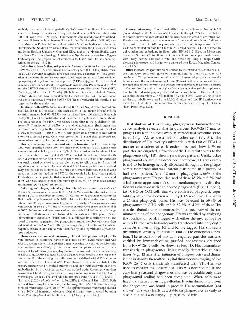
Distribution of Hrs during phagocytosis. Immunofluores-cence analysis revealed that in quiescent RAW264.7 macro-phages Hrs is found exclusively in intracellular vesicular struc-tures (Fig. 1A), as described for other cells (21, 31). Thedistribution of Hrs overlaps substantially with that of EEA1, amarker of a subset of early endosomes (not shown). Whenparticles are engulfed, a fraction of the Hrs redistributes to thephagosome (Fig. 1B), showing a unique pattern. Unlike otherphagosomal constituents described heretofore, Hrs was rarelyfound to be homogeneously dispersed throughout the phago-somal membrane and was instead distributed in a patchy orhalf-moon pattern. After 12 min of phagocytosis, 66% of thephagosomes were Hrs positive, and of these 45.7% � 5.7% hada half-moon appearance. A similar nonhomogeneous distribu-tion was observed with engineered phagocytes (Fig. 1E and I),i.e., CHO or COS cells that were conferred phagocytic capa-bility by stable transfection with Fc�RIIA receptors (13). Aftera 25-min phagocytic pulse, Hrs was detected in 69.8% ofphagosomes in CHO cells and in 52.8% � 4.2% of these Hrswas distributed nonhomogeneously. The specificity of the im-munostaining of the endogenous Hrs was verified by analyzingthe localization of Hrs tagged with either the myc epitope orwith YFP that was heterologously transfected into phagocyticcells. As shown in Fig. 1G and K, the tagged Hrs showed adistribution virtually identical to that of the endogenous pro-tein. The association of Hrs with engulfed particles was alsoverified by immunoblotting purified phagosomes obtainedfrom RAW 264.7 cells. As shown in Fig. 1D, Hrs accumulatestransiently in phagosomes, being readily detectable at earlytimes (e.g., 12 min after initiation of phagocytosis) and dimin-ishing in density thereafter. Digital fluorescence imaging of liveRAW 264.7 cells transiently transfected with YFP-Hrs wasused to confirm this observation. Hrs was never found in thecups lining nascent phagosomes and was detectable only afterphagosomal sealing had been completed. When cells werefixed and stained by using phalloidin, F-actin dissociation fromthe phagosome was found to precede Hrs accumulation (notshown). Hrs was clearly detectable on phagosomes as early as5 to 8 min and was largely depleted by 18 min.
4594 VIEIRA ET AL. MOL. CELL. BIOL.
at Univ of T
oronto on July 4, 2007 m
cb.asm.org
Dow
nloaded from
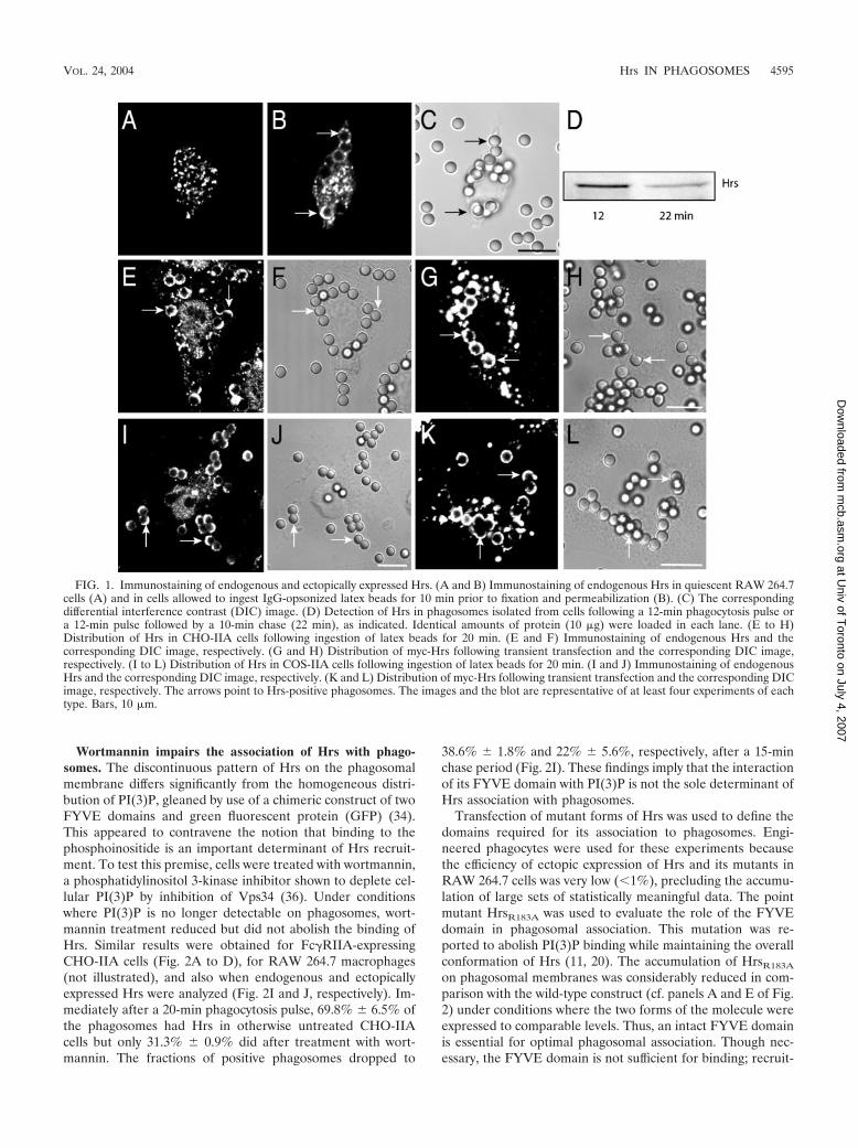
Wortmannin impairs the association of Hrs with phago-somes. The discontinuous pattern of Hrs on the phagosomalmembrane differs significantly from the homogeneous distri-bution of PI(3)P, gleaned by use of a chimeric construct of twoFYVE domains and green fluorescent protein (GFP) (34).This appeared to contravene the notion that binding to thephosphoinositide is an important determinant of Hrs recruit-ment. To test this premise, cells were treated with wortmannin,a phosphatidylinositol 3-kinase inhibitor shown to deplete cel-lular PI(3)P by inhibition of Vps34 (36). Under conditionswhere PI(3)P is no longer detectable on phagosomes, wort-mannin treatment reduced but did not abolish the binding ofHrs. Similar results were obtained for Fc�RIIA-expressingCHO-IIA cells (Fig. 2A to D), for RAW 264.7 macrophages(not illustrated), and also when endogenous and ectopicallyexpressed Hrs were analyzed (Fig. 2I and J, respectively). Im-mediately after a 20-min phagocytosis pulse, 69.8% � 6.5% ofthe phagosomes had Hrs in otherwise untreated CHO-IIAcells but only 31.3% � 0.9% did after treatment with wort-mannin. The fractions of positive phagosomes dropped to
38.6% � 1.8% and 22% � 5.6%, respectively, after a 15-minchase period (Fig. 2I). These findings imply that the interactionof its FYVE domain with PI(3)P is not the sole determinant ofHrs association with phagosomes.
Transfection of mutant forms of Hrs was used to define thedomains required for its association to phagosomes. Engi-neered phagocytes were used for these experiments becausethe efficiency of ectopic expression of Hrs and its mutants inRAW 264.7 cells was very low (�1%), precluding the accumu-lation of large sets of statistically meaningful data. The pointmutant HrsR183A was used to evaluate the role of the FYVEdomain in phagosomal association. This mutation was re-ported to abolish PI(3)P binding while maintaining the overallconformation of Hrs (11, 20). The accumulation of HrsR183A
on phagosomal membranes was considerably reduced in com-parison with the wild-type construct (cf. panels A and E of Fig.2) under conditions where the two forms of the molecule wereexpressed to comparable levels. Thus, an intact FYVE domainis essential for optimal phagosomal association. Though nec-essary, the FYVE domain is not sufficient for binding; recruit-
FIG. 1. Immunostaining of endogenous and ectopically expressed Hrs. (A and B) Immunostaining of endogenous Hrs in quiescent RAW 264.7cells (A) and in cells allowed to ingest IgG-opsonized latex beads for 10 min prior to fixation and permeabilization (B). (C) The correspondingdifferential interference contrast (DIC) image. (D) Detection of Hrs in phagosomes isolated from cells following a 12-min phagocytosis pulse ora 12-min pulse followed by a 10-min chase (22 min), as indicated. Identical amounts of protein (10 �g) were loaded in each lane. (E to H)Distribution of Hrs in CHO-IIA cells following ingestion of latex beads for 20 min. (E and F) Immunostaining of endogenous Hrs and thecorresponding DIC image, respectively. (G and H) Distribution of myc-Hrs following transient transfection and the corresponding DIC image,respectively. (I to L) Distribution of Hrs in COS-IIA cells following ingestion of latex beads for 20 min. (I and J) Immunostaining of endogenousHrs and the corresponding DIC image, respectively. (K and L) Distribution of myc-Hrs following transient transfection and the corresponding DICimage, respectively. The arrows point to Hrs-positive phagosomes. The images and the blot are representative of at least four experiments of eachtype. Bars, 10 �m.
VOL. 24, 2004 Hrs IN PHAGOSOMES 4595
at Univ of T
oronto on July 4, 2007 m
cb.asm.org
Dow
nloaded from
ment of the isolated FYVE domain of Hrs to the phagosomewas negligible (not shown). By contrast, the HrsFYVE-CC con-struct, containing both the coiled-coil (CC) and FYVE do-mains of Hrs, was targeted efficiently to phagosomes (Fig. 2G).Binding is not due solely to the CC domain, since exposure ofthe cells to wortmannin virtually eliminated the interaction(Fig. 2K). It is noteworthy that the extent of the displacementof HrsFYVE-CC from the phagosome induced by wortmanninwas much greater than that noted for full-length Hrs. Weinterpret these findings to mean that yet another domain of theprotein, distinct from the CC and FYVE domains, is involved
in tethering Hrs to phagosomes. The contribution of this thirddomain is most apparent following inhibition of Vps34.
Hrs possesses a region capable of interacting with ubiquity-lated proteins, namely the UIM motif. To test whether thismotif contributes to the association of Hrs with the phagoso-mal membrane, we measured the interaction of UIM-deficientHrs (Hrs�UIM) with phagosomes. Typical results are illustratedin Fig. 2H. UIM-deficient Hrs bound to phagosomes consid-erably less efficiently than did its wild-type counterpart. Den-sitometric analysis of the digital images indicated that omissionof the UIM depressed binding by �75%. Together, these data
FIG. 2. Effects of wortmannin on Hrs recruitment to the phagosomes and localization of Hrs mutants. CHO-IIA were allowed to internalize3-�m-diameter latex beads for 20 min and then fixed and subjected to immunostaining. Where indicated, the cells were pretreated with 100 nMwortmannin or were transfected with epitope-tagged Hrs mutants. (A to D) Distribution of wild-type Hrs in control (A and B) and wortmannin-treated (C and D) cells. (E and F) Distribution of HrsR183A. (G) Distribution of HrsFYVE�CC. (H) Distribution of Hrs�UIM. The images in panelsA, C, E, G, and H are representative confocal fluorescence images. The images in panels B, D, and F are the differential interference contrastimages corresponding to those in panels A, C, and E, respectively. Transfected cells are indicated by asterisks. The solid arrows point toHrs-positive phagosomes, while open arrows indicate Hrs-deficient phagosomes. Bars, 10 �m. Images are representative of at least fourexperiments of each type. (I to K) Quantification of the effect of wortmannin on the phagosomal acquisition of wild-type or mutant Hrs. The cellswere allowed to internalize opsonized beads for 20 min (chase time, 0 min) and then chased for the times indicated in the graph. Shown areendogenous Hrs (I), transfected wild-type Hrs (myc-tagged) (J), and HrsFYVE�CC (K). Empty columns, control cells; black columns, wortmannin-treated cells. Data are means � standard errors of three separate experiments (200 cells were counted in each).
4596 VIEIRA ET AL. MOL. CELL. BIOL.
at Univ of T
oronto on July 4, 2007 m
cb.asm.org
Dow
nloaded from

indicate that at least three regions of Hrs, namely the FYVE,UIM, and CC domains, are required for optimal binding ofHrs to phagosomes.
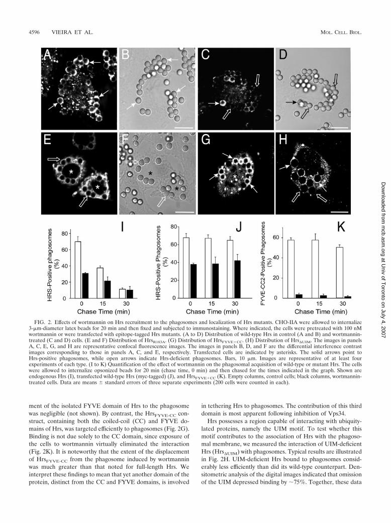
Overexpression of Hrs alters phagosomal maturation. Wenext proceeded to evaluate the possible role of Hrs in phago-somal maturation. To this end, we initially planned to use adominant-negative strategy by transfection of mutant forms ofHrs. However, during the course of control experiments, itbecame obvious that even the overexpression of wild-type Hrsaltered the kinetics of phagosomal maturation. This is appar-ent when comparing the rate of dissociation of endogenous
and ectopically expressed Hrs. (Fig. 2I and J). While wortman-nin inhibits association to a similar extent in the two cases, Hrsdissociates much more slowly when overexpressed. Although asystematic quantitation was not performed, it appeared as ifthe delay in dissociation was proportional to the level of over-expression (not shown). The reduced dissociation of Hrs wasaccompanied by arrested maturation, as indicated by the in-ability of the phagosomes to acquire LAMP-1, a marker of lateendocytic compartments (Fig. 3A to C).
We speculated that overexpression of Hrs may exert thisinhibitory effect by scavenging an inordinately large fraction of
FIG. 3. Effects of Hrs overexpression on LAMP-1 acquisition by phagosomes. CHO-IIA transfected with myc-tagged wild-type Hrs (A to C)or HrsR183A (D to F) were allowed to interact with opsonized latex beads for 20 min, and after unbound beads were washed, the phagosomes wereallowed to mature for 60 min. The cells were then fixed and immunostained with myc (A and D) and LAMP-1 (B and E) antibodies. C and F arethe corresponding DIC images. Transfected cells are identified by asterisks. The solid and open arrows point to LAMP-1-positive and -negativephagosomes, respectively. Images are representative of four experiments of each type. Bars, 10 �m. (G) Quantification of the effects of variousconstructs on LAMP-1 acquisition by phagosomes. CHO-IIA cells either were left untransfected (empty column) or were transfected with wild-typeHrs, HrsR183A, HrsFYVE�CC, the PX domain of p40phox or two tandem FYVE domains of EEA1, as specified. LAMP-1 acquisition by phagosomeswas determined as above. Data are means � standard errors of three experiments (200 cells counted in each).
VOL. 24, 2004 Hrs IN PHAGOSOMES 4597
at Univ of T
oronto on July 4, 2007 m
cb.asm.org
Dow
nloaded from
PI(3)P. To test this hypothesis, cells were transfected withcomparable amounts of HrsR183A, which lacks the ability tobind the inositide. As illustrated in Fig. 3D to F, expression oflarge amounts of HrsR183A had no discernible effect on matu-ration, supporting the notion that scavenging of PI(3)P is re-sponsible for the effects of excess Hrs. Accordingly, we foundthat overexpression of the HrsFYVE-CC construct and of twoother constructs capable of binding PI(3)P (the PX domain ofp40phox and a tandem 2-FYVE domain of EEA1) had sizableinhibitory effects (Fig. 3G).
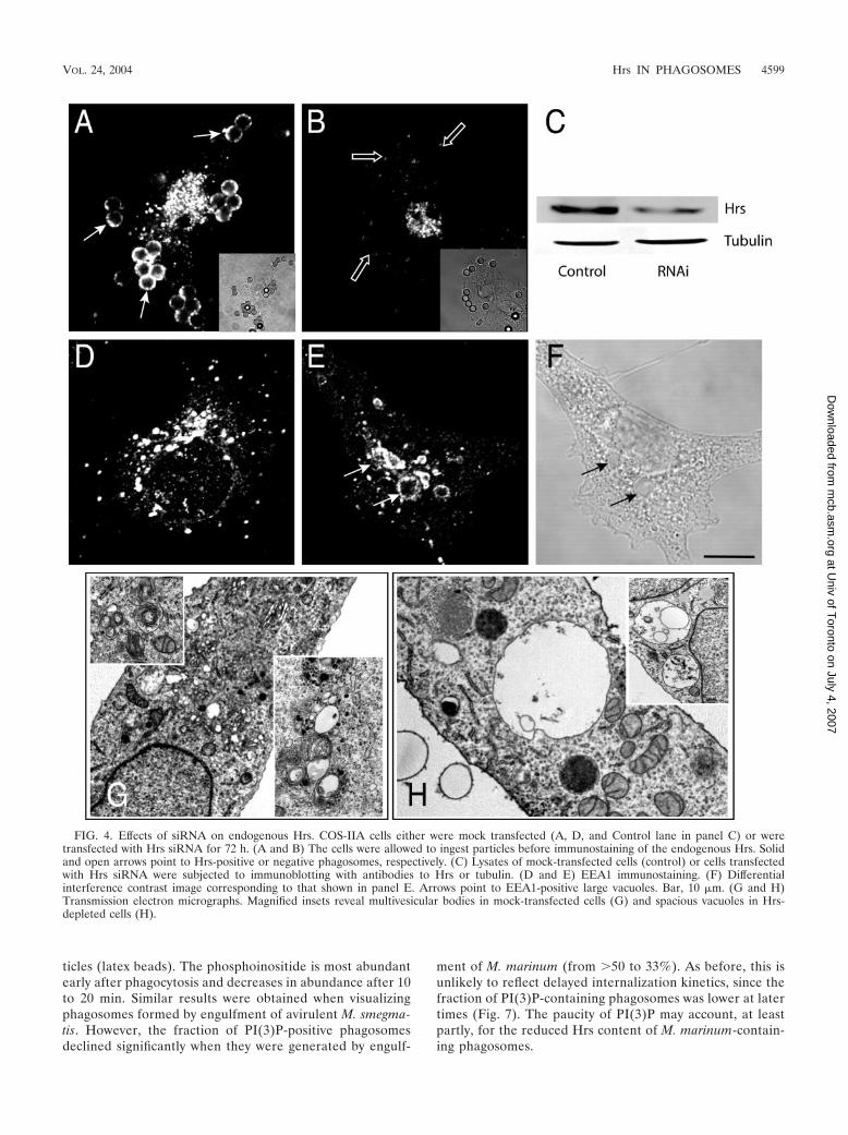
Effect of depletion of endogenous Hrs. While the aboveresults confirm the importance of PI(3)P availability for com-pletion of phagosomal maturation, they also preclude the useof dominant-negative constructs to analyze the putative role ofHrs in the process. As an alternative, we attempted to depletethe endogenous Hrs by using siRNA. We initially confirmedthat synthesis of the protein was arrested by a 72-h treatmentwith the siRNA. As shown in Fig. 4A and B, Hrs was exten-sively depleted in phagocytic COS-IIA cells, as observed byimmunostaining. Unlike the untreated cells, which were allimmunoreactive, approximately 20 to 30% of the cells stainedonly marginally with the antibody to Hrs. This estimate wasconfirmed by immunoblots of whole-cell extracts, whichshowed a 70% decrease in the total Hrs content of the popu-lation (mean from three experiments). The loss was specific,since the levels of tubulin were unaffected (Fig. 4C) and thecells retained their ability to perform phagocytosis at normalrates. The siRNA was ineffective towards RAW 264.7 cells,likely because of their refractoriness to transfection. All sub-sequent experiments were therefore performed with thereadily transfectable COS-IIA cells.
The cells treated with Hrs siRNA exhibited large vacuolesthat were frequently EEA1 positive (Fig. 4E and F). Ultra-structural analysis indicated that the vacuoles were oftenempty or contained infrequent smaller vacuoles within theirlumen but rarely small vesicles. Overall, the number of densemultivesicular structures was smaller in siRNA-treated cellsthan in controls (cf. Fig. 4G and H). This phenotype resemblesthat reported for murine embryonic cells lacking Hrs (16),suggesting that siRNA-treated cells are suitable models tostudy the consequences of selective ablation of Hrs.
The effects of Hrs depletion on phagosomal maturationwere studied next. In cells treated with Hrs siRNA, phagocy-tosis proceeded normally and at early times acquisition ofEEA1 by the phagosomes was indistinguishable from that ofcontrols (76% � 7% and 77% � 6%, respectively, after 20min; Fig. 5A to D and Q). However, EEA1 dissociation fromphagosomes was impaired in Hrs-depleted cells. EEA1 wasclearly enriched in 95.9% � 4.2% of the phagosomes of Hrs-deficient cells even after 3 h, a time when control phagosomesare entirely devoid of EEA1 (Fig. 5E to H). This suggested thatmaturation was arrested at the early phagosomal stage in Hrs-depleted cells. This notion was confirmed by analyzing thepresence of LBPA and the extent of acidification of phago-somes of control and depleted cells. While the vast majority(�80%) of the phagosomes acquire LBPA after 90 min incontrol cells, only 14% � 8% of the phagosomes contained thelysolipid in Hrs-depleted cells (Fig. 5I to L and Q). Moreover,LysoTracker, a fluorescent acidotropic probe accumulated dis-tinctly in phagosomes of normal cells, but poorly, if at all, in
phagosomes of Hrs-deficient cells (Fig. 5M to P). Nearly 90%of normal phagosomes accumulated LysoTracker, but this fig-ure dropped to only 18% � 8% following elimination of Hrs(Fig. 5Q). This implies that the pH of Hrs-depleted phago-somes is higher than that of normal phagosomes. Failure toacidify was not due to an effect of Hrs ablation on the vacuolarATPases responsible for endomembrane acidification, sincemultiple acidic vesicles and vacuoles were observable in thedepleted cells (Fig. 5O). Instead, the impaired acidificationlikely reflects the inability of ATPase-enriched vesicles tomerge with the phagosomes. Jointly, these results suggest thatHrs is essential for the progression of early phagosomes tophagolysosomes.
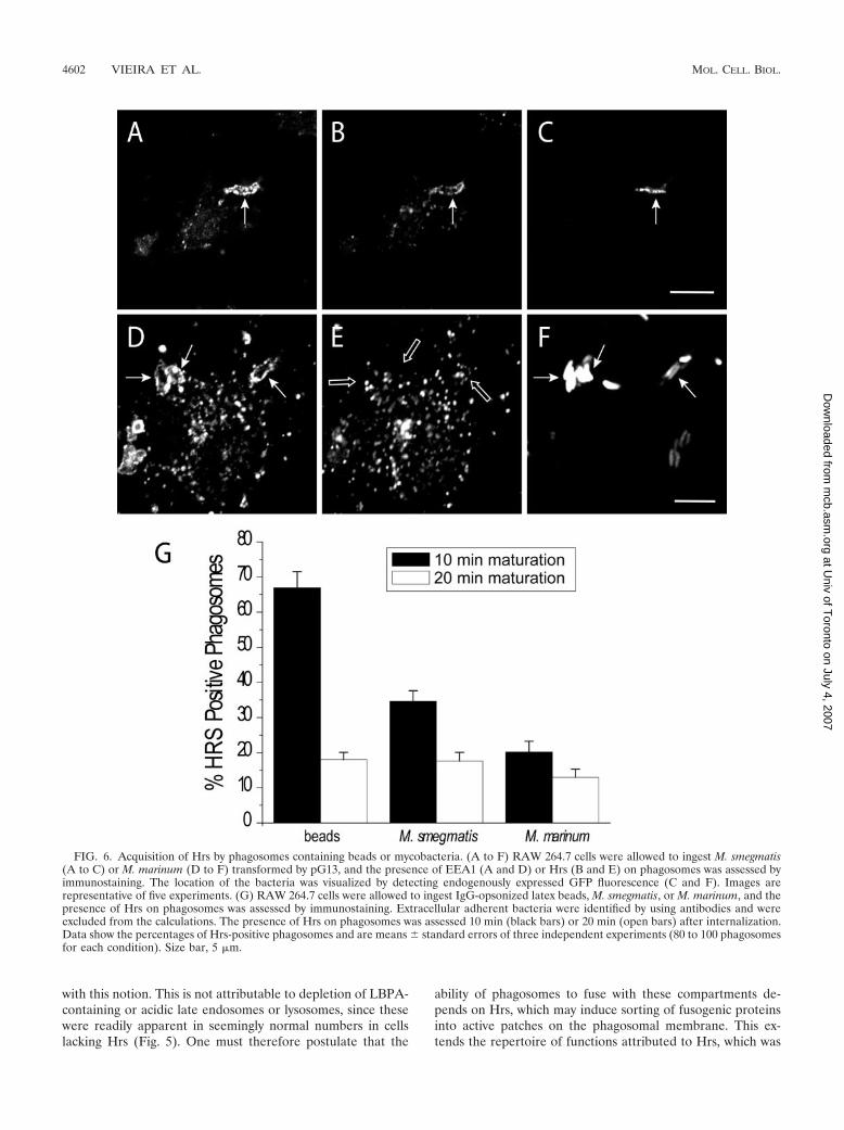
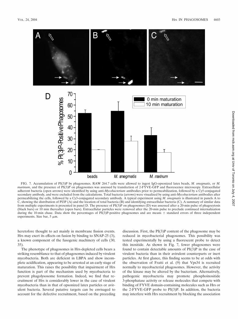
Hrs acquisition by mycobacterium-containing phagosomesis impaired. The stage at which maturation is arrested bydepletion of Hrs is similar to that noted earlier for phagosomescontaining virulent mycobacteria. It is therefore conceivablethat the failure of mycobacterium-containing vacuoles toprogress to phagolysosomes is related to impaired Hrs recruit-ment or function. This hypothesis was tested by comparing theHrs content of phagosomes formed by cells challenged witheither mycobacteria of different degrees of virulence or inertparticles. RAW 264.7 macrophages were exposed either to M.marinum, a pathogen of fish and frogs that is phylogeneticallyrelated to M. tuberculosis (32) and affects maturation in asimilar manner (2), or to the usually avirulent M. smegmatis.Latex beads opsonized with IgG were used as an inert control.Internalization was allowed to occur for various periods oftime, and after fixation and permeabilization, the presence ofHrs in phagosomes was assessed by immunofluorescence. Carewas taken to exclude extracellular, adherent bacteria or beadsfrom the quantitation (see Materials and Methods). As shownin Fig. 6, up to 66.1% of the beads acquired Hrs after 10 minand the fraction of Hrs-positive particles decreased thereafter.As was found for latex beads, the distribution of Hrs on phago-somes containing M. smegmatis was nonhomogeneous (Fig.6B). By contrast, only 20% of the phagosomes containing M.marinum were found to contain Hrs 10 min after internaliza-tion. The slower kinetics of phagocytosis of the bacteria cannotaccount for this difference, because the fraction of Hrs-containing phagosomes decreased further with longer times.When the period of exposure to the bacteria was shortened toless than 10 min, the efficiency of phagocytosis was reduced tothe point where statistical evaluation was compromised. Nev-ertheless, it was qualitatively apparent that the fraction ofphagosomes with Hrs was not higher at earlier times. A signif-icantly higher fraction of phagosomes formed by ingestion ofthe avirulent M. smegmatis had clearly detectable Hrs (Fig. 6).These findings demonstrate a positive correlation between theHrs content of phagosomes and their ability to complete mat-uration.
Because the FYVE domain was shown, as reported above,to be important for Hrs recruitment to phagosomes, defec-tive acquisition of its ligand, PI(3)P, may account for thereduced recruitment of Hrs. This hypothesis was tested bydirectly measuring the accumulation of PI(3)P on phago-somes by use of two tandem FYVE domains fused to GFP,as described earlier (34). The results of these experimentsare shown in Fig. 7. As reported earlier, PI(3)P is readilydetected in phagosomes formed upon ingestion of inert par-
4598 VIEIRA ET AL. MOL. CELL. BIOL.
at Univ of T
oronto on July 4, 2007 m
cb.asm.org
Dow
nloaded from
ticles (latex beads). The phosphoinositide is most abundantearly after phagocytosis and decreases in abundance after 10to 20 min. Similar results were obtained when visualizingphagosomes formed by engulfment of avirulent M. smegma-tis. However, the fraction of PI(3)P-positive phagosomesdeclined significantly when they were generated by engulf-
ment of M. marinum (from 50 to 33%). As before, this isunlikely to reflect delayed internalization kinetics, since thefraction of PI(3)P-containing phagosomes was lower at latertimes (Fig. 7). The paucity of PI(3)P may account, at leastpartly, for the reduced Hrs content of M. marinum-contain-ing phagosomes.
FIG. 4. Effects of siRNA on endogenous Hrs. COS-IIA cells either were mock transfected (A, D, and Control lane in panel C) or weretransfected with Hrs siRNA for 72 h. (A and B) The cells were allowed to ingest particles before immunostaining of the endogenous Hrs. Solidand open arrows point to Hrs-positive or negative phagosomes, respectively. (C) Lysates of mock-transfected cells (control) or cells transfectedwith Hrs siRNA were subjected to immunoblotting with antibodies to Hrs or tubulin. (D and E) EEA1 immunostaining. (F) Differentialinterference contrast image corresponding to that shown in panel E. Arrows point to EEA1-positive large vacuoles. Bar, 10 �m. (G and H)Transmission electron micrographs. Magnified insets reveal multivesicular bodies in mock-transfected cells (G) and spacious vacuoles in Hrs-depleted cells (H).
VOL. 24, 2004 Hrs IN PHAGOSOMES 4599
at Univ of T
oronto on July 4, 2007 m
cb.asm.org
Dow
nloaded from
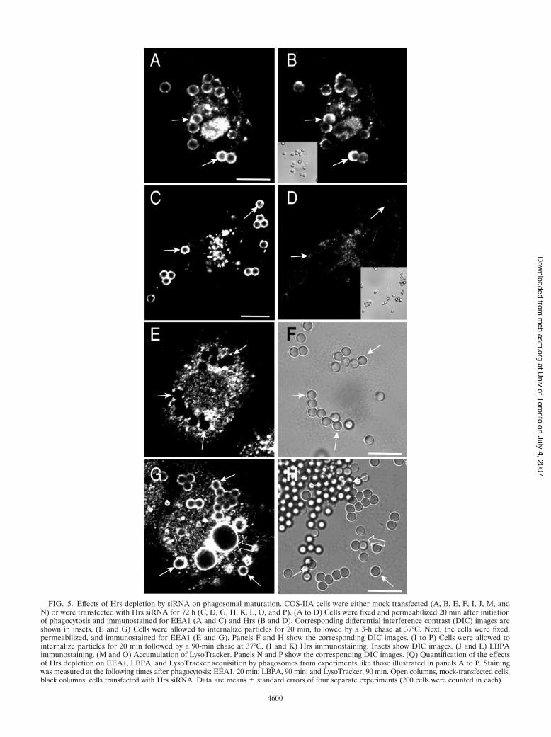
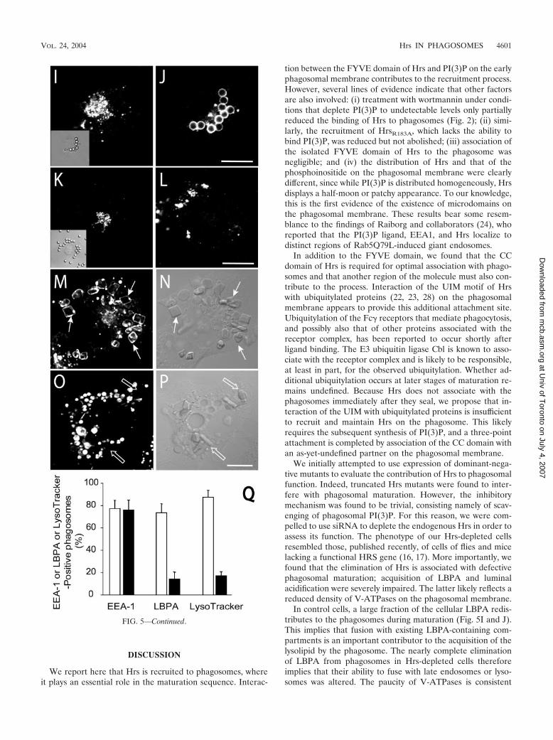
FIG. 5. Effects of Hrs depletion by siRNA on phagosomal maturation. COS-IIA cells were either mock transfected (A, B, E, F, I, J, M, andN) or were transfected with Hrs siRNA for 72 h (C, D, G, H, K, L, O, and P). (A to D) Cells were fixed and permeabilized 20 min after initiationof phagocytosis and immunostained for EEA1 (A and C) and Hrs (B and D). Corresponding differential interference contrast (DIC) images areshown in insets. (E and G) Cells were allowed to internalize particles for 20 min, followed by a 3-h chase at 37°C. Next, the cells were fixed,permeabilized, and immunostained for EEA1 (E and G). Panels F and H show the corresponding DIC images. (I to P) Cells were allowed tointernalize particles for 20 min followed by a 90-min chase at 37°C. (I and K) Hrs immunostaining. Insets show DIC images. (J and L) LBPAimmunostaining. (M and O) Accumulation of LysoTracker. Panels N and P show the corresponding DIC images. (Q) Quantification of the effectsof Hrs depletion on EEA1, LBPA, and LysoTracker acquisition by phagosomes from experiments like those illustrated in panels A to P. Stainingwas measured at the following times after phagocytosis: EEA1, 20 min; LBPA, 90 min; and LysoTracker, 90 min. Open columns, mock-transfected cells;black columns, cells transfected with Hrs siRNA. Data are means � standard errors of four separate experiments (200 cells were counted in each).
4600
at Univ of T
oronto on July 4, 2007 m
cb.asm.org
Dow
nloaded from
DISCUSSION
We report here that Hrs is recruited to phagosomes, whereit plays an essential role in the maturation sequence. Interac-
tion between the FYVE domain of Hrs and PI(3)P on the earlyphagosomal membrane contributes to the recruitment process.However, several lines of evidence indicate that other factorsare also involved: (i) treatment with wortmannin under condi-tions that deplete PI(3)P to undetectable levels only partiallyreduced the binding of Hrs to phagosomes (Fig. 2); (ii) simi-larly, the recruitment of HrsR183A, which lacks the ability tobind PI(3)P, was reduced but not abolished; (iii) association ofthe isolated FYVE domain of Hrs to the phagosome wasnegligible; and (iv) the distribution of Hrs and that of thephosphoinositide on the phagosomal membrane were clearlydifferent, since while PI(3)P is distributed homogeneously, Hrsdisplays a half-moon or patchy appearance. To our knowledge,this is the first evidence of the existence of microdomains onthe phagosomal membrane. These results bear some resem-blance to the findings of Raiborg and collaborators (24), whoreported that the PI(3)P ligand, EEA1, and Hrs localize todistinct regions of Rab5Q79L-induced giant endosomes.
In addition to the FYVE domain, we found that the CCdomain of Hrs is required for optimal association with phago-somes and that another region of the molecule must also con-tribute to the process. Interaction of the UIM motif of Hrswith ubiquitylated proteins (22, 23, 28) on the phagosomalmembrane appears to provide this additional attachment site.Ubiquitylation of the Fc� receptors that mediate phagocytosis,and possibly also that of other proteins associated with thereceptor complex, has been reported to occur shortly afterligand binding. The E3 ubiquitin ligase Cbl is known to asso-ciate with the receptor complex and is likely to be responsible,at least in part, for the observed ubiquitylation. Whether ad-ditional ubiquitylation occurs at later stages of maturation re-mains undefined. Because Hrs does not associate with thephagosomes immediately after they seal, we propose that in-teraction of the UIM with ubiquitylated proteins is insufficientto recruit and maintain Hrs on the phagosome. This likelyrequires the subsequent synthesis of PI(3)P, and a three-pointattachment is completed by association of the CC domain withan as-yet-undefined partner on the phagosomal membrane.
We initially attempted to use expression of dominant-nega-tive mutants to evaluate the contribution of Hrs to phagosomalfunction. Indeed, truncated Hrs mutants were found to inter-fere with phagosomal maturation. However, the inhibitorymechanism was found to be trivial, consisting namely of scav-enging of phagosomal PI(3)P. For this reason, we were com-pelled to use siRNA to deplete the endogenous Hrs in order toassess its function. The phenotype of our Hrs-depleted cellsresembled those, published recently, of cells of flies and micelacking a functional HRS gene (16, 17). More importantly, wefound that the elimination of Hrs is associated with defectivephagosomal maturation; acquisition of LBPA and luminalacidification were severely impaired. The latter likely reflects areduced density of V-ATPases on the phagosomal membrane.
In control cells, a large fraction of the cellular LBPA redis-tributes to the phagosomes during maturation (Fig. 5I and J).This implies that fusion with existing LBPA-containing com-partments is an important contributor to the acquisition of thelysolipid by the phagosome. The nearly complete eliminationof LBPA from phagosomes in Hrs-depleted cells thereforeimplies that their ability to fuse with late endosomes or lyso-somes was altered. The paucity of V-ATPases is consistent
FIG. 5—Continued.
VOL. 24, 2004 Hrs IN PHAGOSOMES 4601
at Univ of T
oronto on July 4, 2007 m
cb.asm.org
Dow
nloaded from
with this notion. This is not attributable to depletion of LBPA-containing or acidic late endosomes or lysosomes, since thesewere readily apparent in seemingly normal numbers in cellslacking Hrs (Fig. 5). One must therefore postulate that the
ability of phagosomes to fuse with these compartments de-pends on Hrs, which may induce sorting of fusogenic proteinsinto active patches on the phagosomal membrane. This ex-tends the repertoire of functions attributed to Hrs, which was
FIG. 6. Acquisition of Hrs by phagosomes containing beads or mycobacteria. (A to F) RAW 264.7 cells were allowed to ingest M. smegmatis(A to C) or M. marinum (D to F) transformed by pG13, and the presence of EEA1 (A and D) or Hrs (B and E) on phagosomes was assessed byimmunostaining. The location of the bacteria was visualized by detecting endogenously expressed GFP fluorescence (C and F). Images arerepresentative of five experiments. (G) RAW 264.7 cells were allowed to ingest IgG-opsonized latex beads, M. smegmatis, or M. marinum, and thepresence of Hrs on phagosomes was assessed by immunostaining. Extracellular adherent bacteria were identified by using antibodies and wereexcluded from the calculations. The presence of Hrs on phagosomes was assessed 10 min (black bars) or 20 min (open bars) after internalization.Data show the percentages of Hrs-positive phagosomes and are means � standard errors of three independent experiments (80 to 100 phagosomesfor each condition). Size bar, 5 �m.
4602 VIEIRA ET AL. MOL. CELL. BIOL.
at Univ of T
oronto on July 4, 2007 m
cb.asm.org
Dow
nloaded from
heretofore thought to act mainly in membrane fission events.Hrs may exert its effects on fusion by binding to SNAP-25 (3),a known component of the fusogenic machinery of cells (30,35).
The phenotype of phagosomes in Hrs-depleted cells bears astriking resemblance to that of phagosomes induced by virulentmycobacteria. Both are deficient in LBPA and show incom-plete acidification, appearing to be arrested at an early stage ofmaturation. This raises the possibility that impairment of Hrsfunction is part of the mechanism used by mycobacteria toprevent phagolysosome formation. Indeed, we find that re-cruitment of Hrs is considerably lower in the case of virulentmycobacteria than in that of opsonized latex particles or avir-ulent bacteria. Several putative targets can be envisaged toaccount for the defective recruitment, based on the preceding
discussion. First, the PI(3)P content of the phagosome may bereduced in mycobacterial phagosomes. This possibility wastested experimentally by using a fluorescent probe to detectthis inositide. As shown in Fig. 7, fewer phagosomes werefound to contain detectable amounts of PI(3)P in the case ofvirulent bacteria than in their avirulent counterparts or inertparticles. At first glance, this finding seems to be at odds withthe observation of Fratti et al. (9) that Vps34 is recruitednormally to mycobacterial phagosomes. However, the activityof the kinase may be altered by the bacterium. Alternatively,pathogenic mycobacteria may promote phosphoinositide3-phosphatase activity or release molecules that compete withbinding of FYVE domain-containing molecules such as Hrs orthe 2-FYVE-GFP probe to PI(3)P. In addition, the bacteriamay interfere with Hrs recruitment by blocking the association
FIG. 7. Accumulation of PI(3)P by phagosomes. RAW 264.7 cells were allowed to ingest IgG-opsonized latex beads, M. smegmatis, or M.marinum, and the presence of PI(3)P on phagosomes was assessed by transfection of 2-FYVE-GFP and fluorescence microscopy. Extracellularadherent bacteria (open arrows) were identified by using anti-Mycobacterium antibodies prior to permeabilization, followed by a Cy5-conjugatedsecondary antibody, and were excluded from the calculations. Total bacteria (arrows) were visualized by using anti-Mycobacterium antibodies afterpermeabilizing the cells, followed by a Cy3-conjugated secondary antibody. A typical experiment using M. smegmatis is illustrated in panels A toC, showing the distribution of PI3P (A) and the location of total bacteria (B) and identifying extracellular bacteria (C). A summary of similar datafrom multiple experiments is presented in panel D. The presence of PI(3)P on phagosomes (D) was assessed after a 20-min pulse of phagocytosis(black bars) or 10 min thereafter (open bars). Extracellular particles were removed after the 20-min pulse to preclude continued internalizationduring the 10-min chase. Data show the percentages of PI(3)P-positive phagosomes and are means � standard errors of three independentexperiments. Size bar, 5 �m.
VOL. 24, 2004 Hrs IN PHAGOSOMES 4603
at Univ of T
oronto on July 4, 2007 m
cb.asm.org
Dow
nloaded from
of the CC domain with its target or by reducing the interactionbetween the UIM motif and phagosomal ubiquitylated substrates.
Multiple mechanisms have been put forward to account forthe effects of mycobacteria on phagosome maturation: reducedcalcium responsiveness (18, 19), impairment of EEA1 recruit-ment (9), and enhanced SNARE degradation (10), along withthe defective Hrs recruitment reported here. These are notmutually exclusive and may act jointly to ensure intraphagoso-mal survival of the bacteria. Other intracellular parasites, likeSalmonella and Shigella, are known to target multiple mecha-nisms of the host cell, often using redundant effectors (1, 12).In the case of mycobacteria, Hrs appears to be an attractivetarget, inasmuch as this protein plays an essential role in theearly-to-late phagosome transition, the very step affected bythe parasite.
ACKNOWLEDGMENTS
M. marinum and pG13 were kind gifts to S.G. from Lucia Parker.This work was supported by the Canadian Institutes of Health Re-
search, the Canadian Arthritis Society, and the National SanatoriumAssociation. O. V. Vieira was the recipient of a postdoctoral fellowshipfrom PRAXIS XX1 (BPD/22039/99). C.C.S. is the recipient of a grad-uate studentship from the Canadian Institutes of Health Research.H.S. was supported by the Norwegian Cancer Society and the NovoNordisk Foundation. S.G. is the current holder of the Pitblado Chairin Cell Biology.
REFERENCES
1. Adam, T. 2001. Exploitation of host factors for efficient infection by Shigella.Int. J. Med. Microbiol. 291:287–298.
2. Barker, L. P., K. M. George, S. Falkow, and P. L. Small. 1997. Differentialtrafficking of live and dead Mycobacterium marinum organisms in macro-phages. Infect. Immun. 65:1497–1504.
3. Bean, A. J., R. Seifert, Y. A. Chen, R. Sacks, and R. H. Scheller. 1997. Hrs-2is an ATPase implicated in calcium-regulated secretion. Nature 385:826–829.
4. Bishop, N., A. Horman, and P. Woodman. 2002. Mammalian class E vpsproteins recognize ubiquitin and act in the removal of endosomal protein-ubiquitin conjugates. J. Cell Biol. 157:91–101.
5. Chin, L. S., M. C. Raynor, X. Wei, H. Q. Chen, and L. Li. 2001. Hrs interactswith sorting nexin 1 and regulates degradation of epidermal growth factorreceptor. J. Biol. Chem. 276:7069–7078.
6. Desjardins, M., J. E. Celis, G. van Meer, H. Dieplinger, A. Jahraus, G.Griffiths, and L. A. Huber. 1994. Molecular characterization of phagosomes.J. Biol. Chem. 269:32194–32200.
7. Desjardins, M., L. A. Huber, R. G. Parton, and G. Griffiths. 1994. Biogenesisof phagolysosomes proceeds through a sequential series of interactions withthe endocytic apparatus. J. Cell Biol. 124:677–688.
8. Elbashir, S. M., J. Harborth, W. Lendeckel, A. Yalcin, K. Weber, and T.Tuschl. 2001. Duplexes of 21-nucleotide RNAs mediate RNA interference incultured mammalian cells. Nature 411:494–498.
9. Fratti, R. A., J. M. Backer, J. Gruenberg, S. Corvera, and V. Deretic. 2001. Roleof phosphatidylinositol 3-kinase and Rab5 effectors in phagosomal biogenesisand mycobacterial phagosome maturation arrest. J. Cell Biol. 154:631–644.
10. Fratti, R. A., J. Chua, and V. Deretic. 2002. Cellubrevin alterations andMycobacterium tuberculosis phagosome maturation arrest. J. Biol. Chem.277:17320–17326.
11. Gaullier, J. M., E. Ronning, D. J. Gillooly, and H. Stenmark. 2000. Interactionof the EEA1 FYVE finger with phosphatidylinositol 3-phosphate and earlyendosomes. Role of conserved residues. J. Biol. Chem. 275:24595–24600.
12. Holden, D. W. 2002. Trafficking of the Salmonella vacuole in macrophages.Traffic 3:161–169.
13. Indik, Z. K., J. G. Park, S. Hunter, M. Mantaring, and A. D. Schreiber. 1995.Molecular dissection of Fc gamma receptor-mediated phagocytosis. Immu-nol. Lett. 44:133–138.
14. Kanai, F., H. Liu, S. J. Field, H. Akbary, T. Matsuo, G. E. Brown, L. C.
Cantley, and M. B. Yaffe. 2001. The PX domains of p47phox and p40phoxbind to lipid products of PI(3)K. Nat. Cell Biol. 3:675–678.
15. Kobayashi, T., M. H. Beuchat, M. Lindsay, S. Frias, R. D. Palmiter, H.Sakuraba, R. G. Parton, and J. Gruenberg. 1999. Late endosomal mem-branes rich in lysobisphosphatidic acid regulate cholesterol transport. Nat.Cell Biol. 1:113–118.
16. Komada, M., and P. Soriano. 1999. Hrs, a FYVE finger protein localized toearly endosomes, is implicated in vesicular traffic and required for ventralfolding morphogenesis. Genes Dev. 13:1475–1485.
17. Lloyd, T. E., R. Atkinson, M. N. Wu, Y. Zhou, G. Pennetta, and H. J. Bellen.2002. Hrs regulates endosome membrane invagination and tyrosine kinasereceptor signaling in Drosophila. Cell 108:261–269.
18. Malik, Z. A., G. M. Denning, and D. J. Kusner. 2000. Inhibition of Ca(2�)signaling by Mycobacterium tuberculosis is associated with reduced phago-some-lysosome fusion and increased survival within human macrophages. J.Exp. Med. 191:287–302.
19. Malik, Z. A., C. R. Thompson, S. Hashimi, B. Porter, S. S. Iyer, and D. J.Kusner. 2003. Cutting edge: Mycobacterium tuberculosis blocks Ca(2�)signaling and phagosome maturation in human macrophages via specificinhibition of sphingosine kinase. J. Immunol. 170:2811–2815.
20. Misra, S., and J. H. Hurley. 1999. Crystal structure of a phosphatidylinositol3-phosphate-specific membrane-targeting motif, the FYVE domain ofVps27p. Cell 97:657–666.
21. Mu, F. T., J. M. Callaghan, O. Steele-Mortimer, H. Stenmark, R. G. Parton,P. L. Campbell, J. McCluskey, J. P. Yeo, E. P. Tock, and B. H. Toh. 1995.EEA1, an early endosome-associated protein. EEA1 is a conserved alpha-helical peripheral membrane protein flanked by cysteine “fingers” and con-tains a calmodulin-binding IQ motif. J. Biol. Chem. 270:13503–13511.
22. Polo, S., S. Sigismund, M. Faretta, M. Guidi, M. R. Capua, G. Bossi, H. Chen,P. De Camilli, and P. P. Di Fiore. 2002. A single motif responsible for ubiquitinrecognition and monoubiquitination in endocytic proteins. Nature 416:451–455.
23. Raiborg, C., K. G. Bache, D. J. Gillooly, I. H. Madshus, E. Stang, and H.Stenmark. 2002. Hrs sorts ubiquitinated proteins into clathrin-coated mi-crodomains of early endosomes. Nat. Cell Biol. 4:394–398.
24. Raiborg, C., K. G. Bache, A. Mehlum, E. Stang, and H. Stenmark. 2001. Hrsrecruits clathrin to early endosomes. EMBO J. 20:5008–5021.
25. Raiborg, C., K. G. Bache, A. Mehlum, and H. Stenmark. 2001. Function ofHrs in endocytic trafficking and signalling. Biochem. Soc. Trans. 29:472–475.
26. Raiborg, C., B. Bremnes, A. Mehlum, D. J. Gillooly, A. D’Arrigo, E. Stang,and H. Stenmark. 2001. FYVE and coiled-coil domains determine the spe-cific localisation of Hrs to early endosomes. J. Cell Sci. 114:2255–2263.
27. Russell, D. G. 2001. Mycobacterium tuberculosis: here today, and heretomorrow. Nat. Rev. Mol. Cell Biol. 2:569–577.
28. Shih, S. C., D. J. Katzmann, J. D. Schnell, M. Sutanto, S. D. Emr, and L.Hicke. 2002. Epsins and Vps27p/Hrs contain ubiquitin-binding domains thatfunction in receptor endocytosis. Nat. Cell Biol. 4:389–393.
29. Simonsen, A., R. Lippe, S. Christoforidis, J. M. Gaullier, A. Brech, J. Callaghan,B. H. Toh, C. Murphy, M. Zerial, and H. Stenmark. 1998. EEA1 links PI(3)Kfunction to Rab5 regulation of endosome fusion. Nature 394:494–498.
30. Sollner, T., S. W. Whiteheart, M. Brunner, H. Erdjument-Bromage, S. Gero-manos, P. Tempst, and J. E. Rothman. 1993. SNAP receptors implicated invesicle targeting and fusion. Nature 362:318–324.
31. Stenmark, H., R. Aasland, B. H. Toh, and A. D’Arrigo. 1996. Endosomallocalization of the autoantigen EEA1 is mediated by a zinc-binding FYVEfinger. J. Biol. Chem. 271:24048–24054.
32. Tonjum, T., D. B. Welty, E. Jantzen, and P. L. Small. 1998. Differentiation ofMycobacterium ulcerans, M. marinum, and M. haemophilum: mapping of theirrelationships to M. tuberculosis by fatty acid profile analysis, DNA-DNA hybrid-ization, and 16S rRNA gene sequence analysis. J. Clin. Microbiol. 36:918–925.
33. Underhill, D. M., and A. Ozinsky. 2002. Phagocytosis of microbes: complex-ity in action. Annu. Rev. Immunol. 20:825–852.
34. Vieira, O. V., R. J. Botelho, L. Rameh, S. M. Brachmann, T. Matsuo, H. W.Davidson, A. Schreiber, J. M. Backer, L. C. Cantley, and S. Grinstein. 2001.Distinct roles of class I and class III phosphatidylinositol 3-kinases in phago-some formation and maturation. J. Cell Biol. 155:19–25.
35. Weber, T., B. V. Zemelman, J. A. McNew, B. Westermann, M. Gmachl, F.Parlati, T. H. Sollner, and J. E. Rothman. 1998. SNAREpins: minimalmachinery for membrane fusion. Cell 92:759–772.
36. Yano, H., S. Nakanishi, K. Kimura, N. Hanai, Y. Saitoh, Y. Fukui, Y.Nonomura, and Y. Matsuda. 1993. Inhibition of histamine secretion bywortmannin through the blockade of phosphatidylinositol 3-kinase in RBL-2H3 cells. J. Biol. Chem. 268:25846–25856.
4604 VIEIRA ET AL. MOL. CELL. BIOL.
at Univ of T
oronto on July 4, 2007 m
cb.asm.org
Dow
nloaded from


![MethodologyforAnti-GeneAnti-IGF-ITherapyof MalignantTumoursdownloads.hindawi.com/journals/chrp/2012/721873.pdf · cleotides, short peptides, and other small molecules [2–4], or](https://img.pdfslide.net/doc/110x75/600dbd1d99baea149a51c9b9/methodologyforanti-geneanti-igf-itherapyof-malig-cleotides-short-peptides-and.jpg)