Embed Size (px)
Citation preview

D.M.Reeve: AAPM Spring Meeting 2012: ACR Breast MRI Accreditation
1
ACR Breast MRI Accreditation Program
ACR Breast MRI Accreditation Program
Donna M. Reeve, MS, DABR, DABMPDonna M. Reeve, MS, DABR, DABMPDepartment of Imaging PhysicsDepartment of Imaging Physics
Educational ObjectivesEducational Objectives
•• Provide an overview of the ACR Breast MRI Accreditation Provide an overview of the ACR Breast MRI Accreditation Program (BMRAP) including personnel qualifications, Program (BMRAP) including personnel qualifications,
i i h li l di i h li l dequipment requirements, the quality control program and equipment requirements, the quality control program and clinical image quality requirements.clinical image quality requirements.
•• Discuss the role of the medical physicist/MRI scientist in Discuss the role of the medical physicist/MRI scientist in the BMRAP application process.the BMRAP application process.
•• Provide clinical examples illustrating common breast MRI Provide clinical examples illustrating common breast MRI artifacts and image quality issues.artifacts and image quality issues.
22

D.M.Reeve: AAPM Spring Meeting 2012: ACR Breast MRI Accreditation
2
ACR Breast MRI Accreditation ProgramACR Breast MRI Accreditation Program
Personnel qualificationsPersonnel qualifications
OutlineOutlineOutlineOutline
Personnel qualificationsPersonnel qualifications
Quality control requirementsQuality control requirements
ACR breast MR image quality assessment ACR breast MR image quality assessment criteriacriteria
Examples of clinical imagesExamples of clinical images Examples of clinical imagesExamples of clinical images
33
ACR Breast MRI Accreditation ProgramACR Breast MRI Accreditation Program
Personnel qualificationsPersonnel qualifications
OutlineOutlineOutlineOutline
Personnel qualificationsPersonnel qualifications
Quality control requirementsQuality control requirements
ACR breast MR image quality assessment ACR breast MR image quality assessment criteriacriteria
Examples of clinical imagesExamples of clinical images Examples of clinical imagesExamples of clinical images
44
D.M.Reeve: AAPM Spring Meeting 2012: ACR Breast MRI Accreditation
3
ACR Breast MRI Accreditation ProgramACR Breast MRI Accreditation Program
ACR Breast Magnetic Resonance Imaging Accreditation ACR Breast Magnetic Resonance Imaging Accreditation Program (BMRAP) launched in May Program (BMRAP) launched in May 2010.2010.
• Separate from the ACR MR Accreditation Program (MRAP)
• Provides accreditation for MR systems used for accreditation for MR systems used for diagnostic breast MR diagnostic breast MR imagingimaging::
D di t d b t MRID di t d b t MRI tt•• Dedicated breast MRI Dedicated breast MRI systemssystems
•• Whole body MRI systems with Whole body MRI systems with detachable detachable tabletable--top breast top breast coil or dedicated tables with coil or dedicated tables with integrated integrated breast coilsbreast coils
55
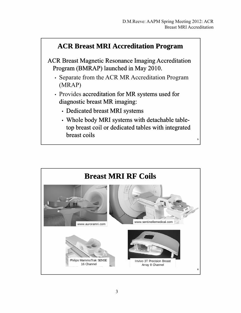
Breast MRI RF CoilsBreast MRI RF Coils
www.sentinellemedical.comwww.auroramri.com
Philips MammoTrak SENSE 16 Channel
Invivo 3T Precision Breast Array 8 Channel
66
D.M.Reeve: AAPM Spring Meeting 2012: ACR Breast MRI Accreditation
4
January 1, 2012: Medicare Improvements for Patients January 1, 2012: Medicare Improvements for Patients and Providers Act of 2008 (and Providers Act of 2008 (MIPPAMIPPA) requires ) requires accreditation for accreditation for outpatient facilities outpatient facilities that furnish the that furnish the
Why get accredited?Why get accredited?Why get accredited?Why get accredited?
p fp ftechnical component of advanced diagnostic imaging technical component of advanced diagnostic imaging procedures (CT, MR, Nuclear Medicine, PET) in order procedures (CT, MR, Nuclear Medicine, PET) in order to receive reimbursement from CMS.to receive reimbursement from CMS.
3 approved accreditation programs: American College 3 approved accreditation programs: American College of Radiology, The Joint Commission, of Radiology, The Joint Commission, IntersocietalIntersocietalAccreditation CommissionAccreditation Commission
ACR ACR BMRAPBMRAP and and MRAPMRAP are separate programs. are separate programs. Scanners performing both general and breast MRI, Scanners performing both general and breast MRI, need to be accredited in both programs in order to be need to be accredited in both programs in order to be reimbursed.reimbursed. 77
Guidance documentsGuidance documents
www.acr.org
88
D.M.Reeve: AAPM Spring Meeting 2012: ACR Breast MRI Accreditation
5
•• Any field strengthAny field strength
•• Coils capable of simultaneous bilateral imagingCoils capable of simultaneous bilateral imaging
MRI System RequirementsMRI System Requirements
•• Must accredit all MR systems at the facility that are Must accredit all MR systems at the facility that are used to perform used to perform diagnosticdiagnostic breast MR imagingbreast MR imaging. Does . Does not include:not include:
•• Dedicated systems used for radiation therapy Dedicated systems used for radiation therapy treatment planningtreatment planning
•• Dedicated interventional MRI systemsDedicated interventional MRI systems
•• Systems used for MRSystems used for MR--guided breast biopsy but not guided breast biopsy but not breast MR imagingbreast MR imaging
99
BMRAP Clinical ImagesBMRAP Clinical Images
• Facilities must submit clinical images and corresponding data for each magnet performing breast MRI examinations at their siteat their site.
• Facilities performing breast MRI must have the capacity to perform mammographic correlation, directed breast ultrasound and MRI-guided intervention, or create a referral arrangement with a cooperating BMRAP accredited facility g p g ythat could provide these services.
• 6 months to acquire clinical exams• No phantom image submission is required at this time.
1010

D.M.Reeve: AAPM Spring Meeting 2012: ACR Breast MRI Accreditation
6
ACR Breast MRI Accreditation ProgramACR Breast MRI Accreditation Program
Step 1: ApplicationStep 1: Application
•• MRI system informationMRI system informationyy
•• Personnel informationPersonnel information
•• $$$ fees$$$ fees
1111
Accreditation feesAccreditation fees
www.acr.org Breast MRI Accreditation Program Requirements, 11/22/20111212
D.M.Reeve: AAPM Spring Meeting 2012: ACR Breast MRI Accreditation
7
ACR Breast MRI Accreditation ProgramACR Breast MRI Accreditation Program
Step 2: Submit test materialsStep 2: Submit test materials•• Clinical* breast MRI exam on CD/DVD BIRADS category 6 Clinical* breast MRI exam on CD/DVD BIRADS category 6
(k h i bi(k h i bi i ) f hi ) f h(known, enhancing, biopsy(known, enhancing, biopsy--proven carcinoma) for each scanner proven carcinoma) for each scanner to be accredited. to be accredited.
•• Test image data formTest image data form
•• Medical physicist’s annual system performance reportMedical physicist’s annual system performance report
•• Quality Assurance QuestionnaireQuality Assurance Questionnaire
*Currently program does not require phantom images*Currently program does not require phantom images
1313
ACR Breast MRI Accreditation ProgramACR Breast MRI Accreditation Program
Personnel qualificationsPersonnel qualifications
OutlineOutlineOutlineOutline
Personnel qualificationsPersonnel qualifications
Quality control requirementsQuality control requirements
ACR breast MR image quality assessment ACR breast MR image quality assessment criteriacriteria
Examples of clinical imagesExamples of clinical images Examples of clinical imagesExamples of clinical images
1414

D.M.Reeve: AAPM Spring Meeting 2012: ACR Breast MRI Accreditation
8
Initial qualifications:• Certification in Radiology or Diagnostic Radiology (ABR, American
O t thi B d f R di l R l C ll f Ph i i d
Personnel Qualifications –Radiologist
Personnel Qualifications –Radiologist
Osteopathic Board of Radiology, Royal College of Physicians and Surgeons of Canada or Le College des Medecins du Quebec)
AND• Supervision, interpretation and reporting of 150 breast MRI exams in
last 36 months or 100 breast MRI exams in a supervised situation.OR
Not Board CertifiedNot Board Certified• Completion of an ACGME or AOA approved diagnostic radiology
residency programAND
• Interpretation and reporting of 100 breast MRI exams in the last 36 months in a supervised situation. 1515
AND15 hours of Cat 1 CME in MRI (including clinical applications of MRI in b t i i MRI tif t f t d i t t ti i th l t 36 th
Personnel Qualifications –Radiologist
Personnel Qualifications –Radiologist
breast imaging, MRI artifacts, safety and instrumentation in the last 36 months.
Continuing Experience:• Upon renewal, 75 breast MRI examinations in prior 24 months.
•• Double reading acceptable (2 or more physicians interpret the same exam)Double reading acceptable (2 or more physicians interpret the same exam)
•• Can reCan re--interpret a prior exam as long as physician did not do the initial readinterpret a prior exam as long as physician did not do the initial read•• Can reCan re--interpret a prior exam as long as physician did not do the initial read.interpret a prior exam as long as physician did not do the initial read.
Continuing Education:5 hours of Category 1 CME in breast MRI in the prior 36 months.
1616
D.M.Reeve: AAPM Spring Meeting 2012: ACR Breast MRI Accreditation
9
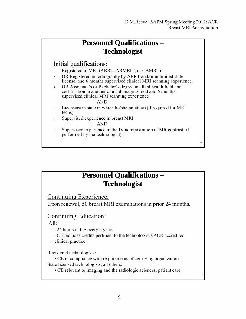
Initial qualifications:1. Registered in MRI (ARRT, ARMRIT, or CAMRT)
OR R i d i di h b ARRT d/ li i d
Personnel Qualifications –Technologist
Personnel Qualifications –Technologist
2. OR Registered in radiography by ARRT and/or unlimited state license, and 6 months supervised clinical MRI scanning experience.
3. OR Associate’s or Bachelor’s degree in allied health field and certification in another clinical imaging field and 6 months supervised clinical MRI scanning experience.
AND• Licensure in state in which he/she practices (if required for MRI
t h )techs)• Supervised experience in breast MRI
AND• Supervised experience in the IV administration of MR contrast (if
performed by the technologist)
1717
Continuing Experience:Upon renewal, 50 breast MRI examinations in prior 24 months.
Personnel Qualifications –Technologist
Personnel Qualifications –Technologist
Continuing Education:All:
• 24 hours of CE every 2 years• CE includes credits pertinent to the technologist’s ACR accredited clinical practice
Registered technologists: • CE in compliance with requirements of certifying organization
State licensed technologists, all others:• CE relevant to imaging and the radiologic sciences, patient care
1818
D.M.Reeve: AAPM Spring Meeting 2012: ACR Breast MRI Accreditation
10
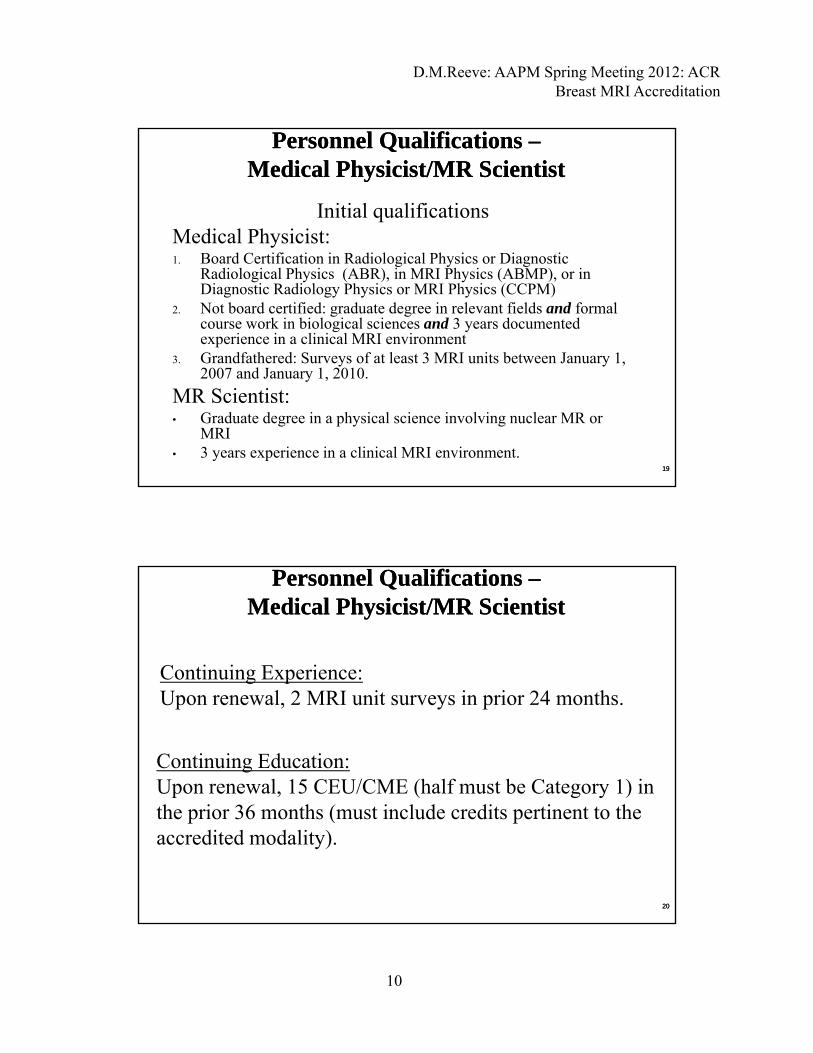
Initial qualificationsMedical Physicist:
Personnel Qualifications –Medical Physicist/MR Scientist
Personnel Qualifications –Medical Physicist/MR Scientist
y1. Board Certification in Radiological Physics or Diagnostic
Radiological Physics (ABR), in MRI Physics (ABMP), or in Diagnostic Radiology Physics or MRI Physics (CCPM)
2. Not board certified: graduate degree in relevant fields and formal course work in biological sciences and 3 years documented experience in a clinical MRI environment
3. Grandfathered: Surveys of at least 3 MRI units between January 1, 2007 and January 1, 2010.
MR Scientist:• Graduate degree in a physical science involving nuclear MR or
MRI• 3 years experience in a clinical MRI environment.
1919
Continuing Experience:
Personnel Qualifications –Medical Physicist/MR Scientist
Personnel Qualifications –Medical Physicist/MR Scientist
Upon renewal, 2 MRI unit surveys in prior 24 months.
Continuing Education:Upon renewal, 15 CEU/CME (half must be Category 1) in h i 36 h ( i l d di i hthe prior 36 months (must include credits pertinent to the
accredited modality).
2020
D.M.Reeve: AAPM Spring Meeting 2012: ACR Breast MRI Accreditation
11
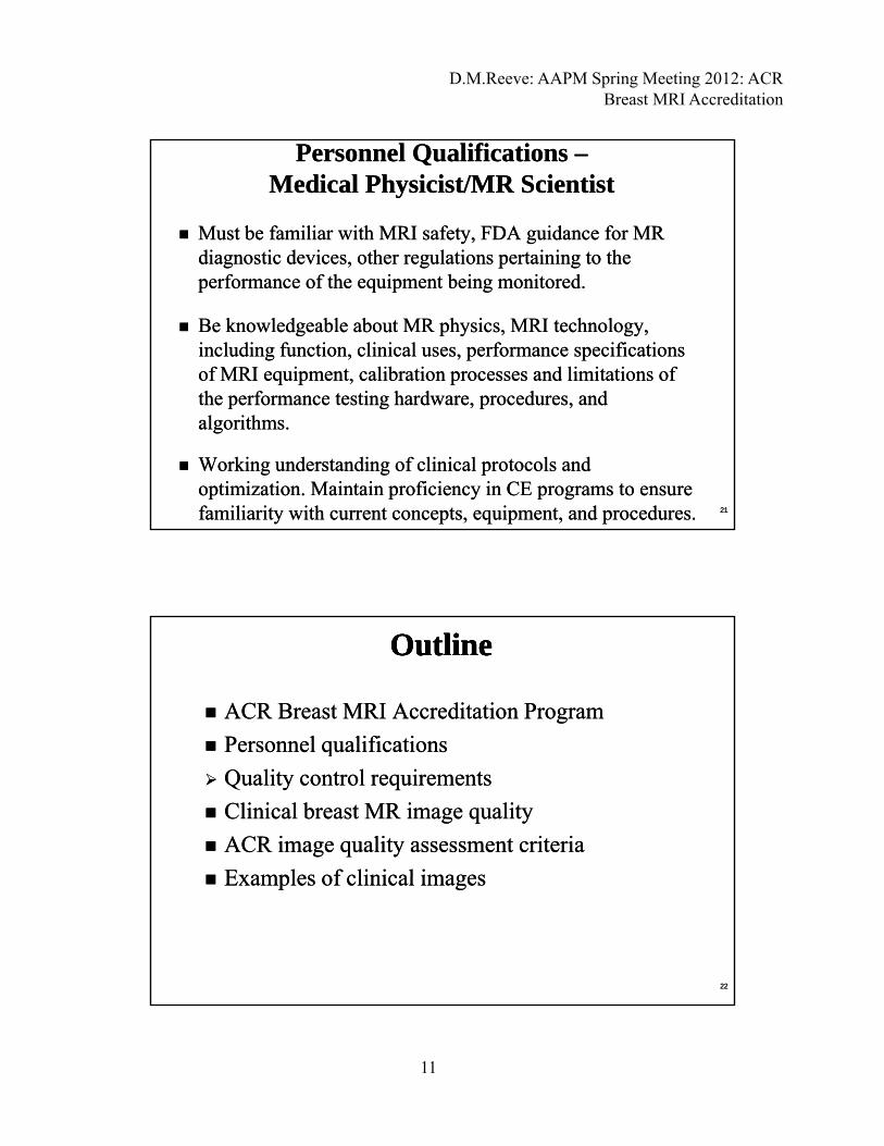
Must be familiar with MRI safety, FDA guidance for MR Must be familiar with MRI safety, FDA guidance for MR diagnostic devices, other regulations pertaining to the diagnostic devices, other regulations pertaining to the
Personnel Qualifications –Medical Physicist/MR Scientist
Personnel Qualifications –Medical Physicist/MR Scientist
performance of the equipment being monitored.performance of the equipment being monitored.
Be knowledgeable about MR physics, MRI technology, Be knowledgeable about MR physics, MRI technology, including function, clinical uses, performance specifications including function, clinical uses, performance specifications of MRI equipment, calibration processes and limitations of of MRI equipment, calibration processes and limitations of the performance testing hardware procedures andthe performance testing hardware procedures andthe performance testing hardware, procedures, and the performance testing hardware, procedures, and algorithms.algorithms.
Working understanding of clinical protocols and Working understanding of clinical protocols and optimization. Maintain proficiency in CE programs to ensure optimization. Maintain proficiency in CE programs to ensure familiarity with current concepts, equipment, and procedures.familiarity with current concepts, equipment, and procedures. 2121
ACR Breast MRI Accreditation ProgramACR Breast MRI Accreditation Program
Personnel qualificationsPersonnel qualifications
OutlineOutlineOutlineOutline
Personnel qualificationsPersonnel qualifications
Quality control requirementsQuality control requirements
Clinical breast MR image quality Clinical breast MR image quality
ACR image quality assessment criteriaACR image quality assessment criteria
Examples of clinical imagesExamples of clinical images Examples of clinical imagesExamples of clinical images
2222
D.M.Reeve: AAPM Spring Meeting 2012: ACR Breast MRI Accreditation
12
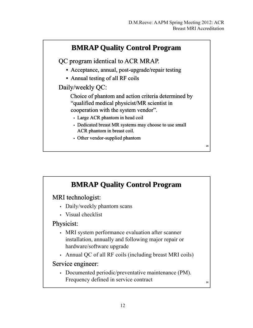
QC program identical to ACR MRAP. QC program identical to ACR MRAP. •• Acceptance, annual, postAcceptance, annual, post--upgrade/repair testingupgrade/repair testing
A l t ti f ll RF ilA l t ti f ll RF il
BMRAP Quality Control ProgramBMRAP Quality Control Program
•• Annual testing of all RF coilsAnnual testing of all RF coils
Daily/weekly QC:Daily/weekly QC:Choice of phantom and action criteria determined by Choice of phantom and action criteria determined by “qualified medical physicist/MR scientist in “qualified medical physicist/MR scientist in cooperation with the system vendor”.cooperation with the system vendor”.
•• Large ACR phantom in head coilLarge ACR phantom in head coil
•• Dedicated breast MR systems may choose to use small Dedicated breast MR systems may choose to use small ACR phantom in breast coil.ACR phantom in breast coil.
•• Other vendorOther vendor--supplied phantomsupplied phantom
2323
BMRAP Quality Control ProgramBMRAP Quality Control Program
MRI technologist: MRI technologist: • Daily/weekly phantom scans
Vi l h kli t• Visual checklist
Physicist: Physicist: • MRI system performance evaluation after scanner
installation, annually and following major repair or hardware/software upgrade
• Annual QC of all RF coils (including breast MRI coils)
Service engineer: Service engineer: • Documented periodic/preventative maintenance (PM).
Frequency defined in service contract2424
D.M.Reeve: AAPM Spring Meeting 2012: ACR Breast MRI Accreditation
13
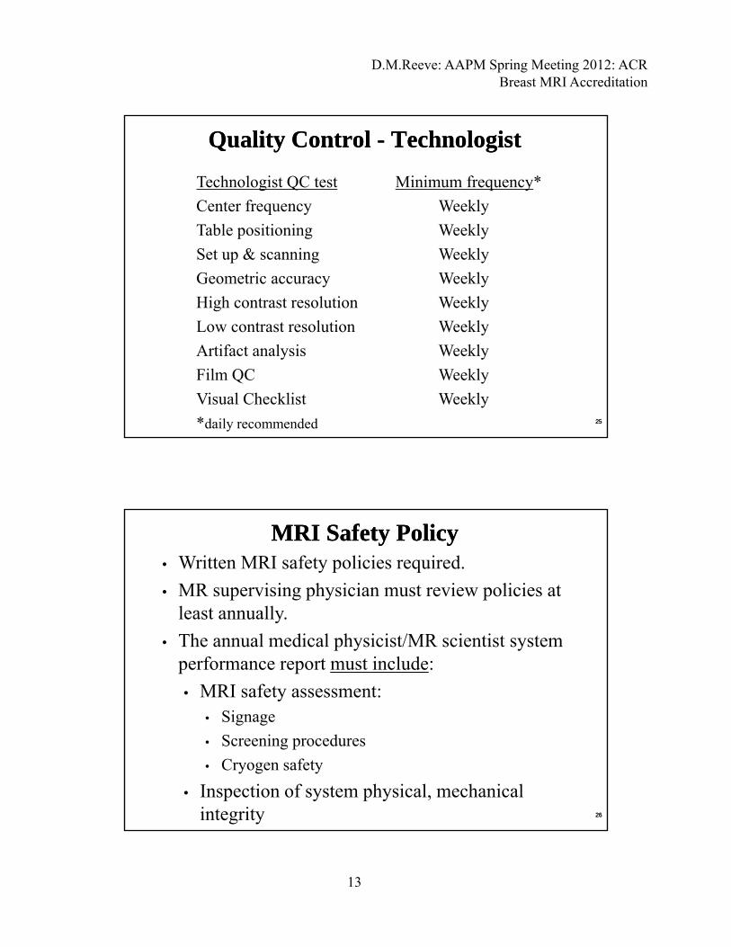
Quality Control - TechnologistQuality Control - Technologist
Technologist QC test Minimum frequency*
Center frequency Weekly
Table positioning Weekly
Set up & scanning Weekly
Geometric accuracy Weekly
High contrast resolution Weekly
Low contrast resolution WeeklyLow contrast resolution Weekly
Artifact analysis Weekly
Film QC Weekly
Visual Checklist Weekly
*daily recommended 2525
MRI Safety PolicyMRI Safety Policy• Written MRI safety policies required.
• MR supervising physician must review policies at least annuallyleast annually.
• The annual medical physicist/MR scientist system performance report must include:
• MRI safety assessment:• Signageg g
• Screening procedures
• Cryogen safety
• Inspection of system physical, mechanical integrity 2626
D.M.Reeve: AAPM Spring Meeting 2012: ACR Breast MRI Accreditation
14
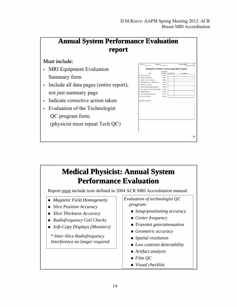
Must include:Must include:
• MRI Equipment Evaluation
Annual System Performance Evaluation Annual System Performance Evaluation reportreport
Annual System Performance Evaluation Annual System Performance Evaluation reportreport
Summary form
• Include all data pages (entire report),
not just summary page
• Indicate corrective action taken
E l ti f th T h l i t• Evaluation of the Technologist
QC program form.
(physicist must repeat Tech QC)
2727
Medical Physicist: Annual System Medical Physicist: Annual System Performance EvaluationPerformance Evaluation
Medical Physicist: Annual System Medical Physicist: Annual System Performance EvaluationPerformance Evaluation
Magnetic Field Homogeneity Evaluation of technologist QC
Report must include tests defined in 2004 ACR MRI Accreditation manual:
Magnetic Field Homogeneity
Slice Position Accuracy
Slice Thickness Accuracy
Radiofrequency Coil Checks
Soft-Copy Displays (Monitors)
Evaluation of technologist QC program:
Setup/positioning accuracy
Center frequency
Transmit gain/attenuation
Geometric accuracy* I Sli R di f
Spatial resolution
Low contrast detectability
Artifact analysis
Film QC
Visual checklist
* Inter-Slice Radiofrequency Interference no longer required
D.M.Reeve: AAPM Spring Meeting 2012: ACR Breast MRI Accreditation
15
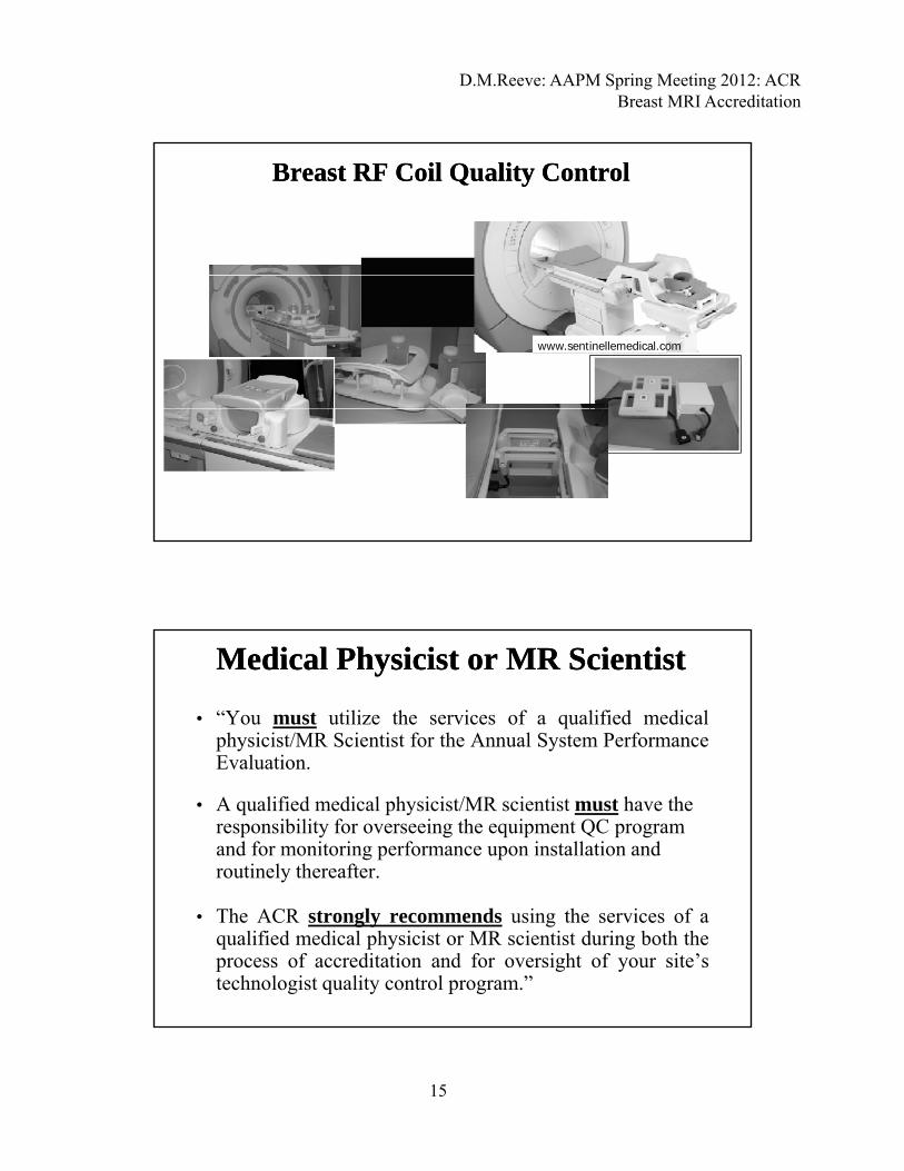
Breast RF Coil Quality ControlBreast RF Coil Quality Control
www.sentinellemedical.com
• “You must utilize the services of a qualified medicalphysicist/MR Scientist for the Annual System PerformanceEvaluation
Medical Physicist or MR ScientistMedical Physicist or MR Scientist
Evaluation.
• A qualified medical physicist/MR scientist must have the responsibility for overseeing the equipment QC program and for monitoring performance upon installation and routinely thereafter.
• The ACR strongly recommends using the services of aqualified medical physicist or MR scientist during both theprocess of accreditation and for oversight of your site’stechnologist quality control program.”
D.M.Reeve: AAPM Spring Meeting 2012: ACR Breast MRI Accreditation
16
Medical Medical Physicist/MRI ScientistPhysicist/MRI ScientistMedical Medical Physicist/MRI ScientistPhysicist/MRI Scientist
Can be very helpful with the technical aspects ofBreast MRI Accreditation process:p
Assist Radiologist with breast MRI protocol development andoptimization. Ensure protocols meet ACR spatial andtemporal resolution requirements.
Review breast MRI cases for image quality and artifacts priorto submission.
ACR Breast MRI Accreditation ProgramACR Breast MRI Accreditation Program
Personnel qualificationsPersonnel qualifications
OutlineOutlineOutlineOutline
Personnel qualificationsPersonnel qualifications
Quality control requirementsQuality control requirements
ACR breast MR image quality assessment ACR breast MR image quality assessment criteriacriteria
Examples of clinical imagesExamples of clinical images Examples of clinical imagesExamples of clinical images
3232
D.M.Reeve: AAPM Spring Meeting 2012: ACR Breast MRI Accreditation
17
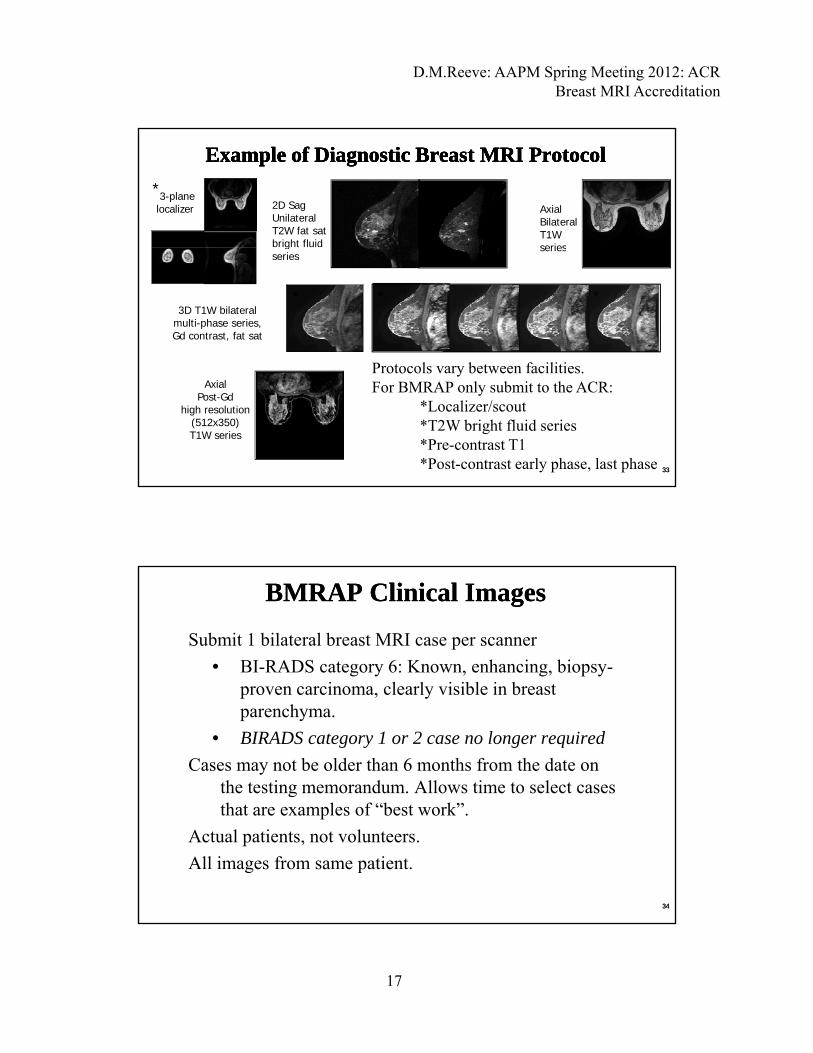
Example of Diagnostic Breast MRI ProtocolExample of Diagnostic Breast MRI ProtocolExample of Diagnostic Breast MRI ProtocolExample of Diagnostic Breast MRI Protocol
3-planelocalizer Axial
BilateralT1W series
2D SagUnilateralT2W fat satbright fluid
* **
3D T1W bilateral multi-phase series,Gd contrast, fat sat
seriesgseries
Protocols vary between facilities
* * *
3333
Axial Post-Gd
high resolution(512x350)T1W series
Protocols vary between facilities.For BMRAP only submit to the ACR:
*Localizer/scout*T2W bright fluid series*Pre-contrast T1*Post-contrast early phase, last phase
Submit 1 bilateral breast MRI case per scanner
• BI-RADS category 6: Known, enhancing, biopsy-i l l i ibl i b t
BMRAP Clinical ImagesBMRAP Clinical Images
proven carcinoma, clearly visible in breast parenchyma.
• BIRADS category 1 or 2 case no longer required
Cases may not be older than 6 months from the date on the testing memorandum. Allows time to select cases th t l f “b t k”that are examples of “best work”.
Actual patients, not volunteers.
All images from same patient.
3434
D.M.Reeve: AAPM Spring Meeting 2012: ACR Breast MRI Accreditation
18
ACR image assessment categories: ACR image assessment categories: AA P l d i t tP l d i t t
BMRAP Clinical ImagesBMRAP Clinical ImagesBMRAP Clinical ImagesBMRAP Clinical Images
A. A. Pulse sequences and image contrastPulse sequences and image contrast
B. B. Positioning and anatomical coveragePositioning and anatomical coverage
C. C. ArtifactsArtifacts
D. D. Spatial and temporal resolutionSpatial and temporal resolution
E. E. Exam identificationExam identification
3535www.acr.orgwww.acr.org Breast MRI Accreditation Program Clinical Image Quality Guide, 9/23/2011Breast MRI Accreditation Program Clinical Image Quality Guide, 9/23/2011
T2, bright fluid series:T2, bright fluid series:
• Bright fluid contrast distinguishable from
A. Pulse sequences and image contrastA. Pulse sequences and image contrast
Bright fluid contrast distinguishable from background
• Must demonstrate sufficient SNR (not too grainy)
3636
D.M.Reeve: AAPM Spring Meeting 2012: ACR Breast MRI Accreditation
19
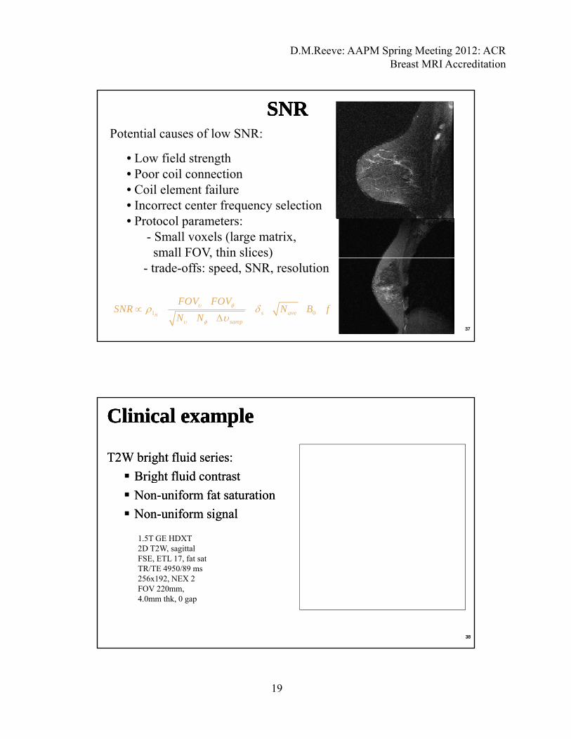
SNRSNRSNRSNRPotential causes of low SNR:
• Low field strength• Poor coil connection• Poor coil connection• Coil element failure• Incorrect center frequency selection• Protocol parameters:
- Small voxels (large matrix, small FOV, thin slices), )
- trade-offs: speed, SNR, resolution
1 0H s ave
samp
FOV FOVSNR N B f
N N
3737
T2W bright fluid series:T2W bright fluid series:
Bright fluid contrastBright fluid contrast
Clinical exampleClinical exampleClinical exampleClinical example
gg
NonNon--uniform fat saturationuniform fat saturation
NonNon--uniform signaluniform signal
1.5T GE HDXT2D T2W, sagittalFSE, ETL 17, fat sat
3838
TR/TE 4950/89 ms256x192, NEX 2FOV 220mm,4.0mm thk, 0 gap

D.M.Reeve: AAPM Spring Meeting 2012: ACR Breast MRI Accreditation
20
T2W bright fluid series:T2W bright fluid series:
Bright fluid contrastBright fluid contrast
Clinical example Clinical example Clinical example Clinical example
gg
Fat saturation fairly uniformFat saturation fairly uniform
GE 1.5T HDXt2D T2W, sagittalFSE, ETL 17, fat satTR/TE 4950/89 ms
3939
TR/TE 4950/89 ms256x192, NEX 2FOV 220mm,4.0mm thk, 0 gap
T1W multiT1W multi--phase series:phase series:• Pre-contrast and post-contrast series: identical scan parameters.
A. Pulse sequences and image contrastA. Pulse sequences and image contrast
• Post-contrast T1W images must either be fat suppressed
or provide subtractions (early and delayed phases)
• IV contrast must be evident in post-contrast images
• Must demonstrate sufficient SNR (not too grainy)
•• If possible should be sequential (i e not “stacked” orIf possible should be sequential (i e not “stacked” or•• If possible, should be sequential (i.e. not stacked or If possible, should be sequential (i.e. not stacked or “interleaved”)“interleaved”)
4040
D.M.Reeve: AAPM Spring Meeting 2012: ACR Breast MRI Accreditation
21
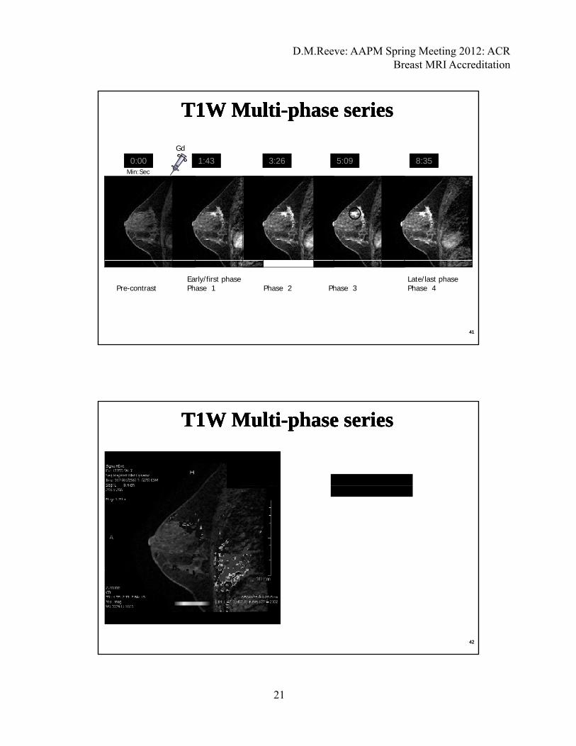
T1W MultiT1W Multi--phase seriesphase seriesT1W MultiT1W Multi--phase seriesphase series
0:00 1:43 3:26 5:09 8:35Min:Sec
Gd
4141
Pre-contrastEarly/first phasePhase 1 Phase 2 Phase 3
Late/last phasePhase 4
T1W MultiT1W Multi--phase seriesphase seriesT1W MultiT1W Multi--phase seriesphase series
4242

D.M.Reeve: AAPM Spring Meeting 2012: ACR Breast MRI Accreditation
22
Subtraction
Multiphase T1 series w/o fat sat: Multiphase T1 series w/o fat sat: subtractionssubtractions
Multiphase T1 series w/o fat sat: Multiphase T1 series w/o fat sat: subtractionssubtractionsPre-contrastPost-contrast
First phase
SubtractionPre-contrastPost-contrast Last phase
4343*Submit pre- and post-contrast series and both subtracted series
T1 weighted dynamic (multiT1 weighted dynamic (multi--phase) phase) series:series:
Clinical example Clinical example Clinical example Clinical example
Uniform signalUniform signal
Uniform fat satUniform fat sat
Low SNR, images grainyLow SNR, images grainy
1.5T GE HDXt3D T1W i l
4444
3D, T1W, sagittalFGRE, fat sat, α 100
TR/TE 4.3/2.0 ms256x256, NEX 0.5FOV 220mm, 2.6 mm thk, 50% overlapSequential
D.M.Reeve: AAPM Spring Meeting 2012: ACR Breast MRI Accreditation
23
• Adequate breast tissue in coil
• Proper positioning of breast tissue
B. Positioning and anatomical coverageB. Positioning and anatomical coverage
• Proper positioning of breast tissue
• Full coverage from axillary tail to inframammary fold
• Absence or minimal skin folds
• Appropriate FOV
4545
• Excessive artifacts can interfere with interpretation
• Some are unavoidable on certain images
C. ArtifactsC. Artifacts
• Some are unavoidable on certain images
• Images do not have to be “artifact free”
• Some are due to pulse sequence errors, inadequate equipment, improper maintenance (PM, QC) of equipmentq p
4646

D.M.Reeve: AAPM Spring Meeting 2012: ACR Breast MRI Accreditation
24
Breast MRI ArtifactsBreast MRI Artifacts
Common artifacts in breast MRICommon artifacts in breast MRI•• MotionMotion•• MotionMotion
•• Truncation artifactsTruncation artifacts
•• Out of volume wrapOut of volume wrap
•• SusceptibilitySusceptibility artifactsartifacts
Signal nonSignal non uniformityuniformity•• Signal nonSignal non--uniformityuniformity
•• Poor or nonPoor or non--uniform fat saturationuniform fat saturation
4747
Motion artifactsMotion artifactsOccur in the phase encoding direction. Caused by cardiac Occur in the phase encoding direction. Caused by cardiac motion, respiration, patient movement. Results in phase motion, respiration, patient movement. Results in phase mismis--mapping in kmapping in k--space due the time delay between space due the time delay between
hh di d i l ddi d i l dphasephase--encoding and signal readout. encoding and signal readout.
4848
D.M.Reeve: AAPM Spring Meeting 2012: ACR Breast MRI Accreditation
25
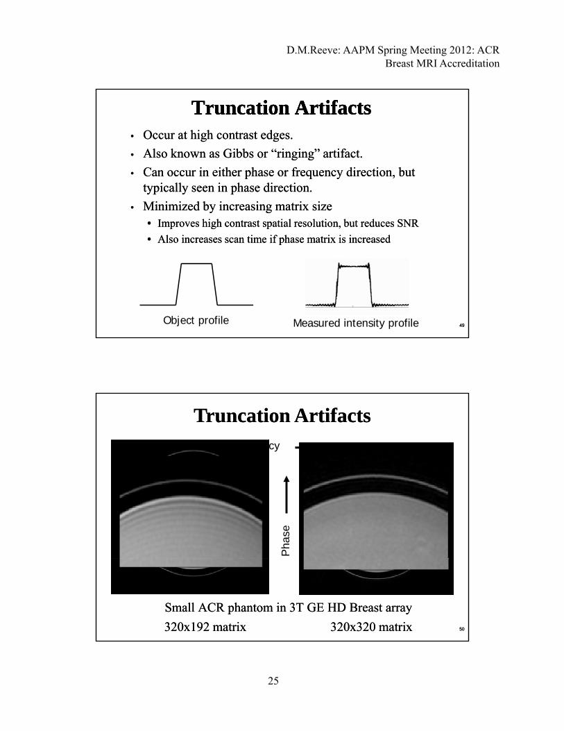
•• Occur at high contrast edges.Occur at high contrast edges.
•• Also Also known known as Gibbs or as Gibbs or “ringing” artifact“ringing” artifact..
•• Can occur in either phase or frequency direction butCan occur in either phase or frequency direction but
Truncation ArtifactsTruncation ArtifactsTruncation ArtifactsTruncation Artifacts
•• Can occur in either phase or frequency direction, but Can occur in either phase or frequency direction, but typically seen in phase direction.typically seen in phase direction.
•• Minimized Minimized by increasing matrix by increasing matrix sizesize•• Improves high contrast spatial resolution, but reduces SNRImproves high contrast spatial resolution, but reduces SNR
•• Also increases scan time if phase matrix is increasedAlso increases scan time if phase matrix is increased
Object profile Measured intensity profile 4949
Truncation ArtifactsTruncation ArtifactsFrequency
Pha
se
Small ACR phantom in 3T GESmall ACR phantom in 3T GE HD Breast array HD Breast array
320x192 matrix320x192 matrix 320x320 matrix320x320 matrix 5050
D.M.Reeve: AAPM Spring Meeting 2012: ACR Breast MRI Accreditation
26
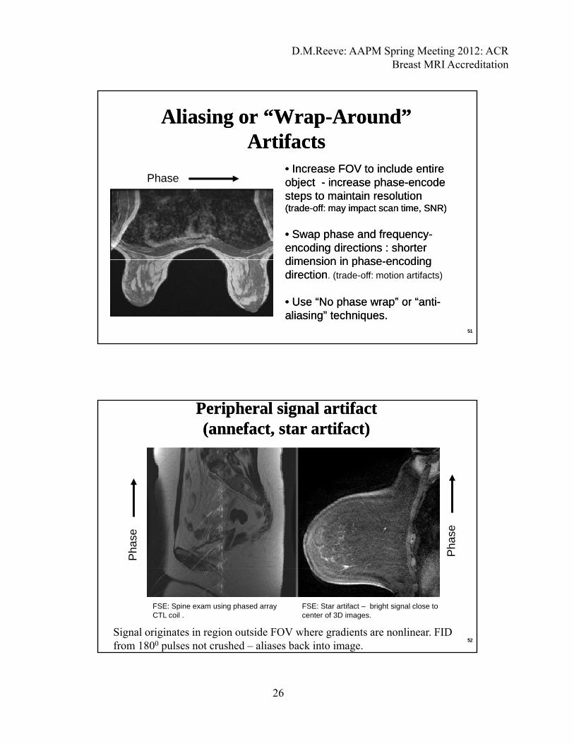
Aliasing or “WrapAliasing or “Wrap--Around” Around” ArtifactsArtifacts
•• Increase FOV to include entire Increase FOV to include entire Ph object object -- increase phaseincrease phase--encode encode
steps to maintain resolution steps to maintain resolution (trade(trade--off: may impact scan time, SNR) off: may impact scan time, SNR)
•• Swap phase and frequencySwap phase and frequency--encoding directions : shorter encoding directions : shorter dimension in phasedimension in phase encodingencoding
Phase
dimension in phasedimension in phase--encoding encoding directiondirection. (trade-off: motion artifacts)
•• Use “No phase wrap” or “antiUse “No phase wrap” or “anti--aliasing” techniques.aliasing” techniques.
5151
Peripheral signal artifact(annefact, star artifact)
Peripheral signal artifact(annefact, star artifact)
Pha
se
Pha
se
FSE: Star artifact – bright signal close tocenter of 3D images.
FSE: Spine exam using phased arrayCTL coil .
Signal originates in region outside FOV where gradients are nonlinear. FID from 1800 pulses not crushed – aliases back into image.
5252
D.M.Reeve: AAPM Spring Meeting 2012: ACR Breast MRI Accreditation
27
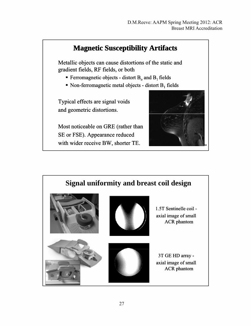
Metallic objects can cause distortions of the static and Metallic objects can cause distortions of the static and gradient fields, RF fields, or bothgradient fields, RF fields, or both F ti bj tF ti bj t di t t Bdi t t B d Bd B fi ldfi ld
Magnetic Susceptibility ArtifactsMagnetic Susceptibility ArtifactsMagnetic Susceptibility ArtifactsMagnetic Susceptibility Artifacts
Ferromagnetic objects Ferromagnetic objects -- distort Bdistort Boo and Band B1 1 fieldsfields
NonNon--ferromagnetic metal objects ferromagnetic metal objects -- distort Bdistort B11 fieldsfields
Typical effects are Typical effects are signal voids signal voids
and and geometric distortionsgeometric distortions..
Most noticeable on GRE (rather thanMost noticeable on GRE (rather than
SE or FSE). Appearance reduced SE or FSE). Appearance reduced
with wider receive BW, shorter TE.with wider receive BW, shorter TE. 5353
Signal uniformity and breast coil design
1.5T 1.5T SentinelleSentinelle coil coil --
axial image of small axial image of small ACR phantomACR phantom
3T GE HD array 3T GE HD array --
axial image of small axial image of small ACR phantomACR phantom
D.M.Reeve: AAPM Spring Meeting 2012: ACR Breast MRI Accreditation
28
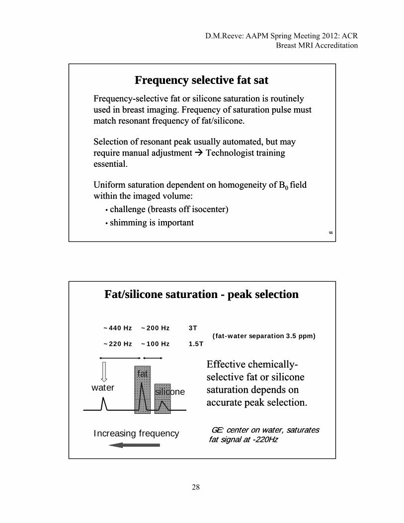
Frequency selective fat satFrequency selective fat sat
FrequencyFrequency--selective fat or silicone saturation is routinely selective fat or silicone saturation is routinely used in breast imaging. Frequency of saturation pulse must used in breast imaging. Frequency of saturation pulse must match resonant frequency of fat/silicone.match resonant frequency of fat/silicone.match resonant frequency of fat/silicone. match resonant frequency of fat/silicone.
Selection of resonant peak usually automated, but may Selection of resonant peak usually automated, but may require manual adjustment require manual adjustment Technologist training Technologist training essential. essential.
U if t ti d d t h it f BU if t ti d d t h it f B fi ldfi ldUniform saturation dependent on homogeneity of BUniform saturation dependent on homogeneity of B0 0 field field within the imaged volume:within the imaged volume:
•• challenge (breasts off challenge (breasts off isocenterisocenter))
•• shimming is importantshimming is important5555
Fat/silicone saturation - peak selectionFat/silicone saturation - peak selection
~200 Hz 3T (fat-water separation 3.5 ppm)
~440 Hz
waterfat
silicone
~220 Hz ~100 Hz 1.5T
Effective chemicallyEffective chemically--selective fat or silicone selective fat or silicone saturation depends on saturation depends on
t k l tit k l ti
Increasing frequency
accurate peak selection. accurate peak selection.
GE: center on water, saturates GE: center on water, saturates fat signal at fat signal at --220Hz 220Hz
D.M.Reeve: AAPM Spring Meeting 2012: ACR Breast MRI Accreditation
29
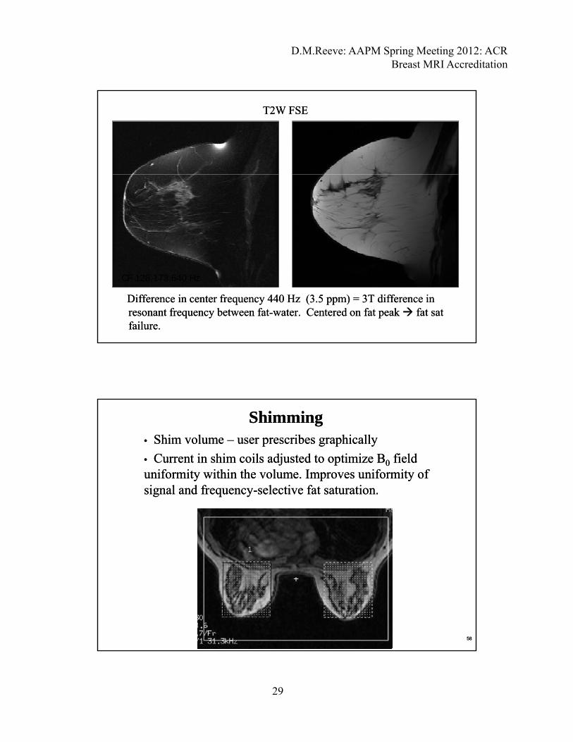
T2W FSET2W FSE
Fat sat failureFat sat failure
Difference in center frequency 440 Hz (3.5 Difference in center frequency 440 Hz (3.5 ppmppm) ) = 3T difference in = 3T difference in resonant frequency between fatresonant frequency between fat--water. water. CCentered on entered on fat peak fat peak fat sat fat sat failure.failure.
CF 128,173,640 Hz CF 128,173,200 Hz
ShimmingShimming•• Shim volume Shim volume –– useruser prescribes graphicallyprescribes graphically
•• Current in shim coils adjusted to optimize BCurrent in shim coils adjusted to optimize B00 field field uniformity within the volume. Improves uniformity of uniformity within the volume. Improves uniformity of signal and frequencysignal and frequency--selective fat saturation.selective fat saturation.
5858

D.M.Reeve: AAPM Spring Meeting 2012: ACR Breast MRI Accreditation
30
Clinical exampleClinical example
•• NonNon--uniform signal uniform signal
•• NonNon--uniform uniform fat suppressionfat suppressionpppp
1.5T GE HDXtSag T2W FSETR =4367ms / TEeff =81 ms echo train length = 17echo train length = 17122 Hz/pixel bandwidth 256x192 matrix, 220 mm FOV4mm thickness/ 0mm gap2 averagesfat sat
5959
Clinical exampleClinical example
•• Uniform fat saturationUniform fat saturation
•• TTruncationruncation artifactsartifacts
•• LLowow SNRSNR
•• Motion artifactsMotion artifacts
3T GE HDXt3D T1W MultiphaseTR =5.4 ms / TE =2.3 ms Flip angle 10O
NEX=0.5244.1 Hz/pixel bandwidth 320x320 matrix, 200 mm FOV2.4mm thickness/ 1.2mm spacing (slices overlap)fat sat
6060

D.M.Reeve: AAPM Spring Meeting 2012: ACR Breast MRI Accreditation
31
Spatial Resolution: Spatial Resolution: Criteria only aCriteria only apply to pre- and post-contrast T1-weighted multi-phase series:
• Acquired (not interpolated) thickness must be ≤ 3mm,
D. Spatial and Temporal ResolutionD. Spatial and Temporal Resolution
Acquired (not interpolated) thickness must be ≤ 3mm, >4.0mm will fail.
• 3-4mm: may fail if there are deficiencies in other categories.
• In-plane resolution must be ≤ 1mm (phase and freq), >1 2mm will fail 1 0-1 2mm may fail if deficiencies in>1.2mm will fail, 1.0 1.2mm may fail if deficiencies in other categories.
• Interslice gap must be ≤ 0mm (i.e. slices either overlap or are contiguous with no gap), >0mm will fail
6161
= FOV / N In-plane pixel size (frequency-encoding direction
Spatial ResolutionSpatial Resolution
encoding direction
= FOV / N In-plane pixel size (phase encoding direction)
6262
slice Prescribed slice thickness (not interpolated)
D.M.Reeve: AAPM Spring Meeting 2012: ACR Breast MRI Accreditation
32
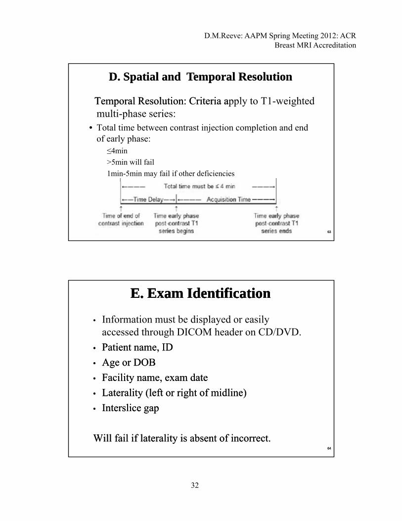
Temporal Resolution: Criteria aTemporal Resolution: Criteria apply to T1-weighted multi-phase series:
D. Spatial and Temporal ResolutionD. Spatial and Temporal Resolution
• Total time between contrast injection completion and end of early phase:
≤4min
>5min will fail
1min-5min may fail if other deficiencies
• Total time = time delay + acquisition time of early phase
6363
• Information must be displayed or easily accessed through DICOM header on CD/DVD.
E. Exam IdentificationE. Exam IdentificationE. Exam IdentificationE. Exam Identification
•• Patient name, IDPatient name, ID
•• Age or DOBAge or DOB
•• Facility name, exam dateFacility name, exam date
•• Laterality (left or right of midline)Laterality (left or right of midline)y ( g )y ( g )
•• IntersliceInterslice gapgap
Will fail if laterality is absent of incorrect.Will fail if laterality is absent of incorrect.6464
D.M.Reeve: AAPM Spring Meeting 2012: ACR Breast MRI Accreditation
33
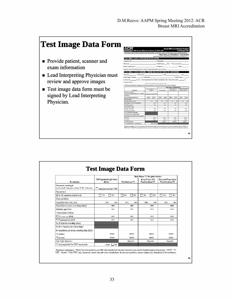
Provide patient, scanner and Provide patient, scanner and exam informationexam information
Test Image Data FormTest Image Data FormTest Image Data FormTest Image Data Form
Lead Interpreting Physician must Lead Interpreting Physician must review and approve imagesreview and approve images
Test image data form must be Test image data form must be signed by Lead Interpreting signed by Lead Interpreting Physician.Physician.
6565
Test Image Data FormTest Image Data FormTest Image Data FormTest Image Data Form
6666
D.M.Reeve: AAPM Spring Meeting 2012: ACR Breast MRI Accreditation
34
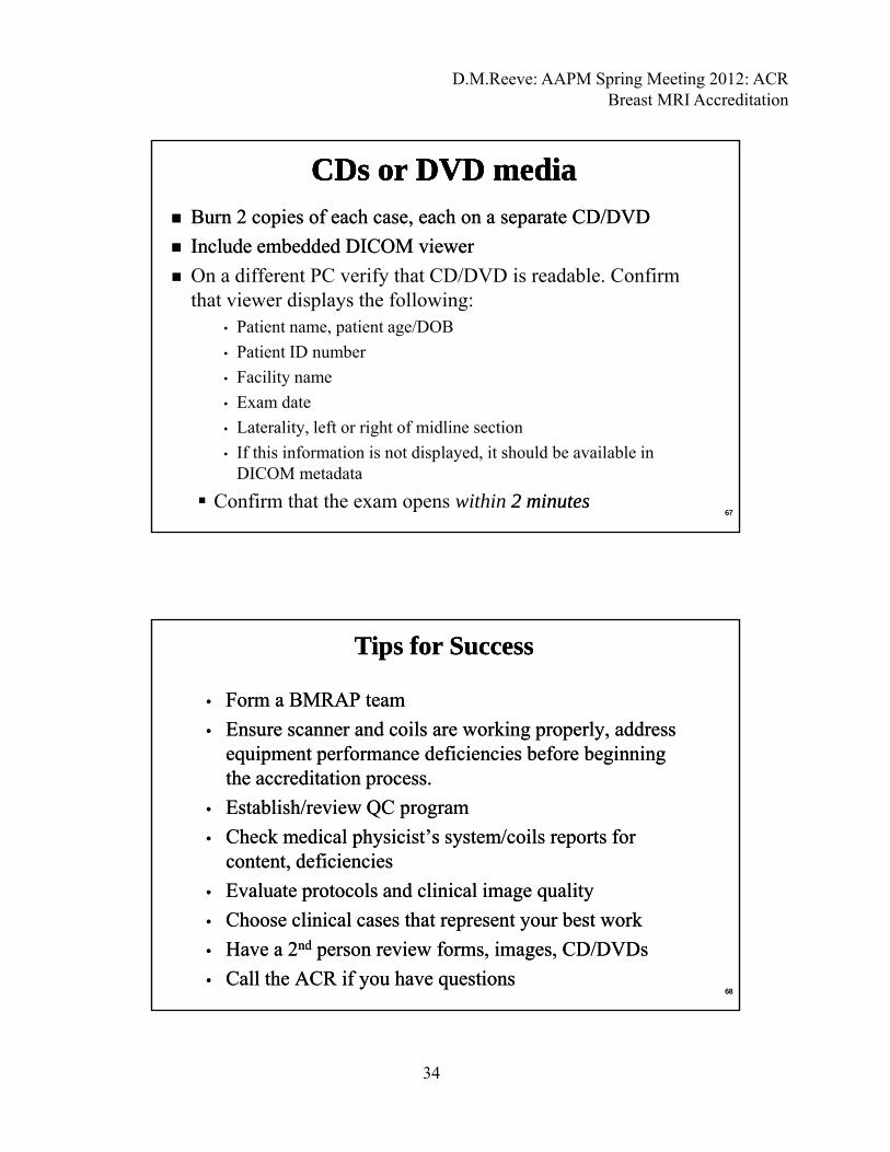
Burn 2 copies of each case, each on a separate CD/DVDBurn 2 copies of each case, each on a separate CD/DVD
Include embedded DICOM viewerInclude embedded DICOM viewer
O diff t PC if th t CD/DVD i d bl C fi
CDs or DVD mediaCDs or DVD mediaCDs or DVD mediaCDs or DVD media
On a different PC verify that CD/DVD is readable. Confirm that viewer displays the following:
• Patient name, patient age/DOB
• Patient ID number
• Facility name
Exam date• Exam date
• Laterality, left or right of midline section
• If this information is not displayed, it should be available in DICOM metadata
Confirm that the exam opens within 2 minutes2 minutes6767
•• Form a BMRAP teamForm a BMRAP team
•• Ensure scanner and coils are working properly, address Ensure scanner and coils are working properly, address i t f d fi i i b f b i ii t f d fi i i b f b i i
Tips for SuccessTips for Success
equipment performance deficiencies before beginning equipment performance deficiencies before beginning the accreditation process.the accreditation process.
•• Establish/review QC programEstablish/review QC program
•• Check medical physicist’s system/coils reports for Check medical physicist’s system/coils reports for content, deficienciescontent, deficiencies
•• Evaluate protocols and clinical image qualityEvaluate protocols and clinical image quality
•• Choose clinical cases that represent your best workChoose clinical cases that represent your best work
•• Have a 2Have a 2ndnd person review forms, images, CD/DVDsperson review forms, images, CD/DVDs
•• Call the ACR if you have questionsCall the ACR if you have questions6868
D.M.Reeve: AAPM Spring Meeting 2012: ACR Breast MRI Accreditation
35
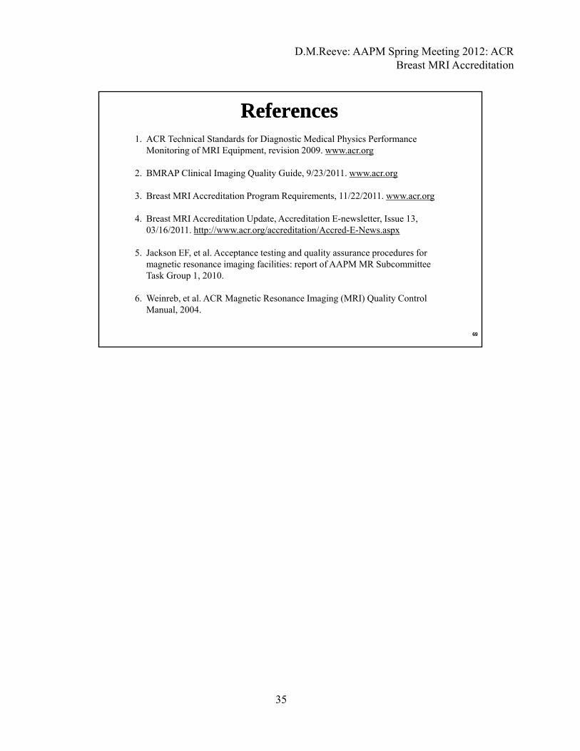
ReferencesReferences1. ACR Technical Standards for Diagnostic Medical Physics Performance
Monitoring of MRI Equipment, revision 2009. www.acr.org
2. BMRAP Clinical Imaging Quality Guide, 9/23/2011. www.acr.orgg g Q y , g
3. Breast MRI Accreditation Program Requirements, 11/22/2011. www.acr.org
4. Breast MRI Accreditation Update, Accreditation E-newsletter, Issue 13, 03/16/2011. http://www.acr.org/accreditation/Accred-E-News.aspx
5. Jackson EF, et al. Acceptance testing and quality assurance procedures for ti i i f iliti t f AAPM MR S b ittmagnetic resonance imaging facilities: report of AAPM MR Subcommittee
Task Group 1, 2010.
6. Weinreb, et al. ACR Magnetic Resonance Imaging (MRI) Quality Control Manual, 2004.
6969


![QuantifyingTumorVascularHeterogeneitywithDynamic Contrast ... · 2015. 7. 29. · ACR Breast Imaging-Reporting and Data System (BI-RADS) MRI lexicon [18]. In cancer treatment, tumor](https://img.pdfslide.net/doc/110x75/5fc9df59a8ef470c23133cae/quantifyingtumorvascularheterogeneitywithdynamic-contrast-2015-7-29-acr.jpg)