Embed Size (px)
Citation preview

8/2/2012
1
ACT IACT IACT IACT I
Image Acquisition:Turning People into Numbers
ACT IACT IACT IACT I
Image Acquisition:Turning People into Numbers
Jeff Siewerdsen, Ph.D.
Department of Biomedical EngineeringJohns Hopkins University
Johns Hopkins UniversitySchools of Medicine and Engineering
Matrices
Modalities
A medical imaging system
is a machine that transforms
people into numbers.
M. Kessler
... performs a measurement.
Projection
CT
MRI
PET
US
Overview
Modalities Acquisition &
Processing
Image Quality
Characteristics
Role in
IGRT / ART
Physical
Principles
Contrast
Mechanism(s)
Physical
Configuration
Processing &
Reconstruction
Spatial
Resolution
Contrast
Resolution
Temporal
Resolution
Real-Time
Near-Real-Time
Offline
Projection
CT
MRI
PET
US

8/2/2012
2
SourceObject
Detector
Processor
DisplayObserver
Acquisition & ProcessingProjection
CT
MRI
PET
US
q(E)
Acquisition & Processing
CT
MRI
PET
US
Projection
Boone & Seibert, Med. Phys. 24(11): 1661 (1997)
µµµµ(x,y,z;E)
Screen-Film (kV) or Cu+Film (MV)
Computed Radiography (CR)
XRII or Flat-Panel Detector
Source DetectorObject
Acquisition & Processing
MRI
PET
US
Contrast Mechanism: Attenuation
I0
Iy
x
Beer’s Law
Line Integral
Contrast
µµµµCT
Projection
( )
( ) ( )∫=
=
∫=−
d
dyyx
dyyxI
Ixp
eII
0
0
,
0
,ln µ
µ
( ) ( )
−
=∆ ∫∫
2010
,,
x
d
x
d
dyyxdyyxp µµ
Kupelian et al.
IJROBP 62(5) (2005)

8/2/2012
3
Acquisition & Processing
MRI
PET
US
Projection
CT
Source
Object
Detector
q(E)
µµµµ(E)
yx
ξξξξ
θθθθ
Sinogram
p(ξξξξ;θθθθ)����RampKernel(ξξξξ)
����RampKernel(ξξξξ)
ξξξξ
θθθθ
Sinogram
p(ξξξξ;θθθθ)
Acquisition & Processing
MRI
PET
US
Projection
CT
Backprojection
# of voxels# of proj
Repeat ××××
Σ
Acquisition & Processing
MRI
PET
US
Projection
Contrast Mechanism: ∆µ∆µ∆µ∆µ
Hounsfield Units (HU)
HU = µ - µwater
µwater1000CT
MDCTCBCT
4D CBCT
Brain (8)
Fat (-100)
Liver (+85)
Breast (-50)
Water (0)
Polyeth (-60)
8/2/2012
4
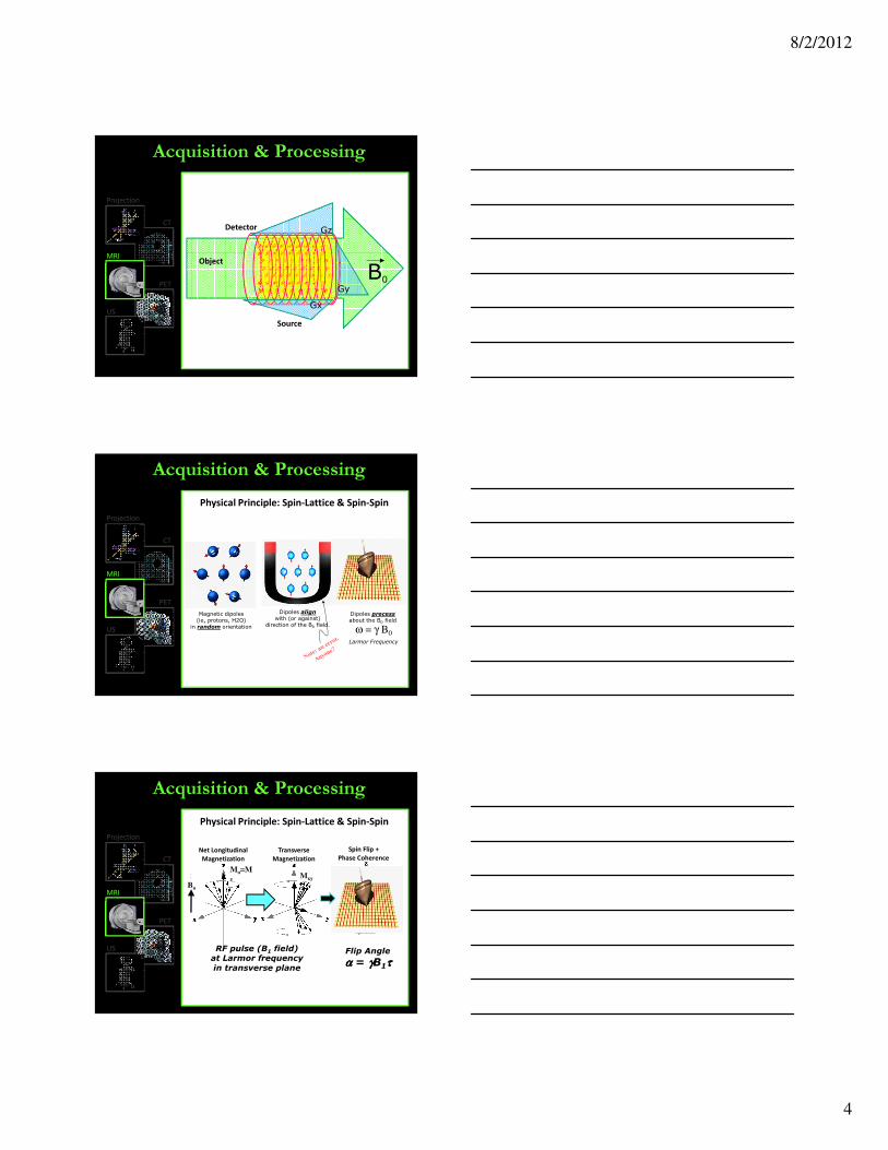
B0
Acquisition & Processing
CT
PET
US
Projection
Source
Object
Gz
Gy
Gx
Detector
MRI
Note
ω = γ B0
Magnetic dipoles(ie, protons, H2O)
in random orientation
Dipoles alignwith (or against)
direction of the B0 field.
Dipoles precessabout the B0 field
Larmor Frequency
Acquisition & Processing
CT
PET
US
Projection
Physical Principle: Spin-Lattice & Spin-Spin
MRI
Note
Mo=Mz
Bo
Acquisition & Processing
CT
PET
US
Projection
RF pulse (B1 field)at Larmor frequency
in transverse plane
Flip Angle
αααα = γγγγB1ττττ
Mxy
Net Longitudinal
Magnetization
Transverse
Magnetization
Spin Flip +
Phase Coherence
Physical Principle: Spin-Lattice & Spin-Spin
MRI

8/2/2012
5
Acquisition & Processing
CT
PET
US
Projection
1 2 3 1 2 3Time Time
Longitudinal Relaxation→ increase in
Longitudinal Magnetization (Mz)
Transverse Relaxation→ decrease in
Transverse Magnetization (Mxy)
T1 Spin-LatticeRelaxation Time
0.63
Physical Principle: Spin-Lattice & Spin-Spin
T2 Spin-SpinRelaxation Time
0.37
MRI
Lon
git
ud
ina
l M
ag
ne
tiza
tio
n
T1 Contrast
Time
Acquisition & Processing
CT
PET
US
Projection
Contrast Mechanism(s): Relaxation Time
Spin-Lattice
∆∆∆∆Mo
Tissue T1 (ms)
Fat 260
Liver 500
White 780
Muscle 870
Gray 920
CSF 2400
MRI
Y Cao (U-Michigan)
Acquisition & Processing
CT
PET
US
Projection
Contrast Mechanism(s): Relaxation Time
Tissue T2 (ms)
Fat 80
Liver 42
White 90
Muscle 45
Gray 100
CSF 160
Tra
nsv
ers
e
Ma
gn
eti
zati
on
Time
∆∆∆∆Mxy
Spin-Spin
T2 Contrast
DWI
T1
Gd
T2
Flair
MRI
T2 (TSE)Images from:
Khoo et al. Br. J. Rad. (1999)
Dawson et al. (PMH)
Ghilezan et al. IJROPB (2001)
T1 (SE)
Y Cao (U-Michigan)

8/2/2012
6
Acquisition & Processing
CT
MRI
US
Projection
PETObject
Detector
Source
Source
Basic Physical Principle
PM
TP
MT
18F→→→→18O+e++νννν
Acquisition & Processing
CT
MRI
US
Projection
PET
Image Reconstruction
ξξξξ
θθθθSinogram p(ξξξξ;θθθθ)
x
yAxial
FBP
PETPET-CT
R. Jeraj (U-Wisconsin)DW Townsend et al, Sem. Nuc. Med. (2003)
Acquisition & Processing
CT
MRI
PET
Projection Source
Detector
Object
Source-Detector
Transducer
Near
Field
Far
Field
Focal
Zone
Tra
nsd
uce
r A
rra
y
Basic Principles
US
Range Equation:
Pulse-Echo Imaging
8/2/2012
7
Acquisition & Processing
CT
MRI
PET
Projection
US
Material:
Air
Lung
Fat
Water
Soft tissue
Liver
Blood
Kidney
Muscle
Bone
v (m/s):
330
600
1460
1480
1540
1555
1560
1565
1600
4080
Bulk modulus (“stiffness”)
Density
Basic PrinciplesPulse-Echo Imaging
Velocity of sound
Z (Mrayls):
0.0004
0.18
1.34
1.48
1.54
1.65
1.65
1.63
1.71
7.80
R (soft tissue):
1.00
0.79
0.07
0.02
0
0.03
0.03
0.03
0.05
0.67
Reflection / Impedance
θi θr
θt
Z1
Z2
Reflectivity
Acoustic
Impedance
Acquisition & Processing
CT
MRI
PET
Projection
US
B-Mode 2D Sector Image
Transducer
Object
Basic Principles
W. Tome (U-Wisconsin) Elekta (ClarityTM)
ProstateCervix
Contrast is higher in CT
than x-ray projections, because:
0%
0%
0%
0%
0%
10
1. CT uses a higher dose.
2. CT uses contrast agents.
3. CT uses lower-energy x-rays.
4. CT has lower noise.
5. Because:

8/2/2012
8
Contrast is higher in CT
than x-ray projections, because:1. CT uses a higher dose.
2. CT uses contrast agents.
3. CT uses lower-energy x-rays.
4. CT has lower noise.
5. Because:
Imaging Performance
Modalities
Projection
CT
MRI
PET
US
Acquisition &
Processing
Image Quality
Characteristics
Role in
IGRT / ART
Physical
Principles
Contrast
Mechanism(s)
Physical
Configuration
Processing &
Reconstruction
Spatial
Resolution
Contrast
Resolution
Temporal
Resolution
Real-Time
Near-Real-Time
Offline
Accuracy (Quantitation)Extent to which the measured value equals the ‘true’ value
Vital to longitudinal imaging, QI:
Monitoring ( SUV → remission)
Diagnosis (BMD → osteoporosis)
Tx planning (µ → dose calculation)
Precision (i.e., “Resolution”)Min interval in {DIM} for which two stimuli can be distinguished
Spatial Resolution→ lp/mm… PSF, LSF, ESF… MTF
(≠ pixel size!)
Contrast Resolution→ contrast… noise… CNR (SDNR)
(≠ a display parameter)
Temporal Resolution→ speed… temporal MTF (≠ fps)
x
themeasuredproperty
t
cuiusmodi “Resolution”
Modalities
Projection
CT
MRI
PET
US
Spatial Resolution
Contrast Resolution
Temporal ResolutionDirect analogue to spatial resolution
(with 1-sided causal response)
101112
13
14
15
16

8/2/2012
9
QuantitativeAccuracy
SpatialResolution
ContrastResolution
TemporalResolution
Imaging Performance
Modalities
Projection
CT
MRI
PET
US
---+++ ---++(but 2D)
Rad
Fluor-
++-
MDCT
CBCT ++ + -+
3D
4D
+ +++ ---+
3D
Cine-
++ + ++++
- ++ - 3D
4D+ +
Role in IGRT / ART
Modalities Acquisition &
Processing
Image Quality
Characteristics
Role in
IGRT / ART
Physical
Principles
Contrast
Mechanism(s)
Physical
Configuration
Processing &
Reconstruction
Spatial
Resolution
Contrast
Resolution
Temporal
Resolution
Real-Time
Near-Real-Time
Offline
Projection
CT
MRI
PET
US
Real-Time
Temporal Scales of Intervention
On-Line
Off-Line
Temporal Scales of Acquisition
CT
MRI
PET
US
Projection Radiography~Instantaneous (exposure time: 10 ms)
… but static
FluoroscopyReal time / dynamic
1 fps… 5 fps… 30-60 fps
Speed governed by:- Frame rate of the detector
also:
- Exposure rate (mA)
- Detector gain (e.g., high-gain XRII)
- Spatial resolution requirement
Temporal Scales of Intervention
Role in IGRT / ART

8/2/2012
10
Role in IGRT / ART
CT
MRI
PET
US
Projection
McJury (NHS) Kuriyama (Yamanishi) Pang (Sunnybrook)Moseley (PMH)
Planning In-Room On-Linac
Projection
PET
US
CT
MRI
MDCTFast: 3 rev / sec (and 64 slices / rev)
→ CT-fluoro
→ 4D CT
CBCTSlow: 0.02 rev sec (60 sec / rev)
→ Full volume (no table motion)
→ Patient motion artifacts
4D CBCTEven slower (>60 – 120 sec / rev)
Many projs + Several motion cycles
→ Retrospective sorting by phase
Role in IGRT / ART
Temporal Scales of Acquisition
Projection
Role in IGRT / ART
PET
US
CT
MRI
Treatment
Planning
Real-Time
Guidance
On-Line
Guidance
Off-Line
Adaptive
8/2/2012
11
Projection
CT
PET
US
MRI
T2 (TSE)
Khoo et al. Br. J. Rad. (1999)
T1 (SE)
L Dawson et al. (PMH)
Role in IGRT / ART
Temporal Scales of Acquisition
3DNotoriously slow (minutes)
Cine SequencesAcquiring one or multiple slices ~1 slice / sec
Fast pulse sequences
Higher SpeedHigher B0 field strength
0.5 T → 1.5 T → 3T
Fast k-space (under-)sampling
HYPR …
Fast pulse sequences
Projection
Role in IGRT / ART
CT
PET
US
MRI
Treatment
Planning
Real-Time
Guidance
On-Line
Guidance
Off-Line
Adaptive
Constantin
(Stanford)
Jaffray
(PMH)
Kessler
(U-Mich)
Fallone
(U-Alberta)
Lagendijk
(Utrecht)
Dempsey
(U-Florida)
Projection
Role in IGRT / ART
CT
MRI
PET
US
Treatment
Planning
Real-Time
Guidance
On-Line
Guidance
Off-Line
Adaptive
Jeraj (U-Wisconsin)
On-Line Imaging
of Activation

8/2/2012
12
Projection
Role in IGRT / ART
CT
MRI
PET
US
Treatment
Planning
Real-Time
Guidance
On-Line
Guidance
Off-Line
Adaptive
Elekta ClarityNomos BATNomos BATBerrang et al.
(BCCA)
Evans et al.
(Dundee)
0%
0%
0%
0%
0%
10
1. Ultrasound
2. MV portal imaging
3. MDCT
4. MRI
5. PET
Which of the following imaging modalities
has the highest temporal resolution?
Reference: The Essential Physics of Medical Imaging,
Jerrold T. Bushberg et al. (Lippincott & Williams, 2002).
Which of the following imaging modalities
has the highest temporal resolution?
1. Ultrasound
2. MV portal imaging
3. MDCT
4. MRI
5. PET
8/2/2012
13
0%
0%
0%
0%
0%
10
1. MDCT
2. MRI
3. Nuclear Medicine
4. All of the above
5. None of the above.
Which of the following modalities is used
exclusively “offline” (outside the treatment room
and on a timescale much greater than the
fractionation schedule)?
1. MDCT
2. MRI
3. Nuclear Medicine
4. All of the above
5. None of the above.
Reference: Image-Guided Radiation Therapy, edited by D. J.
Bourland (Taylor and Francis, New York, 2011).
Which of the following modalities is used
exclusively “offline” (outside the treatment room
and on a timescale much greater than the
fractionation schedule)?
… Acts II and III

8/2/2012
14
0%
0%
0%
0%
0%
10
1. Spatial resolution
2. Integral dose
3. Field of view
4. Contrast resolution
5. Temporal resolution
Which of the following describes the
performance of an imaging system to
discriminate soft tissues?
Reference: The Essential Physics of Medical Imaging,
Jerrold T. Bushberg et al. (Lippincott & Williams, 2002).
1. Spatial resolution
2. Integral dose
3. Field of view
4. Contrast resolution
5. Temporal resolution
Which of the following describes the
performance of an imaging system to
discriminate soft tissues?

8/2/2012
15
0%
0%
0%
0%
0%
10
1. asdf
2. asdf
3. asdf
4. asfd
5. asdf
QuestionTextHere…
Pop-Quiz #1
Reference: Image-Guided Radiation Therapy
Edited by D. J. Bourland (Taylor and Francis, New York, 2011)
1. asdf
2. asdf
3. asdf
4. asfd
5. asdf
QuestionTextHere…
Pop-Quiz #1
0%
0%
0%
0%
0%
10
1. asdf
2. asdf
3. asdf
4. asfd
5. asdf
QuestionTextHere…
Pop-Quiz #2

8/2/2012
16
Reference: Image-Guided Radiation Therapy
Edited by D. J. Bourland (Taylor and Francis, New York, 2011)
1. asdf
2. asdf
3. asdf
4. asfd
5. asdf
QuestionTextHere…
Pop-Quiz #2
0%
0%
0%
0%
0%
10
1. asdf
2. asdf
3. asdf
4. asfd
5. asdf
QuestionTextHere…
Pop-Quiz #3
Reference: Image-Guided Radiation Therapy
Edited by D. J. Bourland (Taylor and Francis, New York, 2011)
1. asdf
2. asdf
3. asdf
4. asfd
5. asdf
QuestionTextHere…
Pop-Quiz #3

8/2/2012
17
282
237
Contrast
Contrast =I1 – I2
(I1 + I2)/2
CT Radiograph
6325 25
252524182219251920 40
20214022 17 3019
Why CCT >> Crad?
CCT =63–25
(63+25)/2=86%
Crad =282–237
(282+237)/2=17%
The main image quality advantage
of CT over radiography is:
0%
0%
0%
0%
0%
10
1. Spatial resolution
2. Contrast resolution
3. Temporal resolution
4. Speed
5. Reimbursement
Reference:
The Essential Physics of Medical ImagingBushberg et al.
The main image quality advantage
of CT over radiography is:
1. Spatial resolution
2. Contrast resolution
3. Temporal resolution
4. Speed
5. Reimbursement

8/2/2012
18
Dr. Tork complains that he cannot see the
trabecular bone details in a CT image.
A reasonable course of action is to:
0%
0%
0%
0%
0%
10
1. Acquire a radiograph.
2. Administer contrast agent.
3. Re-scan at higher mAs.
4. Re-reconstruct with a different filter.
5. Display on a bigger monitor.
1. Acquire a radiograph.
2. Administer contrast agent.
3. Re-scan at higher mAs.
4. Re-reconstruct with a different filter.
5. Display on a bigger monitor.
Dr. Tork complains that he cannot see the
trabecular bone details in a CT image.
A reasonable course of action is to:
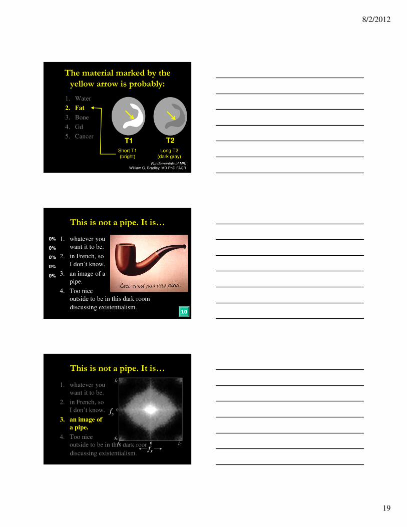
The material marked by the
yellow arrow is probably:
0%
0%
0%
0%
0%
10
1. Water
2. Fat
3. Bone
4. Gd
5. CancerT1 T2
8/2/2012
19
The material marked by the
yellow arrow is probably:
1. Water
2. Fat
3. Bone
4. Gd
5. CancerT1 T2
Short T1(bright)
Long T2(dark gray)
Fundamentals of MRI
William G. Bradley, MD PhD FACR
This is not a pipe. It is…
0%
0%
0%
0%
0%
10
1. whatever you
want it to be.
2. in French, so
I don’t know.
3. an image of a
pipe.
4. Too nice
outside to be in this dark room
discussing existentialism.
in this dark room
discussing existentialism.
This is not a pipe. It is…
1. whatever you
want it to be.
2. in French, so
I don’t know.
3. an image of
a pipe.
4. Too nice
outside to be fC-fC
0
fC
-fC
fx
0
fy

8/2/2012
20
Jeff – Jan-Jakob bridge
Pose a set of unanswered questions:
we have all these images, things moving, … how are
we going to make sense of it and respond (adapt) to
this information in treatment delivers?
bridge

8/2/2012
21
Adaptive Radiation Therapy
Temporal Scales of InterventionTemporal Scales of Intervention
Real timeReal time Off-lineOff-lineOn-lineOn-line
Projection
CT
MRI
PET
US
Role in IGRT / ART
Temporal Scales of Acquisition
… currently slow. So…
Projection
CT
MRI
PET
US
Role in IGRT / ART
Temporal Scales of Acquisition
FastReal-time
Even Faster