Embed Size (px)
Citation preview
A
O
DmA
R
a
Cb
RA
¿E
s
2
ctas Urol Esp. 2016;40(9):564---569
Actas Urologicas Espanolas
www.elsevier.es/actasuro
RIGINAL ARTICLE
oes the design of mini slings anchoring systems reallyatter? A biomechanical comparison between MinircTM and OphiraTM�
.S. Souzaa, P.C.R. Palmaa, F.G.F. Diasa, R.T. Siniscalchib, C.L.Z. Riccettoa,∗
División de Urología, Departamento de Cirugía, Facultad de Ciencias Médicas, Universidad de Campinas-UNICAMP,ampinas, SP, BrazilDivisión of Urología, Facultad de Medicina de Itajubá-FMIt, Itajubá-MG, Brazil
eceived 7 December 2015; accepted 22 February 2016vailable online 17 August 2016
KEYWORDSMini sling;Stress urinaryincontinence;Anchoring system;Biomechanics;Design;Maximum load;Elongation;Fixation;Tensile test
AbstractIntroduction and hypothesis: Currently, a sling implant is the standard treatment for stressurinary incontinence in women. To be effective, they require an adequate anchoring system.The aim of this study is compare biomechanical features of fixation systems of two mini slingsmodels available on the market (OphiraTM and Mini ArcTM) through a tensile test.Materials and methods: Anchoring devices of each sling were surgically implanted in abdom-inal wall of 15 rats divided into three groups of five animals which were arranged accordingto the date of post implant euthanasia on 7, 14 and 30 days. Abdominal walls of rats wereextracted on bloc containing the anchoring system and were submitted to a tensile strengthtest to measure the maximum load and elongation until device avulsion from the tissue. Theresults were compared using Student’s test t and a 5% cut off was considered significant.Results: The OphiraTM mini sling fixation system demanded a greater maximum load and devel-oped a longer stretch for avulsion from the implanted site at all moments evaluated (p valueless than 0.05).Conclusion: There were significant differences in fixation patterns of the anchoring systems,which were exclusively related to their designs. The OphiraTM mini sling fixation device provided
better fixation to the abdominal wall of rats compared to the Mini ArcTM device, even in thelate post-implant period.© 2016 AEU. Published by Elsevier Espana, S.L.U. All rights reserved.� Please cite this article as: Santos-Souza R, Rodrigues-Palma PC, Goulart-Fernandes-Dias F, Teixeira-Siniscalchi R, Zanettini-Riccetto CL.El diseno de sistemas de anclaje mini cabestrillo realmente importa? Una comparación biomecánica entre Mini ArcTMy OphiraTM. Actas Urolsp. 2016;40:564---569.∗ Corresponding author.
E-mail addresses: [email protected], [email protected] (C.L.Z. Riccetto).Abbreviations: SUI, stress urinary incontinence; TVT, tension-free vaginal tape; TOT, transobturator tape; SIMS,
ingle incision mini sling; ML, maximum load; SD, standard deviation.
173-5786/© 2016 AEU. Published by Elsevier Espana, S.L.U. All rights reserved.
Does the design of mini slings anchoring systems really matter? 565
PALABRAS CLAVEMini cabestrillo;Incontinencia urinariade esfuerzo;Sistema de anclaje;Biomecánica;Diseno;Carga máxima;Alargamiento;Fijación;Ensayo de tracción
¿El diseno de sistemas de anclaje mini cabestrillo realmente importa? Unacomparación biomecánica entre Mini ArcTMy OphiraTM
ResumenIntroducción e hipótesis: En la actualidad un implante de cabestrillo es el tratamiento estándarpara la incontinencia urinaria de esfuerzo en mujeres. Para ser eficaces requieren un sistema deanclaje adecuado. El objetivo de este estudio es comparar las características biomecánicas delos sistemas de fijación de 2 modelos de mini cabestrillos disponibles en el mercado (OphiraTM
y Mini ArcTM) a través de un ensayo de tracción.Materiales y métodos: Los dispositivos de anclaje de cada cabestrillo se implantaron quirúr-gicamente en la pared abdominal de 15 ratas divididas en 3 grupos de 5 animales que seorganizaron de acuerdo a la fecha de la eutanasia después del implante en los días 7, 14 y30. Las paredes abdominales de las ratas fueron extraídas en bloque, conteniendo el sistemade anclaje, y se sometieron a una prueba de resistencia tensil para medir la carga máxima yel alargamiento hasta la avulsión del dispositivo desde el tejido. Los resultados se compararonmediante la prueba «t» de Student y un punto de corte del 5% fue considerado significativo.Resultados: El sistema de fijación mini cabestrillo OphiraTM requirió una mayor carga máximay desarrolló un tramo más largo de la avulsión del sitio implantado en todos los momentosevaluados (valor de p inferior a 0,05).Conclusión: Hubo diferencias significativas en los patrones de fijación de los sistemas deanclaje, que fueron exclusivamente relacionadas con sus disenos. El dispositivo de fijaciónde mini cabestrillo OphiraTM proporcionó una mejor fijación a la pared abdominal de ratas encomparación con el dispositivo Mini ArcTM, incluso en el período posterior al implante tardío.© 2016 AEU. Publicado por Elsevier Espana, S.L.U. Todos los derechos reservados.
E
T(aapthe laboratory to have the same length, i.e. 2.7 cm (Fig. 1).They were implanted in the rat’s abdominal wall betweenhypodermis and abdominal fascia. For this procedure, the
Introduction
One-third of women will present involuntary urinary leakageduring their lifetime1 and 1 in 1000 will require surgery dueto stress urinary incontinence (SUI).2 The standard treat-ment of SUI is based on the implant of synthetic midurethralslings, which became popular after the Integral Theoryby Petros and Ulmstem.3 Tension-free vaginal tape (TVT)4
and transobturator tapes (TOT)5,6 have obtained long-lastingresults with cure rates above 80%.
The single incision mini sling (SIMS) is an innovativeanatomical approach that is anchored in the obturator inter-nus muscles bilaterally at the level of the tendinous archthrough a unique vaginal incision. The purpose of SIMS isto offer a minimally invasive treatment for SUI with shortersurgical time plus less bleeding and a rapid patient recovery.They are shorter than former midurethral slings (fewer syn-thetic material) and avoid percutaneous passage, preventinga blind path by insertion guides through the crural area.
Primary fixation is an important feature needed forthe effectiveness of SIMS. Accordingly, the purpose of thisresearch is to compare if the design of mini slings anchor-ing systems can influence their biomechanical properties.We evaluated OphiraTM (Promedon --- Argentina) versus MiniArcTM (American Medical Systems, AMS --- USA, United Statesof America) through in ex vivo animal tensile experiment.
Materials and methods
This work was performed in ex vivo experimental modelusing Wistar rats. This research was approved by the insti-tutional Committee for Ethics in Animal Research (StateUniversity of Campinas --- Unicamp --- Brazil).
F(2p
xperimental protocol
he experiment was conducted in 15 female Wistar150---200 g weight, 8 weeks old and considered youngdults)11 as ex vivo model. Fixation devices of OphiraTM
nd Mini ArcTM mini slings were originally made of poly-ropylene and samples were prepared and conditioned in
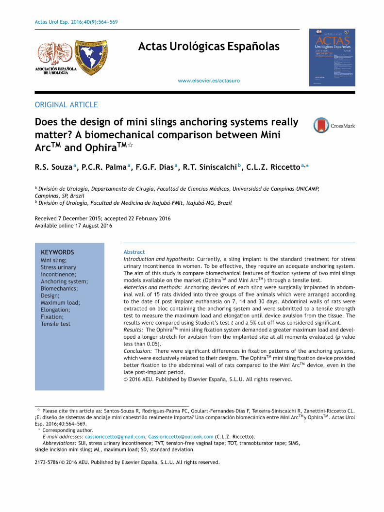
igure 1 This figure shows the fixation devices of Mini ArcTM
top) and OphiraTM (bottom) presenting the same length of.7 cm after being prepared. Note that Mini ArcTM has a singleair of claws, while OphiraTM has a multipoint anchoring device.
566 R.S. Souza et al.
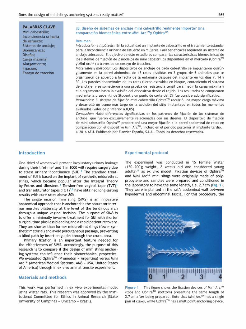
Figure 2 (A) Dissection between hypodermis and anterior abdominal fascia. Note that the linea alba was not violated to isolatet iraTM
a with
absssmmtawaaasscaiop
a7wspwraa
B
T1ihmaTaTamifmf(
S
Emeasures of position, dispersion and graphing. Student’s‘‘t’’-test was used to compare the slings in each time.
wo non-communicated cavities. (B) Detail of insertion of Ophnd removal of abdominal wall blocs. (D) Total thickness blocs
nimals were anesthetized with intravenous 6% sodium bar-ital (30 mg/kg), maintained in horizontal supine position,ubmitted to abdominal trichotomy followed by antisep-is with iodopovidone PVP-I (10%) and placed on sterileurgical fields. A 2 cm transverse suprapubic incision wasade followed by subcutaneous dissection preserving theid tissue fiber --- linea alba (Fig. 2A). This step creates
wo separate cavities where anchoring systems were placedllowing for no contact with each other. The OphiraTM deviceas inserted on the right side according to the standardsdopted (Fig. 2B), while the Mini ArcTM device was system-tically inserted on the left side (they were just put aboventerior muscular fascia freely without any stitch). The inci-ion was closed with Dexon IITM 2---0 (synthetic absorbableutures made of the homopolymer of glycolic acid andoated with polycaprolate, the copolymer of caprolactonend glycolide) avoiding contact between the suture and themplanted material. Post-operative analgesia was made withral paracetamol 100 mg/kg every four hours for the first 12ost-operative hours (3 doses).
Rats were divided into three groups of five animalsccording to the date of devices recovery performed on, 14 and 30 days following implantation. These animalsere sacrificed by induced hypoxia with a lethal dose of
ame anesthetic drug applied intravenously, placed in supineosition and trichotomized. Then, contour abdominal lines
ere made with a standard model marking the areas to beemoved separately (Fig. 2C). Finally, two sagittal halves ofbdominal blocs containing the mini sling anchoring systemsnd tissue were extracted from each animal (Fig. 2D).
Ttaa
anchoring device in the right space. (C) Incision standardizedanchoring systems within its layers.
iomechanics test
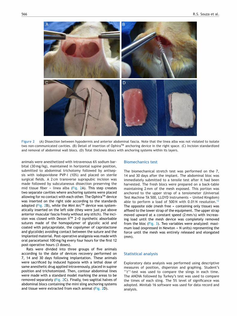
he biomechanical stretch test was performed on the 7,4 and 30 days after the implant. The abdominal bloc wasmmediately submitted to a tensile test after it had beenarvested. The fresh blocs were prepared on a back-tableaintaining 2 mm of the mesh exposed. This portion was
nchored to the upper strap of a tensiometer (Universalest Machine TA 500, LLOYD Instruments --- United Kingdom)ble to perform a load of 500 N with 0.01 N resolution.12
he opposite side (mesh free --- containing only tissue) wasffixed to the lower strap of the equipment. The upper strapoved upward at a constant speed (2 mm/s) with increas-
ng load until the mesh device was completely removedrom the bloc (Fig. 3). Two variables were analyzed: maxi-um load (expressed in Newton --- N units) representing the
orce until the mesh was entirely released and elongatedmm).
tatistical analysis
xploratory data analysis was performed using descriptive
he ANOVA followed by Turkey’s test was used to comparehe times of each sling. The 5% level of significance wasdopted. Minitab 16 software was used for data record andnalysis.
Does the design of mini slings anchoring systems really matter? 567
20
15
10
5
25
20
15
10
5
For
ce v
alue
(N
) fo
r ex
trus
ion
Def
lect
ion
(mm
) of
flxa
tion
devi
ces
MiniArc MiniArc MiniArc
MiniArcMiniArcMiniArc
17 T14 T30
T7 T14 T30
Ophira
Ophira Ophira Ophira
Ophira Ophira
A
B
Ft
tupwoscittgfmt
Figure 3 This figure shows a tensile test applied on abdominalbloc. Note the anchoring system being pulled upward unidirec-tionally with increasing load.
Results
Throughout the tensile test, we obtained the values of max-imum load (ML) and the length of elongation for the avulsionof the anchoring systems from the abdominal bloc.
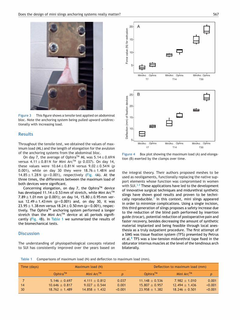
On day 7, the average of OphiraTM ML was 5.14 ± 0.69 Nversus 4.11 ± 0.81 N for Mini ArcTM (p 0.037). On day 14,these values were 10.64 ± 0.81 N versus 9.02 ± 0.54 N (p0.001), while on day 30 they were 18.76 ± 1.48 N and14.85 ± 1.28 N (p < 0.001), respectively (Fig. 4A). At thethree times, the differences between the maximum load ofboth devices were significant.
Concerning elongation, on day 7, the OphiraTM devicehas developed 11.14 ± 0.53 mm of stretch, while Mini ArcTM
7.89 ± 1.01 mm (p 0.001); on day 14, 15.80 ± 0.95 mm ver-sus 12.49 ± 1.43 mm (p < 0.001) and, on day 30, it was23.95 ± 1.38 mm versus 18.24 ± 0.50 mm (p < 0.001), respec-tively. The OphiraTM anchoring system performed a longerstretch than the Mini ArcTM device at all periods signifi-cantly (Fig. 4B). In Table 1 we summarized the results ofthe biomechanical tests.
Discussion
The understanding of physiopathological concepts relatedto SUI has consistently improved over the years based on
aeob
Table 1 Comparisons of maximum load (N) and deflection to max
Time (days) Maximum load (N)
OphiraTM Mini ArcTM p
7 5.146 ± 0.697 4.111 ± 0.812 0.014 10.646 ± 0.817 9.027 ± 0.544 0.030 18.762 ± 1.489 14.858 ± 1.432 <0.0
igure 4 Box plot showing the maximum load (A) and elonga-ion (B) exerted by the clamps over time.
he integral theory. Their authors proposed meshes to besed as neoligaments, functionally replacing the native sup-ort elements whose function was compromised in womenith SUI.3,4 These applications have led to the developmentf innovative surgical techniques and midurethral syntheticlings have shown good results and proven to be techni-ally reproducible.7 In this context, mini slings appearedn order to minimize complications. Using a single incision,his third generation of slings proposes a safety increase dueo the reduction of the blind path performed by insertionuide (trocar), potential reduction of postoperative pain andaster recovery, besides decreasing the amount of syntheticaterial implanted and being feasible through local anes-
hesia as a truly outpatient procedure. The first attempt of SIMS was tissue fixation system (TFS) presented by Petrus
t al.8 TFS was a low-tension midurethral tape fixed in thebturator internus muscles at the level of the tendinous archilaterally.imum load (mm).
Deflection to maximum load (mm)
OphiraTM Mini ArcTM p
37 11.148 ± 0.536 7.982 ± 1.010 0.00101 15.807 ± 0.957 12.494 ± 1.436 <0.00101 23.958 ± 1.382 18.246 ± 0.501 <0.001
5
ctbspisBmmsrdowCwuoqt
iieWtiaishtHrbwcctofiottr
aiioitttrribpf
eao
fgast
slfiiodberOcAm
C
Ttora
n
C
P
t
R
68
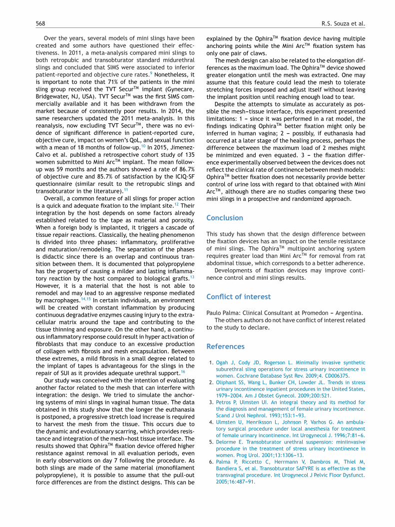
Over the years, several models of mini slings have beenreated and some authors have questioned their effec-iveness. In 2011, a meta-analysis compared mini slings tooth retropubic and transobturator standard midurethrallings and concluded that SIMS were associated to inferioratient-reported and objective cure rates.9 Nonetheless, its important to note that 71% of the patients in the miniling group received the TVT SecurTM implant (Gynecare,ridgewater, NJ, USA). TVT SecurTM was the first SIMS com-ercially available and it has been withdrawn from thearket because of consistently poor results. In 2014, the
ame researchers updated the 2011 meta-analysis. In thiseanalysis, now excluding TVT SecurTM, there was no evi-ence of significant difference in patient-reported cure,bjective cure, impact on women’s QoL, and sexual functionith a mean of 18 months of follow-up.10 In 2015, Jimenez-alvo et al. published a retrospective cohort study of 135omen submitted to Mini ArcTM implant. The mean follow-p was 59 months and the authors showed a rate of 86.7%f objective cure and 85.7% of satisfaction by the ICIQ-SFuestionnaire (similar result to the retropubic slings andransobturator in the literature).11
Overall, a common feature of all slings for proper actions a quick and adequate fixation to the implant site.12 Theirntegration by the host depends on some factors alreadystablished related to the tape as material and porosity.hen a foreign body is implanted, it triggers a cascade of
issue repair reactions. Classically, the healing phenomenons divided into three phases: inflammatory, proliferativend maturation/remodeling. The separation of the phasess didactic since there is an overlap and continuous tran-ition between them. It is documented that polypropyleneas the property of causing a milder and lasting inflamma-ory reaction by the host compared to biological grafts.13
owever, it is a material that the host is not able toemodel and may lead to an aggressive response mediatedy macrophages.14,15 In certain individuals, an environmentill be created with constant inflammation by producingontinuous degradative enzymes causing injury to the extra-ellular matrix around the tape and contributing to theissue thinning and exposure. On the other hand, a continu-us inflammatory response could result in hyper activation ofbroblasts that may conduce to an excessive productionf collagen with fibrosis and mesh encapsulation. Betweenhese extremes, a mild fibrosis in a small degree related tohe implant of tapes is advantageous for the slings in theepair of SUI as it provides adequate urethral support.16
Our study was conceived with the intention of evaluatingnother factor related to the mesh that can interfere withntegration: the design. We tried to simulate the anchor-ng systems of mini slings in vaginal human tissue. The databtained in this study show that the longer the euthanasias postponed, a progressive stretch load increase is requiredo harvest the mesh from the tissue. This occurs due tohe dynamic and evolutionary scarring, which provides resis-ance and integration of the mesh---host tissue interface. Theesults showed that OphiraTM fixation device offered higheresistance against removal in all evaluation periods, even
n early observations on day 7 following the procedure. Asoth slings are made of the same material (monofilamentolypropylene), it is possible to assume that the pull-outorce differences are from the distinct designs. This can beR.S. Souza et al.
xplained by the OphiraTM fixation device having multiplenchoring points while the Mini ArcTM fixation system hasnly one pair of claws.
The mesh design can also be related to the elongation dif-erences as the maximum load. The OphiraTM device showedreater elongation until the mesh was extracted. One mayssume that this feature could lead the mesh to toleratetretching forces imposed and adjust itself without leavinghe implant position until reaching enough load to tear.
Despite the attempts to simulate as accurately as pos-ible the mesh---tissue interface, this experiment presentedimitations: 1 --- since it was performed in a rat model, thendings indicating OphiraTM better fixation might only be
nferred in human vagina; 2 --- possibly, if euthanasia hadccurred at a later stage of the healing process, perhaps theifference between the maximum load of 2 meshes mighte minimized and even equated. 3 --- the fixation differ-nce experimentally observed between the devices does noteflect the clinical rate of continence between mesh models:phiraTM better fixation does not necessarily provide betterontrol of urine loss with regard to that obtained with MinircTM, although there are no studies comparing these twoini slings in a prospective and randomized approach.
onclusion
his study has shown that the design difference betweenhe fixation devices has an impact on the tensile resistancef mini slings. The OphiraTM multipoint anchoring systemequires greater load than Mini ArcTM for removal from ratbdominal tissue, which corresponds to a better adherence.
Developments of fixation devices may improve conti-ence control and mini slings results.
onflict of interest
aulo Palma: Clinical Consultant at Promedon --- Argentina.The others authors do not have conflict of interest related
o the study to declare.
eferences
1. Ogah J, Cody JD, Rogerson L. Minimally invasive syntheticsuburethral sling operations for stress urinary incontinence inwomen. Cochrane Database Syst Rev. 2009;4. CD006375.
2. Oliphant SS, Wang L, Bunker CH, Lowder JL. Trends in stressurinary incontinence inpatient procedures in the United States,1979---2004. Am J Obstet Gynecol. 2009;200:521.
3. Petros P, Ulmsten UI. An integral theory and its method forthe diagnosis and management of female urinary incontinence.Scand J Urol Nephrol. 1993;153:1---93.
4. Ulmsten U, Henriksson L, Johnson P, Varhos G. An ambula-tory surgical procedure under local anesthesia for treatmentof female urinary incontinence. Int Urogynecol J. 1996;7:81---6.
5. Delorme E. Transobturator urethral suspension: miniinvasiveprocedure in the treatment of stress urinary incontinence inwomen. Prog Urol. 2001;13:1306---13.
6. Palma P, Riccetto C, Herrmann V, Dambros M, Thiel M,Bandiera S, et al. Transobturator SAFYRE is as effective as thetransvaginal procedure. Int Urogynecol J Pelvic Floor Dysfunct.2005;16:487---91.
er?
1
1
1
1
Does the design of mini slings anchoring systems really matt
7. Richter HE, Albo ME, Zyczynski HM, Kenton K, Norton PA,Sirls LT, et al. Urinary Incontinence Treatment Network (2010).Retropubic versus transobturator. Midurethral slings for stressincontinence. N Engl J Med. 2010;362:2066---76.
8. Petros PE, Richardson P. Midurethral tissue fixation systemsling --- a micromethod for cure of stress incontinence ---preliminary report. Aust N Z J Obstet Gyn. 2005;45:372---5.
9. Abdel-Fattah M, Ford JA, Lim CP, Madhuvrata P. Single-incisionmini-slings versus standard midurethral slings in surgical man-agement of female stress urinary incontinence: a meta-analysisof effectiveness and complications. Eur Urol. 2011;60:468---80.
10. Mostafa A, Lim CP, Hopper L, Madhuvrata P, Abdel-Fattah M.Single-incision mini-slings versus standard midurethral slings insurgical management of female stress urinary incontinence: an
updated systematic review and meta-analysis of effectivenessand complications. Eur Urol. 2014;65:402---27.11. Jiménez-Calvo J, Montesino-Semper M, Hualde-Alfaro A, Torres-Varas L, Sotil-Arrieta A, Raigoso-Ortega O. Stress urinary
1
569
incontinence surgery with MiniArc: 4-year results. Actas UrolEsp. 2015;39:47---52.
2. Palma P, Siniscalchi RT, Maciel LC, Bigozzi MA, Dal Fabbro I,Riccetto C. Primary fixation of mini slings: a comparative biome-chanical study in vivo. J Bras Urol. 2012;38:253---60.
3. Pierce LM, Rao A, Baumann SS, Glassberg JE, Kuehl TJ, Muir TW.Long-term histologic response to synthetic and biologic graftmaterials implanted in the vagina and abdomen of a rabbitmodel. Am J Obstet Gynecol. 2009;200, 546el---e8.
4. Remes A, Williams DF. Immune response in biocompatibility.Biomaterials. 1992;13:731---43.
5. Wolf MT, Dearth CL, Ranallo CA, LoPresti ST, Carey LE, DalyKA, et al. Macrophage polarization in response to ECM coatedpolypropylene mesh. Biomaterials. 2014;35:6838---49.
6. Gigliobianco G, Regueros SR, Osman NI, Bissoli J, Bullock AJ,Chapple CR, et al. Biomaterials for pelvic floor reconstruc-tive surgery: how can we do better? BioMed Res Int. 2015:968087:20.